Intraoral Radiography: Positioning and Radiation ... - IneedCE.com
Intraoral Radiography: Positioning and Radiation ... - IneedCE.com
Intraoral Radiography: Positioning and Radiation ... - IneedCE.com

You also want an ePaper? Increase the reach of your titles
YUMPU automatically turns print PDFs into web optimized ePapers that Google loves.
Earn<br />
4 CE credits<br />
This course was<br />
written for dentists,<br />
dental hygienists,<br />
<strong>and</strong> assistants.<br />
<strong>Intraoral</strong> <strong>Radiography</strong>:<br />
<strong>Positioning</strong> <strong>and</strong><br />
<strong>Radiation</strong> Protection<br />
A Peer-Reviewed Publication<br />
Written by Gail F. Williamson, RDH, MS<br />
PennWell is an ADA CERP recognized provider<br />
ADA CERP is a service of the American Dental Association to assist dental professionals in identifying<br />
quality providers of continuing dental education. ADA CERP does not approve or endorse individual<br />
courses or instructors, nor does it imply acceptance of credit hours by boards of dentistry.<br />
PennWell is an ADA CERP Recognized Provider<br />
Concerns of <strong>com</strong>plaints about a CE provider may be directed to the provider or to ADA CERP at<br />
www.ada.org/goto/cerp.<br />
Go Green, Go Online to take your course<br />
This course has been made possible through an unrestricted educational grant. The cost of this CE course is $59.00 for 4 CE credits.<br />
Cancellation/Refund Policy: Any participant who is not 100% satisfied with this course can request a full refund by contacting PennWell in writing.
Educational Objectives<br />
Upon <strong>com</strong>pletion of this course, the clinician will be able to<br />
do the following:<br />
1. Underst<strong>and</strong> the various types of intraoral radiographs<br />
that can be taken <strong>and</strong> what these are used for<br />
2. Know how to correctly use the paralleling <strong>and</strong> bisecting<br />
techniques to take intraoral radiographs<br />
3. Know <strong>com</strong>mon errors that occur when taking intra-oral<br />
radiographs <strong>and</strong> how to avoid these<br />
4. Know how to minimize radiation exposure for patients<br />
<strong>and</strong> the operator<br />
Abstract<br />
Several types of intraoral radiographs can be taken. An underst<strong>and</strong>ing<br />
of both the paralleling <strong>and</strong> bisecting techniques<br />
<strong>and</strong> when to use these is necessary. Avoiding <strong>com</strong>mon errors<br />
when taking intraoral radiographs reduces the need for retakes.<br />
Minimizing radiation exposure for patients <strong>and</strong> the<br />
operator is an essential <strong>com</strong>ponent of intraoral radiography.<br />
Introduction<br />
X-rays were discovered in 1895 by Professor Wilhelm Conrad<br />
Roentgen, <strong>and</strong> Dr. Otto Walkhoff is credited with the first<br />
dental radiograph. Until the 1980s, dental radiographs were<br />
typically captured using film. Dr. Frances Mouyens invented<br />
direct digital radiography to take intraoral dental radiographs<br />
in 1984, <strong>and</strong> this technology was introduced into the U.S. in<br />
1989. While the use of digital radiography in dentistry continues<br />
to gain strength, film-based radiographs are still more<br />
<strong>com</strong>mon. The <strong>com</strong>plete transition to digital radiography is<br />
just a matter time.<br />
<strong>Intraoral</strong> dental radiographs fall into two main categories:<br />
bite-wings <strong>and</strong> periapicals. Bite-wing radiographs are the<br />
best diagnostic tool available for the detection of interproximal<br />
caries <strong>and</strong> assessment of alveolar bone levels. Bite-wings<br />
are usually taken in the posterior regions of the mouth.<br />
However, size 1 bite-wings can be taken of the anterior<br />
teeth to assess anterior bone levels. Periapical radiographs<br />
record the entire tooth <strong>and</strong> supporting bone <strong>and</strong> are used to<br />
evaluate the extent of caries <strong>and</strong> periodontal bone loss <strong>and</strong><br />
aid in the diagnosis <strong>and</strong> treatment of root <strong>and</strong> bony pathoses.<br />
Periapicals <strong>and</strong> bite-wings can be <strong>com</strong>bined to form surveys<br />
of varying configurations, for a <strong>com</strong>prehensive view of the<br />
entire dentition. <strong>Intraoral</strong> radiographs can be captured using<br />
film or digital receptors. Digital receptors are available<br />
as wired <strong>and</strong> wireless rigid sensors (CCD — charge-coupled<br />
device; CMOS — <strong>com</strong>plementary metal oxide semiconductor)<br />
<strong>and</strong> photostimulable phosphor plates. Both systems are<br />
<strong>com</strong>puter-based technologies that require specific hardware<br />
<strong>and</strong> software <strong>com</strong>ponents for operation. Digital receptors are<br />
available in sizes <strong>com</strong>parable to film, mostly typically sizes 0,<br />
1, <strong>and</strong> 2.<br />
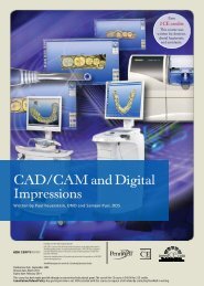
It has been estimated that in 1999 a total of 384 million<br />
sets of radiographs were taken, of which 170 million were a<br />
<strong>com</strong>plete series. 1 This demonstrates the importance <strong>and</strong> value<br />
of radiography in the diagnosis <strong>and</strong> treatment of oral disease.<br />
Number taken (in millions)<br />
Full-mouth series 170.20<br />
Periapical 80.30<br />
Bite-wing 112.80<br />
Panoramic 20.80<br />
Source: ADA. The 1999 Survey of Dental Services Rendered.<br />
Dental radiographs should be prescribed according to<br />
selection criteria guidelines <strong>and</strong> taken only for diagnostic <strong>and</strong><br />
treatment purposes. Selection criteria guidelines are based on<br />
evidence of disease patterns <strong>and</strong> take into consideration the<br />
patient’s medical <strong>and</strong> dental history, clinical signs <strong>and</strong> symptoms<br />
of disease, risk factors, age <strong>and</strong> dentition, <strong>and</strong> new or<br />
recall patient status. Only bite-wing radiographs have timebased<br />
intervals that are determined according to risk factors<br />
for caries. For a <strong>com</strong>plete review of these re<strong>com</strong>mendations,<br />
refer to “The Selection of Patients for Dental Radiographic<br />
Examination, Revised 2004.” 2<br />
Dental radiographs are valuable diagnostic tools when<br />
the image quality is adequate for proper interpretation. Filmbased<br />
<strong>and</strong> digital dental radiographs both require the use of<br />
careful technique <strong>and</strong> precautions to maximize the diagnostic<br />
<strong>and</strong> interpretative value of the radiograph while at the same<br />
time minimizing patient exposure to radiation.<br />
Key Objectives<br />
• Maximize diagnostic value of X-rays<br />
• Minimize patient exposure to radiation<br />
Maximizing the diagnostic value of radiographs starts<br />
with having the correct receptor (film, plate, or sensor) position,<br />
ensuring that the X-ray beam is centered <strong>and</strong> aligned at<br />
the correct vertical <strong>and</strong> horizontal angulations <strong>and</strong> exposed at<br />
the correct time.<br />
<strong>Positioning</strong> Guidelines for<br />
<strong>Intraoral</strong> Radiographs<br />
Accurate positioning is key for diagnostic radiographs <strong>and</strong><br />
helps avoid retakes. <strong>Intraoral</strong> radiographs are taken using<br />
paralleling, bisecting, <strong>and</strong> bite-wing techniques. Devices<br />
used to ac<strong>com</strong>plish this include receptor instruments with<br />
ring guides, st<strong>and</strong>ard biteblocks, <strong>and</strong> bite-wing tabs.<br />
Paralleling Technique<br />
The paralleling technique is used for both periapical <strong>and</strong><br />
bite-wing radiographs <strong>and</strong> is the most accurate technique<br />
for taking these projections. For film or digital radiographs,<br />
the receptor should be placed vertically <strong>and</strong> horizontally<br />
parallel with the teeth that are being radiographed. The<br />
X-ray beam should be directed at right angles to the teeth<br />
<strong>and</strong> receptor.<br />
2 www.ineedce.<strong>com</strong>
image foreshortening <strong>and</strong> elongation that misrepresents the<br />
actual length of all structures including the teeth.<br />
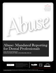
Central Ray Entry Points<br />
In the case of periapical radiographs, the film or digital receptor<br />
should be placed parallel to the full length of the crown<br />
<strong>and</strong> root of the teeth being imaged. The paralleling technique<br />
for bite-wing radiographs is simpler in the sense that the radiograph<br />
is more easily placed in the patient’s mouth even if<br />
the palate is shallow or the patient gags easily.<br />
Film <strong>and</strong> Digital Receptor Instruments<br />
Receptor instruments with X-ray beam ring guides improve<br />
the accuracy of the PID (Position indicating device, or X-ray<br />
cone) alignment to ensure correct beam angulation <strong>and</strong> beam<br />
centering. Receptor instruments <strong>com</strong>bine a receptor holder<br />
with an arm that has an attached ring indicating the position<br />
for the PID. This helps the operator avoid <strong>com</strong>mon errors<br />
by specifically directing the X-ray beam toward the receptor.<br />
Regardless of the instrument used, the placement of the<br />
receptor relative to the teeth must be correct. Instruments are<br />
available for paralleling, bisecting, <strong>and</strong> bite-wing techniques,<br />
as well as for endodontic imaging where endodontic files <strong>and</strong><br />
instruments may otherwise impede proper positioning of the<br />
receptor behind the tooth.<br />
Pupil of eye<br />
Ala of nose<br />
Tip of nose<br />
Nares of nose<br />
Commissure<br />
of lips<br />
Mentum<br />
Cone Cut<br />
Common Errors<br />
Overlap<br />
Outer canthus<br />
Tragus of ear<br />
Great care is necessary when placing the X-ray beam at<br />
right angles to the receptor, to avoid <strong>com</strong>mon errors. Incorrectly<br />
directing the beam in the horizontal plane will result<br />
in overlapping proximal contacts on bite-wing or periapical<br />
radiographs, making them diagnostically useless <strong>and</strong> resulting<br />
in a retake. Similarly, if the X-ray beam is not correctly<br />
centered over the receptor, cone cuts can occur on the image,<br />
with a clear zone where the X-rays did not expose the receptor.<br />
Central ray entry points help to identify the center of the<br />
receptor by using an external l<strong>and</strong>mark. In the case of periapical<br />
radiographs, improper vertical angulation can produce<br />
Foreshortening<br />
Elongation<br />
Rigid digital receptors are more difficult to use initially,<br />
may result in more errors for both periapical <strong>and</strong> bite-wing<br />
radiographs <strong>com</strong>pared to traditional film, <strong>and</strong> can cause<br />
more dis<strong>com</strong>fort for the patient. To avoid these problems,<br />
rigid receptors should be placed close to the midline to aid<br />
proper placement <strong>and</strong> to reduce dis<strong>com</strong>fort. It is particularly<br />
important if a patient has a shallow palate or floor of mouth<br />
to employ this method, both to avoid dis<strong>com</strong>fort <strong>and</strong> to avoid<br />
distortion of the image. The rigid sensors have a slightly<br />
smaller surface area for recording the image than traditional<br />
film does. Therefore, accurate positioning of the receptor<br />
<strong>and</strong> X-ray beam is even more critical to avoid cone cuts <strong>and</strong><br />
crown or apical cut-offs. Due to the sensor’s rigidity, more<br />
errors have been found than with the use of traditional film;<br />
more horizontal placement errors occur posteriorly, <strong>and</strong> more<br />
vertical angulation errors anteriorly. 3 This can be over<strong>com</strong>e<br />
with experience <strong>and</strong> underst<strong>and</strong>ing of the differences between<br />
rigid receptors <strong>and</strong> film. Phosphor plate receptors are<br />
more flexible <strong>and</strong> thinner than the other digital sensors but<br />
have the same dimensions as film, thus making the transition<br />
from film to digital radiography somewhat easier. However,<br />
the plates must be h<strong>and</strong>led carefully, scanned to digitize the<br />
image, <strong>and</strong> exposed to intense light before they can be reused.<br />
www.ineedce.<strong>com</strong> 3
Projection<br />
Or View<br />
Receptor Placement<br />
Teeth<br />
Recorded<br />
Central Ray Entry Point<br />
MAXILLARY PERIAPICALS<br />
Molar<br />
periapical<br />
Place the receptor toward the midline <strong>and</strong> the<br />
biteblock under the 2 nd molar crown, <strong>and</strong> align the<br />
mesial edge of the biteblock between the 1 st <strong>and</strong> 2 nd<br />
molar contact point<br />
1 st , 2 nd , 3 rd molar teeth crowns <strong>and</strong> apices<br />
Point down from the outer canthus (corner)<br />
of the eye to midcheek area<br />
Premolar<br />
periapical<br />
Place the receptor toward the midline <strong>and</strong> the<br />
biteblock under the 2 nd premolar crown, <strong>and</strong> align<br />
the mesial edge of the biteblock between the 1 st <strong>and</strong><br />
2 nd premolar contact point<br />
Distal of the canine, 1 st <strong>and</strong> 2 nd premolar, 1 st molar<br />
crowns <strong>and</strong> apices<br />
Point down from the pupil of the eye to<br />
mid-cheek area<br />
Canine<br />
periapical<br />
Place the receptor lingual to the canine, with the<br />
biteblock centered with the cusp tip<br />
Mesial <strong>and</strong> apex of the canine<br />
Ala (corner) of the nose<br />
Lateral<br />
incisor<br />
periapical<br />
Place the receptor lingual to the lateral incisor <strong>and</strong><br />
the biteblock under the lateral incisor crown<br />
Mesial, distal, <strong>and</strong> apex of the lateral incisor<br />
Nares (nostril) of the nose<br />
Central<br />
incisor<br />
periapical<br />
Place the receptor lingual to the central incisors,<br />
<strong>and</strong> center the biteblock with the central incisor<br />
contact point<br />
Mesial, distal, <strong>and</strong> apices of the central incisors<br />
Tip of the nose<br />
OPTION<br />
Caninelateral<br />
periapical<br />
Place the receptor lingual to the canine <strong>and</strong> lateral;<br />
center the biteblock with the lateral-canine<br />
contact point<br />
Mesial <strong>and</strong> apex of the canine, mesial, distal, <strong>and</strong><br />
apex of the lateral incisor<br />
Ala (corner) of the nose<br />
BITE-WINGS<br />
Molar<br />
bite-wing<br />
Align the mesial edge of the tab between the 1st<br />
<strong>and</strong> 2nd molar contact on the m<strong>and</strong>ible<br />
Maxillary <strong>and</strong> m<strong>and</strong>ibular molar crowns in occlusion<br />
Point down from the outer corner of the eye<br />
to the occusal plane<br />
Premolar<br />
bite-wing<br />
Align the mesial edge of the biteblock between the<br />
1st <strong>and</strong> 2nd premolar contact on the m<strong>and</strong>ible<br />
Distal of the maxillary <strong>and</strong> m<strong>and</strong>ibular canine,<br />
premolar <strong>and</strong> 1st molar crowns in occlusion<br />
Point down from the pupil of the eye to the<br />
occusal plane<br />
MANDIBULAR MOLAR PERIAPICALS<br />
Molar<br />
periapical<br />
Place the receptor toward the tongue, place the<br />
biteblock on the 2 nd molar crown, <strong>and</strong> align the<br />
mesial edge of the biteblock between the 1 st <strong>and</strong> 2 nd<br />
molar contact point<br />
1 st , 2 nd , 3 rd molar teeth crowns <strong>and</strong> apices<br />
Point down from the outer canthus (corner)<br />
of the eye to the mid-m<strong>and</strong>ible area<br />
Premolar<br />
periapical<br />
Place the receptor toward the tongue, place the<br />
biteblock on the 2 nd premolar, <strong>and</strong> align the mesial<br />
edge of the biteblock between the 1 st <strong>and</strong> 2 nd premolar<br />
contact point<br />
Distal of the canine, 1 st <strong>and</strong> 2 nd premolar, 1 st molar<br />
teeth crowns <strong>and</strong> apices<br />
Point down from the pupil of the eye to<br />
mid-m<strong>and</strong>ible area<br />
Caninelateral<br />
periapical<br />
Place the receptor lingual to the canine <strong>and</strong> lateral<br />
with biteblock centered with the contact point<br />
Distal of the lateral <strong>and</strong> mesial of the canine<br />
<strong>and</strong> apices<br />
Point down from the ala (corner) of the nose<br />
to the chin corner<br />
Central<br />
incisor<br />
periapical<br />
Place the receptor lingual to the central incisors,<br />
<strong>and</strong> center the biteblock with the central incisor<br />
contact point<br />
Mesial <strong>and</strong> distal of the central incisors <strong>and</strong> mesial<br />
of the lateral incisors <strong>and</strong> apices<br />
Point down from the tip of the nose to the<br />
chin center<br />
4 www.ineedce.<strong>com</strong>
Receptor<br />
Orientation<br />
Receptor Size<br />
Image<br />
Rough h<strong>and</strong>ling may produce plate scars, result in image<br />
artifacts, <strong>and</strong> necessitate plate replacement, making them less<br />
user-friendly in these instances.<br />
Horizontal placement;<br />
dot toward crown<br />
Size 2<br />
Bite-wing Tabs<br />
For patients who gag easily or children, tab bite-wings are<br />
less cumbersome <strong>and</strong> more <strong>com</strong>fortable for the patient<br />
than instrument holders.<br />
Horizontal placement;<br />
dot toward crown<br />
Size 2<br />
Vertical placement;<br />
dot toward crown<br />
Size 1<br />
Vertical placement;<br />
dot toward crown<br />
Size 1<br />
Vertical placement Size 1 or 2<br />
Vertical placement Size 2<br />
Horizontal or vertical placement;<br />
dot toward m<strong>and</strong>ible<br />
Horizontal or vertical placement;<br />
dot toward m<strong>and</strong>ible<br />
Horizontal placement;<br />
dot toward crown<br />
Horizontal placement;<br />
dot toward crown<br />
Size 2<br />
Size 2<br />
Size 2<br />
Size 2<br />
Correct Bite-wing <strong>Positioning</strong><br />
Position the receptor parallel to the interproximal<br />
spaces, not to the teeth being radiographed;<br />
otherwise, overlapping will occur.<br />
Bite-wing tabs hold the digital receptors or traditional<br />
film in position intraorally. Neither has any directional<br />
capability for PID positioning <strong>and</strong> beam direction. However,<br />
careful placement <strong>and</strong> beam alignment will produce<br />
good results. The vertical angulation is typically set +5°<br />
with the beam centered to the tab. The tab should be<br />
aligned with the teeth contacts, which will indicate the<br />
correct horizontal angulation. Central ray entry points<br />
will help with X-ray beam centering, as will using the<br />
lines on the PID that indicate the direction of the X-rays.<br />
Universal holders are available that can be used for rigid<br />
digital sensors.<br />
Bisecting Technique<br />
The bisecting technique may also be used for periapical<br />
radiographs. In this case, the receptor is placed diagonal<br />
to the teeth. The beam is then directed at a right angle to<br />
a plane that is midway between (bisects) the receptor <strong>and</strong><br />
the teeth. This technique produces less optimal images<br />
because the receptor <strong>and</strong> teeth are not in the same vertical<br />
plane. However, it is a useful alternative technique<br />
when ideal receptor placement cannot be achieved due to<br />
patient trauma or anatomic obstacles such as tori, shallow<br />
palate or shallow floor of the mouth, short frenum, or<br />
narrow arch widths.<br />
Vertical placement Size 1 or 2<br />
Vertical placement Size 1 or 2<br />
www.ineedce.<strong>com</strong> 5
This technique is more operator-sensitive. If the<br />
angle is not correctly bisected, elongation or foreshortening<br />
will occur. A variety of film holders can be used for<br />
different locations in the mouth for accurate positioning<br />
of the receptor. One approach the clinician can use is to<br />
align the PID parallel to the receptor initially <strong>and</strong> then<br />
reduce the vertical angle about ≈10°, which will approach<br />
the bisecting plane. Also, starting angles can be used that<br />
will get the operator close to the bisecting plane in each<br />
area of the mouth. These angles can be aligned using the<br />
angle meter on the side of the X-ray head.<br />
Arch<br />
Maxilla<br />
Molar<br />
+15° to<br />
+25°<br />
M<strong>and</strong>ible +5° to –5°<br />
Premolar<br />
+25° to<br />
+35°<br />
–10° to<br />
–15°<br />
Canine<br />
+40° to<br />
+50°<br />
–10° to<br />
–15°<br />
Incisor<br />
+40° to<br />
+50°<br />
–10° to<br />
–15°<br />
Shallow<br />
Palates<br />
Presence<br />
of tori<br />
Narrow<br />
arches<br />
Edentulous<br />
situations<br />
Endo<br />
Anatomical Variations<br />
• Move receptor towards midline<br />
• Consider using bisecting technique instead of paralleling<br />
technique<br />
• Ensure maxillary tori are between the teeth <strong>and</strong> receptor<br />
• Try to avoid m<strong>and</strong>ibular tori<br />
• Place receptor deeper in mouth if there are m<strong>and</strong>ibular tori,<br />
avoid tipping of receptor<br />
• Consider using bisecting technique instead of paralleling<br />
technique<br />
• Place receptor as far lingually as possible<br />
• For m<strong>and</strong>ibular anterior region, place receptor on dorsum of<br />
tongue<br />
• Use <strong>com</strong>pact size holders with rounded edges<br />
• Consider using bisecting technique instead of paralleling<br />
technique<br />
• Place receptor deeper in mouth<br />
• Place receptor deeper in mouth if necessary to avoid endodontic<br />
instruments<br />
Long PIDs include 12- to 16-inch lengths, but the<br />
st<strong>and</strong>ard 8 inch length PIDs can be used for paralleling<br />
as well. The longer PID length collimators reduce image<br />
magnification <strong>and</strong> improve sharpness <strong>and</strong> result in less<br />
image distortion. Right-angle entry of the X-ray beam<br />
improves anatomic accuracy <strong>and</strong> correct image length.<br />
Special Conditions While <strong>Positioning</strong><br />
Gagging<br />
Gagging patients can be challenging <strong>and</strong> require<br />
patience <strong>and</strong> reassurance from the clinician. It is important<br />
to be organized, pre-set the exposure time,<br />
pre-align the PID, <strong>and</strong> be ready to act quickly. The<br />
most <strong>com</strong>mon area to elicit the gag reflex is the maxillary<br />
molar periapical view. Placement of the receptor<br />
toward the midline <strong>and</strong> away from the soft palate will<br />
reduce the tendency for gagging. There are a variety of<br />
strategies that will help manage the gagging patient:<br />
breathing through the nose, salt on the tongue, distraction<br />
techniques (lifting one leg in the air, bending<br />
the toes toward the body, humming), use of topical anesthetics,<br />
<strong>and</strong> tissue cushions on the receptor. Similar<br />
approaches can be useful when the patient experiences<br />
dis<strong>com</strong>fort from the receptor, particularly the use of<br />
topical anesthetic agents <strong>and</strong> receptor cushions.<br />
<strong>Radiation</strong> Considerations<br />
It is incumbent upon dental professionals to ensure that<br />
in the process of taking dental radiographs, both the<br />
patient <strong>and</strong> the operator are protected as much as possible<br />
from the harmful effects of radiation. It has been<br />
known since shortly after their discovery that X-rays<br />
can result in biological damage. 4 Short-term effects of<br />
radiation result from a high dose over a short period of<br />
time — for example, the severe illness <strong>and</strong> rapid onset<br />
of death following a nuclear bomb explosion. Longterm<br />
effects result from the cumulative effect of low<br />
doses of radiation over an extended period of time <strong>and</strong><br />
can include cancer <strong>and</strong> genetic abnormalities.<br />
The risk of dental radiograph-induced idiopathic<br />
disease is extremely low. To put this in perspective,<br />
full-mouth radiographs (20 films) using F speed film<br />
<strong>and</strong> rectangular collimation equal one to two days of<br />
background radiation. 5 The risk of fatal cancers as a<br />
result of exposure to full-mouth dental X-rays using<br />
E+ speed film has been estimated to be 2.4 per million<br />
patients. 6 Nonetheless, dental professionals must<br />
protect their patients <strong>and</strong> themselves by minimizing<br />
exposure <strong>and</strong> risk.<br />
IV. Minimizing <strong>Radiation</strong> Exposure<br />
There are numerous methods that can be employed to<br />
minimize patients’ exposure to radiation. Together these<br />
methods can significantly reduce patients’ exposure.<br />
Number of Radiographs Taken<br />
Since radiation exposure has a lifetime cumulative<br />
effect, only essential dental radiographs should be<br />
taken. Keeping the total number of radiographs to a<br />
minimum requires an assessment of their necessity<br />
on a patient-by-patient basis. This is the purpose <strong>and</strong><br />
goal of selection criteria.<br />
Retakes contribute to an increased number of radiographs<br />
<strong>and</strong> as a result increased radiation exposure.<br />
Operator technique must be optimal to avoid retakes.<br />
Critical factors include accurate receptor placement,<br />
6 www.ineedce.<strong>com</strong>
proper angulation <strong>and</strong> beam centering, effective patient<br />
management, use of the correct exposure time, <strong>and</strong> careful<br />
processing for film-based imaging.<br />
Processing errors occur only with film <strong>and</strong> result in the<br />
greatest number of retakes, exposing patients to needless<br />
radiation. 7,8 To avoid these, the developer <strong>and</strong> fixer solutions<br />
must be used according to correct time-temperature<br />
regimens <strong>and</strong> renewed <strong>and</strong> replenished regularly along<br />
with provision of regular processing maintenance <strong>and</strong><br />
optimal darkroom conditions.<br />
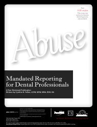
Receptor Selection<br />
For film-based radiography, F speed film is re<strong>com</strong>mended.<br />
The speed of the film depends upon the sensitivity of<br />
the emulsion to the X-ray beam. The faster the film, the<br />
shorter the exposure time <strong>and</strong> the less the total radiation<br />
delivered to the patient. F speed film requires 60% less<br />
exposure time than D speed film does. Digital receptors<br />
are faster than film <strong>and</strong> are 60% faster than E speed film. 9<br />
The table below shows the relative radiation exposure for<br />
different types of film on a scale of 1–10.<br />
Film Speed <strong>and</strong> Relative <strong>Radiation</strong> Exposure<br />
10<br />
8<br />
6<br />
4<br />
2<br />
0<br />
D-film E-film E+ film F film Digital<br />
receptors<br />
Source: Frederiksen NL. Health Physics. In: Pharoah MJ, White SC, eds. Oral<br />
Radiology: Principles <strong>and</strong> Interpretation. 4th ed. St. Louis: Mosby; 2001.<br />
Digital radiographs expose patients to less radiation<br />
on a per-radiograph basis. Additionally, digital radiographs<br />
are in general quicker to take <strong>and</strong> view than<br />
radiographs using film. However, this ease-of-use,<br />
particularly for rigid receptor systems, has been found<br />
to be a factor in a higher number of radiographs taken<br />
when digital radiography is used. 10 As a result, while<br />
the individual radiograph exposes the patient to less<br />
radiation, cumulatively this may not be the case if extra<br />
radiographs are taken. The same study found that the<br />
ease-of-use also resulted in offices being more likely to<br />
take more radiographs.<br />
Studies have found that digital radiographs in general<br />
are as useful as film radiographs for diagnostic purposes.<br />
11,12 Computerized image enhancement of digital<br />
radiographs allows the viewer to change brightness <strong>and</strong><br />
contrast <strong>and</strong> to invert, color, measure, or magnify the<br />
image. The ability to view the image in different formats<br />
may aid in diagnosis <strong>and</strong>, in some cases, <strong>com</strong>pensate for<br />
otherwise less-than-ideal radiographs, making them usable;<br />
13 as such, image enhancement may contribute to a<br />
reduced absolute number of retakes.<br />
Limiting the Number of Radiographs<br />
• Individual patient assessment of necessity <strong>and</strong><br />
number required<br />
• Operator technique to minimize retakes<br />
• Avoiding the temptation to take extra digital<br />
radiographs because of ease-of-use<br />
• Consideration of alternative diagnostic tools<br />
X-ray Beam Filtration <strong>and</strong> Collimation<br />
X-ray beams contain both high-energy <strong>and</strong> low-energy<br />
photons. Low-energy photons would be absorbed by<br />
the patient; to minimize this exposure, beam filtration<br />
is used. It is important to use a machine with a<br />
kilovoltage between 60 <strong>and</strong> 90 kV to reduce radiation<br />
doses to the patient, optimally in the range of 60 to<br />
70 kV. 14<br />
Beam collimation limits the diameter of the beam<br />
at the patient’s face, which should not exceed 7 cm,<br />
or 2.75 inches. Both round <strong>and</strong> rectangular collimators<br />
are available; the rectangular collimator reduces<br />
the beam’s diameter more <strong>and</strong> exposes 60% less tissue<br />
<strong>com</strong>pared to round collimators. 15<br />
Several options are available for rectangular collimation:<br />
semi-permanent rectangular PIDs from the<br />
x-ray machine manufacturer or a secondary removable<br />
rectangular collimator that is affixed to the st<strong>and</strong>ard<br />
round PID.<br />
<strong>Radiation</strong> Protection<br />
Patient Protection<br />
Patients rely upon dental professionals to provide safe<br />
<strong>and</strong> effective treatment. Patient protection includes<br />
the use of lead collars <strong>and</strong> may include the use of lead<br />
aprons. Lead collars are designed to protect the thyroid,<br />
<strong>and</strong> they fit around the patient’s neck. They have<br />
been found to substantially reduce radiation to the<br />
thyroid during dental radiographic examinations. 16<br />
www.ineedce.<strong>com</strong> 7
Operator Protection<br />
Primary radiation is that which is generated at the anode<br />
target, collimated, <strong>and</strong> directed toward the patient<br />
to take the radiograph. To avoid this, the operator must<br />
never st<strong>and</strong> directly in the X-ray beam directed at the<br />
patient, even though it may be tempting to hold a film<br />
in position for a patient having difficulty cooperating<br />
or to help a patient sit still in the correct position.<br />
Patient or film-holding must never be done <strong>and</strong> on a<br />
repeated basis would have a cumulative effect upon<br />
the operator.<br />
Lead aprons are considered optional by the American<br />
Association of Oral <strong>and</strong> Maxillofacial Radiology unless<br />
legally m<strong>and</strong>ated. 17 However, considering the fact that<br />
dental professionals are to <strong>com</strong>ply with the ALARA (As<br />
Low As Reasonably Achievable) principle <strong>and</strong> patients<br />
should be protected as much as possible, providing<br />
patients with added protection through the use of lead<br />
aprons is appropriate. Selection criteria guidelines re<strong>com</strong>mend<br />
patient shielding as an extra precaution during<br />
dental exposures, in particular children, women of<br />
childbearing age, <strong>and</strong> pregnant women. 18 Lead aprons<br />
are available in child <strong>and</strong> adult sizes. Lead aprons are<br />
available with a built-in thyroid collar, in which case a<br />
st<strong>and</strong>-alone lead collar is not required.<br />
The lead contained in lead aprons <strong>and</strong> collars is thin<br />
<strong>and</strong> malleable, <strong>and</strong> if the apron or collar is folded or left<br />
in a heap, the lead can be bent <strong>and</strong> damaged, resulting in<br />
areas of the collar or apron being lead-deficient. Collars<br />
<strong>and</strong> aprons should be hung up to avoid damage.<br />
Annual inspection of lead aprons for defects is m<strong>and</strong>atory,<br />
<strong>and</strong> test results must be recorded. 19 Inspection<br />
should occur immediately if cracks or other damage are<br />
suspected. Testing of lead aprons involves the use of a<br />
radiographic examination (or fluoroscopic examination)<br />
of the apron. If the apron is damaged, it must be appropriately<br />
discarded <strong>and</strong> a new replacement apron used.<br />
Patient <strong>and</strong> Operator Protection from <strong>Radiation</strong> Exposure<br />
• Provide patient with lead collar <strong>and</strong> apron<br />
• Minimize total exposure<br />
Primary <strong>Radiation</strong><br />
• Operator must not st<strong>and</strong> directly in the<br />
primary beam<br />
• Operator must st<strong>and</strong> behind a barrier or<br />
st<strong>and</strong> a minimum of 6 feet from the X-ray<br />
Scatter <strong>Radiation</strong><br />
source <strong>and</strong> at an angle of 90º–135º from<br />
the beam<br />
• Same operator precautions as for scatter<br />
radiation<br />
Leakage <strong>Radiation</strong><br />
• Regular maintenance for X-ray unit<br />
Scatter radiation results from the beam interacting<br />
with the surface of the patient, causing radiation to<br />
bounce as scatter in different directions. The third type<br />
of radiation is leakage that emanates from the X-ray<br />
tube head. To avoid scatter <strong>and</strong> leakage radiation, the<br />
operator must either st<strong>and</strong> behind a barrier or st<strong>and</strong> at a<br />
minimum 6 feet away from the radiation source <strong>and</strong> at an<br />
angle of 90º–135º to the X-ray beam. Barriers need not be<br />
lead-lined. Dental office operatory walls constructed of<br />
drywall are found to be adequate. 20<br />
Operators should <strong>com</strong>ply with the MPD (maximum<br />
permissible dose), to limit their occupational exposure,<br />
to the lesser of either a total effective dose of 5 rems/year<br />
(0.05 Sv); or, the sum of the deep-dose <strong>and</strong> <strong>com</strong>mitted dose<br />
equivalent to any individual organ or tissue other than the<br />
lens of the eye being equal to 50 rems (0.5 Sv). The limit<br />
for pregnant radiation workers is 0.5 rems (5 mSv).<br />
The best method to avoid occupational exposure is to<br />
consistently practice safety rules as described above.<br />
Regular X-ray machine inspection <strong>and</strong> maintenance is<br />
necessary to ensure not only that the machine is delivering<br />
the appropriate radiation to patients, but also to check<br />
for sources of leakage radiation <strong>and</strong> proper filtration <strong>and</strong><br />
collimation <strong>and</strong> if necessary to correct inadequacies.<br />
Summary<br />
Dental radiographs are valuable diagnostic tools <strong>and</strong> expose<br />
the patient to minimal amounts of radiation. Nonetheless,<br />
dental professionals must ensure that both they <strong>and</strong> patients<br />
are protected from the harmful effects of cumulative<br />
exposure to radiation. Patients can be protected through<br />
the use of lead collars <strong>and</strong> aprons <strong>and</strong> by ensuring that only<br />
necessary radiographs are taken <strong>and</strong> that radiation exposure<br />
is kept low. Operator protection involves st<strong>and</strong>ing behind<br />
barriers, avoiding st<strong>and</strong>ing in or near the primary beam,<br />
8 www.ineedce.<strong>com</strong>
<strong>and</strong> regularly maintaining X-ray equipment. One of the<br />
critical factors in minimizing the number of radiographs<br />
is to ensure that retakes are not required due to improper<br />
technique or processing problems. Receptor instruments<br />
are valuable tools that guide the X-ray beam, thereby helping<br />
to increase the accuracy of dental radiography.<br />
Endnotes<br />
1 American Dental Association. 1999 Survey of Services<br />
Rendered.<br />
2 American Dental Association <strong>and</strong> U.S. Department of Health<br />
<strong>and</strong> Human Services. The Selection of Patients for Dental<br />
Radiographic Examination, Revised 2004.<br />
3 Versteeg CH, et al. An evaluation of periapical radiography with a<br />
charge-coupled device. Dentomaxillofac Radiol. 1998;27:97–101.<br />
4 Langl<strong>and</strong> OE, Langlais RP. Early pioneers of oral <strong>and</strong><br />
maxillofacial radiology. Oral Surg Oral Med Oral Pathol Oral<br />
Radiol Endod. 1995;80:496–511.<br />
5 <strong>Radiation</strong> Safety in Dental <strong>Radiography</strong>. Rochester, NY: Eastman<br />
Kodak Company; 1998:2.<br />
6 Frederiksen NL. Health Physics. In: Pharoah MJ, White SC, eds.<br />
Oral Radiology: Principles <strong>and</strong> Interpretation. 4th ed. St. Louis:<br />
Mosby; 2001:49.<br />
7 Yakoumakis EN, et al. Image quality assessment <strong>and</strong> radiation<br />
doses in intraoral radiography. Oral Surg Oral Med Oral Pathol<br />
Oral Radiol Endod. 2001;91(3):362–368.<br />
8 Button TM, Moore WC, Goren AD. Causes of excessive<br />
bite-wing exposure: results of a survey regarding radiographic<br />
equipment in New York. Oral Surg Oral Med Oral Pathol Oral<br />
Radiol Endod. 1999;87(4):513–517.<br />
9 Frederiksen NL. Health Physics. In: Pharoah MJ, White SC, eds.<br />
Oral Radiology: Principles <strong>and</strong> Interpretation. 4th ed. St. Louis:<br />
Mosby; 2001.<br />
10 Berkhout WE, S<strong>and</strong>erink GC, van der Stelt PF. Does digital<br />
radiography increase the number of intraoral radiographs? A<br />
questionnaire study of Dutch dental practices. Dentomaxillofac<br />
Radiol. 2003;32:124–127.<br />
11 Svanaes DB, et al. <strong>Intraoral</strong> storage phosphor radiography for<br />
approximal caries detection <strong>and</strong> effect of image magnification:<br />
Comparison with conventional radiograph. Oral Surg Oral Med<br />
Oral Pathol Oral Radiol Endod. 1996;82:94–100.<br />
12 Naitoh M, et al. Observer agreement in the detection of proximal<br />
caries with direct digital intraoral radiography. Oral Surg Oral<br />
Med Oral Pathol Oral Radiol Endod. 1998;85:107–112.<br />
13 Williamson GF. Digital radiography in dentistry: moving from<br />
film-based to digital imaging. American Dental Assistants<br />
Association Continuing Education Course.<br />
14 Goren AD, et al. Updated quality assurance self-assessment<br />
exercise in intraoral <strong>and</strong> panoramic radiography. Oral Surg Oral<br />
Med Oral Pathol Oral Radiol Endod. 2000;89:369–374.<br />
15 Parameters of Radiologic Care: An Official Report of the American<br />
Academy of Oral <strong>and</strong> Maxillofacial Radiology. Oral Surg Oral<br />
Med Oral Pathol Oral Radiol Endod. 2001;91:498–511.<br />
16 Sikorski PA, Taylor KW. The effectiveness of the thyroid shield<br />
in dental radiology. Oral Surg. 1984;58:225–236.<br />
17 White SC, Heslop EW, et al. Parameters of radiologic care:<br />
An official report of the American Academy of Oral <strong>and</strong><br />
Maxillofacial Radiology. Oral Surg Oral Med Oral Pathol Oral<br />
Radiol Endod. 2001;91(5):498–511.<br />
18 American Dental Association <strong>and</strong> U.S. Department of Health<br />
<strong>and</strong> Human Services. The Selection of Patients for Dental<br />
Radiographic Examination, Revised 2004.<br />
19 Limacher MC, Douglas PS, Germano G, et al. <strong>Radiation</strong> safety<br />
in the practice of cardiology. JACC 1998;31(4):892–913.<br />
20 Razmus TF. The biological effects <strong>and</strong> safe use of radiation. In:<br />
Razmus TF, Williamson GF, eds. Current Oral <strong>and</strong> Maxillofacial<br />
Imaging. Philadelphia, PA: WB Saunders;1996.<br />
Author Profile<br />
Professor Gail F. Williamson, RDH, MS<br />
Professor Gail F. Williamson is a professor of Dental<br />
Diagnostic Sciences in the Department of Oral Pathology,<br />
Medicine, <strong>and</strong> Radiology at Indiana University<br />
School of Dentistry. She serves as Director of Allied<br />
Dental Radiology <strong>and</strong> Couse Director for Dental<br />
Assisting <strong>and</strong> Dental Hygiene Radiology Courses.<br />
Professor Williamson serves on the Council of Sections<br />
Administrative Board of the American Dental<br />
Education Association.<br />
Acknowledgement<br />
Cone cut <strong>and</strong> overlap images from ADTS course, Successful<br />
<strong>Intraoral</strong> <strong>Radiography</strong> by William S. Moore,<br />
DDS, MS<br />
Disclaimer<br />
The author of this course has no <strong>com</strong>mercial ties with the<br />
sponsors or the providers of the unrestricted educational<br />
grant for this course.<br />
Reader Feedback<br />
We encourage your <strong>com</strong>ments on this or any PennWell course.<br />
For your convenience, an online feedback form is available at<br />
www.ineedce.<strong>com</strong>.<br />
www.ineedce.<strong>com</strong> 9
1. _____ is credited with the first<br />
dental radiograph.<br />
a. Professor Roentgen<br />
b. Dr. Hans Blitter<br />
c. Dr. Otto Walkhoff<br />
d. None of the above<br />
2. Only digital radiographs are<br />
currently used in dentistry.<br />
a. True<br />
b. False<br />
3. <strong>Intraoral</strong> radiographs fall into two<br />
main categories: _____.<br />
a. Bite-wings <strong>and</strong> periapicals<br />
b. Bite-wings <strong>and</strong> laterals<br />
c. Panoramic <strong>and</strong> lateral radiographs<br />
d. All of the above<br />
4. In 1999, an estimated _____ sets of<br />
radiographs were taken.<br />
a. 282 million<br />
b. 384 million<br />
c. 462 million<br />
d. 575 million<br />
5. Only _____ radiographs have timebased<br />
intervals that are determined<br />
according to risk factors for caries.<br />
a. Periapical<br />
b. Panoramic<br />
c. Cephalograph<br />
d. Bite-wing<br />
6. The paralleling technique is used<br />
for _____ .<br />
a. Periapical radiographs<br />
b. Bite-wing radiographs<br />
c. Panoramic radiographs<br />
d. a <strong>and</strong> b<br />
7. In the paralleling technique, the<br />
X-ray beam should be directed at<br />
_____ to the teeth <strong>and</strong> receptor.<br />
a. 45 degrees<br />
b. 90 degrees<br />
c. 180 degrees<br />
d. None of the above<br />
8. Receptor instruments<br />
<strong>com</strong>bine _____.<br />
a. A receptor display with an arm that has an<br />
attached rectangle<br />
b. A receptor holder with an arm that has an<br />
attached rectangle<br />
c. A receptor holder with an arm that has an<br />
attached ring<br />
d. None of the above<br />
9. Receptor instruments help the<br />
operator avoid <strong>com</strong>mon errors<br />
by _____.<br />
a. Specifically directing the X-ray beam towards<br />
the receptor<br />
b. Reducing the intensity of the X-ray beam<br />
c. Allowing the operator to rotate the film<br />
d. None of the above<br />
10. Common errors in intraoral<br />
radiographs include _____.<br />
a. Overlapping contacts on bite-wing radiographs<br />
b. Elongation <strong>and</strong> foreshortening on<br />
periapical radiographs<br />
c. Cone cuts<br />
d. All of the above<br />
11. Phosphor plate receptors are _____<br />
than other digital sensors.<br />
a. More flexible<br />
b. Thinner<br />
c. Sturdier<br />
d. a <strong>and</strong> b<br />
Questions<br />
12. Molar periapicals are taken to<br />
record the _____.<br />
a. 1 st , 2 nd <strong>and</strong> 3 rd molar teeth crowns <strong>and</strong> apices<br />
b. 1 st , 2 nd <strong>and</strong> 3 rd molar teeth crowns only<br />
c. Only the surrounding bone<br />
d. None of the above<br />
13. The receptor orientation for a bitewing<br />
radiograph of the premolar<br />
teeth should be _____.<br />
a. Horizontal or vertical with the dot towards<br />
the maxilla<br />
b. Diagonal with the dot towards the m<strong>and</strong>ible<br />
c. Horizontal or vertical with the dot towards<br />
the m<strong>and</strong>ible<br />
d. None of the above<br />
14. The receptor orientation for a periapical<br />
radiograph of the m<strong>and</strong>ibular<br />
central incisors should be ______.<br />
a. Horizontal<br />
b. Diagonal<br />
c. Vertical<br />
d. Any of the above<br />
15. The receptor orientation for a<br />
periapical radiograph of the maxillary<br />
premolars should be _____.<br />
a. Horizontal placement with the dot towards<br />
the crown<br />
b. Vertical placement with the dot towards<br />
the crown<br />
c. Vertical placement with the dot towards the root<br />
d. None of the above<br />
16. The bisecting technique<br />
is ______ <strong>com</strong>pared to the<br />
paralleling technique.<br />
a. Less operator-sensitive<br />
b. More operator-sensitive<br />
c. Easier<br />
d. None of the above<br />
17. The bisecting technique is a<br />
useful alternative to the paralleling<br />
technique if the patient has _____.<br />
a. Tori<br />
b. A shallow palate or floor of mouth<br />
c. Narrow arch width<br />
d. All of the above<br />
18. The most <strong>com</strong>mon area to elicit a<br />
gag reflex is _____.<br />
a. The maxillary molar periapical view<br />
b. The m<strong>and</strong>ibular molar periapical view<br />
c. The molar bite-wing view<br />
d. None of the above<br />
19. If a patient has a shallow palate, it<br />
can help when taking a radiograph<br />
to_____.<br />
a. Consider using the bisecting technique<br />
b. Use a bent film<br />
c. a <strong>and</strong> b<br />
d. None of the above<br />
20. If a patient has a narrow arch, it<br />
can help when taking a radiograph<br />
to _____.<br />
a. Use <strong>com</strong>pact size holders<br />
b. Avoid taking a radiograph<br />
c. Consider using the bisecting technique<br />
d. a <strong>and</strong> c<br />
21. Full mouth radiographs expose<br />
the patient to the same amount<br />
of radiation as ______ of<br />
background radiation.<br />
a. One to two days<br />
b. Three to four days<br />
c. 5 days<br />
d. 10 days<br />
22. A patient’s radiation exposure can<br />
be minimized by _____.<br />
a. Taking only essential radiographs<br />
b. Using a high-speed film or digital radiograph<br />
c. Avoiding errors that would result in retakes<br />
d. All of the above<br />
23. The greatest number of retakes<br />
in intraoral radiography is a result<br />
of _____.<br />
a. Faulty X-ray equipment<br />
b. Processing errors with film radiographs<br />
c. The patient moving while the radiograph is<br />
being taken<br />
d. None of the above<br />
24. Digital radiographs _____.<br />
a. Expose patients to less radiation per radiograph<br />
b. Are quicker to take than traditional<br />
film radiographs<br />
c. Have a greater ease-of-use than traditional<br />
film radiographs<br />
d. All of the above<br />
25. Beam collimation limits the<br />
diameter of the X-ray beam at the<br />
patient’s face, which should not<br />
exceed _____.<br />
a. 3 cm or 1.50 inches<br />
b. 4 cm or 1.75 inches<br />
c. 7 cm or 2.75 inches<br />
d. 9 cm or 2.95 inches<br />
26. Lead collars are designed to<br />
protect _____.<br />
a. The esophagus<br />
b. The thyroid<br />
c. The hypothalamus<br />
d. All of the above<br />
27. The ALARA principle st<strong>and</strong>s<br />
for _____.<br />
a. As Likely As Routinely Assessed<br />
b. As Low As Reasonably Applicable<br />
c. As Low As Reasonably Achievable<br />
d. None of the above<br />
28. _____ inspection of lead aprons<br />
is m<strong>and</strong>atory.<br />
a. Monthly<br />
b. Annual<br />
c. Bi-annual<br />
d. None of the above<br />
29. Operator protection against<br />
primary radiation is achieved<br />
by _____.<br />
a. Not st<strong>and</strong>ing directly in the primary beam<br />
b. Holding the film or sensor at an angle in the<br />
patient’s mouth<br />
c. Wearing a lead collar<br />
d. None of the above<br />
30. _____ can be minimized by regularly<br />
maintaining X-ray equipment.<br />
a. Leakage radiation<br />
b. Seizures<br />
c. Scratches on sensors<br />
d. None of the above<br />
10 www.ineedce.<strong>com</strong>
ANSWER SHEET<br />
<strong>Intraoral</strong> <strong>Radiography</strong>: <strong>Positioning</strong> <strong>and</strong> <strong>Radiation</strong> Protection<br />
Name: Title: Specialty:<br />
Address:<br />
E-mail:<br />
City: State: ZIP:<br />
Telephone: Home ( ) Office ( )<br />
Requirements for successful <strong>com</strong>pletion of the course <strong>and</strong> to obtain dental continuing education credits: 1) Read the entire course. 2) Complete all<br />
information above. 3) Complete answer sheets in either pen or pencil. 4) Mark only one answer for each question. 5) A score of 70% on this test will earn<br />
you 4 CE credits. 6) Complete the Course Evaluation below. 7) Make check payable to PennWell Corp.<br />
Educational Objectives<br />
1. Underst<strong>and</strong> the various types of intraoral radiographs that can be taken <strong>and</strong> what these are used for<br />
2. Know how to correctly use the paralleling <strong>and</strong> bisecting techniques to take intraoral radiographs<br />
3. Know <strong>com</strong>mon errors that occur when taking intra-oral radiographs <strong>and</strong> how to avoid these<br />
4. Know how to minimize radiation exposure for patients <strong>and</strong> the operator<br />
Course Evaluation<br />
Please evaluate this course by responding to the following statements, using a scale of Excellent = 5 to Poor = 0.<br />
1. Were the individual course objectives met? Objective #1: Yes No Objective #3: Yes No<br />
Objective #2: Yes No Objective #4: Yes No<br />
2. To what extent were the course objectives ac<strong>com</strong>plished overall? 5 4 3 2 1 0<br />
3. Please rate your personal mastery of the course objectives. 5 4 3 2 1 0<br />
Mail <strong>com</strong>pleted answer sheet to<br />
Academy of Dental Therapeutics <strong>and</strong> Stomatology,<br />
A Division of PennWell Corp.<br />
P.O. Box 116, Chesterl<strong>and</strong>, OH 44026<br />
or fax to: (440) 845-3447<br />
For immediate results, go to www.ineedce.<strong>com</strong><br />
<strong>and</strong> click on the button “Take Tests Online.” Answer<br />
sheets can be faxed with credit card payment to<br />
(440) 845-3447, (216) 398-7922, or (216) 255-6619.<br />
Payment of $59.00 is enclosed.<br />
(Checks <strong>and</strong> credit cards are accepted.)<br />
If paying by credit card, please <strong>com</strong>plete the<br />
following: MC Visa AmEx Discover<br />
Acct. Number: _______________________________<br />
Exp. Date: _____________________<br />
Charges on your statement will show up as PennWell<br />
4. How would you rate the objectives <strong>and</strong> educational methods? 5 4 3 2 1 0<br />
5. How do you rate the author’s grasp of the topic? 5 4 3 2 1 0<br />
6. Please rate the instructor’s effectiveness. 5 4 3 2 1 0<br />
7. Was the overall administration of the course effective? 5 4 3 2 1 0<br />
8. Do you feel that the references were adequate? Yes No<br />
9. Would you participate in a similar program on a different topic? Yes No<br />
10. If any of the continuing education questions were unclear or ambiguous, please list them.<br />
___________________________________________________________________<br />
11. Was there any subject matter you found confusing? Please describe.<br />
___________________________________________________________________<br />
___________________________________________________________________<br />
12. What additional continuing dental education topics would you like to see?<br />
___________________________________________________________________<br />
___________________________________________________________________ AGD Code 731<br />
PLEASE PHOTOCOPY ANSWER SHEET FOR ADDITIONAL PARTICIPANTS.<br />
AUTHOR DISCLAIMER<br />
The author of this course has no <strong>com</strong>mercial ties with the sponsors or the providers of<br />
the unrestricted educational grant for this course.<br />
SPONSOR/PROVIDER<br />
This course was made possible through an unrestricted educational grant. No<br />
manufacturer or third party has had any input into the development of course content.<br />
All content has been derived from references listed, <strong>and</strong> or the opinions of clinicians.<br />
Please direct all questions pertaining to PennWell or the administration of this course to<br />
Machele Galloway, 1421 S. Sheridan Rd., Tulsa, OK 74112 or macheleg@pennwell.<strong>com</strong>.<br />
COURSE EVALUATION <strong>and</strong> PARTICIPANT FEEDBACK<br />
We encourage participant feedback pertaining to all courses. Please be sure to <strong>com</strong>plete the<br />
survey included with the course. Please e-mail all questions to: macheleg@pennwell.<strong>com</strong>.<br />
INSTRUCTIONS<br />
All questions should have only one answer. Grading of this examination is done<br />
manually. Participants will receive confirmation of passing by receipt of a verification<br />
form. Verification forms will be mailed within two weeks after taking an examination.<br />
EDUCATIONAL DISCLAIMER<br />
The opinions of efficacy or perceived value of any products or <strong>com</strong>panies mentioned<br />
in this course <strong>and</strong> expressed herein are those of the author(s) of the course <strong>and</strong> do not<br />
necessarily reflect those of PennWell.<br />
Completing a single continuing education course does not provide enough information<br />
to give the participant the feeling that s/he is an expert in the field related to the course<br />
topic. It is a <strong>com</strong>bination of many educational courses <strong>and</strong> clinical experience that<br />
allows the participant to develop skills <strong>and</strong> expertise.<br />
COURSE CREDITS/COST<br />
All participants scoring at least 70% (answering 21 or more questions correctly) on the<br />
examination will receive a verification form verifying 4 CE credits. The formal continuing<br />
education program of this sponsor is accepted by the AGD for Fellowship/Mastership<br />
credit. Please contact PennWell for current term of acceptance. Participants are urged to<br />
contact their state dental boards for continuing education requirements. PennWell is a<br />
California Provider. The California Provider number is 4527. The cost for courses ranges<br />
from $49.00 to $110.00.<br />
Many PennWell self-study courses have been approved by the Dental Assisting National<br />
Board, Inc. (DANB) <strong>and</strong> can be used by dental assistants who are DANB Certified to meet<br />
DANB’s annual continuing education requirements. To find out if this course or any other<br />
PennWell course has been approved by DANB, please contact DANB’s Recertification<br />
Department at 1-800-FOR-DANB, ext. 445.<br />
RECORD KEEPING<br />
PennWell maintains records of your successful <strong>com</strong>pletion of any exam. Please contact our<br />
offices for a copy of your continuing education credits report. This report, which will list<br />
all credits earned to date, will be generated <strong>and</strong> mailed to you within five business days<br />
of receipt.<br />
CANCELLATION/REFUND POLICY<br />
Any participant who is not 100% satisfied with this course can request a full refund by<br />
contacting PennWell in writing.<br />
© 2009 by the Academy of Dental Therapeutics <strong>and</strong> Stomatology, a division<br />
of PennWell<br />
www.ineedce.<strong>com</strong> 11