1 Protein Structure Protein Structure
1 Protein Structure Protein Structure
1 Protein Structure Protein Structure

You also want an ePaper? Increase the reach of your titles
YUMPU automatically turns print PDFs into web optimized ePapers that Google loves.
<strong>Protein</strong> <strong>Structure</strong><br />
<strong>Protein</strong> <strong>Structure</strong><br />
I. Introduction of amino acid residues<br />
II. Secondary <strong>Structure</strong> and Backbone Conformation<br />
III. Super-secondary structure<br />
IV. Sidechain Conformation<br />
V. Tertiary <strong>Protein</strong> <strong>Structure</strong> and folds<br />
VI. Quaternary <strong>Structure</strong><br />
VII. Larger <strong>Structure</strong>s<br />
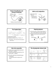
I. Introduction of amino acid residues<br />
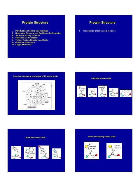
Overview of general properties of 20 amino acids<br />
Aliphatic amino acids<br />
Aromatic amino acids<br />
Sulfur-containing amino acids
Highly polar amino acids<br />
pKa~6.5, it is relatively easy to move protons on and off of the<br />
side chain. It is an ideal residue for protein functional centres.<br />
Histidines are the most common amino acids in protein active<br />
or binding sites. They are very common in metal binding sites<br />
(e.g. zinc), often acting together with Cys or other amino<br />
acids.<br />
Positively charged<br />
Negatively charged<br />
Less polar amino acids<br />
Zinc-finger<br />
Serine Protease<br />
The role of Cys in structure is very dependent on the<br />
cellular location of the protein. Within extracellular<br />
proteins, Cys are frequently involved in disulphide<br />
bonds. These bonds serve mostly to stabilise the protein<br />
structure, and the structure of many extracellular<br />
proteins is almost entirely determined by the topology of<br />
multiple disulphide bonds.<br />
The reducing environment inside cells makes the<br />
formation of disulphide bonds very unlikely.<br />
However, as the most reactive amino acid residue,<br />
Cys plays critical roles both in the structure (i.e.,<br />
meal ion binding) and function (catalysis in active<br />
sites of enzymes).<br />
<strong>Protein</strong> <strong>Structure</strong><br />
I. Introduction of amino acid residues<br />
II. Secondary <strong>Structure</strong> and Backbone Conformation<br />
EGF-like domain<br />
2.1 Peptide Torsion Angles<br />
2.2 The Ramachandran Plot<br />
The figure below shows the three main chain torsion angles of a polypeptide.<br />
These are phi (Φ), psi (Ψ), and omega (Ω).<br />
Allowed regions<br />
Generally allowed regions<br />
Disallowed regions<br />
(except Gly)<br />
L-amino acids generally do not<br />
form left handed α-helix (except<br />
Asn and Asp, of which the<br />
sidechains can form H-bond with<br />
mainchains<br />
The planarity of the peptide bond restricts to 180 degrees (Ω) in very nearly all of<br />
the main chain peptide bonds. In rare cases ~ 10 degrees for a cis peptide bond<br />
which usually involves proline.<br />
GN Ramachandran used computer models of small polypeptides to systematically vary<br />
and with the objective of finding stable conformations. For each conformation, the<br />
structure was examined for close contacts between atoms. Atoms were treated as hard<br />
spheres with dimensions corresponding to their van der Waals radii. Therefore, and<br />
angles, which cause spheres to collide correspond to sterically disallowed<br />
conformations of the polypeptide backbone.
Small changes of backbone<br />
dihedral angle can have large<br />
conformational effects on<br />
polypeptides<br />
Φ, Ψ<br />
-120, 120<br />
-90, 120<br />
Ramachandran plot of a protein containing almost exclusively beta-strands (yellow<br />
dots) and only one helix (red dots). Note how few residues are out of the allowed<br />
regions; and note also that they are almost all Glycines (depicted with a little square<br />
instead of a cross).<br />
-120, 90<br />
2.3 The α-helix.<br />
Properties of the α-helix<br />
5.4 Å<br />
3.6 aa<br />
Pauling and Corey twisted models of polypeptides around to find ways of getting<br />
the backbone into regular conformations which would agree with α-keratin fibre<br />
diffraction data. The most simple and elegant arrangement is a right-handed spiral<br />
conformation known as the 'α-helix'.<br />
1. Every main chain C=O and N-H group is hydrogen-bonded to a peptide bond 4<br />
residues away (i.e. O i to N i+4 ). This gives a very regular, stable arrangement.<br />
2. The peptide planes are roughly parallel with the helix axis and the dipoles<br />
within the helix are aligned, i.e. all C=O groups point in the same direction and<br />
all N-H groups point the other way. Side chains point outward from helix axis<br />
and are generally oriented towards its amino-terminal end.<br />
3. All the amino acids have negative phi and psi angles, typical values being -60<br />
degrees and -50 degrees, respectively.<br />
The majority of α-helices in globular proteins are curved or distorted somewhat compared with the<br />
standard Pauling-Corey model. These distortions arise from several factors including:<br />
1. The packing of buried helices against other secondary structure elements in the core of the<br />
protein.<br />
2. Proline residues induce distortions of around 20 degrees in the direction of the helix axis.<br />
(proline cannot form a regular α-helix due to steric hindrance arising from its cyclic side<br />
chain which also blocks the main chain N atom and chemically prevents it forming a<br />
hydrogen bond). Proline causes two H-bonds in the helix to be broken since the NH group of<br />
the following residue is also prevented from forming a good hydrogen bond. Helices<br />
containing proline are usually long perhaps because shorter helices would be destabilised by<br />
the presence of a proline residue too much.<br />
3. Solvent. Exposed helices are often bent away from the solvent region. This is because the<br />
exposed C=O groups tend to point towards solvent to maximise their H-bonding capacity, i.e.<br />
tend to form H-bonds to solvent as well as N-H groups. This gives rise to a bend in the helix<br />
axis.<br />
3 10 -Helices. Strictly, these form a distinct class of helix but they are always short and<br />
frequently occur at the termini of regular α-helices. The name 3 10 arises because there<br />
are three residues per turn and ten atoms enclosed in a ring formed by each hydrogen<br />
bond (note the hydrogen atom is included in this count). There are main chain<br />
hydrogen bonds between residues separated by three residues along the chain (i.e. O i<br />
to N i+3 ). In this nomenclature the Pauling-Corey α-helix is a 3.6 13 -helix. The dipoles of<br />
the 3 10 -helix are not so well aligned as in the α-helix, i.e. it is a less stable structure<br />
and side chain packing is less favourable.
2.4 The β-sheet<br />
Pauling and Corey derived a model for the conformation of fibrous proteins known as β-keratins. In<br />
this conformation the polypeptide does not form a coil. Instead, it zig-zags in a more extended<br />
conformation than the α-helix. Amino acid residues in the β-conformation have negative Φ angles and<br />
the Ψ angles are positive. Typical values are Φ = -140 degrees and Ψ = 130 degrees.<br />
3.5 Å<br />
7.0 Å<br />
α-helix: 1.5 Å<br />
A section of polypeptide with residues in<br />
the β-conformation is referred to as a β-<br />
strand and these strands can associate by<br />
main chain hydrogen bonding interactions<br />
to form a sheet.<br />
polypeptides in the β-conformation are far more extended than those<br />
in the α-helical conformation<br />
Parallel, anti-parallel and mixed β-sheets<br />
An example of an anti-parallel β-sheet. It emphasises the highly regular pattern of<br />
hydrogen bonds between the main chain NH and CO groups of the constituent strands.<br />
View of the sidechains of an anti-parallel β-sheet. The main chain of the sheet is in<br />
yellow color.<br />
In the classical Pauling-Corey models the parallel β-sheet has somewhat more distorted<br />
and consequently weaker hydrogen bonds between the strands:<br />
β-sheets are very common in globular proteins and most contain less than six<br />
strands. The width of a six-stranded β-sheet is approximately 25 Å. No preference<br />
for parallel or anti-parallel β-sheets is observed, but parallel sheets with less than<br />
four strands are rare, perhaps reflecting their lower stability. Sheets tend to be<br />
either all parallel or all anti-parallel, but mixed sheets do occur.
The Pauling-Corey model of the β-sheet is planar. However, most β-sheets found in<br />
globular protein X-ray structures are twisted. The overall twisting of the sheet results<br />
from a relative rotation of each residue in the strands by 25 degrees per amino acid.<br />
2.5 Reverse turns<br />
Types I and II shown in the<br />
figure are the most<br />
common reverse turns, the<br />
essential difference<br />
between them being the<br />
orientation of the peptide<br />
bond between residues at<br />
(i+1) and (i+2).<br />
Parallel sheets are less twisted than anti-parallel and are always buried. In<br />
contrast, anti-parallel sheets can withstand greater distortions (twisting and β-<br />
bulges) and greater exposure to solvent. This implies that anti-parallel sheets are<br />
more stable than parallel ones which is consistent both with the hydrogen bond<br />
geometry and the fact that small parallel sheets rarely occur<br />
A reverse turn is region of the polypeptide having a hydrogen bond from one main chain carbonyl<br />
oxygen to the main chain N-H group 3 residues along the chain (i.e. O i to N i+3 ). Turns between β-strands<br />
form a special class of turn known as the β-hairpin (see later). Reverse turns are very abundant in<br />
globular proteins and generally occur at the surface of the molecule.<br />
The torsion angles for the residues (i+1) and (i+2) in the two types of turn lie in distinct<br />
regions of the Ramachandran plot.<br />
<strong>Protein</strong> <strong>Structure</strong><br />
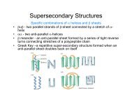
III. Super-secondary structure<br />
Secondary structure elements are observed to combine in specific<br />
geometric arrangements known as motifs or super-secondary structures.<br />
Note that the (i+2) residue of the type II turn lies in a region of the Ramachandran plot<br />
which can only be occupied by glycine. From the diagram of this turn it can be seen<br />
that were the (i+2) residue to have a side chain, there would be steric hindrance with<br />
the carbonyl oxygen of the preceding residue. Hence, the (i+2) residue of type II<br />
reverse turns is nearly always glycine.<br />
3.1 β-hairpins<br />
β-hairpins are one of the simplest super-secondary structures and are widespread in<br />
globular proteins. It connects two anti-parallel β-strands.<br />
Two-residue β-hairpins<br />
The number of amino acid residues in β-hairpins varies from 2-7 aa.<br />
Type I'.The first residue in this turn adopts the left-handed α-helical conformation and<br />
therefore shows preference for glycine, asparagine or aspartate. The second residue of a<br />
type I' turn is nearly always glycine as the required Φ and Ψ angles are well outside the<br />
allowed regions of the Ramachandran plot for amino acids with side chains.<br />
Type II'. The first residue of these turns has a conformation which can only be adopted by<br />
glycine (see Ramachandran plot). The second residue shows a preference for polar amino<br />
acids such as serine and threonine.
Three-residue β-hairpins<br />
Four-residue β-hairpins<br />
Normally the residues at the ends of the two β-strands only make one hydrogen<br />
bond as shown below. The intervening three residues have distinct<br />
conformational preferences as shown in the Ramachandran plot. The first residue<br />
adopts the right-handed α-helical conformation and the second amino acid lies in<br />
the bridging region between between α-helix and β-sheet. Glycine, asparagine or<br />
aspartate are frequently found at the last residue position as this adopts Φ and Ψ<br />
angles close to the left-handed helical conformation.<br />
These are also quite common with the first two residues adopting the α-helical<br />
conformation. The third residue has Φ and Ψ angles which lie in the bridging region<br />
between between α-helix and β-sheet and the final residue adopts the left-handed α-<br />
helical conformation and is therefore usually glycine, aspartate or asparagine.<br />
Longer loop β-hairpins<br />
3.2, β-corners<br />
For these, a wide range of conformations is observed and the general term 'random coil'<br />
is sometimes used. Consecutive anti-parallel β-strands when linked by hairpins form a<br />
super-secondary structure known as the β-meander.<br />
β-strands have a slight right-handed twist such that when they pack side-by-side to<br />
form a β-sheet, the sheet has an overall left-handed curvature. Anti-parallel β-strands<br />
forming a β-hairpin can accommodate a 90 degree change in direction known as a β-<br />
corner. The strand on the inside of the bend often has a glycine at this position while<br />
the other strand can have a β-bulge. The latter involves a single residue in the righthanded<br />
α-helical conformation which breaks the hydrogen bonding pattern of the β-<br />
sheet. This residue can also be in the left-handed helical or bridging regions of the<br />
Ramachandran plot.<br />
3.3 Helix hairpins<br />
3.4 The α-α corner<br />
•The shortest α-helical connections<br />
involve two residues which are<br />
oriented approximately perpendicular<br />
to the axes of the helices. Analysis of<br />
known structures reveals that the first<br />
of these two residues adopts Φ and Ψ<br />
αngles in the bridging or α-helical<br />
regions of the Ramachandran plot.<br />
The second residue is always glycine.<br />
•Three residue loops are also<br />
observed to have conformational<br />
preferences. The first residue<br />
occupies the bridging region of the<br />
Ramachandran plot, the second<br />
adopts the left-handed helical<br />
conformation and the last residue is<br />
in a β-strand conformation.<br />
•Longer loops have more<br />
conformational freedoms.<br />
Short loop regions connecting helices which are roughly perpendicular to one<br />
another are refered to as αα−corners. Efimov has shown that the shortest αα-corner<br />
has its first residue in the left-handed α-helical conformation and the next two<br />
residues in β-strand conformations. This conformation can only be adopted when the<br />
two helices form a right-handed corner. Indeed, if the helices were linked to form a<br />
left-handed corner there would be steric hindrance. This may explain the scarcity of<br />
left-handed αα-corners in protein X-ray structures.
3.5 Helix-turn-helix (EF-hand)<br />
3.6 β-α-β motifs<br />
Anti-parallel β-strands can be linked by short lengths of polypeptide forming β-<br />
hairpin structures. In contrast, parallel β-strands are connected by longer regions of<br />
chain which cross the β-sheet and frequently contain α-helical segments. This motif<br />
is called the β-α-β motif and is found in most proteins that have a parallel β-sheet.<br />
The loop regions linking the strands to the helical segments can vary greatly in<br />
length. The helix axis is roughly parallel with the β-strands and all three elements of<br />
secondary structure interact forming a hydrophobic core<br />
EF-hands are made up from a loop of around 12 residues which has polar and<br />
hydrophobic amino acids at conserved positions. These are crucial for ligating the metal<br />
ion and forming a stable hydrophobic core. Glycine is invariant at the sixth position in<br />
the loop for structural reasons. The calcium ion is octahedrally coordinated by carboxyl<br />
side chains, main chain groups and bound solvent.<br />
(A typical EF-hand sequence: “D-X-D-X-D/S-G-X-X-D/E -X-X-E”)<br />
<strong>Protein</strong> <strong>Structure</strong><br />
The side chain atoms of amino acids are named in the Greek alphabet<br />
according to this scheme<br />
IV. Sidechain Conformation<br />
The side chain torsion angles are named χ 1 (chi1), χ 2 (chi2), χ 3 (chi3), etc.,<br />
as shown below for lysine<br />
The χ 1 angle is subject to certain restrictions which arise from steric<br />
hindrance between the γ side chain atom(s) and the main chain. The<br />
different conformations of the side chain as a function of χ 1 are referred<br />
to as gauche(+), trans and gauche(-). These are indicated in the diagrams<br />
below in which the amino acid is viewed along the Cβ-Cα bond.<br />
Least stable: steric<br />
hindrance between<br />
Cg and N/CO
some conformations that can be adopted by Arginines<br />
<strong>Protein</strong> <strong>Structure</strong><br />
V. Tertiary <strong>Protein</strong> <strong>Structure</strong> and folds<br />
Tertiary structure describes the folding of the polypeptide chain to assemble the<br />
different secondary structure elements in a particular arrangement.<br />
5.1 All-α topologies<br />
5.1.1 The lone helix<br />
5.1.2 The helix-turn-helix motif<br />
The simplest packing arrangement of a domain of two helices is for them to lie<br />
antiparallel, connected by a short loop.<br />
There are a number of examples of small<br />
proteins (or peptides) which consist of little<br />
more than a single helix. A striking<br />
example is alamethicin, a transmembrane<br />
voltage gated ion channel, acting as a<br />
peptide antibiotic.<br />
The structure of the small (63 residue) RNA-binding protein Rop , which is found in<br />
certain plasmids (small circular molecules of double-stranded DNA occurring in<br />
bacteria and yeast) and involved in their replication.<br />
5.1.3 The four-helix bundle myohemeyrthrin<br />
ferritin<br />
cytochrome c'<br />
A number of cytokines consist of four α-helices in a bundle. Here is a diagram of<br />
Interleukin-2, human Growth Hormone, Granulocyte-macrophage colony-stimulating<br />
factor (GM-CSF) and Interleukin-4.
5.1.4 α domains which bind DNA<br />
5.1.5 Other distinctive all-α proteins<br />
the crystal structure of engrailed<br />
homeodomain binding to DNA<br />
the structure of the cro<br />
repressor from phage 434<br />
Annexin V<br />
Calmodulin in complex with a<br />
target peptide<br />
5.2 All-β topologies<br />
5.2.1 β sandwiches and β barrels<br />
<strong>Protein</strong> folds which consist of almost entirely β sheets exhibit a completely or<br />
mostly antiparallel arrangement. Many of these anti-parallel domains consist of<br />
two sheets packed against each other, with hydrophobic side chains forming the<br />
interface. Bearing in mind that side chains of a β-strand point alternately to<br />
opposite sides of a sheet, this means that such structures will tend to have a<br />
sequence of alternating hydrophobic and polar residues.<br />
5.2.1.1 Aligned and orthogonal β sandwiches<br />
orthogonal β sandwiches<br />
5.2.1.2 β barrels<br />
5.2.2.1 The Greek Key topology<br />
Side view Top view Top view showing the<br />
hydrophobic core inside<br />
the barrel<br />
The Greek Key topology, named after a pattern that was common on Greek pottery, is<br />
shown below. Three up-and-down b-strands connected by hairpins are followed by a<br />
longer connection to the fourth strand, which lies adjacent to the first.
5.3 α/β topologies<br />
5.3.1 α/β horseshoe<br />
The most regular and common domain<br />
structures consist of repeating β-α-β<br />
supersecondary units, such that the outer<br />
layer of the structure is composed of α<br />
helices packing against a central core of<br />
parallel β-sheets. These folds are called α/β,<br />
or wound α β.<br />
The β-α-β-α-β subunit, often present in<br />
nucleotide-binding proteins, is named the<br />
Rossman Fold<br />
The structure of the remarkable placental ribonuclease inhibitor (Kobe, B. & Deisenhofer, J.<br />
(1993 ) Nature V.366, 751) takes the concept of the repeating α/β unit to extremes. It is a<br />
cytosolic protein that binds extremely strongly to any ribonuclease that may leak into the<br />
cytosol. Look at the image below and you will see the 17-stranded parallel β sheet curved into<br />
an open horseshoe shape, with 16 α-helices packed against the outer surface. It doesn't form<br />
a barrel although it looks as though it should. The strands are only very slightly slanted, being<br />
nearly parallel to the central `axis'.<br />
5.3.2 α/β barrels<br />
5.3.3 Alpha+Beta Topologies<br />
This is where we collect together all those folds which include significant alpha and<br />
beta secondary structural elements<br />
•SH2 domains<br />
•<strong>Protein</strong> G (prokaryotic<br />
Ig-binding) in blue<br />
Consider a sequence of eight β-α motifs: If the first strand hydrogen bonds to<br />
the last, then the structure closes on itself forming a barrel-like structure. This<br />
is shown in the picture of triose phosphate isomerase.<br />
•Carbonic anhydrase<br />
•Thymidylate synthase<br />
5.4 Small disulphide-rich folds<br />
The members of these families contain a large number of disulphide bonds<br />
which stabilise the fold.<br />
<strong>Protein</strong> <strong>Structure</strong><br />
•EGF-like domain<br />
•Complement C-<br />
module domain<br />
VI. Quaternary <strong>Structure</strong><br />
The quaternary structure is that level of form in which units of tertiary structure<br />
aggregate to form homo- or hetero- multimers. This is found to be remarkably<br />
common, especially in the case of enzymes.<br />
•Kringle domain<br />
•Wheat Plant Toxin; Naja (Cobra) neurotoxin;<br />
green Mamba anticholinesterase.
6.1 Hetero-multimers: different tertiary domains aggregating together to<br />
form a unit<br />
6.2 Homo-multimers: It is more common to find copies of the same<br />
tertiary domain associating non-covalently.<br />
6.3 Larger <strong>Structure</strong>s<br />
The molecular machinery of the cell and indeed of assemblies of cells, rely<br />
on components made from multimeric assemblies of proteins, nucleic<br />
acids, and sugars. A few examples include :-<br />
•Viruses<br />
•Microtubules<br />
•Flagellae<br />
•Fibres of various sorts<br />
•Ribosomes<br />
•Histones<br />
•Gap Junctions