

Westerdykella reniformis sp. nov., producing the ... - IMA Fungus
Westerdykella reniformis sp. nov., producing the ... - IMA Fungus
Westerdykella reniformis sp. nov., producing the ... - IMA Fungus

Create successful ePaper yourself
Turn your PDF publications into a flip-book with our unique Google optimized e-Paper software.
doi:10.5598/imafungus.2012.03.02.11<br />
<strong>IMA</strong> <strong>Fungus</strong> · volume 3 · no 2: 189–201<br />
<strong>Westerdykella</strong> <strong>reniformis</strong> <strong>sp</strong>. <strong>nov</strong>., <strong>producing</strong> <strong>the</strong> antibiotic metabolites<br />
melinacidin IV and chetracin B<br />
Ghada A. Ebead 1 , David P. Overy 2,3 , Fabrice Berrué 2,3 , and Russell G. Kerr 1,2,3<br />
1<br />
Department of Biomedical Sciences, Atlantic Veterinary College, University of Prince Edward Island, 550 University Ave., Charlottetown, PEI,<br />
Canada, C1A 4P3; corre<strong>sp</strong>onding author e-mail: rkerr@upei.ca<br />
2<br />
Department of Chemistry, University of Prince Edward Island, 550 University Ave., Charlottetown, PEI, Canada, C1A 4P3<br />
3<br />
Nautilus Biosciences Canada, Duffy Research Center (NRC-INH), 550 University Ave., Charlottetown, PEI, Canada, C1A 4P3<br />
ARTICLE<br />
Abstract: <strong>Westerdykella</strong> <strong>reniformis</strong> Ebead & Overy <strong>sp</strong>. <strong>nov</strong>. is described based on morphology and phylogenetic<br />
analyses using ITS, nLSU rDNA, and β-tubulin gene sequences. <strong>Westerdykella</strong> <strong>reniformis</strong> is characterized by<br />
<strong>the</strong> production of cleisto<strong>the</strong>cioid ascomata, containing small globose to subglobose asci with 32, aseptate, dark<br />
colored, pronouncedly reniform asco<strong>sp</strong>ores having a concave central groove. The isolate was obtained from<br />
a red alga (Polysiphonia <strong>sp</strong>.) collected from <strong>the</strong> tidal zone in Canada at low tide. Organic extracts enriched<br />
in extrolites, obtained from fermentation on a rice-based media, inhibited <strong>the</strong> growth of methicillin-resistant<br />
Staphylococcus aureus (MRSA), vancomycin-resistant Enterococcus faecium (VRE), S. warneri, and Proteus<br />
vulgaris. Presented here is <strong>the</strong> identification of <strong>the</strong> compounds re<strong>sp</strong>onsible for <strong>the</strong> observed antimicrobial activity,<br />
<strong>the</strong> taxonomic description of W. <strong>reniformis</strong>, and a dichotomous key to <strong>the</strong> known <strong>sp</strong>ecies of <strong>Westerdykella</strong> based<br />
on macro- and micromorphological characters.<br />
Key words:<br />
antimicrobial screening<br />
Ascomycota<br />
ITS phylogeny<br />
multigene phylogeny<br />
Sporormiaceae<br />
Article info: Submitted: 15 September 2012; Accepted: 5 December 2012; Published: 11 December 2012.<br />
INTRODUCTION<br />
While screening organic solvent extracts of isolates of<br />
algicolous fungi obtained from Prince Edward Island<br />
(Canada) for antimicrobial activity, we found several strains<br />
with unique ITS rDNA gene sequences and associated<br />
extracts having antibiotic activity. Of particular interest was<br />
an isolate that was phylogenetically related to <strong>the</strong> genus<br />
<strong>Westerdykella</strong> within <strong>the</strong> family Sporormiaceae. Taxa<br />
in Sporormiaceae occur worldwide, e<strong>sp</strong>ecially on dung,<br />
but also as endophytes and as soil saprobes. The family<br />
currently comprises seven genera representing around<br />
100 <strong>sp</strong>ecies: Chaetopreussia, Pleophragmia, Preussia,<br />
Sporormia, Sporormiella, Spororminula, and <strong>Westerdykella</strong><br />
(Kruys et al. 2006, Lumbsch & Huhndorf 2007, Kruys &<br />
Wedin 2009).<br />
The genus <strong>Westerdykella</strong>, first described by Stolk in 1955,<br />
was named after Johanna Westerdijk, <strong>the</strong> founding director<br />
of what is now <strong>the</strong> KNAW-CBS Fungal Biodiversity Centre<br />
in Utrecht, The Ne<strong>the</strong>rlands (Stolk 1955). <strong>Westerdykella</strong><br />
<strong>sp</strong>ecies occur worldwide on a variety of substrates including<br />
soil, mud, dung, and plant material (Clum 1955, Ito & Nakagiri<br />
1995, Stolk 1955, Cain 1961, Rai & Tewari 1962, Malloch<br />
& Cain 1972). Kruys & Wedin (2009) retypified <strong>the</strong> genus,<br />
and distinguished it from o<strong>the</strong>r genera in <strong>the</strong> family by <strong>the</strong><br />
production of cleisto<strong>the</strong>cioid ascomata containing small asci<br />
(< 50 µm tall) with a short or almost absent stipe, encasing<br />
one-celled asco<strong>sp</strong>ores without germ slits.<br />
Species delineation within <strong>the</strong> genus historically has been<br />
based primarily on asci and asco<strong>sp</strong>ore shape. Originating<br />
with <strong>the</strong> description of <strong>the</strong> ex-type strain, W. ornata<br />
(Stolk 1955), to date nine <strong>sp</strong>ecies have been described<br />
within<strong>Westerdykella</strong>: W. ornata, W. angulata, W. aurantiaca,<br />
W. cylindrica, W. di<strong>sp</strong>ersa, W. globosa, W. multi<strong>sp</strong>ora, W.<br />
nigra, and W. purpurea. Over time and through various<br />
taxonomic revisions, several <strong>sp</strong>ecies of <strong>the</strong> genera Preussia,<br />
Pycnidiophora, and Eremodothis have been reclassified in<br />
<strong>Westerdykella</strong>. Pycnidiophora multi<strong>sp</strong>ora was <strong>the</strong> first taxon<br />
to be transferred into <strong>the</strong> genus by Cejp & Milko (1964).<br />
Subsequently, Arx (1973) reclassified Preussia cylindrica<br />
in <strong>the</strong> genus due to <strong>the</strong> production of cylindrical, larger<br />
asco<strong>sp</strong>ores and <strong>the</strong> presentation of an asexual Phomalike<br />
state, and also P. nigra due to <strong>the</strong> production of short<br />
cylindrical asci ellipsoidal asco<strong>sp</strong>ores, and <strong>the</strong> absence of<br />
a conidial state. Subsequently, Preussia purpurea was also<br />
transferred to <strong>the</strong> genus by Arx (1975) due to <strong>the</strong> production<br />
of an orange pigment in culture, non-ostiolate ascomata<br />
with often a central columnar body and asco<strong>sp</strong>ores without<br />
germ pores. Ito & Nakagiri (1995) added P. globosa on <strong>the</strong><br />
basis of <strong>the</strong> production of asci containing 32 asco<strong>sp</strong>ores,<br />
each having a single semicircular <strong>sp</strong>iral ridge on <strong>the</strong><br />
<strong>sp</strong>ore surface, and so conforming to <strong>the</strong> generic concept<br />
© 2012 International Mycological Association<br />
You are free to share - to copy, distribute and transmit <strong>the</strong> work, under <strong>the</strong> following conditions:<br />
Attribution:<br />
You must attribute <strong>the</strong> work in <strong>the</strong> manner <strong>sp</strong>ecified by <strong>the</strong> author or licensor (but not in any way that suggests that <strong>the</strong>y endorse you or your use of <strong>the</strong> work).<br />
Non-commercial: You may not use this work for commercial purposes.<br />
No derivative works: You may not alter, transform, or build upon this work.<br />
For any reuse or distribution, you must make clear to o<strong>the</strong>rs <strong>the</strong> license terms of this work, which can be found at http://creativecommons.org/licenses/by-nc-nd/3.0/legalcode. Any of <strong>the</strong> above conditions can be waived if you get<br />
permission from <strong>the</strong> copyright holder. Nothing in this license impairs or restricts <strong>the</strong> author’s moral rights.<br />
volume 3 · no. 2 189
Ebead et al.<br />
ARTICLE<br />
of <strong>Westerdykella</strong> as described by Stolk (1955). From a<br />
multilocus phylogenetic study based of ITS and nLSU rDNA,<br />
mtSSU and β-tubilin gene sequences, Kruys & Wedin (2009)<br />
reclassified Pycnidiophora di<strong>sp</strong>ersa and P. aurantiaca in <strong>the</strong><br />
genus. Fur<strong>the</strong>rmore, Eremodothis angulata was found to be<br />
phylogenetically related to <strong>Westerdykella</strong>, de<strong>sp</strong>ite <strong>producing</strong><br />
eight pyramidal star-shaped asco<strong>sp</strong>ores per ascus compared<br />
to <strong>the</strong> typical 32 asco<strong>sp</strong>ores of o<strong>the</strong>r <strong>Westerdykella</strong> <strong>sp</strong>ecies,<br />
and was <strong>the</strong>refore also reclassified within it.<br />
In this study, a unique <strong>Westerdykella</strong> isolate from<br />
algae collected in <strong>the</strong> littoral zone was evaluated for<br />
morphological similarity to o<strong>the</strong>r taxa of <strong>the</strong> genus and<br />
for phylogenetic relatedness within Sporormiaceae. Multigene<br />
phylogenies were constructed using data sets of<br />
ITS and nLSU rDNA and β-tubulin gene sequences. Two<br />
extrolites were re<strong>sp</strong>onsible for <strong>the</strong> observed antibiotic<br />
activity and <strong>the</strong>se metabolites were isolated and identified<br />
from organic extracts of rice-based medium fermentations<br />
of <strong>the</strong> isolate. Presented here is <strong>the</strong> taxonomic description<br />
of <strong>the</strong> <strong>nov</strong>el <strong>Westerdykella</strong> <strong>sp</strong>ecies, W. <strong>reniformis</strong> <strong>sp</strong>. <strong>nov</strong>.,<br />
a dichotomous key to <strong>the</strong> known <strong>sp</strong>ecies of <strong>the</strong> genus based<br />
on macro- and micromorphological characteristics, and<br />
<strong>the</strong> isolation and identification of <strong>the</strong> antibiotic secondary<br />
metabolites melinacidin IV and chetracin B, production<br />
which is particular feature of W. <strong>reniformis</strong>.<br />
MATERIALS AND METHODS<br />
Sample collection<br />
Algal material (Polysiphonia <strong>sp</strong>.) was collected by G.A.E.<br />
from <strong>the</strong> shoreline at Point Prim, Prince Edward Island<br />
(46’04”N, 62’59”W) at low tide on 4 June 2009. Immediately<br />
after removal from <strong>the</strong> sea, <strong>the</strong> algal sample was deposited<br />
in a sterile plastic bag and seawater was added. The sample<br />
was kept cold until arrival at <strong>the</strong> laboratory where it was<br />
maintained at 4 °C until <strong>the</strong> following day. Before being<br />
processed, <strong>the</strong> sample was shaken three times with sterile<br />
seawater in order to wash <strong>the</strong> surface free of any adhering<br />
particulate material.<br />
Sample plating and fungal isolation<br />
The sample was homogenized in sterile seawater and<br />
<strong>the</strong> resulting homogenate was plated on a 9 cm Petri dish<br />
containing YM media (Yeast extract Malt agar; 2 g yeast<br />
extract, 10 g malt extract, 10 g glucose, 20 g agar, 50 mg<br />
chloramphenicol, 18 g Instant Ocean in 1 L Millipore H 2<br />
O)<br />
and in<strong>sp</strong>ected daily for fungal growth. The plates were<br />
incubated at 22 °C for 5 d and <strong>the</strong>n examined under <strong>the</strong><br />
dissecting microscope. Emerging fungal colonies were<br />
transferred via a flame-sterilized needle to ano<strong>the</strong>r Petri dish<br />
containing YM. After obtaining a pure isolate, seed inoculum<br />
was prepared by excising cubes (1–3 mm 3 ) from an actively<br />
growing culture into 15 mL of yeast extract-maltose medium<br />
(10 g peptone, 40 g maltose, 10 g yeast extract, 18 g Instant<br />
Ocean and 1 g agar in 1 L Millipore H 2<br />
O) in a 50 mL test tube<br />
and incubated at 22 °C, 200 rpm for 5 d, after which 500 µL of<br />
mycelial su<strong>sp</strong>ension was removed for DNA extraction and <strong>the</strong><br />
remainder reserved to inoculate fermentations.<br />
Culture characteristics and morphology<br />
For morphological and molecular comparisons, six<br />
<strong>Westerdykella</strong> isolates were obtained from CBS: W. cylindrica<br />
CBS 454.72, W. di<strong>sp</strong>ersa CBS 297.56, W. multi<strong>sp</strong>ora CBS<br />
391.51, W. nigra CBS 416.72, W. ornata CBS 379.55, and W.<br />
rapa-nuiensis ined. CBS 604.97. For macro-morphological<br />
comparisons, fungi were grown on OA (Oatmeal Agar; 30 g<br />
oatmeal, 15 g agar in 1 L Millipore H 2<br />
O), Mannitol Soya agar<br />
(20 g mannitol, 20 g soya flour, 20 g agar in 1L Millipore H 2<br />
O)<br />
and Rice agar (75 g brown rice, 20 g agar in 1 L Millipore<br />
H 2<br />
O) at 22 °C and <strong>the</strong>ir growth rates were measured and<br />
colonies were evaluated after 7 and 14 d of incubation.<br />
Colour descriptions were qualified using Kornerup &<br />
Wanscher (1978). Measurements were repeated twice. For<br />
micro-morphological measurements and photographs, fungal<br />
structures from 26 d-old cultures were mounted on glass<br />
slides in lactic acid; photographs were taken while viewing<br />
using ei<strong>the</strong>r bright field or phase contrast microscopy. For<br />
measurements, a Leica DME light microscope with phase<br />
contrast optics accompanied by a Leica EC3 camera (Leica<br />
Microsystems, Switzerland), was used at 100× magnification<br />
and a total of 25 asco<strong>sp</strong>ores and 25 asci were measured<br />
from crushed mounts and <strong>the</strong> dimension range (minimum and<br />
maximum) and average were determined (measurements<br />
were adjusted to <strong>the</strong> nearest 0.5 microns to avoid false<br />
impression of accuracy). Bright-field photomicrographs were<br />
obtained with a Carl Zeiss microscope, Axio Imager A1m<br />
model with a HRc Axiocam digital camera and AxioVision v.<br />
3.1 software (Carl Zeiss, Heerbrugg, Switzerland).<br />
Salt tolerance testing<br />
Isolate RKGE 35 was point-inoculated onto OA media and OA<br />
media with artificial seawater (+ASW; 18 g L -1 Instant Ocean)<br />
and plates were incubated at 22 °C. Radial growth rates and<br />
colony features were noted after 7 d and 14 d of incubation.<br />
Fermentation and extraction<br />
Strains were fermented on a rice-based medium (10 g brown<br />
rice; 50 mL YNB (6.7 g YNB + 5 g sucrose in 1 L Millipore<br />
H 2<br />
O)) in 250 ml Erlenmeyer flasks. The brown rice medium<br />
was autoclaved twice for 20 min at 121 °C, first with only<br />
brown rice, which was allowed to cool before YNB was<br />
added and <strong>the</strong> mixture was autoclaved again. Flasks were<br />
inoculated with 1.5 mL of seed inoculum. An uninoculated<br />
control flask was used to in<strong>sp</strong>ect medium purity and to be<br />
used as a negative control for antimicrobial screening. All<br />
experiments were incubated under stationary conditions at<br />
22 °C for 21 d.<br />
After 21 d of incubation, fermentations were extracted by<br />
first disrupting <strong>the</strong> fungal colony using a sterile <strong>sp</strong>atula and<br />
adding 30 mL EtOAc:MeOH (1:1), followed by shaking at 50<br />
rpm for 1 h at room temperature. Organic extracts were <strong>the</strong>n<br />
vacuum-filtered through Whatman #3 filter paper and dried<br />
using a GeneVac vacuum evaporating system (model: EZ-2<br />
MK2) prior to fractionation. Extracts were fractionated on<br />
Thermo HyperSep C-18 Sep Pack columns (500 mg C-18,<br />
6 mL column volume) using a vacuum manifold by eluting<br />
with 14 mL of each of <strong>the</strong> following solvent combinations:<br />
8:2 H 2<br />
O:MeOH (fraction 1), 1:1 H 2<br />
O:MeOH (fraction 2),<br />
2:8 H 2<br />
O:MeOH (fraction 3), EtOH (fraction 4), and 1:1<br />
190 ima fUNGUS
Antibiotic <strong>producing</strong> <strong>Westerdykella</strong> <strong>reniformis</strong> <strong>sp</strong>. <strong>nov</strong>.<br />
MeOH:DCM (fraction 5). The eluent representing fractions<br />
2–5 were retained and using a GeneVac (model: EZ-2 MK2)<br />
evaporating system, weighed and submitted for antimicrobial<br />
testing, and analyzed by LC/HRMS using a Kinetex 1.7 µm<br />
C18 UPLC column (Phenomenex, 50 × 2.1 mm) and Accela<br />
Thermo equipment coupled with MS-ELSD-UV detection<br />
(Orbitrap Excactive mass <strong>sp</strong>ectrometer fitted with an ESI<br />
source, PDA, and LT-ELSD Sedex 80 (Sedere)).<br />
Additional fermentation of strain RKGE 35 was carried<br />
out in 10 Erlenmeyer flasks and following <strong>the</strong> same growth<br />
conditions and extraction protocol as described above in<br />
order to obtain sufficient material to determine <strong>the</strong> structural<br />
identity of <strong>the</strong> secondary metabolites re<strong>sp</strong>onsible for <strong>the</strong><br />
observed antimicrobial activity. The resulting EtOAc:MeOH<br />
extract was partitioned between EtOAc:H 2<br />
O (1:1) and<br />
<strong>the</strong> organic layer (EtOAc) was dried under vacuum. The<br />
resulting gum was resu<strong>sp</strong>ended in a biphasic solvent mixture<br />
of Hexane:MeOH:H 2<br />
O (6:7:2) and <strong>the</strong> H 2<br />
O:MeOH layer<br />
after evaporation was subjected to a flash chromatography<br />
using bulk C-18 to yield to five fractions: 9:1 H 2<br />
O:MeOH,<br />
1:1 H 2<br />
O:MeOH, 2:8 H 2<br />
O:MeOH, EtOH, acetone, and 1:1<br />
MeOH:DCM. The 2:8 H 2<br />
O:MeOH fraction was fur<strong>the</strong>r<br />
fractionated on normal phase silica by using automated<br />
medium pressure chromatography system (Combiflash<br />
Rf200 (Teledyne Isco)) to yield to 15 fractions. Fractions<br />
3–5 were purified by semi-preparative normal phase HPLC<br />
(Phenomenex Luna Silica, 250 × 10 mm, 5 μm) with isocratic<br />
conditions using 15% CHCl 3<br />
:MeOH (9:1) in 85 % CHCl 3<br />
and a<br />
flow rate of 2.5 ml min -1 to afford melanicidin IV and chetracin<br />
B.<br />
Antimicrobial bioassay<br />
All microbroth antibiotic susceptibility testing was carried<br />
out in 96-well plates in accordance with Clinical Laboratory<br />
Standards Institute testing standards (Ferraro, 2003) using<br />
<strong>the</strong> following pathogens: methicillin-resistant Staphylococcus<br />
aureus ATCC 33591 (MRSA), S. warneri ATCC 17917,<br />
vancomycin-resistant Enterococcus faecium EF379 (VRE),<br />
Pseudomonas aeruginosa ATCC 14210, Proteus vulgaris<br />
ATCC 12454, and Candida albicans ATCC 14035. Extract<br />
fractions and pure compounds were tested in triplicate<br />
against each organism. Extract fractions were resu<strong>sp</strong>ended<br />
in sterile 20 % DMSO and assayed at 250 µg/mL with a<br />
final well volume concentration of 2 % DMSO while pure<br />
compounds were serially diluted to generate a range of<br />
twelve concentrations (128 µg mL -1 to 0.0625 µg mL -1 ) in a<br />
final well volume concentration of 2% DMSO. Each plate<br />
contained eight uninoculated positive controls (media +<br />
20 % DMSO), eight untreated negative controls (Media<br />
+ 20 % DMSO + organism), and one column containing a<br />
concentration range of a control antibiotic (vancomycin for<br />
MRSA, and S. warneri, rifampicin for VRE, gentamycin for<br />
P. aeruginosa, ciprofloxacin for P. vulgaris, or nystatin for C.<br />
albicans). The optical density of <strong>the</strong> plate was recorded using<br />
a BioTek Synergy HT plate reader at 600 nm at time zero and<br />
<strong>the</strong>n again after incubation of <strong>the</strong> plates for 22 h at 37 °C.<br />
After subtracting <strong>the</strong> time zero OD600 from <strong>the</strong> final reading,<br />
<strong>the</strong> percentages of microorganism survival relative to vehicle<br />
control wells were calculated.<br />
DNA extraction and PCR amplification<br />
Genomic DNA was obtained from all strains using <strong>the</strong> fast<br />
DNA extraction kit (FASTDNA SPIN KIT FOR SOIL®, MP<br />
Biomedicals) according to <strong>the</strong> manufacturer’s protocols.<br />
Double-stranded copies of <strong>the</strong> ITS and nLSU rRNA gene<br />
and <strong>the</strong> β-tubulin gene were obtained by polymerase chain<br />
reaction (PCR) amplifications using 50 µL of reaction mixture<br />
consisting of 25 µL of Econo Taq ® PLUS GREEN 2× Master<br />
Mix (Lucigen), 17 µL of sterile ddH 2<br />
O, 2 µL of each primer<br />
and 4 µL of genomic DNA. Reactions were run in a Biometra<br />
<strong>the</strong>rmocycler using <strong>the</strong> following settings for ITS amplicon<br />
generation: an initial denaturation step at 96 °C for 3 min,<br />
35 cycles consisting of denaturation at 96 °C for 45 s, primer<br />
annealing at 54.5 °C for 45 s and extension at 72 °C for 1<br />
min. The PCR was completed with a final extension step of<br />
10 min at 72 °C. Amplification protocols were similar for both<br />
β-tubulin and nLSU rDNA genes with <strong>the</strong> exception of <strong>the</strong><br />
employed annealing temperatures: 58 °C for β-tubulin and for<br />
50 °C nLSU. Primers used for <strong>the</strong> ITS rDNA gene were ITS-<br />
1 and ITS-4 (White et al. 1990), for <strong>the</strong> β-tubulin gene were<br />
BT1819R and BT2916 (Miller & Hundorf 2005); for <strong>the</strong> nLSU<br />
rDNA gene were LROR and LR7 (Vilglys & Hester 1990,<br />
Rehner & Samuels 1994). PCR amplicons were checked for<br />
correct length and concentration by electrophoresis in 1 %<br />
agarose gel in 1× TAE buffer (Tris Base 2.42 g, glacial acetic<br />
acid 0.572 lL, 0.5 M EDTA 1 mL; add ddH 2<br />
O to 500 mL.<br />
DNA sequencing and sequence alignment<br />
The ITS, nLSU, and β-tubulin amplicons were sent to a<br />
commercial sequencing facility (Eurofins MWG Biotech) and<br />
sequenced on a 3730xl DNA Analyzer coupled with BigDye<br />
Terminator v. 3.1 Cycle Sequencing reagents, Applied<br />
Biosystems (ABI). The generated sequences were compared<br />
with o<strong>the</strong>r fungal DNA sequences from NCBI’s GenBank<br />
sequence database using a Blastn search algorithm.<br />
Phylogenetic analysis of <strong>the</strong> ITS rDNA gene were performed<br />
using <strong>the</strong> software Molecular Evolutionary Genetics Analysis<br />
v. 5 (MEGA5) (Tamura et al. 2011). Sequence data generated<br />
in this study were aligned with additional sequences of<br />
representative <strong>Westerdykella</strong> <strong>sp</strong>p. as well as several<br />
isolates belonging to Sporormiaceae and o<strong>the</strong>r Pleo<strong>sp</strong>orales<br />
available in GenBank (Table 1). In total, 34 sequences were<br />
aligned using <strong>the</strong> ClustalW algorithm, with a DNA Gap Open<br />
Penalty = 15.0, DNA Gap Extension Penalty = 6.66 and a<br />
delay divergent cutoff of 30 %. For result optimization, <strong>the</strong><br />
alignments were refined by manual correction when needed.<br />
The evolutionary history was inferred using <strong>the</strong> neighborjoining<br />
method employing <strong>the</strong> maximum composite likelihood<br />
model using pairwise deletion and <strong>the</strong> clade stability was<br />
evaluated using <strong>the</strong> bootstrap method (n = 2000 bootstrap<br />
replications). Novel sequences were accessioned in<br />
GenBank under accession numbers JX235699–JX235707.<br />
A multigene phylogeny was constructed using 23 isolates<br />
(Table 1). Relevant sequence data were downloaded from<br />
GenBank and used to construct aligned and trimmed ITS, nLSU<br />
and β-tubulin data matrices in MEGA5. A Bayesian analysis<br />
was performed using MrBayes 3.2 (Ronquist et al. 2012) with<br />
<strong>the</strong> following settings: nst = 6, <strong>the</strong>refore using GTR (General<br />
Time Reversible) model; rates = invgama, setting acrosssite<br />
rate variation for gamma distribution with a proportion of<br />
ARTICLE<br />
volume 3 · no. 2<br />
191
Ebead et al.<br />
ARTICLE<br />
Table 1. Sequences included in this study, newly generated sequences are highlighted in bold.<br />
Species Isolate Origin GenBank accession no.<br />
ITS 28S β-tubulin<br />
Herpotrichia juniper CBS 468.64 Switzerland, Pinus mugo GQ203759 DQ384093 GQ203681<br />
Pleo<strong>sp</strong>ora herbarum ATCC 11681 USA, onion leaf AF229479 AF382386 AY749032<br />
Preussia australis Lundqvist 20884-a France, rabbit dung GQ203773 GQ203732 GQ203695<br />
P. funiculata Huhndorf 2577 USA, porcupine dung GQ203762 GQ203722 GQ203685<br />
P. isomera CBS 388.78 Venezuela, cow dung GQ203763 GQ203723 GQ203686<br />
P. lignicola CBS 363.69 Ne<strong>the</strong>rlands, rabbit dung GQ203783 DQ384098 GQ203703<br />
P. tenerifae CBS 354.86 Tenerife, rabbit dung GQ203794 GQ203752 GQ203713<br />
P. terricola CBS 317.65 Honduras, Musa sapientum GQ203765 GQ203725 GQ203688<br />
P. terricola CBS 527.84 Tanzania, elephant dung GQ203764 GQ203724 GQ203687<br />
P. typharum CBS 107.69 Japan, deer dung GQ203766 GQ203726 GQ203689<br />
P. vulgaris Strid 18884 Sweden, hare dung GQ203767 GQ203727 GQ203690<br />
Sporormia fimetaria Lundqvist 2302-c Sweden, cow dung GQ203768 GQ203728 GQ203691<br />
Sporomiella affinis Lundqvist 17739-j Denmark, rabbit dung GQ203770 – –<br />
S. dakotensis Thulin 2570-g Ethiopia, cow dung GQ203776 – –<br />
S. heptamera Lundqvist 3090-b Sweden, horse dung GQ203778 – –<br />
S. irregularis Lundqvist 16568-f Hungary, cow dung GQ203780 GQ203739 GQ203700<br />
S. leporina Lundqvist 19873-a Sweden, hare dung GQ203781 – –<br />
S. pulchella Richardson MJR67/01 USA, dung GQ203789 – –<br />
S. vexans UME23 Sweden, moose dung GQ203793 – –<br />
Tremato<strong>sp</strong>haeria hetero<strong>sp</strong>ora CBS 644.86 Switzerland, Iris <strong>sp</strong>. GQ203795 AY016369 GQ203714<br />
<strong>Westerdykella</strong> angulata IMI 090323 India, rice-field soil GQ203758 GQ203720 GQ203680<br />
W. angulata CBS 610.74 India, rice-field soil GQ203757 – –<br />
W. aurantiaca IMI 086825 India, mud AY943057 – –<br />
W. aurantiaca FNBR-03 India, soil JN118571 – –<br />
W. cylindrica ATCC 24077 = CBS 454.72 Kenya, cow dung AY943056 AY004343 JX235707<br />
W. di<strong>sp</strong>ersa CBS 297.56 Virginia, damp seedlings GQ203797.1 GQ203753.1 GQ203716.1<br />
W. di<strong>sp</strong>ersa CBS 156.67 Nigeria, soil DQ468016 – –<br />
W. di<strong>sp</strong>ersa CBS 508.75 Armenia, salt-marsh soil GQ203798 – –<br />
W. di<strong>sp</strong>ersa CBS 712.71 The Ne<strong>the</strong>rlands, greenhouse DQ468031 – –<br />
soil<br />
W. globosa IFO 32588 India, soil AY943046 – –<br />
W. multi<strong>sp</strong>ora CBS 383.69 France, saline soil GQ203799 GQ203754 GQ203717<br />
W. multi<strong>sp</strong>ora CBS 391.51 Japan AY943048 – –<br />
W. nigra CBS 416.72 Pakistan, soil GQ203800 GQ203755 GQ203718<br />
W. nigra ATCC 12756 AY943049 – –<br />
W. ornata CBS 379.55 Mozambique, mangrove mud GQ203801 AY853401 GQ203719<br />
W. purpurea CBS 297.75 Togo, sandy soil AY943050 – –<br />
W. purpurea HN6-5B China, mangrove FJ624258 – –<br />
W. rapa-nuiensis ined. CBS 604.97 Chile, soil JX235699 JX235703 JX235705<br />
W. <strong>reniformis</strong> RKGE35 = DAOM 242243 Canada, red algae JX235700 JX235704 JX235706<br />
Verruculina enalia CBS 304.66 Liberia, drift wood GQ203796 AY016363 GQ203715<br />
invariant sites; MCMC heated chain set with nchains = 4 and<br />
temp = 0.2, ngen = 500 000, samplefreq = 100, sumt burnin =<br />
1250; <strong>the</strong> analysis was continued for 500 000 generations in<br />
order obtain an average standard deviation of <strong>sp</strong>lit frequencies<br />
below 0.01. The first 25 % of sampled trees were discarded<br />
as burn-in. Resulting trees were viewed in FigTree v. 1.3.1.<br />
Sequence alignments and trees presented were deposited in<br />
TreeBASE (accession number 13676).<br />
RESULTS<br />
Sequencing analysis<br />
In order to verify <strong>the</strong> taxonomic placement of isolate<br />
RKGE 35, gDNA was extracted and amplified by PCR<br />
for different genes resulting in sequence lengths of 472,<br />
1295 and 935 nucleotides for <strong>the</strong> ITS rDNA, nLSU rDNA,<br />
and β-tubulin genes re<strong>sp</strong>ectively. The Blastn search for<br />
192 ima fUNGUS
Antibiotic <strong>producing</strong> <strong>Westerdykella</strong> <strong>reniformis</strong> <strong>sp</strong>. <strong>nov</strong>.<br />
<strong>Westerdykella</strong> di<strong>sp</strong>ersa CBS 297.56<br />
<strong>Westerdykella</strong> di<strong>sp</strong>ersa CBS 156.67<br />
<strong>Westerdykella</strong> multi<strong>sp</strong>ora CBS 391.51<br />
65<br />
<strong>Westerdykella</strong> di<strong>sp</strong>ersa CBS 712.71<br />
98<br />
<strong>Westerdykella</strong> rapa-nuiensis ined. CBS 604.97<br />
74 <strong>Westerdykella</strong> di<strong>sp</strong>ersa CBS 508.75<br />
99 <strong>Westerdykella</strong> aurantiaca FNBR-03<br />
<strong>Westerdykella</strong> aurantiaca IMI 086825<br />
<strong>Westerdykella</strong> multi<strong>sp</strong>ora CBS 383.69<br />
100 <strong>Westerdykella</strong> nigra CBS 416.72<br />
<strong>Westerdykella</strong> nigra ATCC 12756<br />
100 <strong>Westerdykella</strong> angulata IMI 090323<br />
<strong>Westerdykella</strong> angulata CBS 610.74<br />
98 <strong>Westerdykella</strong> purpurea CBS 297.75<br />
81<br />
<strong>Westerdykella</strong> purpurea HN6-5B<br />
<strong>Westerdykella</strong> globosa IFO 32588<br />
99<br />
<strong>Westerdykella</strong> cylindrica CBS 454.72<br />
51 <strong>Westerdykella</strong> <strong>reniformis</strong> RKGE35=DAOM 242243<br />
<strong>Westerdykella</strong> ornata CBS 379.55<br />
Sporormia fimetaria Lundqvist2302-c<br />
Sporormiella affinis Lundqvist17739-j<br />
92 Sporormiella heptamera Lundqvist3090b<br />
99<br />
Sporormiella vexans UME23<br />
Sporormiella leporina Lundqvist19873-a<br />
Sporormiella irregularis Lundqvist 16568-f<br />
Preussia isomera CBS 388.78<br />
Preussia tenerifae CBS 354.86<br />
78<br />
Sporormiella dakotensis Thulin 2570-g<br />
Sporormiella pulchella Richardson MJR93/01<br />
Preussia australis Lundqvist 20884-a<br />
Preussia lignicola CBS 363.69<br />
Preussia vulgaris Strid 18884<br />
100<br />
Preussia funiculata Huhndorf 2577<br />
76<br />
Preussia typharum CBS 107.69<br />
Tremato<strong>sp</strong>haeria hetero<strong>sp</strong>ora CBS 644.86<br />
Herpotrichia juniperi CBS 468.64<br />
Pleo<strong>sp</strong>ora herbarum ATCC 11681<br />
Verruculina enalia CBS 304.66<br />
ARTICLE<br />
59<br />
54<br />
59<br />
58<br />
96<br />
0.02<br />
Fig. 1. Bootstrap consensus tree inferred from 2000 replicates using <strong>the</strong> neighbor-joining method based on ITS rDNA sequences. The percentage<br />
of replicate trees (> 50 %) in which <strong>the</strong> associated taxa clustered toge<strong>the</strong>r in <strong>the</strong> bootstrap tests of 2000 replicates are shown next to <strong>the</strong><br />
branches. Evolutionary distances were computed using <strong>the</strong> maximum composite likelihood method and are in <strong>the</strong> units of <strong>the</strong> number of base<br />
substitutions per site. The tree was rooted with Verruculina enalia (CBS 304.66).<br />
<strong>the</strong> sequences showed that isolate RKGE 35 is classified<br />
within <strong>the</strong> genus <strong>Westerdykella</strong>. For <strong>the</strong> ITS region, <strong>the</strong><br />
closest sequence matches with 95 % maximum identity<br />
and complete coverage were to those of W. ornata CBS<br />
379.55 (AY943045.1; matching 455/477 bases with 7<br />
gaps), W. di<strong>sp</strong>ersa CBS 297.56 (AY943055.1; matching<br />
454/477 bases with 6 gaps), and W. aurantiaca IMI 08625<br />
(AY943048.1; matching 452/477 bases with 6 gaps). For<br />
<strong>the</strong> amplified nLSU region, <strong>the</strong> closest sequence matches<br />
with complete coverage were to W. angulata IMI 090323<br />
(GQ203720.1; matching 1281/1296 bases with 1 gap) with<br />
99 % maximum identity followed by W. cylindrica ATCC<br />
24077 (NG027595.1; matching 1266/1269 bases with 1<br />
gap) with 98 % maximum identity. For <strong>the</strong> β-tubulin gene, <strong>the</strong><br />
closest sequence matches with complete coverage were to<br />
W. di<strong>sp</strong>ersa CBS 297.56 (GQ203716.1; matching 876/941<br />
bases with 6 gaps) with 93 % maximum identity and W.<br />
ornata CBS 379.55 (GQ203719.1; matching 869/945 bases<br />
with 10 gaps) with 92 % maximum identity as well as with<br />
W. angulata IMI090323 (GQ203680.1; matching 368/926<br />
bases with 6 gaps) with 94 % maximum identity and only<br />
98 % coverage.<br />
Phylogenetic analyses<br />
The ITS rDNA gene was analysed to determine <strong>the</strong> relative<br />
evolutionary history of isolate RKGE 35 with multiple isolates<br />
of o<strong>the</strong>r representative <strong>Westerdykella</strong> <strong>sp</strong>p. The evolutionary<br />
history was inferred by <strong>the</strong> bootstrap consensus tree (Fig.<br />
1) constructed using <strong>the</strong> neighbour-joining method and 2000<br />
bootstrap replicates. The analysis involved 39 sequences<br />
volume 3 · no. 2<br />
193
Ebead et al.<br />
Herpotrichia juniperi CBS 468.64<br />
ARTICLE<br />
<strong>Westerdykella</strong> angulata IMI 090323<br />
64<br />
100 <strong>Westerdykella</strong> di<strong>sp</strong>ersa CBS 297.56<br />
100 <strong>Westerdykella</strong> rapa-nuiensis ined. CBS 604.97<br />
72<br />
<strong>Westerdykella</strong> multi<strong>sp</strong>ora CBS 383.69<br />
98<br />
<strong>Westerdykella</strong> ornata CBS 379.55<br />
100<br />
<strong>Westerdykella</strong> <strong>reniformis</strong> RKGE35=DAOM 242243<br />
100<br />
<strong>Westerdykella</strong> cylindrica CBS 454.72<br />
<strong>Westerdykella</strong> nigra CBS 416.72<br />
Sporormia fimetaria Lundqvist 2302-c<br />
70 Preussia funiculata Huhndorf 2577<br />
100<br />
Preussia typharum CBS 107.69<br />
Preussia vulgaris Strid 18884<br />
Preussia australis Lundqvist 20884<br />
Preussia lignicola CBS 363.69<br />
Preussia isomera CBS 388.78<br />
100 Preussia terricola CBS 527.84<br />
Preussia terricola CBS 317.65<br />
Preussia tenerifae CBS 354.86<br />
Sporormiella irregularis Lundqvist 16568-f<br />
Tremato<strong>sp</strong>haeria hetero<strong>sp</strong>ora CBS 644.86<br />
Pleo<strong>sp</strong>ora herbarum CBS 191.86<br />
Verruculina enalia CBS 304.66<br />
100<br />
52<br />
89<br />
86<br />
99<br />
85<br />
89<br />
99<br />
63<br />
64<br />
0.05<br />
Fig. 2. Consensus tree inferred from a Bayesian analysis of ITS and nLSU rDNA and β-tubulin gene sequences. Bayesian posterior probabilities<br />
are given as % values at <strong>the</strong> nodes. The tree was rooted with Verruculina enalia (CBS 304.66).<br />
and included 409 nucleotide positions in <strong>the</strong> final dataset<br />
with an overall mean distance calculated as 0.137 with a<br />
standard error of 0.013. The genus <strong>Westerdykella</strong> formed<br />
a well-supported monophyletic clade distinct from o<strong>the</strong>r<br />
members of Sporormiaceae. Within <strong>the</strong> <strong>Westerdykella</strong> clade,<br />
sequences from individual isolates formed distinct <strong>sp</strong>ecies<br />
groups with high bootstrap support; however, evolutionary<br />
relatedness of <strong>sp</strong>ecies within <strong>the</strong> genus was difficult to infer<br />
due to separation with low associated bootstrap values.<br />
Isolate RKGE 35 formed a sister clade to that of W. cylindrica,<br />
represented by isolate CBS 454.72 (= ATCC 24077). Isolate<br />
CBS 604.97, representing <strong>the</strong> unpublished <strong>sp</strong>ecies W. rapanuiensis<br />
ined., clustered toge<strong>the</strong>r within <strong>the</strong> W. di<strong>sp</strong>ersa<br />
clade along with <strong>the</strong> W. multi<strong>sp</strong>ora isolate CBS 391.51.<br />
Evolutionary history within <strong>the</strong> genus <strong>Westerdykella</strong> was<br />
also inferred by a multigene Bayesian analysis involving<br />
sequences of <strong>the</strong> ITS and nLSU rDNA and β-tubulin genes<br />
from 23 strains. The aligned dataset consisted of 417<br />
nucleotides from <strong>the</strong> ITS rDNA, 872 nucleotides from <strong>the</strong><br />
nLSU rDNA and 511 nucleotides from <strong>the</strong> β-tubulin gene<br />
sequences. Convergence was assumed as an average<br />
standard deviation of <strong>sp</strong>lit frequencies of 0.007432 was<br />
achieved following 500 000 generations. From <strong>the</strong> generated<br />
phylogenetic tree (Fig. 2), representative isolate sequences of<br />
<strong>Westerdykella</strong> <strong>sp</strong>ecies once again clustered toge<strong>the</strong>r forming<br />
a distinct clade with 100 % Bayesian posterior probability<br />
support. Isolate RKGE 35 clustered within <strong>the</strong> <strong>Westerdykella</strong><br />
clade, forming its own discrete lineage with 100 % posterior<br />
probability support. Isolate CBS 604.97 representing <strong>the</strong> not<br />
yet formally named W. rapa-nuiensis clustered toge<strong>the</strong>r and<br />
most proximal to W. di<strong>sp</strong>era (CBS 297.56).<br />
Antimicrobial metabolite identification<br />
Two successive orthogonal fractionations (reverse phase<br />
<strong>the</strong>n normal phase) of <strong>the</strong> MeOH:H 2<br />
O extract obtained after<br />
liquid-liquid partitions yielded three fractions (3–5) exhibiting<br />
strong antimicrobial activities. Chemical profiling by LC-<br />
HRMS coupled to a universal detector (ELSD) suggested<br />
that two major compounds were re<strong>sp</strong>onsible for <strong>the</strong> observed<br />
biological activities (Fig. 3). The interpretation of <strong>the</strong> HRMS<br />
data indicated <strong>the</strong> molecular formulae C 30<br />
H 28<br />
N 6<br />
O 8<br />
S 4<br />
(m/z<br />
729.09076 [M+H] + , Δ -2.2 ppm) and C 30<br />
H 28<br />
N 6<br />
O 8<br />
S 5<br />
(m/z<br />
761.06274 [M+H] + , Δ -2.3 ppm) re<strong>sp</strong>ectively and was in<br />
agreement with <strong>the</strong> observed isotopic pattern and <strong>the</strong><br />
presence of sulfur atoms. After searches in databases<br />
Antibases and SciFinder, <strong>the</strong> two prominent components<br />
with antimicrobial properties were identified as <strong>the</strong> known<br />
metabolites melinacidin IV and chetracin B (Fig. 4) which<br />
belong to <strong>the</strong> important class of biologically active metabolites:<br />
epipolythiodioxopiperazines (ETPs) (Argoudelis & Mizsak<br />
1977, Li et al. 2012). This conclusion was fur<strong>the</strong>r confirmed<br />
by 1 H NMR analysis after <strong>the</strong> purification of both metabolites<br />
by normal phase HPLC.<br />
194 ima fUNGUS
Antibiotic <strong>producing</strong> <strong>Westerdykella</strong> <strong>reniformis</strong> <strong>sp</strong>. <strong>nov</strong>.<br />
3.56<br />
milliVolts<br />
300<br />
250<br />
200<br />
150<br />
3.17<br />
NL:<br />
3.35E2<br />
ELSD<br />
ARTICLE<br />
100<br />
0.48<br />
Relative Abundance<br />
Relative Abundance<br />
50<br />
100<br />
80<br />
60<br />
40<br />
20<br />
0<br />
100<br />
80<br />
60<br />
40<br />
*<br />
3.24<br />
3.40<br />
3.60<br />
*<br />
3.66<br />
Relative Abundance<br />
Relative Abundance<br />
100<br />
50<br />
[M+H] +<br />
729.09076<br />
[M+Na] +<br />
0<br />
720 730 740 750 760<br />
m/z<br />
100<br />
50<br />
melinacidin IV<br />
751.07135<br />
[M+H] +<br />
761.06274<br />
[M+Na] +<br />
chetracin B<br />
783.04559<br />
0<br />
200 300 400<br />
nm<br />
20<br />
0<br />
0<br />
750 760 770 780 790 200 300 400<br />
m/z<br />
nm<br />
0<br />
0.0 0.5 1.0 1.5 2.0 2.5 3.0 3.5 4.0 4.5 5.0 5.5 6.0 6.5 7.0 7.5 8.0 8.5 9.0 9.5 10.0<br />
Time (min)<br />
Relative Absorbance<br />
Relative Absorbance<br />
100<br />
50<br />
100<br />
50<br />
NL:<br />
4.54E5<br />
m/z= 729.0900-<br />
729.0950<br />
NL:<br />
1.02E5<br />
m/z= 761.0600-<br />
761.0650<br />
Fig. 3. ELSD (top) and single ion monitoring LCMS traces (middle, bottom) of fraction 3 (2:8 H 2<br />
O:MeOH) generated from rice fermentations of<br />
W. <strong>reniformis</strong>. Due to differences in tubing length, <strong>the</strong>re is a 4 sec delay between <strong>the</strong> ELSD and MS detectors. High resolution mass <strong>sp</strong>ectra and<br />
UV absorbance traces (200–400 nm) are provided confirming <strong>the</strong> production of melinacidin IV and chetracin B. *Denotes possible artifacts due<br />
to reverse phase chromatography or presence of analogs.<br />
Bioactivity<br />
Microbroth dilution antibiotic susceptibility was determined at<br />
a concentration of 250 µg mL -1 for all of <strong>the</strong> fractions generated<br />
from rice fermentation extracts of <strong>the</strong> representative<br />
<strong>Westerdykella</strong> strains. Antibiotic activity was observed for<br />
all of <strong>the</strong> strains tested and <strong>the</strong> more potent antimicrobial<br />
re<strong>sp</strong>onse was observed in fraction 3 (2:8 H 2<br />
O:MeOH)<br />
(summarized in Table 2). It is notable that none of <strong>the</strong>se<br />
fractions inhibited <strong>the</strong> growth of Pseudomonas aeruginosa<br />
and <strong>the</strong> yeast Candida albicans at a concentration of 250<br />
µg mL -1 . Gram positive antibiotic activity against methicillinresistant<br />
Staphylococcus aureus (MRSA) and S. warneri was<br />
observed for all of <strong>the</strong> strains tested whereas activity against<br />
vancomycin-resistant Enterococcus faecium (VRE) and <strong>the</strong><br />
Gram negative bacterium Proteus vulgaris was exhibited only<br />
for <strong>the</strong> strain RKGE 35. Strain RKGE 35 exhibited a distinct<br />
antibiotic phenotype relative to <strong>the</strong> o<strong>the</strong>r strains tested.<br />
This observation was fur<strong>the</strong>r confirmed by <strong>the</strong> comparison<br />
of <strong>the</strong> LC-HRMS data. Indeed, melinacidin derivatives were<br />
exclusively detected for <strong>the</strong> isolate RKGE 35 and constituted<br />
<strong>the</strong> main components of fraction 3 (2:8 H 2<br />
O:MeOH). LC-<br />
HRMS analysis of <strong>the</strong> extract fractions of <strong>the</strong> remaining<br />
<strong>Westerdykella</strong> strains examined confirmed <strong>the</strong> absence of<br />
melinacidin IV and chetracin B from both fraction 3 and fraction<br />
4. Ra<strong>the</strong>r, <strong>the</strong> metabolite profiles of <strong>the</strong>se o<strong>the</strong>r strains for<br />
fraction 3 and fraction 4 were dominated by <strong>the</strong> presence of<br />
fatty acids characterized by <strong>the</strong> molecular formulae C 16<br />
H 32<br />
O 2<br />
,<br />
C 18<br />
H 34<br />
O 2<br />
, and C 18<br />
H 32<br />
O 2<br />
and oxidized fatty acids characterized<br />
by <strong>the</strong> molecular formulae C 18<br />
H 34<br />
O 3<br />
and C 18<br />
H 32<br />
O 3.<br />
Fur<strong>the</strong>r<br />
purification and identification of <strong>the</strong> metabolites re<strong>sp</strong>onsible<br />
for <strong>the</strong> antibacterial effect observed from <strong>the</strong> remaining<br />
<strong>Westerdykella</strong> strains has not been followed up here as it<br />
beyond <strong>the</strong> intended scope of this manuscript. Additional<br />
antimicrobial testing was carried out on purified melinacidin A<br />
and chetracin B to determine minimal inhibitory concentration<br />
(MIC) and half maximal inhibitory concentration (IC 50<br />
) values<br />
volume 3 · no. 2<br />
195
Ebead et al.<br />
O<br />
O<br />
ARTICLE<br />
HO<br />
HO<br />
O<br />
O<br />
dykellic acid<br />
HO<br />
HO<br />
O<br />
O<br />
gelastatin B<br />
O<br />
N<br />
S 2<br />
N<br />
H<br />
N<br />
O<br />
H<br />
N<br />
O<br />
N<br />
S 2<br />
N<br />
H<br />
N<br />
O<br />
H<br />
N<br />
HO<br />
O<br />
gelastatin A<br />
O O<br />
HO<br />
OH<br />
HO<br />
OH<br />
O<br />
N<br />
S 2<br />
N<br />
O<br />
O<br />
N<br />
S 3<br />
N<br />
O<br />
O<br />
O<br />
melinacidin IV<br />
OH<br />
chetracin B<br />
OH<br />
O<br />
lanomycin<br />
NH 2<br />
HO<br />
O<br />
HO<br />
O<br />
O<br />
O<br />
O<br />
HO<br />
O<br />
O<br />
O<br />
HO<br />
O<br />
O<br />
O<br />
O<br />
O<br />
H<br />
N<br />
glucolanomycin<br />
O<br />
OH<br />
OH<br />
HO<br />
auranticin A<br />
O<br />
auranticin B<br />
OH<br />
OH<br />
Fig. 4. Chemical structures of biologically active secondary metabolites produced by <strong>Westerdykella</strong> <strong>sp</strong>ecies.<br />
Table 2. Observed biological activity, presented as a percentage of inhibition, of fraction 3 (2:8 H 2<br />
O:MeOH) generated from organic extracts of<br />
rice fermentations of various <strong>Westerdykella</strong> <strong>sp</strong>p. against various pathogens tested at 250 µg mL -1 in a microbroth dilution assay (results of values<br />
less than 50 % were not included).<br />
Species Strain MRSA VRE P. aeruginosa P. vulgaris S. warneri C. albicans<br />
W. <strong>reniformis</strong> RKGE35 = DAOM 242243 100 100 – 99 100 –<br />
W. ornata CBS 379.55 87 – – – 74 –<br />
W. nigra CBS 416.72 93 – – – 84 –<br />
W. multi<strong>sp</strong>ora CBS 383.69 87 – – – 73 –<br />
W. cylindrica CBS 454.72 74 – – – – –<br />
W. di<strong>sp</strong>ersa CBS 297.56 97 – – – 86 –<br />
W. rapa-nueinsis ined. CBS 604.97 93 – – – 82 –<br />
Table 3. Biological activity of melinacidin IV and chetracin B against <strong>the</strong> drug resistant Gram-positive bacteria, methicillin-resistant Staphylococcus<br />
aureus (MRSA) and vancomycin-resistant Enterococcus faecium (VRE) and <strong>the</strong> Gram-negative bacterium Proteus vulgaris along with antibiotics<br />
tested as a positive control. All assays were run in triplicate, averaged and activity values are expressed in µM. If an assay was not performed,<br />
table entry was left blank. MIC: minimal inhibitory concentration. IC 50<br />
: half maximal inhibitory concentration.<br />
MRSA VRE P. vulgaris<br />
Compound MIC IC 50<br />
MIC IC 50<br />
MIC IC 50<br />
melinacidin IV 0.7 0.1 22 6 175.8 71.4<br />
chetracin B 0.7 0.2 10.5 1.1 168.4 76.3<br />
vancomycin 1.4 0.6 – – – –<br />
rifampicin – – 2.4 1 – –<br />
ciprofloxacin – – – – 0.024 0.012<br />
196 ima fUNGUS
Antibiotic <strong>producing</strong> <strong>Westerdykella</strong> <strong>reniformis</strong> <strong>sp</strong>. <strong>nov</strong>.<br />
of <strong>the</strong> compounds against MRSA, VRE and P. vulgaris<br />
(summarized in Table 3). Both melinacidin IV and chetracin<br />
B were slightly more efficacious than vancomycin against<br />
MRSA and were considerably less efficacious than rifampicin<br />
and ciprofloxacin against VRE and P. vulgaris re<strong>sp</strong>ectively.<br />
Taxonomy<br />
The <strong>Westerdykella</strong> isolate RKGE 35 was clearly distinguished<br />
from o<strong>the</strong>r <strong>Westerdykella</strong> <strong>sp</strong>ecies studied based on DNA<br />
sequence comparisons of three gene regions, and growth<br />
inhibition to both Gram positive and Gram negative bacteria<br />
due to <strong>the</strong> production of melinacidin IV and chetracin B.<br />
Additional differences in both macro- and micro-morphological<br />
characters were also observed from those of <strong>the</strong> closest<br />
phylogenetically related <strong>sp</strong>ecies, confirming this isolate as<br />
representing a new taxon:<br />
<strong>Westerdykella</strong> <strong>reniformis</strong> G.A. Ebead & D.P. Overy,<br />
<strong>sp</strong>. <strong>nov</strong>.<br />
MycoBank MB800917<br />
(Fig. 5)<br />
Etymology: The <strong>sp</strong>ecies name reflects <strong>the</strong> pronounced<br />
reniform (kidney shape) of <strong>the</strong> asco<strong>sp</strong>ores.<br />
Diagnosis: Colonies appressed, velvety, faint brown on<br />
oatmeal agar; attaining 16 mm diam after 7 d at 22 °C;<br />
<strong>producing</strong> distinct, glabrous, brownish black cleisto<strong>the</strong>cia<br />
after 26 d; containing globose to subglobose, rarely ovoid<br />
asci (12–18 × 11–17 µm); each bearing 32, black, glabrous,<br />
reniform asco<strong>sp</strong>ores with a distinct central grove (2–4 × 4–6<br />
µm); pycnidal stage unknown.<br />
Type: Canada: Prince Edward Island: Point Prim (46’04”N,<br />
62’59”W), from Polysiphonia <strong>sp</strong>. collected from littoral tidal<br />
zone at low tide, 4 June 2009, G.A. Ebead, (DAOM 242243 –<br />
holotype; culture ex-type RKGE35).<br />
Description: Cleisto<strong>the</strong>cia discrete, occurring in <strong>the</strong> upper layer<br />
of <strong>the</strong> culture medium, normally forming underneath a dense<br />
mat of hyphae exuding clear exudates, globose, glabrous<br />
and brownish black to black. Ascus initials somewhat clavate,<br />
asci later becoming globose to subglobose, occasionally<br />
ovoid when mature, measuring 12–18 (av. 14.7) × 10.5 – 16.5<br />
(av. 12.8) µm, containing 32 asco<strong>sp</strong>ores. Asco<strong>sp</strong>ores 2.5–4<br />
(av. 2.9) × 4 – 6 (av. 4.9) µm, black, glabrous, pronouncedly<br />
reniform in shape, having a central groove on <strong>the</strong> concave<br />
side, no oil droplets or germ-slits observed, germinating<br />
readily in 24 h at 22 °C. No conidial stage observed.<br />
Colony morphology: Colonies on oatmeal agar slow growing,<br />
attaining 16 mm diam in 7 d, and 40 mm diam in 14 d at 22<br />
°C. Mycelial development appressed, velvety, with no or little<br />
aerial hyphae. The colonies are faint brown in colour (6E4), <strong>the</strong><br />
reverse brown (6E4) to dark grey-black in parts according to<br />
age of <strong>the</strong> colony. In older cultures (26 d), forming white mycelial<br />
aggregates covering brownish black cleisto<strong>the</strong>cia, overall<br />
texture of <strong>the</strong> culture varies with density of <strong>the</strong> cleisto<strong>the</strong>cia<br />
and <strong>the</strong> extent of hyphal overgrowth. Colonies on oatmeal agar<br />
(with artificial seawater) attaining 20 mm diam in 7 d and 40<br />
mm diam in 14 d at 22 °C, colony and reverse faint brown<br />
(6E4) and mycelia appressed to <strong>the</strong> agar surface, cleisto<strong>the</strong>cia<br />
absent at 26 d, appearing later after 32 d. Colonies on mannitol<br />
soya agar slow growing, attaining 12 mm diam in 7 d and 36<br />
mm diam in 14 d at 22 °C, mycelia appressed and velvety,<br />
greyish brown (6D4), later becoming darker (6E4) with age,<br />
aerial mycelia present. Concentric circles of black cleisto<strong>the</strong>cia<br />
apparent upon review of <strong>the</strong> colony reverse after 26 d. Colonies<br />
on rice agar, reaching 15 mm diam after 7 d and 44 mm diam<br />
after 14 d at 22 °C, dense, floccose aerial mycelia apparent,<br />
reverse progressing from a lighter to darker brown with age<br />
(6E4–6F4), cleisto<strong>the</strong>cia absent after 26 d.<br />
ARTICLE<br />
Key to <strong>the</strong> known <strong>sp</strong>ecies of <strong>Westerdykella</strong><br />
As previously evaluated by Kruys & Wedin (2009), <strong>the</strong> morphological characteristics of ascus shape and dimensions, along with<br />
asco<strong>sp</strong>ore shape, dimensions and ornamentation were found to be diagnostic in distinguishing <strong>sp</strong>ecies within <strong>the</strong> genus. The<br />
following dichotomous key was produced to facilitate <strong>the</strong> morphological identification of <strong>Westerdykella</strong> <strong>sp</strong>ecies.<br />
1 Asco<strong>sp</strong>ores ornamented; asci 32-<strong>sp</strong>ored .......................................................................................................................... 2<br />
Asco<strong>sp</strong>ores not ornamented; asci 8- or 32-<strong>sp</strong>ored............................................................................................................ 3<br />
2 (1) Asco<strong>sp</strong>ores globose with semicircular ridge; asci subglobose-ovoid ..................................................................... globosa<br />
Asco<strong>sp</strong>ores globose with 4–5 <strong>sp</strong>iral bands, asci subglobose-elliptical .................................................................... ornata<br />
3(1) Asco<strong>sp</strong>ores reniform, cylindrical, or subglobose; asci 32-<strong>sp</strong>ored ..................................................................................... 4<br />
Asco<strong>sp</strong>ores angular with rounded ends, asci globose, 8-<strong>sp</strong>ored .......................................................................... angulata<br />
4(3) Asco<strong>sp</strong>ores reniform ......................................................................................................................................................... 5<br />
Asco<strong>sp</strong>ores subglobose or cylindrical ............................................................................................................................... 6<br />
5 (4) Asci globose; pycnidial state present; conidia globose to pyriform ....................................................................... di<strong>sp</strong>ersa<br />
Asco<strong>sp</strong>ores with a pronounced central groove; asci globose to subglobose, sometimes ovoid;<br />
pycnidial state absent ..................................................................................................................................... <strong>reniformis</strong><br />
volume 3 · no. 2<br />
197
Ebead et al.<br />
ARTICLE<br />
6 (4) Asci clavate to cylindrical-clavate ...................................................................................................................................... 7<br />
Asci globose to ellipsoidal ................................................................................................................................................. 8<br />
7 (6) Asco<strong>sp</strong>ores ovoid to cylindrical; asci cylindrical-clavate to ovoid; pycnidial state present;<br />
conidia ovoid to ellipsoidal ............................................................................................................................... cylindrica<br />
Asco<strong>sp</strong>ores ellipsoid, rarely with one mid septum; asci distinctly clavate; pycnidial state absent .............................. nigra<br />
8 (6) Pycnidial state absent ....................................................................................................................................................... 9<br />
Pycnidial state present; conidia oblong; asco<strong>sp</strong>ores ovoid to cylindrical;asci ellipsoidal to pyriform ................ aurantiaca<br />
9 (8) Colonies violet to purple; cleisto<strong>the</strong>cia 120–208 µm diam; asco<strong>sp</strong>ores ellipsoid; asci globose to subglobose ....... purpurea<br />
Colonies ochraceous to salmon; cleisto<strong>the</strong>cia 150–600 µm diam; asco<strong>sp</strong>ores cylindrical with rounded ends;<br />
asci globose to subglobose ........................................................................................................................... multi<strong>sp</strong>ora<br />
DISCUSSION<br />
The new taxon, <strong>Westerdykella</strong> <strong>reniformis</strong>, conforms to<br />
<strong>the</strong> classical morphological characterization of <strong>the</strong> genus<br />
<strong>Westerdykella</strong>, including <strong>the</strong> production of cleisto<strong>the</strong>cioid<br />
ascomata containing small asci with an almost absent ascus<br />
stipe, and each ascus containing 32, 1-celled, dark-coloured<br />
asco<strong>sp</strong>ores lacking germ slits. Phylogenetic analyses using<br />
ITS and combined ITS and nLSU rDNA and β-tublin genes<br />
confirmed <strong>the</strong> placement in <strong>Westerdykella</strong>.<br />
Morphologically, W. <strong>reniformis</strong> is differentiated from both<br />
W. ornata and W. globosa on asco<strong>sp</strong>ore characters, as both<br />
those <strong>sp</strong>ecies produce globose, ornate asco<strong>sp</strong>ores (Stolk<br />
1955, Ito & Nakagiri 1995) while W. <strong>reniformis</strong> produces<br />
reniform asco<strong>sp</strong>ores lacking ornamentation. <strong>Westerdykella</strong><br />
<strong>reniformis</strong> is also easily distinguished from W. cylindrica and<br />
W. nigra as both <strong>sp</strong>ecies produce clavate asci (Cain 1961,<br />
Malloch & Cain 1972), while <strong>the</strong> asci of W. <strong>reniformis</strong> are<br />
globose to subglobose. Both <strong>the</strong> asci and asco<strong>sp</strong>ores of W.<br />
<strong>reniformis</strong> are morphologically most similar to those of W.<br />
di<strong>sp</strong>ersa, W. multi<strong>sp</strong>ora, and W. purpurea in both shape and<br />
dimension ranges (Cain 1961); however, phylogenetically W.<br />
<strong>reniformis</strong> is distinct from W. di<strong>sp</strong>ersa, W. multi<strong>sp</strong>ora, and<br />
W. purpurea in both <strong>the</strong> ITS and <strong>the</strong> multigene analyses.<br />
Additionally, W. di<strong>sp</strong>ersa produces a pycnidial asexual<br />
morph (Clum 1955), which is absent in W. <strong>reniformis</strong>. CBS<br />
604.97, previously classified and deposited but yet not validly<br />
published under <strong>the</strong> name W. rapa-nueinsis, also has reniform<br />
asco<strong>sp</strong>ores; however based on phylogenetic analyses of all<br />
three genes sequenced and compared, W. rapa-nueinsis<br />
was phylogenetically distinct from W. <strong>reniformis</strong> and most<br />
similar to W. di<strong>sp</strong>ersa. Moreover, CBS 604.97 presented<br />
a pycnidial asexual morph comparable to W. di<strong>sp</strong>ersa.<br />
Based on micromorphological observations and <strong>the</strong> ITS and<br />
multigene phylogenetic comparisons, CBS 604.97 should be<br />
considered as a strain of W. di<strong>sp</strong>ersa. CBS 391.51, identified<br />
as W. multi<strong>sp</strong>ora, was also found to cluster with W. di<strong>sp</strong>ersa<br />
in both our ITS phlyogenetic analysis as well as a previous<br />
ITS-nLSU phylogeny (Kruys & Wedin, 2009), suggesting<br />
that <strong>the</strong> strain has been misidentified. In order to confirm this<br />
synonmy, a morphological comparison of this strain to that of<br />
<strong>the</strong> ex-type strain of W. di<strong>sp</strong>ersa is warranted.<br />
<strong>Westerdykella</strong> <strong>sp</strong>ecies have been isolated from a wide<br />
variety of environmental substrates, including soil/sediment,<br />
and dung and plant debris. Our isolate was obtained from<br />
algal debris collected from <strong>the</strong> littoral zone at low tide.<br />
Growth measurement with W. <strong>reniformis</strong> on OA media<br />
varying in salt concentrations demonstrated that this fungus<br />
is capable of growing and <strong>sp</strong>orulating in both a saline and<br />
non-saline environment. The ability of this fungus to grow and<br />
<strong>sp</strong>orulate in <strong>the</strong> absence of salt suggests that this fungus is<br />
not obligate marine (as defined by Kohlmeyer & Kohlmeyer<br />
1979); an obligate marine fungus must be able to grow<br />
and <strong>sp</strong>orulate exclusively in a marine or estuarine habitat.<br />
Although <strong>Westerdykella</strong> <strong>sp</strong>ecies are commonly isolated<br />
from terrestrial environments, <strong>the</strong>y have also been isolated<br />
from both aquatic, estuarine and marine environments.<br />
In particular, W. aurantiaca and W. multi<strong>sp</strong>ora have been<br />
isolated from mangrove sediments (Lee & Baker 1973, Poch<br />
& Gloer 1991) while W. di<strong>sp</strong>ersa has been isolated from a<br />
saline lake in Egypt (El-Sharouny et al. 2009). <strong>Westerdykella</strong><br />
di<strong>sp</strong>ersa and W. multi<strong>sp</strong>ora have also been isolated from low<br />
salinity and fresh water environments, both from sediment<br />
samples from lakes (Mishra 1995), and river delta flood plains<br />
(Bettucci et al. 2002) and estuaries (da Silva et al. 2003).<br />
Fur<strong>the</strong>rmore, <strong>the</strong>y occurred as endophytes within <strong>the</strong> leaves<br />
of <strong>the</strong> freshwater lake reed Phragmites australis (Angelini<br />
et al. 2012). Therefore <strong>sp</strong>ecies of <strong>the</strong> genus <strong>Westerdykella</strong><br />
appear to be wide<strong>sp</strong>read and most likely play a saprobic role<br />
in <strong>the</strong> decomposition of plant organic material within <strong>the</strong>se<br />
ecosystems.<br />
Several research groups have previously examined<br />
<strong>Westerdykella</strong> isolates for <strong>the</strong> production of bioactive<br />
compounds (Fig. 4). Dykellic acid is an apoptosis inhibitor,<br />
isolated from W. multi<strong>sp</strong>ora with indications as a <strong>the</strong>rapeutic<br />
in a range of apotopsis-mediated diseases, such as hepatitis,<br />
neurodegeneration, and stroke (Lee et al. 1999a, 2003).<br />
Dykellic acid inhibited Ca 2+ influx, Ca 2+ -activated DNA<br />
endonuclease activity and suppressed ca<strong>sp</strong>ase-3 protease<br />
activation preventing <strong>the</strong> cell from entering <strong>the</strong> execution of<br />
apoptosis (Lee et al. 2003). The gelastatins (A and B) are<br />
stereoisomers structurally related to dykellic acid that were<br />
isolated from <strong>the</strong> same strain of W. multi<strong>sp</strong>ora (Lee et al.<br />
1997). A mixture of <strong>the</strong> isomers was found to selectively inhibit<br />
198 ima fUNGUS
Antibiotic <strong>producing</strong> <strong>Westerdykella</strong> <strong>reniformis</strong> <strong>sp</strong>. <strong>nov</strong>.<br />
ARTICLE<br />
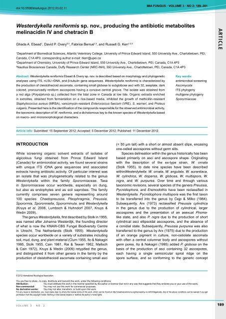
Fig. 5. Macro and micro-morphology of <strong>Westerdykella</strong> <strong>reniformis</strong> (RKGE 35 = DAOM 242243). A. Colony grown on oatmeal agar (left) and<br />
oatmeal agar with sea salts (right) at 14d. B–C. Asco<strong>sp</strong>ores. D–F. Asci.<br />
<strong>the</strong> metalloproteinase gelatinase MMP-2 (involved in <strong>the</strong><br />
cleavage of type IV collagen), demonstrating reversible and<br />
competitive inhibition of <strong>the</strong> enzyme; <strong>the</strong> mixture has <strong>the</strong>refore<br />
been proposed as lead compounds for <strong>the</strong> development as<br />
an antimetastatic agent (Lee et al. 1999b). Lanomycin and<br />
glucolanomycin, were two antifungal metabolites isolated<br />
from liquid fermentations of W. di<strong>sp</strong>ersa inhibiting growth of<br />
various dermatophytes and some <strong>sp</strong>ecies of Candida, but<br />
volume 3 · no. 2<br />
199
Ebead et al.<br />
ARTICLE<br />
were inactive against C. albicans, A<strong>sp</strong>ergillus flavus and<br />
Gram-positve and Gram-negative bacteria (O’Sullivan et al.<br />
1992). Antifungal activity of lanomycin and glucolanomycin<br />
was attributed to <strong>the</strong> inhibition of lanosterol 14α-demethylase,<br />
suggesting a similar mode of action to <strong>the</strong> azole and bistriazole<br />
class of antifungal agents (O’Sullivan et al. 1992).<br />
From this survey, culture extracts generated from each<br />
of <strong>the</strong> <strong>Westerdykella</strong> <strong>sp</strong>ecies strains tested, demonstrated<br />
an antibiotic effect. Antibiotic activity was first associated<br />
with <strong>the</strong> genus <strong>Westerdykella</strong> from a mangrove isolate of<br />
W. aurantiaca. The depsidones, auranticins A and B, were<br />
isolated and demonstrated to have activity against both <strong>the</strong><br />
Gram-positive bacteria Staphylococcus aureus and Bacillus<br />
subtilis, with auranticin A being more potent in antibiotic<br />
activity compared to auranticin B using a disc diffusion assay<br />
(Poch & Gloer 1991). In our survey, antibiosis, measured<br />
as growth inhibition, was assayed against several Grampositive<br />
and Gram-negative bacteria, including <strong>the</strong> drug<br />
resistant pathogens MRSA and VRE, and <strong>the</strong> pathogenic<br />
yeast, C. albicans. All of <strong>the</strong> <strong>Westerdykella</strong> isolates tested<br />
inhibited growth of <strong>the</strong> Gram-positive bacteria MRSA and S.<br />
warneri; however activity against VRE and Proteus vulgaris<br />
was unique to W. <strong>reniformis</strong>. LC-HRMS analysis confirmed<br />
<strong>the</strong> absence of auranticin A and B in each of <strong>the</strong> fraction 3’s<br />
obtained from W. cylindrica, W. di<strong>sp</strong>ersa, W. multi<strong>sp</strong>ora, W.<br />
nigra, W. ornata, and W. <strong>reniformis</strong>. The observed biological<br />
activity of fraction 3 derived from culture extracts from W.<br />
<strong>reniformis</strong> was attributed to <strong>the</strong> production of melinacidin<br />
IV and chetracin B, which was found to be exclusive within<br />
<strong>the</strong> genus to W. <strong>reniformis</strong>. Melinacidin derivatives have<br />
been reported previously from a variety of different fungi:<br />
Acrostalagmus luteoalbus (syn. A. cinnabarinus; Argoudelis<br />
& Mizsak 1977), Chaetomium nigricolor (syn. C. abuense;<br />
Saito et al. 1985), Cladobotryum <strong>sp</strong>. (Feng et al. 2003),<br />
and Oidiodendron truncatum (Li et al. 2012); indicating that<br />
melinacidin production is not uncommon, nor limited to a<br />
particular taxonomic order. Both melinacidin IV and chetracin<br />
B are epipolythiodioxopiperazines, an important class of<br />
biologically active metabolites which possess a wide variety<br />
of biological activities, including antiproliferative, cytotoxic,<br />
immunomodulatory, antiviral, and antimicrobial activities (Li<br />
et al. 2012). We have reported potent antibiotic activity of<br />
melinacidin IV against <strong>the</strong> drug resistant bacteria methicillinresistant<br />
S. aureus and vancomycin-resistant Enterococcus<br />
faecium for <strong>the</strong> first time.<br />
ACKNOWLEDGEMENTS<br />
We gratefully acknowledge financial support from <strong>the</strong> Natural<br />
Sciences and Engineering Council of Canada (NSERC), Canada<br />
Research Chair Program, University of Prince Edward Island, Atlantic<br />
Canada Opportunities Agency (funding from <strong>the</strong> AIF program), and<br />
Jeanne and Jean-Louis Lévesque Foundation. G.A.E. was supported<br />
by a scholarship from <strong>the</strong> Egyptian Cultural and Educational Mission<br />
Sector, Ministry of Scientific Research, Egypt. We also acknowledge<br />
experimental assistance from Martin Lanteigne who carried out all<br />
antimicrobial assays.<br />
REFERENCES<br />
Angelini P, Rubini A, Gigante D, Reale L, Pagiotti R, Venanzoni R<br />
(2012) The endophytic fungal communities associated with <strong>the</strong><br />
leaves and roots of <strong>the</strong> common reed (Phragmites australis) in<br />
Lake Trasimeno (Perugia, Italy) in declining and healthy stands.<br />
Fungal Ecology 5: 683–693.<br />
Argoudelis AD, Mizsak SA (1977) Melinacidins II, III and IV structural<br />
studies. Journal of Antibiotics 30: 468−473.<br />
Arx JA von (1975) On Thielavia angulata and some recently described<br />
Thielavia <strong>sp</strong>ecies. Kavaka 3: 33–36.<br />
Arx JA von (1973) Ostiolate and non-ostiolate Pyrenomycetes.<br />
Proceedings van de Koninklijke Nederlandse Akademie van<br />
Wetenschappen, Section C 76: 289–296.<br />
Bettucci L, Malvarez I, Dupont J, Bury E, Roquebert M-F (2002)<br />
Paraná river delta wetlands soil microfungi. Pedobiologia,<br />
International Journal of Soil Biology 46: 606–623.<br />
Bunyard BA, Nicholson MS, Royse DJ (1994) A systematic<br />
assessment of Morchella using RFLP analysis of <strong>the</strong> 28S<br />
ribosomal RNA gene. Mycologia 86: 762–772.<br />
Cain RF (1961) Studies on soil fungi III. New <strong>sp</strong>ecies of Coniochaeta,<br />
Chaetomidium and Thielavia. Canadian Journal of Botany 39:<br />
1231–1235.<br />
Cejb K, Milko AA (1964) Genera of <strong>the</strong> Eurotiaceae with 32<br />
asco<strong>sp</strong>ores. I. <strong>Westerdykella</strong>. Česká Mykologie 18: 82–84.<br />
Clum FM (1955) A new genus in A<strong>sp</strong>ergillaceae. Mycologia 47: 899–<br />
901.<br />
da Silva M, Umbuzeiro GA, Pfenning LH, Canhos VP, E<strong>sp</strong>osito E<br />
(2003) Filamentous fungi isolated from estuarine sediments<br />
contaminated with industrial discharges. Soil and Sediment<br />
Contamination 12: 345–356.<br />
El-Sharouny H, Gherbawy YAMH, Abdel-Aziz F (2009) Fungal<br />
diversity in brackish and saline lakes in Egypt. Nova Hedwigia<br />
89: 437–450.<br />
Feng Y, Blunt JW, Cole ALJ, Cannon JF, Robinson WT, Monro<br />
MHG (2003) Two <strong>nov</strong>el cytotoxic cyclodepsipeptides from a<br />
mycoparasitic Cladobotryum <strong>sp</strong>. Journal of Organic Chemistry<br />
68: 2002–2005.<br />
Ferraro MJ (2003) Methods for dilution antimicrobial susceptibility<br />
test for bacteria that grow aerobically: approved standards M7–<br />
A6. Wane, PA: Committee for Clinical Laboratory Standards.<br />
Ito T, Nakagiri A (1995) <strong>Westerdykella</strong> globosa, a proposal for a new<br />
combination. Mycoscience 36: 361–363.<br />
Kohlmeyer J, Kohlmeyer E (1979) Marine Mycology: <strong>the</strong> higher fungi.<br />
New York: Academic Press.<br />
Kornerup A, Wanscher JH (1978) Methuen Handbook of Colour: 3 rd<br />
edn. London: Eyre Methuen.<br />
Kruys A, Eriksson OE, Wedin M (2006) Phylogenetic relationships of<br />
coprophilous Pleo<strong>sp</strong>orales (Dothideomycetes, Ascomycota), and<br />
<strong>the</strong> classification of some bitunicate taxa of unknown position.<br />
Mycological Research 110: 527–536.<br />
Kruys A, Wedin M (2009) Phylogenetic relationships and an<br />
assessment of traditionally used taxonomic characters in <strong>the</strong><br />
Sporormiaceae (Pleo<strong>sp</strong>orales, Dothideomycetes, Ascomycota),<br />
utilising multi-gene phylogenies. Systematics and Biodiversity 7:<br />
465–478.<br />
Lee BKH, Baker GE (1973) Fungi associated with <strong>the</strong> roots of red<br />
mangrove, Rhizophora mangle. Mycologia 65: 894–906.<br />
Lee H, Lee C, Chung M, Chun H, Rhee J, Kho Y (1999a) Dykellic<br />
acid, a <strong>nov</strong>el apoptosis inhibitor from <strong>Westerdykella</strong> multi<strong>sp</strong>ora<br />
200 ima fUNGUS
Antibiotic <strong>producing</strong> <strong>Westerdykella</strong> <strong>reniformis</strong> <strong>sp</strong>. <strong>nov</strong>.<br />
F50733. Tetrahedron Letters 40: 6949–6950.<br />
Lee S, Youk E, Lee H, Kho Y, Kim H, Kim S (2003) Dykellic acid<br />
inhibits drug-induced ca<strong>sp</strong>ase-3-like protease activation.<br />
Biochemical and Biophysical Research Communications 302:<br />
539–544.<br />
Lee HJ, Chung MC, Lee CH, Yun BS, Chun HK, Kho YH (1997)<br />
Gelastatins A and B, new inhibitors of gelatinase A from<br />
<strong>Westerdykella</strong> multi<strong>sp</strong>ora F50733. The Journal of Antibiotics 50:<br />
357–359.<br />
Lee H, Chung M, Lee C, Chun H, Rhee J, Kho Y (1999b) Gelastatins,<br />
new inhibitors of matrix metalloproteinases from <strong>Westerdykella</strong><br />
multi<strong>sp</strong>ora F50733. Annals of <strong>the</strong> New York Academy of<br />
Sciences 878: 635–637.<br />
Li L, Li D, Luan Y, Gu Q, Zhu T (2012) Cytotoxic metabolites from <strong>the</strong><br />
Antarctic psychrophilic fungus Oidiodendron truncatum. Journal<br />
of Natural Products 75: 920-927.<br />
Lumbsch H, Huhndorf S (2007) Whatever happened to <strong>the</strong><br />
pyrenomycetes and loculoascomycetes? Mycological Research<br />
111: 1064–1074.<br />
Malloch D, Cain RF (1972) New <strong>sp</strong>ecies and combinations of<br />
cleisto<strong>the</strong>cial Ascomycetes. Canadian Journal of Botany 50:<br />
61–72.<br />
Miller AN, Huhndorf SM (2005) Multi-gene phylogenies indicate<br />
ascomal wall morphology is a better predictor of phylogenetic<br />
relationships than asco<strong>sp</strong>ore morphology in <strong>the</strong> Sordariales<br />
(Ascomycota, Fungi). Molecular Phylogenetics and Evolution 35:<br />
60–75.<br />
Mishra RC (1995) Distribution of microfungi in <strong>the</strong> transitional zone<br />
of a subtropical lake in India. Canadian Journal of Botany 73:<br />
512–516.<br />
O’Sullivan J, Phillipson DW, Kirsch DR, Fisher SM, Lai MH, Trejo<br />
WH (1992) Lanomycin and glucolanomycin, antifungal agents<br />
produced by Pycnidiophora di<strong>sp</strong>ersa. I. Discovery, isolation and<br />
biological activity. Journal of Antibiotics 45: 306–312.<br />
Poch GK, Gloer JB (1991) Auranticins A and B: two new depsidones<br />
from a mangrove isolate of <strong>the</strong> fungus Preussia aurantiaca.<br />
Journal of Natural Products 54: 213–217.<br />
Rai JN, Tewari JP (1963) On some isolates of <strong>the</strong> genus Preussia<br />
Fuckel from Indian soils. Proceedings of <strong>the</strong> Indian Academy of<br />
Science 57: 45–55.<br />
Rehner SA, Samuels GJ (1994) Taxonomy and phylogeny of<br />
Gliocladium analysed from nuclear large subunit ribosomal DNA<br />
sequences. Mycological Research 98: 625-634.<br />
Ronquist F, Teslenko M, van der Mark P, Ayres DL, Darling A, Hohna<br />
S, Larget B, Liu L, Suchard MA, Huelsenbeck JP (2012) MrBayes<br />
3.2: eficient Bayesian phylogenetic inference and model choice<br />
across a large model <strong>sp</strong>ace. Systematic Biology 61: 539–542.<br />
Saito T, Koyama K, Natori S, Iitaka Y (1985) Chetracin A, a new<br />
epipolythiodioxopiperazine having a tetrasulfide bridge from<br />
Chaetomium abuense and C. retardatum. Tetrahedron Letters<br />
26: 4731–4734.<br />
Stolk AC (1955) Emericellopsis minima <strong>sp</strong>. <strong>nov</strong>. and <strong>Westerdykella</strong><br />
ornata gen. <strong>nov</strong>., <strong>sp</strong>. <strong>nov</strong>. Transactions of British Mycological<br />
Society 38: 419–424.<br />
Tamura K, Peterson D, Peterson N, Stecher G, Nei M, Kumar S<br />
(2011) MEGA5: molecular evolutionary genetics analysis using<br />
maximum likelihood, evolutionary distance, and maximum<br />
parsimony methods. Molecular Biology and Evolution 28: 2731–<br />
2739.<br />
Vilgalys R, Hester M (2012) Rapid genetic identification and<br />
mapping of enzymatically amplified ribosomal DNA from several<br />
Cryptococcus <strong>sp</strong>ecies. Journal of Bacteriology 172: 4238–4246.<br />
White TJ, Bruns T, Lee S, Taylor J (1990) Amplification and direct<br />
sequencing of fungal ribosomal RNA genes for phylogenetics.<br />
In: Innis MA, Gelfand DH, Sninsky JJ, White TJ (eds), PCR<br />
Protocols: a guide to methods and applications: 315–322. San<br />
Diego: Academic Press.<br />
ARTICLE<br />
volume 3 · no. 2<br />
201


