DYNAMIC ORGANIZATION AND FUNCTION OF BIOMEMBRANES
DYNAMIC ORGANIZATION AND FUNCTION OF BIOMEMBRANES DYNAMIC ORGANIZATION AND FUNCTION OF BIOMEMBRANES
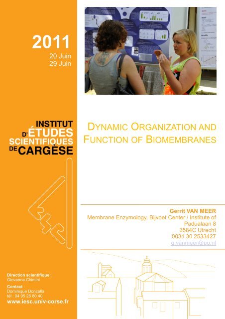
2011 20 Juin 29 Juin DYNAMIC ORGANIZATION AND FUNCTION OF BIOMEMBRANES Gerrit VAN MEER Membrane Enzymology, Bijvoet Center / Institute of Padualaan 8 3584C Utrecht 0031 30 2533427 g.vanmeer@uu.nl Direction scientifique : Giovanna Chimini Contact : Dominique Donzella tél : 04 95 26 80 40 www.iesc.univ-corse.fr
- Page 2 and 3: FEBS/EMBO Advanced Lecture Course B
- Page 4 and 5: not done so already, please contact
- Page 6 and 7: 2. Air France Bus. Busses leave Roi
- Page 8 and 9: Saturday June 25 9h 00 Reinhard Jah
- Page 10 and 11: Cellular lipidomics: what are the q
- Page 12 and 13: Patricia Bassereau Patricia studied
- Page 14 and 15: ChemBioChem, 11, 848-865 Some pape
- Page 16 and 17: Recommended reading Lipid sorting -
- Page 18 and 19: The amino acid sequence of a protei
- Page 20 and 21: Recommended reading Lecture 1 Dobso
- Page 22 and 23: McLean, P.J., Klucken, J., Shin, Y.
- Page 24 and 25: Name: Tom A. Rapoport Full address:
- Page 26 and 27: mechanism may operate for hexameric
- Page 28 and 29: HOW THE ER GETS INTO SHAPE Tom A. R
- Page 30 and 31: Profiles and abstracts of main and
- Page 32 and 33: The Sec14-Superfamily and Mechanism
- Page 34 and 35: Name : COSSART Pascale Full address
- Page 36 and 37: Name Sandrine ETIENNEMANNEVILLE F
- Page 38 and 39: Cytoskeleton rearrangements during
- Page 40 and 41: Van den Bogaart, G.,, Thutupalli, S
- Page 42 and 43: TOM KIRCHHAUSEN HARVARD MEDICAL SCH
- Page 44 and 45: 3. What types of vesicles exist and
- Page 46 and 47: 2. Dynamics of endocytosis Cargese
- Page 48 and 49: References Review: Kirchhausen, T.
- Page 50 and 51: Nikolaus (Klaus) Pfanner Institute
2011<br />
20 Juin<br />
29 Juin<br />
<strong>DYNAMIC</strong> <strong>ORGANIZATION</strong> <strong>AND</strong><br />
<strong>FUNCTION</strong> <strong>OF</strong> <strong>BIOMEMBRANES</strong><br />
Gerrit VAN MEER<br />
Membrane Enzymology, Bijvoet Center / Institute of<br />
Padualaan 8<br />
3584C Utrecht<br />
0031 30 2533427<br />
g.vanmeer@uu.nl<br />
Direction scientifique :<br />
Giovanna Chimini<br />
Contact :<br />
Dominique Donzella<br />
tél : 04 95 26 80 40<br />
www.iesc.univ-corse.fr
FEBS/EMBO Advanced Lecture Course<br />
BIOMEMBRANE <strong>DYNAMIC</strong>S: FROM<br />
MOLECULES TO CELLS<br />
June 20-30, 2011<br />
Cargèse-Corsica-France<br />
Institut d'Etudes Scientifiques Cargèse<br />
(Corse)<br />
1
General Information<br />
LOCATION<br />
The lectures, poster sessions, and discussions will be held from<br />
9.00h until 12.30h and from 16.30h until approximately 20.00h<br />
starting on Tuesday June 21 at the Institut d'Etudes Scientifiques<br />
de Cargèse. The Institute is located at walking distance from the<br />
village (going towards Ajaccio): 40 min. via the main road or 15<br />
min. via a "goat path". Bring a pocket-torch and good walking shoes<br />
as this path is dark at night, steep and rough.<br />
Address:<br />
Institut d'Etudes Scientifiques de Cargèse,<br />
F - 20130 Cargèse, France<br />
Telephone # : + 33 4 95 26 80 40<br />
Fax # : + 33 4 95 26 80 45<br />
REGISTRATION<br />
All participants must register on site, starting 8.30h on Tuesday June 21.<br />
TRAVEL<br />
Participants are expected to arrive on Monday, June 20 and to leave on Thursday, June 30.<br />
Cargèse is located on the west coast of Corsica, 50 km north of Ajaccio.<br />
As was indicated on the registration form, a group flight (AF 4502) from Paris Orly West to<br />
Ajaccio has been organized. All tickets for this group flight are electronic. Flight details will be<br />
sent to you by E-mail in the week before the meeting: please check your E-mails regularly.<br />
For on-line check-in follow the instructions from Air France: print your e-ticket and show it<br />
together with your passport at the AirFrance counter for flight AF4502 in the airport.<br />
PLEASE MAKE SURE THAT YOU HAVE PLENTY <strong>OF</strong> TIME INBETWEEN YOUR<br />
ARRIVAL IN PARIS <strong>AND</strong> THE DEPARTURE <strong>OF</strong> THE GROUP FLIGHT FROM ORLY<br />
WEST TO AJACCIO. REMEMBER THAT SECURITY CONTROLS AT THE AIRPORTS<br />
ARE VERY STRICT <strong>AND</strong> THAT YOU MUST ARRIVE AT THE AIRPORT WELL BEFORE<br />
DEPARTURE TIME (14:50H).<br />
Ajaccio - Cargèse:<br />
A chartered bus will transfer the group flight participants from Ajaccio airport to Cargèse. The<br />
"Imperial Tour" busses will leave from the parking lot at the opposite side of the airport<br />
concourse from the baggage claim section. Participants arriving on earlier flights can also<br />
take this bus, provided they have made a reservation (j.a.f.opdenkamp@uu.nl). The busses<br />
will leave at about 16.40h.<br />
Transport can be also organized for participants arriving in Ajaccio after 16.30h. If you have<br />
2
not done so already, please contact Jos Op den Kamp.<br />
Public busses from Ajaccio to Cargèse leave twice a day (except on Sunday) at 7.30 a.m.<br />
and 15.30 p.m. from the Gare Routière near the port. Participants, not arriving with the group<br />
flight or with one of the chartered busses or taxis, should report at the Institute on Monday.<br />
Ajaccio - Paris:<br />
Transfer to the Ajaccio airport on Thursday June 30 will be arranged later during the course.<br />
ACCOMMODATION<br />
Housing will be taken care of by the staff of the Institute following, as much as possible, the<br />
preferences you indicated on the registration form. Most students will be accommodated in<br />
the Institute in rooms with double occupancy. Others, opting for hotel accomodation and/or<br />
single rooms will stay in the village.<br />
Reservations will be made from Monday June 20 until Thursday June 30 only.<br />
MEALS<br />
Breakfast will be served only for those participants living in the Institute<br />
Lunch will be served for all participants immediately after the last morning lecture. Please<br />
contact Jos Op den Kamp, upon arrival or during the first day, if your accompanying friend(s)<br />
or relative(s) would like to join this lunch. The charge for the 8 lunches is 80 Euro per person.<br />
Students should find their dinner in local restauarants. The village has several good and<br />
inexpensive restaurants, a bakery and two minimarkets where you can buy food. Please note<br />
that shops are closed after lunch until 4.30pm.<br />
SOCIAL ACTIVITIES<br />
A welcome party for participants and accompanying persons will be held at the end of the<br />
first working day. Other social events will be announced during the meeting. These will<br />
include a free bus tour into the local countryside on Sunday, a boat trip up the coast to see<br />
the magnificent red cliffs of Piana on Saturday evening (a modest contribution to costs will be<br />
requested from students), and student sketches followed by a farewell party on the last day.<br />
FINANCES<br />
A registration fee, as described in the letter of acceptance, is necessary to complete your<br />
registration. If you have not paid already, please do so during the course or transfer the<br />
registration fee, without costs for the receiver, to the following account :<br />
Account #: 3783.81.733<br />
BIC or SWIFT #: RABO NL 2U<br />
IBAN #: NL86 RABO 0378 3817 33<br />
J.A.F. Op den Kamp / FEBS<br />
Rabobank<br />
P.O. Box 9<br />
3730 AA De Bilt<br />
The Netherlands.<br />
3
The fare for the flights from and to Paris can be paid in advance using the account mentioned<br />
above, or during the meeting.<br />
Participants accommodated in a hotel must pay the hotel manager directly.<br />
PROGRAMME<br />
A programme and abstracts of the lectures and posters are included herewith. Printed copies<br />
of the lecture and poster programme will be distributed upon registration for the course.<br />
Please print a copy to take with you to Cargèse from the abstracts you are interested in. We<br />
do not plan to distribute printed versions of abstracts during the course. It is also possible to<br />
view the abstracts etc. via internet. Computer facilities are excellent at the Institute<br />
Posters will be presented during three sessions (according to alphabetical order) and a<br />
number of posters will be selected for an oral presentation. There are no specific rules for<br />
size etc. of your poster.<br />
Students are encouraged to bring a short (25 minutes maximum) presentation of their work in<br />
PowerPoint or Keynote format in case their posters are selected for oral presentation. Please<br />
save your presentation on a memory stick or a CD.<br />
ADDITIONAL ARRANGEMENTS<br />
• If you want to rent a car, please arrange this in advance via a travel agent or upon arrival<br />
at the airport.<br />
• Please arrange your own travel and/or health insurance. The organizers do not assume<br />
any responsibility for this or any other liability.<br />
• Cargèse does not have a travel agency. Please make personal travel arrangements<br />
before you arrive in Corsica.<br />
• Some shops do not accept payment by credit card. There are several cash dispensers in<br />
the village.<br />
• Please make sure that you have the appropriate visa and a valid passport.<br />
AIRPORT TRANSFERS<br />
Most international flights arrive in Paris at Roissy-Charles de Gaulle airport in the north of<br />
Paris. The group flight to Ajaccio leaves from Orly airport in the south of Paris. The absolute<br />
minimum travelling time between the two airports is 90 minutes, but it usually takes much<br />
longer, according to traffic and other conditions. Trains from Germany and The Netherlands<br />
arrive at Gare du Nord. Trains from Switzerland and Italy arrive at Gare de l'Est or Gare de<br />
Lyon.<br />
To transfer from your point of arrival to Orly, please use one of the following services.<br />
1. RER line B from Roissy to Orly. Take the shuttle train or walk to the SNCF train station and<br />
buy a ticket for Orly airport. Take the train as far as Anthony Station, then change to the<br />
monorail train to the airport. Trains leave Roissy airport every 10 minutes and monorail trains<br />
leave Anthony station every 6 minutes and take 8 minutes to reach Orly. Get off the monorail<br />
at the first stop, Orly West terminal.<br />
4
2. Air France Bus. Busses leave Roissy airport for Orly Airport every 30 minutes. Buy a ticket<br />
from the Air France Bus desk in the airport terminal or (sometimes) from the driver. Air<br />
France Busses also leave Invalides and Montparnasse Railway Station in Paris for Orly every<br />
15 minutes. You can reach these bus stops by the metro. Buy a bus ticket from the bus<br />
driver. Get off the bus at the Orly West terminal.<br />
3. OrlyBus. This city bus leaves Denfert Rochereau station (on RER line B) for Orly airport<br />
every 15-20 minutes. By a ticket from the ticket office in front of the train station. Get off the<br />
bus at the Orly West terminal.<br />
On arrival at Orly West, go to the area in front of the check-in desk for flight AF 4502. If you<br />
miss the group flight because of transfer delays take the next available flight (you will have to<br />
buy a full-price ticket).<br />
REMEMBER THAT SECURITY CONTROLS AT THE AIRPORTS ARE VERY STRICT <strong>AND</strong><br />
THAT YOU MUST ARRIVE AT THE AIRPORT WELL BEFORE DEPARTURE TIME<br />
5
LECTURE PROGRAMME<br />
Tuesday June 21<br />
8h 30 Registration<br />
8h 45 Welcome, presentation of students, faculty and staff,<br />
introduction to the course (Jos Op den Kamp, Gerrit van Meer)<br />
9h 15 Tom Rapoport. Lecture 1: Mechanism of protein transport across membranes<br />
10h 15 Coffee break<br />
10h 45 Gerrit van Meer. Lecture 1: Cellular lipidomics: what are the questions?<br />
11h 45 Janet Shaw. Lecture 1: Mitochondrial function and dysfunction: the role of<br />
membrane remodeling machineries<br />
12h 30 Lunch and free afternoon<br />
16h 00 Poster session 1 with refreshments<br />
19h 00 Welcome drinks<br />
Wednesday June 22<br />
9h 00 William Wickner. Lecture 1: Fusion of Biological Membranes<br />
10h 00 Pascale Cossart. Lecture 1: Interactions between bacteria and cells<br />
11h 00 Coffee break<br />
11h 30 Klaus Pfanner. Lecture 1: Dynamic machineries for importing mitochondrial<br />
proteins<br />
12h 30 Lunch; tutorial 2 and free afternoon<br />
16h 00 Poster session 1 and refreshments: vote for selection for best poster in<br />
session 1<br />
18h 00 Patricia Bassereau. Lecture 1: Physical basis for membrane traffic<br />
Thursday June 23<br />
9h 00 Ulrich Hartl. Lecture 1: Mechanisms of chaperone-assisted protein folding<br />
and membrane translocation<br />
10h 00 Tommy Kirchhausen. Lecture 1: Molecular basis for membrane traffic<br />
11h 00 Coffee break<br />
11h 30 Vytas Bankaitis. Lecture 1: The Sec14-Superfamily and Mechanisms of<br />
Crosstalk Between Lipid Metabolism and Lipid Signaling<br />
12h 30 Lunch and free afternoon<br />
16h 30 Poster session 2 and refreshments,<br />
18h 00 EMBO Young investigator lecture: Sandrine Etienne-Manneville.<br />
Cytoskeleton rearrangements during cell migration<br />
Friday June 24<br />
9h 00 EMBO PLENARY LECTURE: Reinhard Jahn. SNAREs — engines for<br />
membrane fusion<br />
10h 00 Pascale Cossart. Lecture 2: Interactions between bacteria and cells 2<br />
11h 00 Coffee break<br />
11h 30 Patricia Bassereau. Lecture 2: Membrane curvature and traffic: quantitative<br />
approaches<br />
12h 30 Lunch and free afternoon<br />
16h 30 Poster session 2 and refreshments: vote for best poster in session 2<br />
18h 00 EMBO Women in Science Lecture: Petra Schwille. Lecture 1: Women in<br />
science – exercising freedom<br />
6
Saturday June 25<br />
9h 00 Reinhard Jahn. Lecture: Exocytosis of synaptic vesicles in neurons<br />
10h 00 Jan Tommassen. Lecture 1: The bacterial outer membrane: Biogenesis of LPS<br />
11h 00 Coffee break<br />
11h 30 Petra Schwille. Lecture 2: Minimal systems for membrane-associated cellular<br />
processes<br />
12h 30 Lunch and free afternoon and evening<br />
Boatride<br />
Sunday June 26 Free<br />
Bus trip<br />
Monday June 27<br />
9h 00 William Wickner. Lecture 2: Mechanisms of yeast homotypic vacuole fusion<br />
10h 00 Jan Tommassen. Lecture 2: Biogenesis of the outer membrane: outer<br />
membrane proteins<br />
11h 00 Coffee break<br />
11h 30 Klaus Pfanner. Lecture 2: Sorting of mitochondrial proteins: from<br />
proteomics to functional mechanisms<br />
12h 30 Lunch and free afternoon<br />
16h 00 Poster session 3 and refreshments.<br />
18h 00 Janet Shaw. Lecture 2: Moving Mitochondria: Establishing Distribution of an<br />
Essential Organelle<br />
19h 00 Tom Kirchhaussen. Lecture 2: Dynamics of endocytosis<br />
Tuesday June 28<br />
9h 00 Ulrich Hartl. Lecture 2: Protein misfolding and disease<br />
10h 00 Joost Holthuis. Lecture 1: to be announced<br />
11h 00 Coffee break<br />
11h 30 Vytas Bankaitis. Lecture 2: The Secret Lives of Lipid Transfer Proteins<br />
12h 30 Lunch and free afternoon<br />
16h 30 Poster session 3 and refreshments: vote for best poster in session 3<br />
18h 00 Manajit Hayer-Hartl. Lecture 1: Role of chaperones and AAA+ ATPases in<br />
the assembly and conformational modulation of hexadecameric Rubisco<br />
19h 00 William Wickner. Special Session: How to Get a Life in the Life Sciences.<br />
Wednesday June 29<br />
9h 00 Tom Rapoport. Lecture 2: How the ER gets into shape<br />
10h 00 Student presentations<br />
11h 00 Coffee break<br />
11h 30 Student presentations<br />
12h 30 Lunch, preparation of student sketches and free afternoon<br />
18h 00 Special session Student sketches and prizes<br />
Concluding remarks by the chairs<br />
20h 00 Farewell party<br />
7
FACULTY PR<strong>OF</strong>ILES<br />
Course Directors and Organizing Committee<br />
Gerrit van Meer<br />
Faculty of Science<br />
Utrecht University<br />
Budapestlaan 63584 CD Utrecht, The Netherlands<br />
Tel. +31 302531385<br />
g.vanmeer@uu.nl<br />
Gerrit van Meer studied biochemistry at Utrecht University where he obtained his PhD on lipid<br />
translocation across plasma membranes with Laurens van Deenen in 1981. He then spent 5 years at<br />
EMBL Heidelberg working as a postdoc with Kai Simons on lipid polarity and lipid sorting in<br />
epithelial cells, resulting in the (in)famous lipid raft hypothesis in 1987. He independently<br />
continued this work in the Cell Biology department of Utrecht University Medical School, where the<br />
group stumbled on the lipid translocation activity of multidrug transporters. After 10 year he<br />
moved to Cell Biology at the medical school of the University of Amsterdam, where they discovered<br />
a hitherto unknown function of glycolipids in pigmentation. In 2001 the group moved to the<br />
Chemistry Department of Utrecht University where, together with his colleagues in the Bijvoet<br />
Center for biomolecular research, Joost Holthuis and Toon de Kroon, Gerrit was interested in how<br />
cells use lipids for their vital functions. His group resolved the glycolipid function in pigmentation<br />
as being a consequence of the action of glycolipids on the acidification of cellular organelles.<br />
Objects of study were lipid flippases, lipid rafts and lipidprotein interactions. Presently, Gerrit is<br />
dean of the Faculty of Science at Utrecht University.<br />
Representative publications<br />
van Meer, G., Stelzer, E.H.K., Wijnaendts‐van‐Resandt, R.W. and Simons, K. (1987) Sorting of sphingolipids in<br />
epithelial (Madin‐Darby canine kidney) cells. J. Cell Biol. 105, 1623‐1635.<br />
van Helvoort, A., Smith, A.J., Sprong, H., Fritzsche, I., Schinkel, A.H., Borst, P., and van Meer, G. (1996) MDR1<br />
P‐glycoprotein is a lipid translocase of broad specificity, while MDR3 P‐glycoprotein specifically translocates<br />
phosphatidylcholine. Cell 87, 507‐517.<br />
Sprong, H., Degroote, S., Claessens, T., van Drunen, J., Oorschot, V., Westerink, B.H.C., Hirabayashi, Y.,<br />
Klumperman, J., van der Sluijs, P., and van Meer, G. (2001) Glycosphingolipids are required for sorting of<br />
melanosomal proteins in the Golgi complex. J. Cell Biol. 155, 369‐380.<br />
van Meer, G. (2005) Cellular Lipidomics. EMBO J. 24, 3159‐3165.<br />
van Meer, G., Halter, D., Sprong, H., Somerharju, P., and Egmond, M.R. (2006) ABC lipid transporters:<br />
extruders, flippases, or flopless activators? FEBS Lett. 580, 1171‐1177.<br />
Halter, D., Neumann, S., van Dijk, S.M., Wolthoorn, J., de Mazière, A.M., Vieira, O.V., Mattjus, P., Klumperman,<br />
J., van Meer, G. and Sprong, H. (2007) Pre‐ and post‐Golgi translocation of glucosylceramide in glycosphingolipid<br />
synthesis. J. Cell Biol. 179, 101‐115.<br />
Groux‐Degroote, S., van Dijk, S.M., Wolthoorn, J., Neumann, S., Theos, A.C., De Mazière, A.M., Klumperman, J.,<br />
van Meer, G., and Sprong, H. (2008) Glycolipid dependent sorting of melanosomal from lysosomal membrane<br />
proteins by lumenal determinants. Traffic 9, 951‐963.<br />
van Meer G., de Kroon A.I. (2011). Lipid map of the mammalian cell. J Cell Sci 124, 5‐8.<br />
8
Cellular lipidomics: what are the questions?<br />
Gerrit van Meer<br />
Lipidomics is a new term to describe a scientific field that is significantly broader than<br />
lipidology, the science of lipids. Besides lipidology, lipidomics covers the lipid‐metabolizing<br />
enzymes and lipid transporters, their genes and regulation; it covers the quantitative<br />
determination of lipids in space and time, and also includes the study of lipid function. Because<br />
lipidomics is concerned with all lipids and their enzymes and genes, it faces the formidable<br />
challenge to develop enabling technologies to comprehensively measure the expression, location<br />
and regulation of lipids, enzymes and genes in time. The second challenge is to devise<br />
information technology that allows the construction of metabolic maps by browsing through<br />
connected databases containing the subsets of data in lipid structure, lipid metabolomics,<br />
proteomics, genomics. In addition, to understand lipid function, on the one hand we need a<br />
broad range of imaging techniques to define where exactly the relevant events happen in the cell<br />
and subcellular organelles, and on the other hand we need a thorough understanding of how<br />
lipids physically interact, especially with proteins. The final challenge is to apply this knowledge<br />
in the diagnosis, monitoring and cure of lipid‐related diseases.<br />
Cells have thousands of different lipids. To make them, break them and transport them<br />
they probably need a thousand enzymes or more. So, it is very likely that (groups of) lipids serve<br />
unique functions. Over the years it has become clear that such functions can be both structural<br />
and in signaling. While the biophysicists have made great progress on the structure side, many<br />
important discoveries have been made on the roles of lipids in signal transduction pathways.<br />
Finally, unexpected insights have been obtained from the elucidation of the genetic causes of a<br />
number of inherited lipid‐related diseases. In all this, it must be realized that the functionality of<br />
lipids is determined not just by their presence as measured by a mass spectrometer, but by their<br />
local concentration, which varies between organelles, between the two leaflets of the lipid<br />
bilayer and even within the lateral plane of the membrane. Therefore, to obtain insights in lipid<br />
function one needs a multidisciplinary approach, in which approaches from chemistry and<br />
biophysics are combined with genetics and cell biology.<br />
The various intracellular membranes have different protein and lipid compositions. In view of<br />
the rapid transport between these membranes via vesicles, how do cells introduce selectivity in these<br />
pathways? How is lipid transport involved in lipid signaling? How do cells deal with lipids? Starting<br />
from simple principles we have to try and understand the basic organization of lipids in the cell. There<br />
are still major open questions concerning how the cell regulates its lipid homeostasis. After that we<br />
will introduce more detail in the system to find out how the highly specific functions of lipids can be<br />
carried out against the underlying framework of the bulk lipid organization.<br />
van Meer, G. (2005). Cellular lipidomics. Embo J 24, 3159‐3165.<br />
Wenk, M. R. (2005). The emerging field of lipidomics. Nat Rev Drug Discov 4, 594‐610.<br />
van Meer G., de Kroon A.I. (2011). Lipid map of the mammalian cell. J Cell Sci 124, 5‐8.<br />
9
Cellular lipid transport and disease<br />
Gerrit van Meer<br />
Realizing that cellular membranes have different lipid compositions but that they are connected<br />
by a variety of transport pathways to be traveled by lipid molecules, the question arises as to<br />
what imposes specificity onto those pathways. Strong biophysical evidence supports the idea<br />
that the basis for the selective transport of lipids resides in the aggregation of sphingolipids and<br />
cholesterol into microdomains or "rafts". These rafts would then be expected to play a role in<br />
protein sorting, and in signal transduction at the plasma membrane. However, many questions<br />
remain to be answered.<br />
Whereas lipids with small polar groups like cholesterol and diacylglycerol spontaneously<br />
translocate across lipid bilayers, this is generally not the case for lipids with large and charged<br />
headgroups like the regular membrane phospholipids and glycolipids. Two families of ATPases have<br />
been identified that can move lipids across membranes, the ABC transporters and the P-type ATPases<br />
of the aminophospholipid translocase subfamily. Finally, various protein classes have been found<br />
involved in moving lipids as monomers between membranes through the cytosol, and more and more<br />
evidence indicates that also these activities are highly regulated. An important question is how these<br />
proteins contribute to the dynamic lipid organization in the various organelles.<br />
Finally, there are many lipid-related diseases for which the genetic cause has now been<br />
identified. However, even knowing the protein that is reponsible, and even having ideas on how those<br />
proteins function, it often remains unclear how the defect leads to pathology. In addition, many lipidrelated<br />
diseases like cardiovascular disease and type 2 diabetes are multifactorial. Also in those cases<br />
we don't really understand the lipid part of the disease process: What parameter is disturbed in the lipid<br />
organization and how does the disturbance lead to disease? The answers to these questions will be very<br />
important in trying to find cures for these wide-spread diseases.<br />
Eggeling, C., Ringemann, C., Medda, R., Schwarzmann, G., Sandhoff, K., Polyakova, S., Belov, V.N.,<br />
Hein, B., von Middendorff, C., Schonle, A., and Hell, S.W. (2009). Direct observation of the<br />
nanoscale dynamics of membrane lipids in a living cell. Nature 457, 1159‐1162.<br />
Halter, D., Neumann, S., van Dijk, S.M., Wolthoorn, J., de Maziere, A.M., Vieira, O.V., Mattjus, P.,<br />
Klumperman, J., van Meer, G., and Sprong, H. (2007). Pre‐ and post‐Golgi translocation of<br />
glucosylceramide in glycosphingolipid synthesis. J Cell Biol 179, 101‐115.<br />
Holthuis, J. C., and Levine, T. P. (2005). Lipid traffic: floppy drives and a superhighway. Nat Rev<br />
Mol Cell Biol 6, 209‐220.<br />
Maxfield, F.R., van Meer, G. (2010). Cholesterol, the central lipid of mammalian cells. Curr Opin<br />
Cell Biol 22, 422‐429.<br />
Munro, S. (2003). Lipid rafts: elusive or illusive? Cell 115, 377‐388.<br />
van Meer, G., Halter, D., Sprong, H., Somerharju, P., and Egmond, M. R. (2006). ABC lipid<br />
transporters: extruders, flippases, or flopless activators? FEBS Lett 580, 1171‐1177.<br />
10
Patricia Bassereau<br />
Patricia studied physics and solid state physics at the University of Montpellier, France.<br />
She received a “thèse de 3ème cycle” (short PhD) in 1985 and her PhD in 1990 in Soft<br />
Condensed Matter at the Montpellier University, working on the structure of surfactant-based<br />
phases (highly swollen lamellar phases and sponges phases). She could show that the<br />
stability of these lyotropic smectics with large periodicity was due to repulsive force of<br />
entropic origin (Helfrich force). She entered the CNRS in 1986 in Montpellier (GDPC). In<br />
1992, she was visiting scientist at the Almaden IBM Center (San Jose-USA) and worked on<br />
the structure of thin polymer films. In 1993, she moved to the Curie Institute where she<br />
initially investigated the interactions of soluble proteins with polymer monolayers. Since 15<br />
years, she has been working in the field of "physics for cell biology". She has developed a<br />
multidisciplinary approach to understand the role of lipid membranes and physical<br />
parameters in important cellular functions such as intracellular trafficking, endo/exocytosis,<br />
transmembrane ion transport, or cell adhesion. In 1999, she received her habilitation and in<br />
2002, she was promoted Directrice de Recherche. Presently, she is a group leader at the<br />
Curie Institute and works on complex model membranes mimicking biologically relevant<br />
systems (Giant Unilamellar Vesicles and membrane nanotubes). She has a long-standing<br />
tradition to collaborate with both theoretician physicists and cell biologists.<br />
Main contributions to biology: In the past years, her group contributed to show that active ion<br />
pumps induce an amplification of membrane fluctuations and a reduction of the membrane<br />
tension, and that similar effects are observed during fusion of small liposomes with GUV. She<br />
is now studying signal propagation in membrane tubes containing voltage-gated ion<br />
channels. The group also studies membrane deformation mechanisms by proteins or<br />
colloids. In collaboration with L. Johannès, they have shown a new clathrin-independent<br />
endocytosis mechanism mediated by toxins. Moreover, the group demonstrated the physical<br />
mechanism underlying the formation of membrane nanotubes by molecular motors. Recently,<br />
the group has investigated the role of membrane curvature in lipid/protein sorting (coll. B.<br />
Goud) and in dynamin assembly. Currently, they study the mechanism of membrane<br />
deformation and scission induced by various proteins involved in trafficking.<br />
Recent relevant publications<br />
Callan-Jones A., Sorre B., Bassereau P. (2011) Curvature-driven lipid sorting in biomembranes (review), Cold<br />
Spring Harbor Perspectives in Biology, 3, a004648<br />
Safouane M., Berland L., Callan-Jones A., Sorre B., Römer W., Johannes L., Toombes G. E., Bassereau P.<br />
(2010) Lipid co-sorting mediated by Shiga toxin induced tubulation Traffic, 11, 1519–1529<br />
Roux A., Koster G., Lenz M., Sorre B., Manneville J.-B., Nassoy P., Bassereau P. (2010) Membrane curvature<br />
controls dynamin polymerization, Proc. Natl. Acad. Sci. U.S.A, 107, 4141-4146<br />
Römer W., Pontani L.-L., Sorre B., Rentero C., Berland L., Chambon V., Lamaze C., Bassereau P., Sykes C.,<br />
Gaus K., Johannes L. (2010) Actin dynamics drive membrane reorganization and scission in clathrin independent<br />
endocytosis, Cell, 140, 540-553<br />
Sorre B., Callan-Jones A., Manneville J.-B., Nassoy P., Joanny J. F., Prost J., Goud B., Bassereau P. (2009)<br />
Curvature-Driven Lipid Sorting Needs Proximity to a Demixing Point and Is Aided by Proteins, Proc. Natl. Acad.<br />
Sci. U.S.A (on line)<br />
Sens P., Johannes L., Bassereau P. (2008) Biophysical Approaches to Protein-Induced Membrane Deformations<br />
in Trafficking (review), Curr. Opin. Cell Biol., 20, 476–482<br />
Römer W., Berland L., Chambon V., Gaus K., Windschiegl B., Tenza D., Aly M., Fraisier V., Florent J.-C., Perrais<br />
D., Lamaze C., Raposo G., Steinem C., Sens P., Bassereau P., Johannes L. (2007) Shiga Toxin Induces Tubular<br />
Membrane Invaginations for Its Uptake into Cells, Nature, 450, 670-675<br />
11
Patricia Bassereau<br />
Lecture 1: Physical basis for membrane traffic<br />
Endocytosis, exocytosis, membrane transport between intracellular compartments, virus<br />
or toxin entry or exit out of the cell, all these processes imply to deform membranes.<br />
Membrane deformation mechanisms of cell membranes by proteins are currently actively<br />
studied in the cell biology context. But, there is a long history of membrane physics, which<br />
can help to better address this question. For more than 30 years, physicists have worked on<br />
developing theories and model systems in order to model cell membranes. They have<br />
started, for sake of simplicity, with one-component membranes. It was proposed, and it has<br />
been well verified since then, that the mechanics of fluid membranes could be well described<br />
using only two mechanical parameters: the bending rigidity of the membrane, quantifying the<br />
energy required to curve it, and the membrane tension that is related to the energy necessary<br />
for stretching. The different vesicle shapes were deduced from this approach. Very rapidly,<br />
more complexity was added in the problem: the effects of asymmetry in the membrane were<br />
introduced together with the notion of spontaneous and global curvature and the interplay<br />
between membrane curvature and the presence of inclusions or lipid domains in the<br />
membrane investigated. Most of these effects have been studied using in vitro model<br />
systems. Initially, biophysicists were studying simple cell systems such as red blood cells to<br />
study the physical properties of membranes. But, later different techniques of preparation<br />
were established, and membranes with different geometries and controlled composition were<br />
gradually available, among them Giant Unilamellar Vesicles (GUV), allowing for a direct<br />
comparison with theoretical models. In this talk, we will first briefly review these fundamental<br />
bases of membrane physics and illustrate them with some experimental examples.<br />
In a second part on the talk, I will focus on membrane deformations induced by proteins<br />
and show how this question can be addressed from a physical point of view, and how some<br />
physical parameters such as membrane tension and line tension can affect these<br />
deformations. For this purpose, I will discuss a few examples coming from my lab and from<br />
other groups, based on GUVs, where membrane deformations induced by proteins relevant<br />
for membrane traffic, were observed mimicking deformations observed in vivo. In particular, I<br />
will show that the B-subunits of Shiga toxin or Cholera Toxin, binding to their lipid receptors,<br />
Gb3 or GM1 respectively, incorporated in GUV membrane, induce local negative<br />
spontaneous curvature and form tubular invaginations, in absence of any other cellular<br />
machinery. Membrane nanotubes can also be formed when a local pulling force in exerted on<br />
the membrane of GUVs, for instance by kinesin motors walking along microtubules. These<br />
nanotubes are stable but interestingly, if lipid domains are formed, line tension on the edge of<br />
the domains can lead to a spontaneous fission of these curved structures, even in the<br />
absence of specialized proteins.<br />
Recommended reading<br />
Reference books:<br />
-Lipowsky R., Sackmann E. (1995) Structure and dynamics of membranes: from cells to vesicles. (Elsevier<br />
North Holland, Amsterdam)<br />
- Safran S. (2003) Statistical thermodynamics of surfaces, interfaces, and membranes (Westview Press)<br />
Reviews:<br />
-McMahon H. T., Gallop J. L. (2005) "Membrane curvature and mechanisms of dynamic cell membrane<br />
remodelling", Nature, 438, 590-596<br />
-Zimmerberg J., Kozlov M. M. (2005) "How proteins produce cellular membrane curvature", Nat. Rev. Mol. Cell<br />
Biol.,<br />
-Sens P., Johannes L., Bassereau P. (2008) "Biophysical approaches to protein-induced membrane<br />
deformations in trafficking", Curr. Opin. Cell Biol., 20, 476–482<br />
- Walde P., Cosentino K., Engel H., Stano P. (2010) Giant vesicles: preparations and applications,<br />
12
ChemBioChem, 11, 848-865<br />
Some papers:<br />
- Helfrich W. (1973) "Elastic properties of lipid bilayers : theory and possible experiments", Z. Naturforsch.,<br />
28c, 693-703<br />
- Leibler S. (1986) "Curvature instability in membranes", J. Phys., 47, 507-516<br />
- Baumgart T., Hess S. T., Webb W. W. (2003) "Imaging coexisting fluid domains in biomembrane models<br />
coupling curvature and line tension", Nature, 425, 821-824<br />
- Leduc C., Campas O., Zeldovich K., Roux A., Jolimaitre P., Bourel-Bonnet L., Goud B., Joanny J. F.,<br />
Bassereau P., Prost J. (2004) "Cooperative extraction of membrane nanotubes by molecular motors", Proc.<br />
Natl. Acad. Sci. U.S.A, 101, 17096-17101<br />
- Roux A., Cuvelier D., Nassoy P., Prost J., Bassereau P., Goud B (2005) Role of Curvature and Phase<br />
Transition in Lipid Sorting and Fission of Membrane Tubules . EMBO J. 24, 1537-1545<br />
- Römer W., Berland L., Chambon V., et al (2007) Shiga toxin induces tubular membrane invaginations for its<br />
uptake into cells, Nature, 450, 670-675<br />
- Römer W., Pontani L.-L., Sorre B., et al (2010) Actin dynamics drive membrane reorganization and scission in clathrin<br />
independent endocytosis, Cell, 140, 540-553<br />
13
Patricia Bassereau<br />
Lecture 2: Membrane curvature and traffic: quantitative approaches<br />
Similar to proteins, most membrane lipids are transported by carriers (vesicles or tubules)<br />
with typical 50-100nm diameters that bud off from a donor membrane. During budding,<br />
sorting occurs: some lipids and proteins are selectively incorporated into these transport<br />
intermediates. It has been proposed that constituents can be dynamically sorted due to<br />
membrane curving during vesicle or tube formation. For some proteins such as Arf-GAP,<br />
dynamin and BAR-domain proteins such as amphiphysin or endophilin, curvature-dependent<br />
binding processes have already been reported. In order to test the curvature-induced lipid<br />
sorting hypothesis, we have set-up a technique with which we can continuously tune<br />
membrane curvature and simultaneously detect lipid or protein concentration with confocal<br />
microscopy. We use membrane nanotubes pulled from Giant Vesicles (GUV); the tube<br />
diameter (15-500 nm) can directly be controlled by micropipette aspiration, which sets<br />
membrane tension . The force f on the tube is simultaneously directly measured with optical<br />
tweezers. The tube radius R can be measured from R f 4 .<br />
I will show in this talk, that curvature-induced lipid sorting only occurs if the membrane is<br />
close to a demixing point. This is probably relevant for cell membranes, following<br />
observations reported on membrane blebs. For other lipid compositions, lipid mixing entropy<br />
is dominant, as the tube is connected to a membrane reservoir. In addition, for compositions<br />
close to a phase separation, lipid sorting is further amplified when even a low fraction of lipids<br />
is clustered upon cholera toxin binding suggesting that lipid-clustering proteins may play an<br />
important role in curvature-induced sorting in biological membranes<br />
Another aspect of the role of curvature in membrane trafficking can be studied with these<br />
nanotubes. We will compare the mechanical effects and the binding curvature dependence of<br />
two proteins involved in clathrin mediated endocytosis: dynamin and amphiphysin. These 2<br />
proteins have been shown in vivo to show up at a late stage of the budding process, just<br />
before fission thus when the bud neck is very narrow and the membrane curvature high.<br />
Dynamin is a protein, which assembles in helical structures around the neck of vesicles<br />
during budding and induces fission upon GTP hydrolysis. We will show that, at physiological<br />
concentrations, dynamin assembly can occur only when the neck diameter is below a<br />
threshold value. This curvature-dependent polymerization mechanism guaranties a correct<br />
timing for carrier budding in cells. In addition, when assembled, dynamin is able to constrict<br />
the membrane tube down to R=10nm, corresponding to the dynamin internal radius<br />
measured by EM. A final important result of this study is that, although dynamin is able to<br />
spontaneously tubulate membranes at high concentration, the same protein has a curvaturedependent<br />
polymerization process at lower concentrations. A similar but continuous dual<br />
behavior dependent on protein density on the membrane has also been measured with<br />
amphiphysin, a N-BAR domain protein. We will show, using the same in vitro assay, that this<br />
protein is a curvature sensor at very low density, has an increasing constricting effect when<br />
the protein on the GUV density increases due to the spontaneous curvature it induces to the<br />
lipid membrane and eventually, at high density sets the tube radius at about 7 nm<br />
independently of the membrane tension. This suggests that at high density, (above typically<br />
20-25%) amphiphysin is able to form a scaffold on the tube. These two examples show that<br />
curvature–sensing and curvature-inducing functions are 2 facets on the same proteinmembrane<br />
interactions that correspond to different protein density ranges.<br />
14
Recommended reading<br />
Lipid sorting<br />
- Seifert U. (1993) "Curvature-induced lateral phase segregation in two-component vesicles", Phys. Rev. Lett., 70,<br />
1335-1338<br />
-Mukherjee S., Maxfield F. R. (2000) "Role of membrane organization and membrane domains in endocytic lipid<br />
trafficking", Traffic, 1, 203-211<br />
- van Meer G., Sprong H. (2004) "Membrane lipids and vesicular traffic", Curr. Opin. Cell Biol., 16, 373-378<br />
- Sorre B., Callan-Jones A., Manneville J.-B., Nassoy P., Joanny J. F., Prost J., Goud B., Bassereau P. (2009)<br />
Curvature-driven lipid sorting needs proximity to a demixing point and is aided by proteins, Proc. Natl Acad. Sci. USA,<br />
106, 5622-5626<br />
- Callan-Jones A., Sorre B., Bassereau P. (2011) Curvature-driven lipid sorting in biomembranes, Cold Spring Harbor<br />
Perspectives in Biology, 3, a004648<br />
Proteins and curvature<br />
- Antonny B. (2006) "Membrane deformation by protein coats", Curr. Opin. Cell Biol., 18, 386-394<br />
- Ramachandran R., Schmid S. L. (2008) "Real-time detection reveals that effectors couple dynamin's GTP-dependent<br />
conformational changes to the membrane", EMBO J., 27, 27-37<br />
- Roux A., Koster G., Lenz M., Sorre B., Manneville J.-B., Nassoy P., Bassereau P. (2010) Membrane<br />
curvature controls dynamin polymerization, Proc. Natl Acad. Sci. USA, 107, 4141-4146<br />
- Sorre B., Callan-Jones A., Manzi J., Goud B., Prost J., Bassereau P., Roux A. (in revision) Duality of a N-BAR<br />
Domain Protein: Amphiphysin-1 Senses Membrane Curvature and Deforms Membrane,<br />
15
Franz-Ulrich Hartl<br />
Director Department of Cellular Biochemistry<br />
Max-Planck Institute of Biochemistry<br />
Am Klopferspitz 18<br />
82152 Martinsried, Germany<br />
uhartl@biochem.mpg.de<br />
Ulrich Hartl studied Medicine at Heidelberg University. After receiving his doctoral degree in<br />
Biochemistry in 1985, he moved to the laboratory of Prof. Walter Neupert in Munich, where he worked<br />
on protein import into mitochondria, first as a post-doctoral fellow and from 1987 to 1991 as a group<br />
leader. In 1988, Ulrich began to work on molecular chaperones and demonstrated, together with Arthur<br />
Horwich, the basic role of chaperones in assisting protein folding. The period in Walter Neupert’s<br />
department was interrupted by a stay in Prof. William Wickner’s laboratory at UCLA (1989/1990), where<br />
he worked on the mechanism of bacterial protein export. After returning to Munich, Ulrich received his<br />
Habilitation in Biochemistry and soon after accepted an offer from Sloan-Kettering Cancer Center in New<br />
York to join the newly-founded department of Prof. James Rothman as an Associate Member. Since<br />
then he has collaborated closely with his wife Dr. Manajit Hayer-Hartl. Between 1991 and 1997 they<br />
worked mainly on protein folding in the bacterial and eukaryotic cytosol. They reconstituted the pathway<br />
of chaperone-assisted folding in which the Hsp70 and the GroEL chaperone systems cooperate and<br />
discovered that GroEL and its co-factor GroES provide a cage for single protein molecules to fold<br />
unimpaired by aggregation. In 1993 Ulrich was promoted to Member with tenure, and in 1994 became<br />
an Investigator of the Howard Hughes Medical Institute. In 1997, he returned to Munich to head the<br />
Department of Cellular Biochemistry at the Max Planck Institute of Biochemistry.<br />
At MPIB Ulrich continues to investigate the mechanisms of cellular protein folding using a range of<br />
methods from cell biology, biochemistry and structural biology. In addition, he initiated research into<br />
neurodegenerative diseases caused by protein misfolding and aggregation. A more recent interest<br />
includes the effects of aging on molecular chaperone functions and the role of chaperones in the folding<br />
and assembly of membrane proteins.<br />
Recent publications<br />
1. Hartl, F.U. and Hayer-Hartl, M. (2002). Molecular chaperones in the cytosol: From nascent chain to folded<br />
protein. Science 295, 1852-1858.<br />
2. Kerner, M.J., Naylor, D.J., Ishihama, Y., Maier, T., Chang, H.-C., Stines, A.P., Georgopoulos, C., Frishman, D.,<br />
Hayer-Hartl, M., Mann, M. and Hartl, F.U. (2005). Proteome-wide analysis of chaperonin-dependent protein<br />
folding in Escherichia coli. Cell 122, 209-220.<br />
3. Tang, Y.-C., Chang, H.-C., Roeben, A., Wischnewski, D., Wischnewksi, N., Kerner, M.J., Hartl, F.U. and<br />
Hayer-Hartl, M. (2006). Structural features of the GroEL-GroES nano-cage required for rapid folding of<br />
encapsulated protein. Cell 125, 903-914.<br />
4. Kaiser, C., Chang, H.-C., Agashe, V.R., Lakshmipathy, S.K., Etchells, S.A., Hartl, F.U. and Barral, J.M. (2006).<br />
Real-time observation of Trigger factor function on translating ribosomes. Nature 444, 455-460.<br />
5. Behrends, C., Langer, C.A., Boteva, R., Böttcher, U., Stemp, M.J., Schaffar, G., Vasudeva Rao, B., Giese, A.,<br />
Kretzschmar, H., Siegers, K. and Hartl, F.U. (2006). Chaperonin TRiC promotes the assembly of polyQ<br />
expansion proteins into non-toxic oligomers. Molecular Cell 23, 887-897.<br />
6. Sharma, S., Chakraborty, K., Müller, B.K., Astola, N., Tang, Y.-C., Lamb, D.C., Hayer-Hartl, M. and Hartl, F.U.<br />
(2008). Monitoring protein conformation along the pathway of chaperonin-assisted protein folding.<br />
Cell 133, 142-153.<br />
7. Olzscha, H., Schermann, S.M., Woerner, A.C., Pinkert, S., Hecht, M.H., Tartaglia, G.G., Vendruscolo, M.,<br />
Hayer-Hartl, M., Hartl, F.U., and Vabulas, R.M. (2011). Amyloid-like aggregates sequester numerous<br />
metastable proteins with essential cellular functions. Cell 144, 67-78.<br />
The cellular machinery of protein folding and assembly<br />
Franz-Ulrich Hartl, uhartl@biochem.mpg.de<br />
16
The amino acid sequence of a protein contains all the information necessary to specify its<br />
native, three-dimensional conformation. Many purified proteins, when denatured to random<br />
coil-like structures, can refold spontaneously in vitro driven by small differences in the<br />
Gibbs free energy between the unfolded and native states. It was assumed that in vivo the<br />
folding (acquisition of tertiary structure) and assembly (acquisition of quaternary structure)<br />
of newly synthesized polypeptides also occur by an essentially spontaneous process<br />
without the help of additional components. This view has changed radically over the last 15<br />
years as a result of the discovery of an essential cellular protein machinery that assists in<br />
protein folding and assembly in an energy-dependent manner. Molecular chaperone<br />
proteins constitute the main components of this machinery. Their role in folding has mostly<br />
been investigated with soluble proteins, but chaperones also participate in the folding and<br />
assembly of membrane proteins, such as the cystic fibrosis chloride channel (CFTR). The<br />
structure and function of two main classes of molecular chaperones, the Hsp70s (and their<br />
cofactors) and the chaperonins, as well as their functional cooperation in cellular folding<br />
pathways are now reasonably well understood. Additionally, it has become clear that<br />
molecular chaperones can suppress the cellular toxicity of misfolded proteins that cause<br />
diseases such as Parkinson’s disease or Huntington’s disease.<br />
17
Lecture 1<br />
Franz-Ulrich Hartl<br />
Mechanisms of chaperone-assisted protein folding and membrane translocation<br />
The main role of molecular chaperones in protein folding is to prevent misfolding and<br />
aggregation of non-native states in the highly crowded cellular environment, both during de<br />
novo folding and in conditions of conformational stress (e.g. heat stress) where some<br />
preexistent proteins begin to denature. A subset of chaperones act co-translationally in<br />
folding by shielding hydrophobic segments exposed by nascent polypeptides on<br />
ribosomes. These factors cooperate with other chaperones that act downstream in the<br />
folding or membrane targeting/translocation of proteins.<br />
Prokaryotic and eukaryotic cells contain chaperone factors that bind directly to the<br />
ribosome close to the polypeptide exit tunnel, including trigger factor (TF) in bacteria and<br />
possibly NAC (nascent chain associated complex) in eukaryotic cells. The reaction<br />
mechanism of these components will be discussed with TF serving as an example. TF has<br />
overlapping functions with the E. coli Hsp70, DnaK, which interacts with nascent chains but<br />
does not bind to the ribosome. In addition, TF has specific functions in stabilizing a subset<br />
of outer membrane proteins for export. TF and NAC are not ATP regulated.<br />
The Hsp70s (~70 kDa proteins) are perhaps the most important chaperones. They<br />
have ATPase and peptide binding activities in their N-terminal and C-terminal domains,<br />
respectively. They recognize heptapeptides enriched in hydrophobic amino acid residues<br />
that are presented by unfolded or non-native polypeptides such as nascent chains on<br />
ribosomes. Peptide binding and release occurs through an ATP-dependent reaction cycle<br />
that is regulated by protein cofactors (Hsp40s, GrpE, Bag, Hsp110). The main role of the<br />
Hsp70 system in the folding of newly synthesized polypeptides is to prevent misfolding and<br />
aggregation until either the fully synthesized chain or a domain thereof is capable of<br />
productive folding. In addition, Hsp70s participate in a number of cellular pathways,<br />
including protein translocation across membranes and protein degradation via the<br />
proteasome. Their role in these reactions is to maintain proteins in an unfolded, nonaggregated<br />
state.<br />
In contrast to the Hsp70s, the chaperonins are cylindrical complexes that form a cagelike<br />
structure in which single protein molecules are transiently enclosed for folding to<br />
proceed unimpaired by aggregation. These folding machines occur in all three domains of<br />
life and function immediately downstream of the nascent chain-binding chaperones. They<br />
are sub-divided into two distantly related groups with overall similar architecture: group I<br />
chaperonins are found in bacteria (GroEL), mitochondria (Hsp60) and chloroplasts (cpn60),<br />
and group II chaperonins in archaea (thermosome) and the eukaryotic cytosol (TRiC/CCT).<br />
E. coli GroEL, the best studied chaperonin, is involved in the folding of ~250 proteins<br />
(~10% of the cytosolic proteome). It is composed of two stacked heptameric rings of ~60<br />
kDa subunits enclosing a central cavity. The apical domains of the subunits expose<br />
hydrophobic amino acid residues towards the ring cavity for the binding of partially folded<br />
polypeptides exposing complementary hydrophobic surfaces. Folding initiates when the<br />
bound polypeptide is displaced into the central cavity upon binding of the co-factor GroES.<br />
GroES is a heptameric ring of ~10 kDa subunits that covers the ends of the GroEL cylinder<br />
and causes a 90 o clock-wise movement of the apical GroEL domains, disrupting their<br />
interactions with the polypeptide substrate. As a result, the wall of the GroEL cavity<br />
changes from hydrophobic to hydrophilic and the substrate is encapsulated in a folding<br />
chamber, large enough for proteins up to ~60 kDa. Binding and release of GroES is<br />
regulated by the GroEL ATPase and depends on an intricate network of allosteric<br />
interactions within and between the GroEL rings. Recent observations suggest that the<br />
chaperonin cage exerts an effect of steric confinement on the enclosed protein substrate,<br />
thereby destabilizing misfolded states and accelerating the overall folding reaction for some<br />
proteins.<br />
18
Recommended reading<br />
Lecture 1<br />
Dobson, C.M. and Karplus, M. (1999): The fundamentals of protein folding: bringing<br />
together theory and experiment. Current Opinion Struct. Biol. 9, 92-101.<br />
Hartl, F.U. and Hayer-Hartl, M. (2002). Molecular chaperones in the cytosol: From nascent<br />
chain to folded protein. Science 295, 1852-1858.<br />
Hartl, F.U. and Hayer-Hartl, M. (2009). Converging concepts of protein folding in vitro and in<br />
vivo. Nat Struct Mol Biol. 16, 574-581.<br />
Chang, H.-C., Tang, Y.-C., Hayer-Hartl, M. and Hartl, F.U. (2007). SnapShot: Molecular<br />
Chaperones, Part I. Cell 128, 212.<br />
Tang, Y.-C., Chang, H.-C., Hayer-Hartl, M. and Hartl, F.U. (2007). SnapShot: Molecular<br />
Chaperones, Part II. Cell 128, 412.e1.<br />
Ostermann. J., Horwich, A., Neupert, W. and Hartl, F.U. (1989). Protein folding in<br />
mitochondria requires complex formation with hsp60 and ATP hydrolysis. Nature 341,<br />
125-130.<br />
Langer, T., Lu, C., Echols, H., Flanagan, J., Hayer, M.K. and Hartl, F.U. (1992). Successive<br />
action of DnaK, DnaJ and GroEL along the pathway of chaperone-mediated protein<br />
folding. Nature 356, 683-689.<br />
Frydman, J., Nimmesgern, E., Ohtsuka, K. and Hartl, F.U. (1994). Folding of nascent<br />
polypeptide chains in a high molecular mass assembly with molecular chaperones.<br />
Nature 370, 111-117.<br />
Brinker, A., Pfeifer, G., Kerner, M.J., Naylor D.J., Hartl, F.U. and Hayer-Hartl, M. (2001).<br />
Dual function of protein confinement in chaperone-assisted protein folding. Cell 107,<br />
223-233.<br />
Agashe, V.R., Guha, S., Chang, H.-C., Genevaux, P., Hayer-Hartl, M., Stemp, M.,<br />
Georgopoulos, C., Hartl, F.U., and Barral, J.M. (2004). Function of trigger factor and<br />
DnaK in multi-domain protein folding: Increase in yield at the expense of folding speed.<br />
Cell 117, 199-209.<br />
Kerner, M.J., Naylor, D.J., Ishihama, Y., Maier, T., Chang, H.-C., Stines, A.P.,<br />
Georgopoulos, C., Frishman, D., Hayer-Hartl, M., Mann, M. and Hartl, F.U. (2005).<br />
Proteome-wide analysis of chaperonin-dependent protein folding in Escherichia coli.<br />
Cell 122, 209-220.<br />
Tang, Y.-C., Chang, H.-C., Roeben, A., Wischnewski, D., Wischnewksi, N., Kerner, M.J.,<br />
Hartl, F.U. and Hayer-Hartl, M. (2006). Structural features of the GroEL-GroES nanocage<br />
required for rapid folding of encapsulated protein. Cell 125, 903-914.<br />
Kaiser, C., Chang, H.-C., Agashe, V.R., Lakshmipathy, S.K., Etchells, S.A., Hartl, F.U. and<br />
Barral, J.M. (2006). Real-time observation of Trigger factor function on translating<br />
ribosomes. Nature 444, 455-460.<br />
Saschenbrecker, S., Bracher, A., Vasudeva Rao, K., Vasudeva Rao, B., Hartl, F.U., and<br />
Hayer-Hartl, M. (2007). Structure and function of RbcX, a specific assembly chaperone<br />
for hexadecameric Rubisco. Cell 129, 1189-1200.<br />
Sharma, S., Chakraborty, K., Müller, B.K., Astola, N., Tang, Y.-C., Lamb, D.C., Hayer-Hartl,<br />
M. and Hartl, F.U. (2008). Monitoring protein conformation along the pathway of<br />
chaperonin-assisted protein folding. Cell 133, 142-153.<br />
Chakraborty, K., Chatila, M., Sinha, J., Shi, Q., Poschner, B.C., Sikor, M., Jiang, G., Lamb,<br />
D.C., Hartl, F.U., and Hayer-Hartl, M. (2010). Chaperonin-catalyzed rescue of entropically<br />
trapped states in protein folding. Cell 142, 112-122.<br />
19
Lecture 2<br />
Franz-Ulrich Hartl<br />
Protein misfolding and disease<br />
Besides their fundamental role in de novo folding, molecular chaperones are critical in the<br />
maintenance of protein structure under stress conditions (protein homeostasis, or<br />
‘proteostasis’) and in the cellular defence against aberrantly folded proteins that are<br />
recognized as the cause of late on-set neurodegenerative diseases. Protein misfolding as<br />
the cause of disease occurs mainly in the cytosol and in the secretory pathway and may<br />
affect soluble or membrane proteins. In principle, misfolding can result in a loss of function<br />
(e.g. in the case of CFTR) or in a toxic gain of function (dominantly inherited diseases like<br />
Huntington’s disease). The major agents of toxicity in the latter group are probably soluble<br />
oligomers of misfolded, beta-sheet rich proteins that give rise to the formation fibrillar<br />
aggregates of higher order, called amyloid. Chaperones can interfere with amyloid<br />
formation at different steps of the aggregation pathway. For example, in cell culture and<br />
model organisms chaperones of the Hsp70 class prevent the formation of toxic, soluble<br />
oligomers of polyQ-expanded huntingtin and instead support the formation of benign<br />
oligomers. Hsp70-mediated prevention of aggregate formation and cytotoxicity of alphasynuclein<br />
in models of Parkinson’s disease has also been observed.<br />
Based on recent findings, the regulation of chaperones and stress proteins is intimately<br />
connected with the genetic pathways underlying the process of cellular aging. Cellular<br />
chaperone capacity appears to gradually decline during aging, suggesting that the<br />
manifestation of neurodegenerative diseases results from an imbalance between the<br />
production of misfolded proteins and the ability of neurons to deal with these potentially<br />
toxic species. We therefore believe that searching for means to upregulate the chaperone<br />
network may provide a generic therapeutic strategy for the group of neurodegenerative<br />
diseases caused by protein misfolding and aggregation. We and others have provided<br />
proof of principle for this idea by demonstrating that treatment of mammalian cells with the<br />
Hsp90 inhibitor geldanamycin (GA) causes a powerful induction of chaperone proteins that<br />
prevents protein aggregation and toxicity. GA and its derivatives activate the transcription<br />
factor HSF1, the major regulator of stress proteins, by displacing it from the chaperone<br />
Hsp90. These drugs are also being tested for the treatment of cancer. Celastrols are<br />
another promising class of drugs that activate HSF1. The regulatory network of stress<br />
proteins and other components of protein quality control should be explored to identify<br />
additional drug targets for chaperone upregulation. A potentially negative effect of<br />
chaperone upregulation is the inhibition of apoptosis which may favour the development of<br />
cancer, but such effects may be controlled by aiming at a moderate increase in chaperone<br />
levels. Treatment with stress protein inducers should probably occur for short periods at a<br />
time with intermittent recovery periods.<br />
Recommended reading<br />
Lecture 2<br />
Balch WE, Morimoto RI, Dillin A, Kelly JW. (2008). Adapting proteostasis for disease<br />
intervention. Science 319, 916-919.<br />
Auluck, P.K., Chan, H.Y.E., Trojanowski, J.Q., Lee, V.M.Y. and Bonini, N.M. (2002)<br />
Chaperone suppression of alpha-synuclein toxicity in a Drosophila model for<br />
Parkinson's disease. Science, 295, 865-868.<br />
Behrends, C., Langer, C.A., Boteva, R., Böttcher, U., Stemp, M.J., Schaffar, G., Vasudeva<br />
Rao, B., Giese, A., Kretzschmar, H., Siegers, K. and Hartl, F.U. (2006). Chaperonin<br />
TRiC promotes the assembly of polyQ expansion proteins into non-toxic oligomers.<br />
Molecular Cell, 23, 887-897.<br />
20
McLean, P.J., Klucken, J., Shin, Y. and Hyman, B.T. (2004) Geldanamycin induces Hsp70<br />
and prevents alpha-synuclein aggregation and toxicity in vitro. Biochemical &<br />
Biophysical Research Communications, 321, 665-669.<br />
Muchowski, P.J., Schaffar, G., Sittler, A., Wanker, E.E., Hayer-Hartl, M.K. and Hartl, F.U.<br />
(2000) Hsp70 and Hsp40 chaperones can inhibit self-assembly of polyglutamine<br />
proteins into amyloid-like fibrils. Proceedings of the National Academy of Sciences of<br />
the United States of America, 97, 7841-7846.<br />
Sakahira, H., Breuer, P., Hayer-Hartl, M.K. and Hartl, F.U. (2002) Molecular chaperones as<br />
modulators of polyglutamine protein aggregation and toxicity. Proceedings of the<br />
National Academy of Sciences of the United States of America, 99, 16412-16418.<br />
Schaffar, G., Breuer, P., Boteva, R., Behrends, C., Tzvetkov, N., Strippel, N., Sakahira, H.,<br />
Siegers, K., Hayer-Hartl, M. and Hartl, F.U. (2004) Cellular toxicity of polyglutamine<br />
expansion proteins: Mechanism of transcription factor deactivation. Molecular Cell, 15,<br />
95-105.<br />
Sittler, A., Lurz, R., Lueder, G., Priller, J., Hayer-Hartl, M.K., Hartl, F.U., Lehrach, H. and<br />
Wanker, E.E. (2001) Geldanamycin activates a heat shock response and inhibits<br />
huntingtin aggregation in a cell culture model of Huntington's disease. Human<br />
Molecular Genetics, 10, 1307-1315.<br />
Westerheide, S.D. and Morimoto, R.I. (2005) Heat shock response modulators as<br />
therapeutic tools for diseases of protein conformation. Journal of Biological Chemistry,<br />
280, 33097-33100.<br />
Olzscha, H., Schermann, S.M., Woerner, A.C., Pinkert, S., Hecht, M.H., Tartaglia, G.G.,<br />
Vendruscolo, M., Hayer-Hartl, M., Hartl, F.U., and Vabulas, R.M. (2011). Amyloid-like<br />
aggregates sequester numerous metastable proteins with essential cellular functions. Cell<br />
144, 67-78.<br />
21
Name<br />
Jos Op den Kamp<br />
Email: j.a.f.opdenkamp@uu.nl<br />
Jos Op den Kamp studied biochemistry and microbiology at Utrecht University where he<br />
obtained his PhD on the structure of bacterial phospholipids with Laurens van Deenen in 1968.<br />
After a postdoctoral year with Arthur Kornberg at Stanford University he returned to Utrecht<br />
and stayed there throughout the rest of his career. His work concentrated mainly on the<br />
localization and –asymmetric‐ distribution of phospholipids in a variety of biological membrane<br />
systems, including normal and malaria infected erythrocytes, bacterial plasma membranes and<br />
cultured hart muscle cells. At a later stage lipid peroxidation was studied.<br />
He performed his research in collaboration with a seizable number of graduate students, post<br />
docs and visiting scientists from all over the world. Throughout the years he got more and more<br />
involved in teaching both in Utrecht and in various institutes in Europe. One of those, the Science<br />
Faculty of the University of Coimbra, Portugal, appointed him as professor.<br />
During his international contacts, visits and participation in meetings and courses he realised<br />
that training in membranology was defective in most cases. Courses and meetings were<br />
organised on very specific aspects of membrane constitutents, membrane structure or<br />
membrane function but a coherent overview of the complete system was missing in most cases.<br />
For example, courses on lipid composition, structure, distribution and function were organised<br />
without any information about membrane proteins. Together with Ben de Kruijff, Kai Simons,<br />
Claude Lazdunski and Bill Lennarz he started in 1987 the first Biomembrane Course in Cargese,<br />
Corsica, in which we succeeded to bring together experts from different areas of membrane<br />
research and to present a more coherent view of the complexity of biological membranes.<br />
This, apparently, was a successful approach and the course has been organised since then every<br />
two years. You are participating now in the thirteenth edition. Altogether more than 1000<br />
graduate students and postdocs attended the courses. Initially the courses were sponsored by<br />
the NATO Scientific Affairs Division, later on by FEBS and EMBO. Additional sponsoring was<br />
obtained frequently from pharmaceutical companies.<br />
It should be emphasized that the larger part of the successs of the courses is due to the<br />
enthousiastic contributions of several well known experts in the feld some of which form a<br />
“permanent staff” of the course during the past 25 years.<br />
22
Name:<br />
Tom A. Rapoport<br />
Full address:<br />
Harvard Medical School/HHMI<br />
Dept. Of Cell Biology<br />
240 Longwood Avenue<br />
Boston, MA 02115<br />
Email: tom_rapoport@hms.harvard.edu<br />
I am a Professor of Cell Biology at Harvard Medical School (since January, 1995) and a Howard<br />
Hughes Medical Institute Investigator (since July, 1997). I received my Ph.D. degree from<br />
Humboldt University, Berlin (East Germany), and my "Habilitation" from the same institution.<br />
Before assuming my current position, I was Professor of Cell Biology at the Academy of Sciences<br />
of East Germany and later at the Max Delbrück Center for Molecular Medicine.<br />
I received my Ph.D. for work in enzymology. Together with Reinhart Heinrich, I developed the<br />
theory that is now called “Metabolic Control Analysis” (MCA). At the same time, I worked on the<br />
molecular cloning of the cDNA for carp insulin. This brought me into the field of protein<br />
translocation which has occupied me ever since. After my move to the U.S., we have worked in<br />
several different areas including molecular motors, DNA transport across membranes, uptake of<br />
cholera toxin into cells, mRNA transport, chaperones, and others.<br />
Currently, my laboratory is interested in three major projects: One project concerns the<br />
translocation of proteins across membranes. We have identified a protein‐conducting channel<br />
and determined its X‐ray structure. A variety of biochemical techniques are being used to<br />
understand how polypeptides are moved through the channel. A second project addresses the<br />
mechanism by which misfolded proteins of the endoplasmic reticulum (ER) are transported back<br />
into the cytosol and degraded by the proteasome (ERAD). Finally, my lab is interested in the<br />
question of how the morphology of the ER is generated.<br />
23
MECHANISMS <strong>OF</strong> PROTEIN TRANSPORT ACROSS MEMBRANES<br />
Tom A. Rapoport, Ph.D.<br />
Protein transport across the ER membrane in eukaryotes and across the cytoplasmic<br />
membranes in prokaryotes occurs through a protein‐conducting channel formed from the<br />
heterotrimeric Sec61p/SecY complex. The complex consists of an ‐subunit, that spans the<br />
membrane ten times, and two small subunits ( and ), that each contains one essential transmembrane<br />
segment.<br />
The crystal structure of the SecY channel has given significant insight into the mechanism of<br />
translocation. The structure indicates that a single copy of the Sec61p/SecY complex forms the<br />
pore of the channel. The resting channel has an hourglass shape, with a cytoplasmic funnel that<br />
is empty and an extracellular funnel that is filled with a short helix, the plug. The plug is<br />
displaced during translocation. The constriction of the hourglass channel is formed by a pore<br />
ring of amino acids that project their side chains radially inwards. In E. coli, all six pore residues<br />
are isoleucines. The structure suggests that a signal sequence opens the channel by inserting its<br />
hydrophobic part into the lateral gate, which likely destabilizes interactions that keep the plug in<br />
the center of SecY. The structure also suggests mechanisms by which trans‐membrane segments<br />
of nascent membrane proteins integrate into the lipid phase. Several predictions of the crystal<br />
structure have been confirmed. Disulfide crosslinking experiments show that the plug, a short<br />
helix in the center of the SecY molecule, moves out of the way during translocation. Disulfide<br />
crosslinking and electron microscopy experiments show that the translocation pore is located at<br />
the center of a single SecY molecule.<br />
The channel itself is passive; it needs to associate with partners that provide a driving force for<br />
translocation. Depending on the partner, the channel can function in three different pathways: 1.<br />
Cotranslational translocation in which the ribosome is the major partner; 2. Posttranslational<br />
translocation in eukaryotes in which the Sec62/63p membrane protein complex and lumenal<br />
BiP (Kar2p) are the partners; and 3. Posttranslational translocation in prokaryotes in which the<br />
cytosolic ATPase SecA is the partner.<br />
In cotranslational translocation, the ribosome binds to the Sec61/SecY channel, allowing the<br />
polypeptide chain to be transferred from the ribosome to the channel and on to the ER lumen or<br />
extracellular space as it is being elongated. This mode is also used for the integration of most<br />
membrane proteins.<br />
Post-translational translocation in eukaryotes employs a ratcheting mechanism to move a polypeptide<br />
chain through the channel. BiP (Kar2p) starts out in its ATP form with an open peptide-binding<br />
pocket. Upon interaction with the J-domain of Sec63p, rapid ATP hydrolysis is induced, and the<br />
peptide-binding pocket closes around the incoming polypeptide chain. The bound BiP molecule<br />
prevents back-sliding of the polypeptide into the cytosol, This process is repeated until the polypeptide<br />
chain is completely across the membrane. Finally, nucleotide exchange re-opens the binding pocket<br />
and BiP is released from the polypeptide chain. A Brownian ratchet is sufficient to move a polypeptide<br />
chain into the ER.<br />
Post‐translational translocation in bacteria uses the cytosolic ATPase SecA to push the<br />
polypeptide through the channel. The structure of SecA bound to the SecY complex suggests how<br />
the ATPase moves polypeptides through the channel. SecA could capture a polypeptide through<br />
a "clamp", which is located right on top of the SecY pore. A two‐helix finger of SecA reaches into<br />
the cytoplasmic opening of the SecY channel. It contacts the polypeptide chain and is postulated<br />
to move up and down, thereby pushing the polypeptide into the channel. A tyrosine at the<br />
fingertip appears to be essential for the interaction with the polypeptide chain. The clamp would<br />
tighten when the two‐helix finger resets, preventing the chain from moving backwards. A similar<br />
24
mechanism may operate for hexameric ATPases that move polypeptide chains (e.g. the 19S<br />
proteasome, the Clp's, p97, FtsH).<br />
Recent experiments have employed intact E. coli cells to address the question of how the channel<br />
can transport large polypeptides, and yet be impermeable to small molecules, such as ions or<br />
metabolites. The resting channel is sealed by both the pore ring and the plug. During<br />
translocation, when the plug is displaced, the pore ring forms a gasket‐like seal around the<br />
translocating polypeptide. When the translocating chain leaves the channel, either after<br />
termination of translocation, or after lateral release of a hydrophobic trans‐membrane segment<br />
into the lipid phase, the plug returns and re‐seals the channel.<br />
Recommended reading:<br />
Blobel, G., and Dobberstein, B. (1975). Transfer of proteins across membranes. I. Presence of<br />
proteolytically processed and unprocessed nascent immunoglobulin light chains on membrane‐bound<br />
ribosomes of murine myeloma. J. Cell Biol. 67: 835‐851.<br />
Deshaies, R. J., and Schekman, R. (1987). A yeast mutant defective at an early stage in import of<br />
secretory protein precursors into the endoplasmic reticulum. J. Cell Biol. 105, 633‐645.<br />
Simon, S. M., and Blobel, G. (1991). A protein‐conducting channel in the endoplasmic reticulum. Cell 65,<br />
371‐380.<br />
Görlich, D., and Rapoport, T. A. (1993). Protein translocation into proteoliposomes reconstituted from<br />
purified components of the endoplasmic reticulum membrane. Cell 75, 615‐630.<br />
Crowley, K. S., Liao, S. R.,Worrell, V. E.,Reinhart, G. D. and Johnson, A. E. (1994). Secretory<br />
proteins move through the endoplasmic reticulum membrane via an aqueous, gated pore. Cell<br />
78, 461‐471.<br />
Walter, P. and Johnson, A.E. (1994). Signal sequence recognition and protein targeting to the<br />
endoplasmic reticulum membrane. Ann Rev. Cell Biol. 10, 87‐119.<br />
Matlack, K. E. S., Misselwitz, B., Plath, K., and Rapoport, T. A. (1999) BiP acts as a molecular ratchet<br />
during posttranslational transport of prepro-a-factor across the ER membrane. Cell 97, 553-564.<br />
Heinrich, S. U., Mothes, W., Brunner, J., and Rapoport, T. A. (2000) The Sec61p complex mediates<br />
the integration of a membrane protein by allowing lipid partitioning of the transmembrane domain.<br />
Cell 102, 233-244.<br />
Menetret, J.-F., Neuhof, A., Morgan, D. G., Plath, K., Rademacher, M., Rapoport, T. A. and Akey, C.<br />
W. (2000) The structure of ribosome-channel complexes engaged in protein translocation. Mol. Cell 6,<br />
1219-1232.<br />
Van den Berg, B., Clemons, W. M., Collinson, I., Modis, Y., Hartmann, E., Harrison, S. C., and<br />
Rapoport, T. A. (2004) X-ray structure of a protein-conducting channel. Nature 427, 36-44.<br />
Osborne, A. and Rapoport, T.A. (2007). Protein translocation is mediated by oligomers of the SecY<br />
complex with one SecY copy forming the channel. Cell 129, 97‐110.<br />
Zimmer, J., Nam, Y., and Rapoport, T.A. (2008) Structure of a complex of the ATPase SecA and the<br />
protein‐translocation channel. Nature 455, 936‐945.<br />
25
Bauer, B.W. and Rapoport, T.A. (2009) Proc. Natl. Acad. Sci. U S A, 106 (49), 20800‐05. Probing the<br />
polypeptide path in SecA‐mediated protein translocation.<br />
Frauenfeld, J., Gumbart, J., Sluis, E,O., Funes, S., Gartmann, M., Beatrix, B., Mielke, T., Berninghausen, O.,<br />
Becker, T., Schulten, K., and Beckmann, R. (2011) Nat. Struct. Mol. Biol. 18 (5), 614‐21.<br />
Park, E., and Rapoport, T.A. (2011) Nature, in press. Preserving the membrane barrier for small<br />
molecules during bacterial protein translocation.<br />
26
HOW THE ER GETS INTO SHAPE<br />
Tom A. Rapoport, Ph.D.<br />
Many organelles have characteristic shapes. A particularly striking example is the endoplasmic<br />
reticulum (ER), which consists of both sheets, found in the inner and outer nuclear membrane as<br />
well as in the peripheral ER, and of tubules that are interconnected by three‐way junctions to<br />
form a network. The entire ER is a continuous membrane system with a common luminal space.<br />
How the different morphologies are generated and maintained and how these morphologies<br />
correlate with different functions is only poorly understood.<br />
We have started to investigate how the tubular ER is generated and maintained. Previous work<br />
has demonstrated that tubules can be generated by being pulled out of membrane reservoirs by<br />
molecular motors that move along microtubule or actin filaments, or by the tips of filaments as<br />
these grow by polymerization. However, the alignment of membrane tubules with the filaments<br />
of the cytoskeleton is not perfect, the ER network does not retract upon deplomerization of actin<br />
filaments in yeast and retracts only slowly upon depolymerization of microtubules in mammals,<br />
and ER tubules can also be generated from vesicles. Thus, other mechanisms are likely<br />
responsible for stabilizing the tubules.<br />
We have used an in vitro assay to address the mechanism of ER tubule formation. Xenopus egg<br />
membranes, consisting of small vesicles, served as starting material. These vesicles can fuse to<br />
generate an elaborate ER network, a process that can be followed visually with hydrophobic<br />
fluorescent dyes or by the efflux of Ca 2+ ions from the ER lumen. Based on the inhibitory effect of<br />
sulfhydryl reagents and antibodies, we found that ER network formation requires the integral<br />
membrane protein reticulon 4a (Rtn4a). This protein belongs to the ubiquitous reticulon family,<br />
whose members contain the reticulon domain, a segment of ~200 amino acids that contains two<br />
relatively long hydrophobic segments (30‐35 residues), which each sit in the membrane as<br />
hairpins. The reticulons are largely restricted to the tubular ER, even when highly<br />
overexpressed. They interact with DP1/Yop1, a conserved integral membrane protein that also<br />
localizes to the tubular ER. DP1/Yop1 also has two relatively long hydrophobic segments that sit<br />
in the membrane as hairpins. The simultaneous absence of the reticulons (Rtn1 and Rtn2) and<br />
Yop1 in yeast converts the tubular cortical ER into sheets, indicating that these proteins are<br />
essential for ER tubule formation. The absence of the abundant Rtn1 and Yop1 proteins has the<br />
same effect, and the overexpression of the less abundant Rtn2 protein can restore ER tubules,<br />
indicating that abundance of these proteins is important for ER tubule formation. Purified yeast<br />
Rtn1 or Yop1, when reconstituted with pure lipids, can form tubules in vitro. Together, these<br />
results indicate that the reticulons and DP/Yop1 are both necessary and sufficient for the<br />
formation of membrane tubules. These data thus give first insight into how the shape of an<br />
organelle is generated and maintained.<br />
We postulate that the reticulons and DP1/Yop1p have a wedge‐like shape in the membrane that<br />
would allow them to induce the high curvature that is characteristic for tubules in cross section.<br />
In addition, these proteins form oligomers and could thus form arc‐like scaffolds around the<br />
tubules.<br />
Next, we addressed the mechanism by which the tubules are connected to form a network. In<br />
mammalian cells, this homotypic fusion reaction likely requires the atlastins (ATLs), dynaminlike<br />
membrane‐bound GTPases. The ATLs interact with the tubule‐shaping proteins, localize to<br />
the tubular ER, and are required for proper network formation in vivo and in vitro. Depletion of<br />
the ATLs or overexpression of dominant‐negative forms inhibits tubule interconnections. The<br />
group of J. McNew has shown that purified Drosophila ATLmediates GTP‐dependent fusion in<br />
vitro. Recent crystal structures of the purified cytoplasmic domain of ATL suggest how ATLs<br />
mediate fusion. Two molecules sitting in apposing membranes first form a dimer. Following GTP<br />
27
hydrolysis and Pi release, a conformational change occurs, which pulls the membrane together<br />
so that they can fuse. The Sey1p GTPase in S. cerevisiae is likely a functional ortholog of the<br />
atlastins; it shares the same signature motifs and membrane topology and interacts genetically<br />
and physically with the tubule‐shaping proteins. Sey1p also appears to be a fusogen. However, S.<br />
cerevisiae seems to have an alternative mechanism to fuse ER membranes. Our results indicate<br />
that formation of the tubular ER network depends on conserved dynamin‐like GTPases. Since<br />
atlastin‐1 mutations cause a common form of hereditary spastic paraplegia, we suggest ER<br />
shaping defects, specifically defects in ER fusion, as a novel neuropathogenic mechanism.<br />
Recommended reading:<br />
Du, Y., Ferro‐Novick, S., and Novick, P. (2004) J. Cell Sci. 117, 2871‐78. Dynamics and inheritance<br />
of the endoplasmic reticulum.<br />
Baumann, O., and Walz, B. (2001) Int. Rev. Cytol. 205, 149‐214. Endoplasmic reticulum of animal<br />
cells and its organization into structural and functional domains.<br />
Voeltz, G.K., Prinz, W.A., Shibata, Y., Rist, J.M., and Rapoport, T.A. (2006) Cell 124, 573‐86. A class of<br />
membrane proteins shaping the tubular endoplasmic reticulum.<br />
Shibata, Y., Voeltz, G.K., and Rapoport, T.A. (2006) Cell 126, 435‐439. Rough sheets and smooth<br />
tubules.<br />
Voeltz, G.K., and Prinz, W.A. (2007) Nat. Rev. Mol. Cell Biol. 8, 258‐64.<br />
Sheets, ribbons and tubules ‐ how organelles get their shape.<br />
Hu, J., Shibata, Y., Voss, C., Shemesh, T., Li, Z., Coughlin, M., Kozlov, M.M., Rapoport, T.A., and Prinz, W.A.<br />
(2008) Science 319, 1247‐1250. Membrane proteins of the endoplasmic reticulum induce highcurvature<br />
tubules.<br />
Hu, J., Shibata, Y., Zhu, P.‐P., Voss, C., Rismanchi, N., Prinz, W.A., Rapoport, T.A., and Blackstone, C.<br />
(2009) Cell 138, 549‐561. A class of dynamin‐like GTPases involved in the generation of the tubular<br />
ER network.<br />
Orso, G., Pendin, D., Liu, S., Tosetto, J., Moss, T.J., Faust, J.E., Micaroni, M., Egorova, A., Martinuzzi, A.,<br />
McNew, J.A., and Daga, A. (2009) Nature 460, 978‐83.<br />
Bian, X., Klemm, R.W., Liu, T.Y., Zhang, M., Sun, S., Sui, X., Liu, X., Rapoport, T.A., and Hu, J. (2011), Proc.<br />
Natl. Acad. Sci. U S A, 108(10), 3976‐3981. Structures of the atlastin GTPase provide insight into<br />
homotypic fusion of endoplasmic reticulum membranes.<br />
28
Profiles and abstracts of main and guest lectures<br />
Vytas A. Bankaitis<br />
Department of Cell & Developmental Biology<br />
108 Isaac M. Taylor Hall<br />
108 Taylor Street<br />
School of Medicine<br />
University of North Carolina at Chapel Hill<br />
Chapel Hill, NC 27599-7090, USA<br />
TEL: 919-962-9870<br />
vytas@med.unc.edu<br />
Vytas obtained his PhD in 1984 from the University of North Carolina School of Medicine where,<br />
under the direction of Philip J. Bassford, he studied protein translocation in the gram negative<br />
bacterium Escherichia coli. His doctoral studies demonstrated that it is the hydrophobicities of signal<br />
peptides that are the primary determinants of their functionality, and that some proteins with<br />
defective signal peptides are nonetheless recognized by cells as being destined for export. After a<br />
short postdoctoral stint at the California Institute of Technology with Scott Emr, where he isolated<br />
and characterized yeast mutants defective in protein transport to the vacuole, Vytas began his<br />
independent career at the University of Illinois. There he, in collaboration with Phil Bassford,<br />
exploited the E. coli system to demonstrate that SecB is an essential chaperone required for posttranslational<br />
export of maltose-binding protein from the cytoplasm, and that it is this factor that is<br />
engaged by export-defective MBP. His lab also discovered that the yeast SEC14 gene encodes an<br />
essential phosphatidylinositol transfer protein (PITP) required for membrane trafficking and that<br />
lesions in specific, yet nonessential, lipid biosynthetic pathways bypass the Sec14 requirement. Those<br />
studies provided the first demonstration that lipids play an active role in the constitutive activity of<br />
the secretory pathway. After six years he moved to the University of Alabama Medical Center where<br />
his laboratory discovered a role for oxysterol binding proteins (OSBPs) in regulating Golgi and<br />
endosomal trafficking functions in yeast.<br />
Presently, Vytas is Professor and Chair of the Department of Cell & Developmental Biology at the<br />
University of North Carolina School of Medicine. His major interest is how lipids and lipid<br />
metabolism interface with membrane trafficking and cell growth control, particularly from the<br />
standpoint of the PITPs and OSBPs. Ongoing projects in the laboratory derive from a<br />
multidisciplinary approach that encompasses biochemical characterization of novel members of the<br />
metazoan PITP family, and the application of genetic, molecular, structural, and biophysical<br />
approaches to detailed structural and functional analyses of PITPs and OSBPs.<br />
Representative publications:<br />
Schaaf, G., Dynowski, M., Mousley, C.J., Shah, S.D., Yuan, P., Winklbauer, E., de Campos,<br />
M.K.F., Trettin, K., Quinones, M.-C., Smirnova, T., Yanagisawa, L.L., Ortlund, E., and Bankaitis, V.A.<br />
2011. Resurrection of a functional phosphatidylinositol transfer protein from a pseudo-Sec14 scaffold<br />
by directed evolution. Mol. Biol. Cell 22: 892-905.<br />
Ile, K.E., Kassen, S., Cao, C., Vihtehlic, T., Shah, S.D., Huijbregts, R.P.H., Alb, J.G.,<br />
Jr., Stearns, G.W., Brockerhoff, S.E., Hyde,D.R., and Bankaitis, V.A. 2010. The zebrafish class 1<br />
phosphatidylinositol transfer protein family: PITP isoforms and double cone cell outer segment<br />
integrity in retina. Traffic 11: 1151 - 1167.<br />
Schaaf, G., Ortlund, E.A., Tyeryar, K.R., Mousley, C.J., Ile, K.E., Garrett, T.A., Ren, J., Woolls, M.J.,<br />
Raetz, C.R.H., Redinbo, M.R., and Bankaitis, V.A. 2008. Functional anatomy of phospholipid binding<br />
and regulation of phosphoinositide homeostasis by proteins of the Sec14 superfamily. Mol. Cell 29:<br />
191- 206.<br />
29
Mousley, C.J., Tyeryar, K., Ile, K.E., Schaaf, G., Brost, R., Boone, C., Guan, X., Wenk, M.R., and<br />
Bankaitis, V.A. 2008. Trans-Golgi network and endosome dynamics connect ceramide homeostasis<br />
with regulation of the unfolded protein response and TOR signaling in yeast. Mol. Biol. Cell 19: 4785-<br />
4803.<br />
Liu, Y., Boukhelifa, M., Tribble, E., Morin-Kensicki, E., Uetrecht, A., Bear, J.E., and Bankaitis, V.A.<br />
2008. The Sac1 phosphoinositide phosphatase regulates Golgi membrane morphology and mitotic<br />
spindle organization in mammals. Mol. Biol. Cell 19: 3080-3096.<br />
Ile, K.E., Schaaf, G., and Bankaitis, V.A. 2006. Phosphatidylinositol transfer proteins and cellular<br />
nanoreactors for lipid signaling. Nature Chem. Biol. 2: 576-583.<br />
Slessareva, J.E., Routt. S.M., Temple, B., Bankaitis, V.A., and Dohlman, H.G. 2006. G protein<br />
activation of a PtdIns 3-kinase at the endosome. Cell 126: 191-203.<br />
Vincent, P., Chua, M., Nogue, F., Fairbrother, A., Mekheel, H., Xu, Y., Allen, N., Bibikova, T.N., Gilroy,<br />
S., and Bankaitis, V.A. 2005. A Sec14p-nodulin domain phosphatidylinositol transfer protein polarizes<br />
membrane growth of Arabidopsis root hairs. Journal of Cell Biology 168: 801-812.<br />
Sha, B., Phillips, S.E., Bankaitis, V.A. and Luo, M. 1998. Crystal structure of the Saccharomyces<br />
cerevisiae phosphatidylinositol transfer protein Sec14p. Nature 391: 506-510.<br />
Cleves, A. E., T. P. McGee, E. A. Whitters, K. Champion, J. R. Aitken, W. Dowhan, M. Goebl, and V. A.<br />
Bankaitis. 1991. Mutations in the CDP-choline pathway for phospholipid biosynthesis bypass the<br />
requirement for an essential phospholipid transfer protein. Cell 64: 789-800.<br />
Bankaitis, V. A., J. F. Aitken, A. E. Cleves, and W. Dowhan. 1990. An essential role for a phospholipid<br />
transfer protein in yeast Golgi function. Nature 347: 561-562.<br />
Collier, D. N., V. A. Bankaitis, J. B. Weiss, and P. J. Bassford, Jr. 1988. The anti-folding activity of<br />
SecB promotes the export of the Escherichia coli maltose-binding protein. Cell 53: 273-283.<br />
Bankaitis, V. A., B. A. Rasmussen, and P. J. Bassford, Jr. 1984. Intragenic suppressor mutations that<br />
restore export of maltose binding protein with a truncated signal peptide. Cell 37: 243-252<br />
30
The Sec14-Superfamily and Mechanisms of Crosstalk Between Lipid Metabolism and<br />
Lipid Signaling<br />
Vytas A. Bankaitis<br />
Department of Cell & Developmental Biology, Lineberger Comprehensive Cancer Center, School<br />
of Medicine, University of North Carolina, Chapel Hill 27599‐7090 USA<br />
Lipid signaling pathways define central mechanisms through which eukaryotic cells respond to<br />
extracellular cues and regulate the spatial and temporal activities of major intracellular systems.<br />
An effective lipid signaling program relies on an orchestrated coupling between lipid<br />
metabolism, lipid organization, and the action of protein machines that execute appropriate<br />
downstream reactions. Recent advances suggest new insights into the mechanisms by which<br />
Sec14‐like lipid binding proteins imprint the lipid metabolome onto the control of<br />
phosphoinositide signaling. Using membrane trafficking control as primary context, I will<br />
explore the idea that the Sec14‐protein superfamily defines a set of modules engineered for the<br />
sensing of specific aspects of lipid metabolism and subsequent transduction of ‘sensing’<br />
information to a phosphoinositide‐driven ‘execution phase’. In this manner, the Sec14–<br />
superfamily connects diverse territories of the lipid metabolome with phosphoinositide<br />
signaling in a productive ‘crosstalk’ between these two systems. I will describe a physical<br />
picture of what ‘crosstalk’ means in the context of Sec14‐like proteins and the coordination of<br />
lipid metabolism with membrane trafficking. Mechanisms of crosstalk, where non‐enzymatic<br />
proteins integrate metabolic cues with the action of interfacial enzymes, represent<br />
unappreciated regulatory themes in lipid signaling.<br />
31
The Secret Lives of Lipid Transfer Proteins<br />
Vytas A. Bankaitis<br />
Department of Cell & Developmental Biology, Lineberger Comprehensive Cancer Center, School<br />
of Medicine, University of North Carolina, Chapel Hill 27599‐7090 USA<br />
Why are compartment maturation mechanisms favored over stable compartment modes as the<br />
principle upon which the eukaryotic secretory pathway is engineered? I will explore the idea<br />
that the former affords a flexibility of signaling that is not reproduced by the latter. I use the<br />
interplay between two trans‐Golgi network (TGN)/endosomal lipid transfer proteins (Sec14 and<br />
Kes1) as a paradigm for addressing this question. Any productive signaling circuit obviously<br />
requires the involvement of factors which generate, and subsequently propagate, the signal.<br />
Such a system also requires the balanced, and tunable, action of antagonistic signal‐dampening<br />
activities. Kes1, and other oxysterol binding protein (OSBP) superfamily members, command<br />
interest because of their involvements in membrane and lipid trafficking through trans‐Golgi<br />
network (TGN) and endosomal systems. Yet, OSBPs remain enigmatic because reliable<br />
functional assays with which to study them are lacking. I will discuss our recent findings that<br />
Kes1 represents a sterol‐regulated rheostat for dampening TGN/endosomal<br />
phosphatidylinositol‐4‐phosphate signaling. This rheostat, in turn, modulates TOR activation by<br />
amino acids, and attenuates gene expression driven by Gcn4 ‐‐ the major transcriptional<br />
activator of the general amino acid control pathway. Repression of Gcn4 activity as transcription<br />
factor involves TGN/endosome‐derived sphingolipid signaling. These results describe novel<br />
conceptual frameworks for interpreting how Kes1, other Kes1‐like OSBPs, and compartment<br />
maturation mechanisms, are functionally integrated into eukaryotic cell physiology.<br />
32
Name : COSSART Pascale<br />
Full address : Institut Pasteur, 25 rue du Docteur Roux 75015 Paris<br />
Email: pcossart@pasteur.fr<br />
After studying chemistry in Lille (France), Pascale Cossart obtained a master degree at Georgetown<br />
University, Washington DC, in 1971. Back in France she obtained her PhD in Paris, in 1977 working with<br />
G. Cohen in the Pasteur Institute where she is still now. Her thesis project was the amino‐acid sequence<br />
determination of an E coli enzyme. During her postdoctorate with M. Yaniv, she sequenced the gene<br />
encoding this enzyme, thrA, the first gene sequenced in the Pasteur Institute. In the early 80’s, she<br />
switched to the study of DNA‐protein interactions and together with B. Gicquel, collaborated with J.<br />
Beckwith in Harvard to analyze the site‐specificity of the E. coli cyclic AMP binding protein. In 1986, she<br />
started a group within the Unit of Génie Microbiologique headed by Julian Davies and embarked on her<br />
work on Listeria monocytogenes, an intracellular bacterium responsible for food borne infections that she<br />
had chosen as a model to study intracellular parasitism. Her work has used a combination of innovative<br />
approaches to unravel the mechanisms underlying Listeria infection. Listeria is now one of the most<br />
documented bacterial pathogens and a reference in infection biology.<br />
P. Cossart is Professor at the Pasteur Institute, Head of the Unit « Interactions Bactéries ‐Cellules »,<br />
Inserm Unit U604 and INRA USC2020. Since 2000, she is an international scholar of the Howard Hughes<br />
Medical Institute. In 2009, she was awarded an « ERC advanced Grant ». P. Cossart has received a number<br />
of awards including the L’Oreal Prize for women in Science (1998), the Louis Pasteur Gold Medal of the<br />
Swedish Society of Medicine (2000), the Robert Koch Prize (2007), the Louis Jeantet Prize for Medicine<br />
(2008). She is an elected member of EMBO (1995), of the French Academy of Sciences(2002), of<br />
"Deutsche Akademie der Naturforscher Leopoldina" (2001). She is a foreign member of the American<br />
National Academy of Sciences(2009) and of the Royal Society (2010).<br />
Major publications<br />
‐ J.L. Gaillard, P. Berche, C. Frehel, E. Gouin and P. Cossart (1991). Entry of L. monocytogenes into cells is mediated<br />
by internalin, a repeat protein reminiscent of surface antigens from Gram‐positive cocci. Cell, 65 : 1127‐1141.<br />
‐ C. Kocks, E. Gouin, M. Tabouret, P. Berche, H. Ohayon and P. Cossart (1992). Listeria monocytogenes‐induced<br />
actin assembly requires the actA gene product, a surface protein. Cell, 68 : 521‐531.<br />
‐ S. Cudmore, P. Cossart, G. Griffiths and M. Way (1995). Actin‐based motility of vaccinia virus. Nature, 378 : 636‐<br />
638.<br />
‐ J. Mengaud, H. Ohayon, P. Gounon, R. M. Mege and P. Cossart (1996). E‐cadherin is the receptor for internalin, a<br />
surface protein required for entry of Listeria monocytogenes into epithelial cells. Cell, 84 : 923‐932.<br />
‐ P. Cossart, P. Boquet, S. Normark and R. Rappuoli (1996). Cellular Microbiology emerging. Science, 271 : 315‐<br />
316.<br />
‐ K. Ireton, B. Payrastre, H. Chap, W. Ogawa, H. Sakaue, M. Kasuga and P. Cossart (1996). A role for<br />
Phosphoinositide 3‐kinase in bacterial invasion. Science, 274 : 780‐782.<br />
‐ B.B. Finlay, and P. Cossart (1997). Exploitation of mammalian host cell functions by bacterial pathogens.<br />
Science, 276 : 718‐725.<br />
‐ M. Lecuit, S. Dramsi, C. Gottardi, M. Fedor‐Chaiken, B. Gumbiner and P. Cossart (1999). A single amino‐acid in E‐<br />
cadherin responsible for host‐specificity towards the human pathogen Listeria monocytogenes. EMBO J., 18 :<br />
3956‐3963.<br />
‐ M. Marino, L. Braun, P. Cossart and P. Ghosh (1999) . Structure of the InlB Leucine‐rich repeats, a domain that<br />
triggers host cell invasion by the bacterial pathogen. Mol. Cell., 4 : 1063‐1072.<br />
‐ M. Lecuit, S. Vandormael-Pournin, Jean Lefort, M. Huerre, P. Gounon, C. Dupuy, C. Babinet and<br />
P. Cossart (2001). A transgenic model for listeriosis: role of internalin in crossing the intestinal<br />
barrirer. Science, 292 : 1722-1725.<br />
‐ P. Glaser, xx authors , J. Wehland, and P. Cossart (2001). Comparative genomics of Listeria<br />
species. Science, 294 : 849-853.<br />
‐ J. Johansson, P. Mandin, A. Renzoni, C. Chiaruttini, M. Springer and P. Cossart (2002). An RNA<br />
Thermosensor Controls Expression of Virulence Genes in Listeria monocytogenes. Cell, 110 : 551-<br />
561.<br />
33
‐ E. Gouin, C. Egile, P. Dehoux, V. Villiers, J. Adams, F. Gertler, R. Li and P. Cossart (2004). The RickA Protein of<br />
Rickettsia conorii activates the Arp2/3 complex Nature, 427 : 457‐461.<br />
‐ M. Lecuit, D.M. Nelson, S. D. Smith, H. Khun, M. Huerre, M.C. Vacher‐Lavenu, J. I. Gordon and P. Cossart. (2004).<br />
Targeting and crossing of the human maternofetal barrier by Listeria monocytogenes is dependent upon<br />
interactions between bacterial internalin and trophoblast E‐cadherin. Proc.Natl.Acad.Sci., USA, 101‐16 : 6152‐<br />
6157.<br />
‐ P. Cossart and P. Sansonetti. (2004). Bacterial Invasion : The paradigms of enteroinvasive pathogens. Science.<br />
304 : 242‐248.<br />
‐ S. Sousa, D. Cabanes, C. Archambaud, F. Colland, E. Lemichez, M. Popoff, S. Boisson‐Dupuis, E. Gouin, M. Lecuit, P.<br />
Legrain and P. Cossart. (2005). ARHGAP10 is necessary for ‐catenin recruitment at adherens junctions and for<br />
Listeria invasion. Nat. Cell. Biol., 7 : 954‐960<br />
‐ E.Veiga and P. Cossart. (2005) Listeria hijacks the clathrin‐dependent endocytic machinery to invade<br />
mammalian cells. Nat. Cell. Biol., 7 : 894‐900.<br />
‐ J.J. Martinez, S. Seveau, E. Veiga‐Chacon, S. Matsuyama and P. Cossart (2005).Identification of Ku70, a component<br />
of the DNA‐dependent protein kinase, as a receptor involved in Rickettsia conorii invasion of mammalian cells.<br />
Cell, 123 : 1013‐1023.<br />
‐ J. Pizarro‐Cerda and P. Cossart. (2006). Bacterial adhesion and entry into host cells. Cell, 124 : 715‐727.<br />
‐ M. Hamon, H. Bierne and P. Cossart (2006). Listeria monocytogenes : a multifaceted model. Nat. Rev. Microbiol.,<br />
4 : 423‐434.<br />
‐ I. Boneca,, O. Dussurget , D. Cabanes, MA Nahori, S. Sousa, M. Lecuit, E. Psylinakis, V. V. Bouriotis , JP Hugot, M.<br />
Giovannini, A. Coyle, J. Bertin, A. Namane, JC Rousselle, N. Cayet, MC Prevost, V. Balloy, M. Chignard, D. Philpott,<br />
P. Cossart* , SE Girardin (2007). A critical role for peptidoglycan N‐deacetylation in Listeria evasion from the<br />
host innate immune system. Proc Natl Acad Sci USA 104 : 997‐1002.<br />
‐ M. A. Hamon, E. Batsche, B. Regnault, T. Nam Tham, S. Seveau, C. Muchardt and P. Cossart (2007) Histone<br />
modifications induced by a family of bacterial toxins. Proc Natl Acad Sci USA, 104:13467‐72<br />
‐ E. Veiga, J A Guttman, M Bonazzi, E. Boucrot, A. Toledo‐Arana, A. E. Lin, J. Enninga, J, Pizzaro‐Cerda, B. Brett<br />
Finlay, T. Kirchhausen, P. Cossart (2007). Invasive and adherent bacterial pathogens co‐opt host cell clathrin<br />
forinfection. Cell Host & Microbe, 15 : 340‐351<br />
‐ A. Toledo‐Arana, O. Dussurget, G. Nikitas, N. Sesto, H. Guet‐Revillet, D. Balestrino, E. Loh, J. Gripenland, T.<br />
Tiensuu, K. Vaitkevicius, M. Barthelemy, M. Vergassola, M.A. Nahori, G. Soubigou, B. Régnault, J.Y. Coppée, M.<br />
Lecuit, J. Johansson, P. Cossart (2009). The Listeria transcriptional landscape from saprophytism to virulence.<br />
Nature, 459 : 950‐6<br />
‐ H. Bierne, T. Nam Tham, E. Batsche, A. Dumay, M Leguillou, S Kerneis‐Golsteyn, B. Regnault, J. Seeler, C. Muchardt,<br />
J. Feunteun, P. Cossart (2009). Human BAHD1 promotes heterochromatic gene silencing. Proc Natl Acad Sci<br />
USA, 106 : 13826‐13831<br />
‐ E. Loh, O. Dussurget, J. Gripenland, P. Mandin, F. Repoila, C. Buchreiser, P. Cossart*, and J. Johansson* (2009). A<br />
trans‐acting riboswitch controls expression of the virulence regulator PrfA in Listeria. Cell, 139: 770‐9<br />
‐ D. Ribet, M. Hamon, E. Gouin, M.‐A. Nahori, F. Impens, H. Neyret‐Kahn, K. Gevaert, J. Vandekerckhove, A. Dejean<br />
and P. Cossart (2010). Listeria monocytogenes impairs SUMOylation for efficient infection. Nature, 464 : 1192‐<br />
1195<br />
‐ E. Gouin, M. Adib‐Conquy, D. Balestrino, M.‐A. Nahori, V. Villiers, F. Colland, S. Dramsi, O. Dussurget and P.<br />
Cossart (2010). The Listeria monocytogenes InlC protein interferes with innate immune responses by targeting<br />
the IκB kinase α. Proc Natl Acad Sci USA, 107 : 17333‐8<br />
‐ S. Mostowy, M. Bonazzi, M.‐A. Hamon, T. N. Tham, A. Mallet, M. Lelek, E. Gouin, C. Demangel, R. Brosch, C. Zimmer,<br />
A. Sartori, M. Kinoshita, M. Lecuit and P. Cossart (2010). Entrapment of Intracytosolic Bacteria by Septin Cages.<br />
Cell Host Microbe, 8 :433‐44<br />
‐ F. Stavru, F. Bouillaud, A. Sartori, D. Ricquier, and P. Cossart (2011). Listeria monocytogenes transiently alters<br />
mitochondrial dynamics during infection. Proc Natl Acad Sci USA, 108 : 3612‐7<br />
‐ A. Lebreton, G.Lakisic, V. Job, L. Fritsch, T.‐N.Tham, A. Camejo, P.‐J. Matteï, B. Regnault, M.‐A. Nahori, D. Cabanes,<br />
A. Gautreau, S. Ait‐Si‐Al, A. Dessen, P. Cossart and H. Bierne (2011). A Bacterial Protein Targets the BAHD1<br />
Chromatin Complex to Stimulate Type III Interferon Response. Science, 331 : 1319‐21<br />
34
Name Sandrine ETIENNEMANNEVILLE<br />
Full address<br />
Cell Polarity, Migration and Cancer Unit<br />
Institut Pasteur<br />
25 rue du Dr Roux<br />
75724 Paris cedex 15, France<br />
Email: sandrine.etienne‐manneville@pasteur.fr<br />
Married, 3 children<br />
CNRS DR2 position<br />
Part‐time professor at Ecole Polytechnique, Palaiseau<br />
Diplomas<br />
2006: Diploma for direction of research, Paris7 university<br />
1998: PhD in Immunology, Paris7 university.<br />
1995: Teaching diploma: Agrégation de Sciences de la Vie et de la Terre.<br />
1994: M. Sc in Immunology Magistère Interuniversitaire de Biologie-Biochimie<br />
1991- 1994 : Biology student at « Ecole Normale Supérieure » of Paris<br />
1993: B. Sc in Biology and Biochemistry.<br />
Research experience<br />
Since 2011<br />
Head of Pasteur Unit, Institut Pasteur CNRS (Paris, France)<br />
“Cell Polarity, Migration and Cancer”.<br />
20062010 Head of a G5 group, Institut Pasteur CNRS (Paris, France)<br />
“Cell Polarity and Migration”.<br />
20032005 CR1 CNRS , Institut Curie (Paris, France)<br />
Head of laboratory : Prof. D. Louvard<br />
“Tumor suppressor genes in astrocyte polarization and migration”<br />
19992003 Postdoc, MRC‐LMCB, University College London (London, UK)<br />
Prof. A. Hall<br />
“Rho GTPases and downstream signals in astrocyte polarization and migration”<br />
19951999 PhD student, Institut Cochin de Génétique moléculaire (Paris, France).<br />
Dr P.‐O. Couraud<br />
“Molecular mechanisms of leukocyte infiltration across the bloodbrain barrier”<br />
1994 Undergraduate Research, Johns Hopkins University (Baltimore, MD).<br />
Prof. S. Desiderio<br />
“Rag2 function in S. pombe”<br />
1991 Undergraduate Research, University of Southern Califormia (Los Angeles, CA).<br />
Supervisor: Dr. Baudry<br />
“Superoxidedismutase properties of salenmanganese complexes”<br />
Teaching experience<br />
- Professor at Ecole Polytechnique (Cell Biology)<br />
- Cell Biology courses in 1st and 2 nd year of magistère (ENS, Paris),<br />
in master program and in European PhD programs.<br />
Honors and awards<br />
2007 Young Investigator Award EMBO (EMBO YIP)<br />
2006 Award of the Schlumberger Foundation<br />
2006 CNRS bronze medal, best prize for talented and promising young researcher.<br />
2004 CNRS ATIPE<br />
2000 Long‐term EMBO fellowship<br />
Scientific Interests<br />
My main interest is to understand the molecular mechanisms that regulate cell migration<br />
in health and disease and in particular during cancer. Cell polarization plays a central role in the<br />
35
initiation and the regulation of cell migration. Directed as well as random cell migration is<br />
associated with polarization of the cellular machinery in order to define a leading protrusive front<br />
and a retracting rear. The role of cell polarity is highlighted by the systematic alteration of<br />
polarity in invasive cancer cells. We hypothesize that initial perturbations of polarity signaling<br />
pathways may be responsible for the abnormal migration of cancer cells.<br />
An essential goal of our research project is to decipher the molecular mechanisms<br />
regulating polarity and migration in normal cells. We also investigate how alterations of polarity<br />
and migration pathways may contribute to the acquisition of an invasive phenotype. To address<br />
these questions, we focus our study on a physiologically model, primary astrocytes and astrocytederived<br />
tumors gliomas.<br />
Selected Publications<br />
S. Etienne, P. Adamson, J. Greenwood, A. D. Strosberg, S. Cazaubon, P.O. Couraud. ICAM1 signaling<br />
pathways associated with Rho activation in microvascular brain cells. 1998. J. Immunol. 161 : 5755‐<br />
5761.<br />
S. Etienne‐Manneville, N. Chaverot, A. D. Strosberg, P. O. Couraud. ICAM1coupled signaling pathways in<br />
astrocytes converge to CREB phosphorylation and TNFa secretion. 1999. J. Immunol. 163 : 668‐674.<br />
S. Etienne, S. Bourdoulous, A. D. Strosberg, P. O. Couraud. MHC Class II engagement in brain endothelial<br />
cells induces PKAdependent interleukin6 secretion and phosphorylation of cAMPresponse element<br />
binding protein. 1999. J. Immunol. 163 : 3636‐3641.<br />
S.Etienne‐Manneville, J. B. Manneville, P. Adamson, J. Greenwood, P. O. Couraud. ICAM1coupled<br />
cytoskeleton rearrangements and lymphocyte migration across brain endothelium involve intracellular<br />
calcium signaling in brain endothelial cells. 2000. J. Immunol. 165 : 3375‐3383.<br />
S. Etienne‐Manneville, A. Hall. Integrinmediated activation of Cdc42 controls cell polarity in migrating<br />
astrocytes through PKCz. 2001. Cell 106 : 489‐498.<br />
S. Etienne‐Manneville, A. Hall. Rho GTPases in cell biology. 2002. Nature 420 : 629‐635.<br />
S. Etienne‐Manneville, A. Hall. Cdc42 regulates GSK3b and adenomatous polyposis coli (APC) to control<br />
cell polarity. 2003. Nature 421 : 753‐756.<br />
S. Etienne‐Manneville, A. Hall. Cdc42, Par6, aPKC and cell polarity. 2003. Curr. Opin. Cell Biol. 15 : 67‐<br />
72.<br />
S. Etienne‐Manneville. Cdc42 and the regulation of cell polarity. 2004. J. Cell Sci. 117 : 1291‐1300<br />
S. Etienne‐Manneville, J.‐B. Manneville, S. Nicholls , A. Ferenczi, A. Hall. 2005. Cdc42 and Par6‐PKC<br />
regulate the spatially localized association of Dlg1 and APC to control cell polarization. J.Cell Biol. 170:<br />
895‐901.<br />
N. Osmani, N. Vitale, J.‐P. Borg, S. Etienne‐Manneville. 2006. Scrib controls Cdc42 localization and<br />
activity to promote cell polarization during astrocyte migration. Curr. Biol. 16(24):2395‐405.<br />
S. Etienne‐Manneville. Polarity proteins in glial cell functions. 2008. Curr. Opin. Neurobiol. 18(5):488‐<br />
94.<br />
S. Etienne‐Manneville. Tumor suppressors in cell migration and invasion. 2008. Oncogene.<br />
27(55):6970‐80.<br />
Dupin, E. Camand, N. Osmani, S. Etienne‐Manneville. 2009. Classical cadherins control nucleus and<br />
centrosome position and cell polarity. J. Cell Biol. 185(5):779‐86.<br />
S. Etienne‐Manneville. 2010. From signaling pathways to microtubule dynamics: the key players. Curr.<br />
Opin. Cell. Biol. 22 (1): 104‐111<br />
J.B. Manneville eq , M. Jehanno eq , S. Etienne‐Manneville. 2010. Dlg1 regulates dynein association with<br />
microtubules to control microtubule anchoring and cell polarization. J. Cell Biol. 191(3): 585‐98<br />
N. Osmani, F. Peglion, P. Chavrier, S. Etienne‐Manneville. 2010 Arf6 controls cell polarity by regulating<br />
Cdc42 trafficking and activation. J. Cell Biol. 191(7):1261‐9.<br />
Dupin, Y. Sakamoto, S. Etienne‐Manneville. 2011. Cytoplasmic intermediate filaments mediate actindriven<br />
nucleus positioning. J. Cell Sci. 124:865‐72.<br />
36
Cytoskeleton rearrangements during cell migration<br />
Sandrine Etienne-Manneville<br />
Institut Pasteur, Cell polarity, migration and cancer unit and CNRS URA 2582, 25 rue du Dr<br />
Roux, 75724 Paris cedex 15, France.<br />
Cell migration, like cell division and differentiation involves the coordinated regulation of the<br />
different cytoskeletal elements. Microfilaments, microtubules and intermediate filaments are<br />
all required for directed astrocyte migration during which they fulfill different and<br />
complementary functions. Initiation of migration is associated with the formation of long<br />
stress fibers that parallel the direction of migration and a rearward flow of transverse actin<br />
cables. Microtubules elongate in the forming protrusion and the microtubule network together<br />
with the centrosome localizes in front of the nucleus towards the leading edge of the cells. In<br />
astrocytes intermediate filaments, essentially formed of GFAP, nestin and vimentin, elongate<br />
together with microtubules within the forming protrusion. They also interact with the actin<br />
cytoskeleton to control nucleokinesis. We have identified several signaling pathways<br />
responsible for the coordinated regulation of the three cytoskeletons and essential for<br />
astrocyte polarization and migration. Alterations of these pathways in cancer are likely to<br />
have dramatic consequences on cancer cell migration and tumor invasion.<br />
37
Name: Reinhard Jahn<br />
Full address:<br />
Department of Neurobiology,<br />
MaxPlanckInstitute for Biophysical Chemistry, D37077<br />
Göttingen/Germany<br />
Email: rjahn@gwdg.de<br />
Education<br />
1970‐1976 Studies in Biology and Chemistry, Universities of Freiburg and Göttingen<br />
1976 Diploma (Biology), Staatsexamen (Biology, Chemistry)<br />
1981 Dr. rer. nat. (Biology) University of Göttingen<br />
1981‐1982 Postdoc (Laboratory of H.D. Söling, Göttingen)<br />
1983‐1985 Postdoc (Laboratory of P.Greengard, New York)<br />
1990 Habilitation (Biochemistry, Faculty of Chemistry, LMU Munich)<br />
Professional Career<br />
1985‐1986 Assistant Professor, The Rockefeller University, New York (NY, USA)<br />
1986‐1991 Junior group leader, Max‐Planck‐Institute for Psychiatry, Martinsried /Munich (Germany)<br />
1991‐1997 Associate (and since 1995 Full) Professor of Pharmacology and Cell Biology with tenure,<br />
Yale University School of Medicine, New Haven (CT, USA), Assoc. Investigator, Howard Hughes Medical<br />
Institute<br />
1997‐ Director, Dept. Neurobioloy, MPI Biophysical Chemistry, Göttingen (Germany)<br />
1997‐2001 Adjunct Professor of Pharmacology, Yale University School of Medicine<br />
2001‐ Adjunct Professor of Biology, University of Göttingen<br />
2001‐2002 Executive Director, MPI for Biophysical Chemistry<br />
Fellowships, Awards, and Honors<br />
Scholarship of the "Studienstiftung des deutschen Volkes" (1970‐1976), Postdoctoral Fellowship of the<br />
DFG (1983‐1985), Max‐Planck‐Research Prize (1990), Gottfried‐Wilhelm‐Leibniz Prize of the DFG (2000),<br />
Ernst‐Jung Prize for Medicine (2006), Aschoff Lecture Award (2007), Sir Bernhard Katz Award of the<br />
Biophysical Society (2008), Science prize of the State of Lower Saxony (2010)<br />
Professional activities<br />
Editorial Board memberships: Neuron, J. Neurosci. (2008), EMBO J., EMBO Reports (2010), J. Biol. Chem.<br />
(term ending 2011)<br />
Advisory boards: Max‐Delbrück Centre (Berlin, chairman of the board, ‐2009), ZMBH (Heidelberg, ‐2008),<br />
Institute of Genetics (Cologne), Medical Faculty (Univ. Dresden), EMBL, A*STAR (Singapore).<br />
Reviewer Services: Elected Reviewer of the German Research Foundation: Review Panel Molecular and<br />
Cell Biology (2000‐2008, chair of the panel 2000‐2006), Minerva‐Weizmann Committee (chairman of the<br />
board, 2000‐2009). Panel LS1 (Biophysics/Molecular Biology, panel chair) of the European Research<br />
Council, ad‐hoc reviewer and member of review and site visiting teams for the NIH, MRC, EMBL, DFG,<br />
Wissenschaftsrat, Elite‐Netzwerk Bayern, and others.<br />
Elected member: EMBO, Leopoldina (German National Academy of Sciences), Senate of the German<br />
Research Foundation (term 2008‐2011)<br />
Selected publications<br />
38
Van den Bogaart, G.,, Thutupalli, S., Risselada, J.H., Meyenberg, K., Holt, M., Riedel, D., Diederichsen, U.,<br />
Herminghaus, S., Grubmüller, H., Jahn, R.(2011) Synaptotagmin‐1 may be a distance regulater acting<br />
upstream of SNARE nucleation. Nature Struct. Mol. Biol., in press.<br />
Van den Bogaart, G., Holt, M.G., Bunt, G., Riedel, D., Wouters, F.S., Jahn, R.(2010) One SNARE complex is<br />
sufficient for membrane fusion. Nature Struct Mol Biol. 17, 358‐365.<br />
Stein, A., Weber, G., Wahl, M.C., Jahn, R.(2009) Helical extension of the neuronal SNARE complex into the<br />
membrane. Nature 460, 525‐52.<br />
Holt, M., Riedel, D., Stein, A., Schuette, C., Jahn, R. (2008) Synaptic vesicles are constitutively active fusion<br />
machines, which function independently of Ca 2+ .Curr. Biol.18, 715‐722.<br />
Stein, A., Radhakrishnan, A., Riedel, D., Fasshauer, D., Jahn, R. (2007) Synaptotagmin activates membrane<br />
fusion through a Ca 2+ ‐dependent trans‐interaction with phospholipids. Nature Struct. Mol. Biol. 14,<br />
904 – 911.<br />
Zwilling, D., Cypionka,A., Pohl, W., Fasshauer, D., Walla, P.J., Wahl, M.C., Jahn, R. (2007) Early endosomal<br />
SNAREs form a structurally conserved SNARE complex and fuse liposomes with multiple topologies.<br />
EMBO J. 26, 9‐18.<br />
Takamori, S., Holt, M., , Stenius, K. , Lemke, E.A., Grønborg, M., Riedel, D., Urlaub, H., Schenck, S., Brügger, B.,<br />
Ringler, P., Müller, S.A., Rammner, B., Gräter, F., Hub, J.S., De Groot, B.L., Mieskes, G., Moriyama, Y.,<br />
Klingauf, J., Grubmüller, H., Heuser, J., Wieland, F., Jahn, R.(2006) Molecular anatomy of a trafficking<br />
organelle. Cell 127, 831‐846.<br />
Brandhorst, D., Zwilling, D., Rizzoli, S.O., Lippert, U., Lang, T., Jahn, R. (2006) Homotypic fusion of early<br />
endosomes: SNAREs do not determine fusion specificity. Proc. Natl. Acad. Sci. USA 103, 2701‐2706.<br />
Willig., K.I., Rizzoli, S.O., Westphal, V., Jahn, R.*, Hell, S. (2006) STED‐microscopy reveals that the synaptic<br />
vesicle protein synaptotagmin remains clustered after exocytosis. Nature 440:935‐939<br />
(*corresponding author).<br />
Takamori, S., Rhee, J.‐S., Rosenmund, C., Jahn, R. (2000) Identification of a vesicular glutamate transporter<br />
that defines a glutamatergic phenotype in neurons. Nature 407, 189 – 194.<br />
39
Exocytosis of synaptic vesicles in neurons<br />
Reinhard Jahn<br />
Department of Neurobiology, MaxPlanckInstitute for Biophysical Chemistry, D37077<br />
Göttingen/Germany<br />
Abstract<br />
Neurotransmitter release is mediated by Ca 2+ ‐dependent exocytosis of synaptic vesicles.<br />
Exocytotic membrane fusion is mediated by the SNARE proteins synaptobrevin/VAMP, syntaxin<br />
1, and SNAP‐25 that are substrates of the Tetanus and Botulinum neurotoxin proteases. Upon<br />
membrane contact, the vesicular SNARE synaptobrevin forms complexes with the plasma<br />
membrane‐resident SNAREs SNAP‐25 and syntaxin 1, which pulls the membranes together and<br />
initiates fusion. SNARE assembly is controlled by several additional proteins including the<br />
calcium sensor synaptotagmin, complexin, and the SM protein Munc‐18. We have focused on<br />
understanding the mechanisms of SNARE assembly and SNARE‐induced fusion using structural<br />
and biochemical approaches and in‐vitro fusion reactions with native and artificial membranes.<br />
Furthermore, we use quantitative approaches for studying the composition of synaptic vesicles<br />
and the presynaptic plasma membrane.<br />
40
TOM KIRCHHAUSEN<br />
HARVARD MEDICAL SCHOOL<br />
DEPARTMENT <strong>OF</strong> CELL BIOLOGY<br />
200 LONGWOOD AVE<br />
BOSTON MA 02115<br />
Phone: 617.713.8888<br />
Email: KIRCHHAUSEN@CRYSTAL.HARVARD.EDU<br />
We study the molecular mechanisms that underlie the cell's sorting machineries linked to clathrin<br />
responsible for receptor-mediated endocytosis and for secretion, and how they are high jacked by<br />
toxins, viruses and bacterial pathogens to enter cells. We also study how during cell division, cells<br />
control their organelle architecture.<br />
Our structural studies led to the first X-ray crystal structure determination of clathrin and the AP-1<br />
endosomal clathrin adaptor, the mode of interaction of ß-arrestins and adaptors with clathrin and the<br />
linkage between the clathrin machinery and the non-canonical Wnt-signaling pathway. We also used<br />
cryo-electronmicroscopy to visualize a complete clathrin coat at 8 Å resolution and thereby unveiled<br />
the basic structure of the triskelion leg, established the way triskelions pack when they form the clathrin<br />
coat, and figured out how auxilin and Hsc70 mediate the ATP-dependent uncoating step.<br />
Our fluorescent live-cell imaging microscopy techniques are geared to gather in 3D and real time,<br />
quantitative, "single-object" data from hundreds of uniquely identified clathrin coated pits and coated<br />
vesicles, tracked while they are assembling, recruiting cargo and uncoating, using as probes clathrin, AP-<br />
2, auxilin, dynamin and other molecules fused to fluorescent proteins such as EGFP, and fluorescently<br />
tagged cargo such as transferrin, LDL, viruses and bacteria. With these type of dynamic studies we<br />
integrate molecular snapshots obtained at high resolution with live-cell processes, to generate<br />
‘molecular movies' aimed towards obtaining new frameworks for analyzing some of the molecular<br />
contacts and switches that participate in the regulation, availability, and intracellular traffic of the many<br />
molecules involved in signal transduction, immune responsiveness, lipid homeostasis, cell-cell recognition<br />
and organelle biogenesis.<br />
I love science, nice people, windsurfing, dancing and good meals. I received my Ph.D. in Biophysics from<br />
the Instituto Venezolano de Investigaciones Cientificas and my post-doctoral training at Harvard where I<br />
am now Professor of Cell Biology.<br />
A few papers:<br />
1. Boucrot, E., and Kirchhausen, T. (2007). Endosomal recycling controls plasma membrane area during mitosis.<br />
PNAS 104, 7939.<br />
2. Saffarian, S., Cocucci, E., and Kirchhausen, T. (2009). Distinct dynamics of endocytic clathrin-coated pits and<br />
coated plaques. PLoS Biol 7, e1000191.<br />
3. Lu, L., Ladinsky, M.S., and Kirchhausen, T. (2009). Cisternal organization of the endoplasmic reticulum during<br />
mitosis. Mol Biol Cell 20, 3471.<br />
4. Cureton, D.K., Massol, R.H., Whelan, S.P., and Kirchhausen, T. (2010). The length of vesicular stomatitis virus<br />
particles dictates a need for actin assembly during clathrin-dependent endocytosis. PLoS Pathog 6.<br />
5. Xing, Y., Bocking, T., Wolf, M., Grigorieff, N., Kirchhausen, T., and Harrison, S.C. (2010). Structure of clathrin<br />
coat with bound Hsc70 and auxilin: mechanism of Hsc70-facilitated disassembly. EMBO J 29, 655.<br />
6. Bocking, T., Aguet, F., Harrison, S.C., and Kirchhausen, T. (2011). Single-molecule analysis of a molecular<br />
disassemblase reveals the mechanism of Hsc70-driven clathrin uncoating. Nat Struct Mol Biol 18, 295.<br />
A recent review:<br />
7. Kirchhausen, T. (2009). Imaging endocytic clathrin structures in living cells. Trends Cell Biol. 6, 380.
1. Molecular basis for membrane traffic<br />
Cargese June 2011<br />
Tom Kirchhausen<br />
Harvard Medical School, Dept. of Cell Biology, 200 Longwood Av., Boston, MA 02115<br />
kirchhausen@crystal.harvard.edu<br />
This lecture outlines what is known about cargo selection, coat assembly and coat disassembly<br />
for clathrin coated vesicles.<br />
Vesicular carrier membrane-based transport of proteins and lipids along endocytic or secretory<br />
pathways is a hallmark of eukaryotic cells. The membrane fluxes along these pathways are<br />
extraordinarily large and fast. Current estimates indicate a complete turnover in 1 hr or less of<br />
most protein and lipid constituents of the plasma membrane of a mammalian cell kept in tissue<br />
culture conditions.<br />
1. The general problem, traffic without disorder.<br />
The membrane-based traffic is very selective. Only a subset of the proteins and lipids in the<br />
donor membrane are allowed into the transport vesicle, effectively preventing the<br />
homogenization of membrane components and permitting the plasma membrane and also<br />
membranous organelles to maintain their distinct identities throughout the life of the cell.<br />
Traffic between intracellular compartments requires a special mechanism to transport proteins,<br />
lipids and cargo molecules from a donor to an acceptor organelle. Vesicles that bud off the donor<br />
membrane and fuse with an acceptor membrane mediate this process. This solves the problem of<br />
selection, because the cargo molecules are concentrated into the vesicles before the vesicle<br />
separates from the donor membrane, and solves the problem of disorder, because it prevents the<br />
intermixing of membrane compartments.<br />
2. How to make a vesicle: molecular mechanism based on the evolution of biological<br />
engines to transport membrane proteins and cargo molecules.<br />
Vesicular traffic involves invagination of specialized portions of the donor membrane and their<br />
pinching to form carrier vesicles of different forms and sizes. A number of traffic pathways have<br />
been defined, major protein elements identified, and the structures of several of the key<br />
components determined at atomic or molecular resolution.<br />
The first step in (soluble) cargo traffic is binding of the cargo to the lumenal domain of its<br />
transmembrane receptor. This is followed by the interaction of the cytoplasmic domain of the<br />
receptor with a protein complex that either triggers or stabilizes the formation of a protein coat,<br />
in turn leading to the formation of a relatively stable membrane domain that will eventually<br />
become a vesicular carrier. This coupling of cargo concentration and coat formation ensures<br />
efficient cargo loading into the assembling carrier. The neck of the vesicle then narrows, and the<br />
vesicle finally pinches off the membrane. At this point the protein coat is removed, and the<br />
vesicular carrier is targeted to the acceptor compartment, eventually fusing with the target<br />
membrane.<br />
1<br />
5/31/11
3. What types of vesicles exist and how do they work.<br />
The best-studied traffic pathways are those that use carrier vesicles clearly identifiable by their<br />
coats, such as those using clathrin and its partners, COPI or COPII. These proteins make<br />
molecular machineries that carry out a programmed set of sequential interactions, leading to<br />
capture of cargo, membrane deformation and budding of vesicles, uncoating, fusion with a target<br />
membrane, and recycling of the coat components.<br />
• Clathrin coated carriers: first and best studied vesicle-mediated transport system. The clathrin<br />
pathway has two major routes, from the plasma membrane to early endosomes, and between the<br />
trans Golgi network and endosomes.<br />
• COPI carriers: vesicle-mediated transport between the endoplasmic reticulum (ER) and the<br />
Golgi apparatus (primarily from the Golgi to ER and between Golgi cisternae)<br />
• COPII carriers: vesicle-mediated secretory transport from the ER to the Golgi.<br />
• Caveolae containing carriers: least understood. Defined operationally by their morphology<br />
(little caves, pockets or recess), and biochemically by their resistance to detergent extraction and<br />
by their ‘contents’ (lipids [gangliosides, cholesterol], caveolin, acylated proteins (hetero-trimeric<br />
G, src, fyn, lck, folate receptor, prion, GPI-proteins, SOS, Grb2, EGFR, etc). The mechanism(s)<br />
regulating their assembly, traffic, fusion, etc. remains to be determined.<br />
4. How coats form: analysis that focuses on the formation of clathrin coats.<br />
Biochemical and cell biological live-cell data obtained together with recent high-resolution<br />
studies performed by x-ray crystallography and by cryoelectron microscopy on components of<br />
the clathrin pathways, establish a molecular rationale for the formation of clathrin-based carriers.<br />
Assembly of clathrin coats<br />
Characteristics<br />
• Assembly is very fast (seconds) in vivo and in model systems<br />
• Error free and cooperative<br />
• Involves many assembly units (>100)<br />
How does it work?<br />
• 3-dimensional shape of the clathrin triskelion<br />
• Packing of the triskelion into the clathrin lattice<br />
• Molecular structure of a clathrin coat<br />
• Regulation of assembly: initiation, stabilization (cargo recruitment, clathrin/adaptor/lipid<br />
interactions<br />
Disassembly of clathrin coats<br />
2<br />
5/31/11
Characteristics<br />
• Energy (ATP) dependent, very fast, coordinated<br />
• Happens in coated vesicles and not in coated pits<br />
• Hsc70, auxilin and ATP<br />
How does it work?<br />
• Topology change (formation of the budded vesicle) leads to a change in lipid composition<br />
(lipid signal) to trigger recruitment of auxilin : budding sensor<br />
• Recruitment of Hsc70:ATP coordinated by the co-chaperone auxilin<br />
• Distributed distortion of the coat induced by auxilin and Hsc70<br />
• ATP hydrolysis and release of the clathrin coat<br />
References<br />
Reviews:<br />
Kirchhausen, T., Three ways to make a vesicle, Nature Reviews Molecular Cell Biology, 1, 187-<br />
198 (2000).<br />
Kirchhausen, T. (2002). Clathrin adaptors really adapt. Cell 109, 413-416.<br />
Lee, M. C., Miller, E. A., Goldberg, J., Orci, L., and Schekman, R. (2004). Bi-directional protein<br />
transport between the ER and Golgi. Annu Rev Cell Dev Biol 20, 87-123.<br />
Primary papers:<br />
Collins, B.M., A.J. McCoy, H.M. Kent, P.R. Evans, and D.J. Owen. 2002. Molecular<br />
architecture and functional model of the endocytic AP2 complex. Cell. 109:523-35.<br />
Fotin, A., Y. Cheng, N. Grigorieff, T. Walz, S.C. Harrison, and T. Kirchhausen. 2004. Structure<br />
of an auxilin-bound clathrin coat and its implications for the mechanism of uncoating.<br />
Nature. 432:649-53.<br />
Fotin, A., Y. Cheng, P. Sliz, N. Grigorieff, S.C. Harrison, T. Kirchhausen, and T. Walz. 2004.<br />
Molecular model for a complete clathrin lattice from electron cryomicroscopy. Nature.<br />
432:573-9.<br />
Massol, R.H., W. Boll, A.M. Griffin, and T. Kirchhausen. 2006. A burst of auxilin<br />
recruitment determines the onset of clathrin-coated vesicle uncoating. Proc Natl Acad Sci<br />
U S A. 103:10265-70.<br />
Böcking, T., F. Aguet, S.C. Harrison, and T. Kirchhausen. 2011. Single-molecule analysis of a<br />
molecular disassemblase reveals the mechanism of Hsc70-driven clathrin uncoating. Nat<br />
Struct Mol Biol. 2011; doi:10.1038/nsmb.1985<br />
Xing, Y., T. Bocking, M. Wolf, N. Grigorieff, T. Kirchhausen, and S.C. Harrison. 2010.<br />
Structure of clathrin coat with bound Hsc70 and auxilin: mechanism of Hsc70-facilitated<br />
disassembly. EMBO J. 29:655-65.<br />
3<br />
5/31/11
2. Dynamics of endocytosis<br />
Cargese June 2011<br />
Tom Kirchhausen<br />
Harvard Medical School, 200 Longwood Av., Boston, MA 02115<br />
kirchhausen@crystal.harvard.edu<br />
This lecture shows how use of improved methods for visualization of small vesicles and<br />
molecules as they form and move inside live cells, and the discovery and use of small molecules<br />
that can enter cells and act acutely and specifically on given steps of a selected pathway can be<br />
extremely helpful for the mechanistic understanding of complex systems within living<br />
organisms.<br />
1. The clathrin system<br />
Clathrin coated pits and vesicles are transient molecular machines. The biochemical and<br />
structural approaches, however powerful, can only provide snapshots or averaged global<br />
information about the properties of objects within a heterogeneous population. They are not<br />
sufficient to resolve important steps in coated vesicle formation, uncoating and cargo<br />
recruitment. Live-cell imaging methods are the most promising way to get essential missing<br />
information, such as the localization of components during a given step, the order in which<br />
components are incorporated or released, and how the composition of an assembling vesicle<br />
affects its behavior.<br />
The “life cycle” of a clathrin coated vesicle, from coat assembly and cargo loading to coat<br />
disassembly and cargo delivery, involves dozens of structural and regulatory proteins and lipids,<br />
often in small amounts, participating in a number of highly regulated events on a rapid time scale<br />
(seconds). Their study poses particular challenges, both for the strategies required to trap a<br />
defined state of a dynamic assembly and for the technologies needed to follow its normal cycle<br />
in cells. Recent developments in fluorescent live-cell imaging techniques geared for singlemolecule<br />
detection, used in combination with object-identification algorithms, and fast acting<br />
small-molecule based perturbations now allow sufficient temporal and spatial resolution to<br />
follow the life of a single clathrin coated pit. It is thus possible to obtain information about the<br />
behavior of a specific coated pit or vesicle by figuring out when, for how long and in what<br />
quantity are specific proteins recruited during the life of a coated pit.<br />
Endocytosis is a particularly favorable membrane-traffic pathway for studies of this kind,<br />
because labeled cargo can be prepared and followed, thus identifying functional events. Using<br />
this powerful approach, it is possible to gather quantitative, “single-object” data from hundreds<br />
of uniquely identified clathrin coated pits and coated vesicles, tracked while they are assembling,<br />
recruiting cargo and uncoating, using as probes clathrin, AP-2, epsin, CALM and dynamin fused<br />
to fluorescent EGFP, mRFP, cherry, tomato, dsRED, etc, and fluorescently tagged cargo such as<br />
LDL, viruses and transferrin.<br />
Keen’s group was the first to tag clathrin coats with an EGFP-light-chain-A (LCa) fusion protein<br />
and to visualize clathrin clusters at the plasma membrane of living cells. Their results, those of<br />
4<br />
5/31/11
other groups, and especially new data obtained by analyzing hundreds of coated pits and vesicles<br />
forming at the cell surface suggest a complex set of phenomena that will be described in detail in<br />
this lecture.<br />
We can formally distinguish several stages in the formation of clathrin coated pits/vesicles. Each<br />
of the steps is very fast; the whole cycle occurs in about a minute. (1) initiation, (2) growth, (3)<br />
stabilization and generation of (4) clathrin coated pits, (5) budding, (6) uncoating, (7) traffic.<br />
Alternative pathways also occur resulting in the generation of (8) abortive clathrin coated pits<br />
and of (11) clathrin coated plaques.<br />
The model for canonical pits separates the mechanism of initiation (probably stochastic) from the<br />
process of cargo recognition and loading. It allows a variety of different cargo adaptors to<br />
participate equally, rather than making one a privileged initiator, avoiding the need to postulate<br />
differentiated budding sites on the membrane. The assembly of plaques is developmentally<br />
conserved (present in yeast cells) and intersects with actin and the dynamics of its cytoskeleton,<br />
in contexts associated with synaptic transmission, viral entry and bacterial pathogenesis.<br />
2. Implications for cell size control during cell division, neurotransmission and<br />
pathogenesis.<br />
3. Where are we heading and what can we expect<br />
Current research is attempting to define the remaining components that participate in clathrin<br />
pathways (and others such as the COP systems). Structural and functional comparisons between<br />
the elements of each of these systems have proved to be very useful. Efforts to understand other<br />
routes of traffic are just starting to bear fruit, by a combination of genetics, biochemistry, livecell<br />
imaging, and high-resolution studies. Soon there should be a better understanding of how<br />
molecules traffic between all membrane-bound intracellular compartments, whether by vesicular<br />
carriers of by vesiculo-tubular structures. With the complete list in hand, it should be possible,<br />
for any given pathway, to perform the appropriate biochemical characterization and to obtain a<br />
mechanistic description, at atomic resolution, of the interactions regulating the traffic. One is<br />
struck by the complexity of these pathways, by the large number of components and by the even<br />
larger number of interactions and regulating steps that are used to control the proper membrane<br />
flow. Until recently, it was thought that relatively simple genetic manipulations such as gene<br />
disruptions would provide direct clues for function for any given protein component. The multi<br />
component character of the coated-vesicle based pathways frequently allows for compensation,<br />
however, sometimes to the extent that only a weak cellular phenotype is manifest, even in a<br />
multiple knockout. In some cases, it will be possible to obtain important and useful information<br />
by over-expression of defective proteins or of molecules modified in a fixed state in their normal<br />
cycle.<br />
5<br />
5/31/11
References<br />
Review:<br />
Kirchhausen, T. 2009. Imaging endocytic clathrin structures in living cells. Trends Cell Biol.<br />
19:596-605.<br />
Traub, L.M. 2009. Clathrin couture: fashioning distinctive membrane coats at the cell surface.<br />
PLoS Biol. 7:e1000192.<br />
First live-cell imaging paper focusing on the clathrin system:<br />
Gaidarov, I., F. Santini, R.A. Warren, and J.H. Keen. 1999. Spatial control of coated-pit<br />
dynamics in living cells. Nature Cell Biology. 1:1-7.<br />
More advanced imaging of the clathrin endocytic system:<br />
Ehrlich, M., W. Boll, A. Van Oijen, R. Hariharan, K. Chandran, M.L. Nibert, and T.<br />
Kirchhausen. 2004. Endocytosis by random initiation and stabilization of clathrin-coated<br />
pits. Cell. 118:591-605.<br />
Massol, R. H., Boll, W., Griffin, A. M., and Kirchhausen, T. (2006). A burst of auxilin<br />
recruitment determines the onset of clathrin-coated vesicle uncoating. Proc Natl Acad Sci<br />
U S A 103, 10265-10270.<br />
Merrifield, C. J., Perrais, D., and Zenisek, D. (2005). Coupling between clathrin-coated-pit<br />
invagination, cortactin recruitment, and membrane scission observed in live cells. Cell<br />
121, 593-606.<br />
Saffarian, S., E. Cocucci, and T. Kirchhausen. 2009. Distinct dynamics of endocytic clathrincoated<br />
pits and coated plaques. PLoS Biol. 7:e1000191.<br />
Taylor, M.J., D. Perrais, and C.J. Merrifield. 2011. A high precision survey of the molecular<br />
dynamics of mammalian clathrin-mediated endocytosis. PLoS Biol. 9:e1000604.<br />
Loerke, D., M. Mettlen, S.L. Schmid, and G. Danuser. 2011. Measuring the hierarchy of<br />
molecular events during clathrin-mediated endocytosis. Traffic. 10.1111/j.1600-<br />
0854.2011.01197.x<br />
Fast perturbations and their use to study endocytosis:<br />
Boucrot, E., Saffarian, S., Massol, R., Kirchhausen, T., and Ehrlich, M. (2006). Role of lipids<br />
and actin in the formation of clathrin-coated pits. Exp Cell Res 312, 4036-4048.<br />
Macia, E., Ehrlich, M., Massol, R., Boucrot, E., Brunner, C., and Kirchhausen, T. (2006).<br />
Dynasore, a cell-permeable inhibitor of dynamin. Dev Cell 10, 839-850.<br />
Newton, A. J., Kirchhausen, T., and Murthy, V. N. (2006). Inhibition of dynamin completely<br />
blocks compensatory synaptic vesicle endocytosis. Proc Natl Acad Sci U S A 103,<br />
17955-17960.<br />
Of more general interest:<br />
Carreno, S., A.E. Engqvist-Goldstein, C.X. Zhang, K.L. McDonald, and D.G. Drubin. 2004.<br />
Actin dynamics coupled to clathrin-coated vesicle formation at the trans-Golgi network. J<br />
Cell Biol. 165:781-8.<br />
Motley, A., N.A. Bright, M.N. Seaman, and M.S. Robinson. 2003. Clathrin-mediated<br />
endocytosis in AP-2-depleted cells. J Cell Biol. 162:909-18.<br />
6<br />
5/31/11
Boucrot, E., S. Saffarian, R. Zhang, and T. Kirchhausen. 2010. Roles of AP-2 in clathrinmediated<br />
endocytosis. PloS one. 5:e10597.<br />
Boucrot, E., and T. Kirchhausen. 2008. Mammalian Cells Change Volume during Mitosis. PloS<br />
one. 3:e1477.<br />
7<br />
5/31/11
Nikolaus (Klaus) Pfanner<br />
Institute of Biochemistry<br />
and Molecular Biology<br />
University of Freiburg<br />
Stefan‐Meier‐Str. 17<br />
D‐79104 Freiburg, Germany<br />
Tel. ++49‐761 203 5224<br />
Nikolaus.Pfanner@biochemie.uni‐freiburg.de<br />
Klaus Pfanner studied medicine at the University of Munich, Germany. He started<br />
experimental work in the lab of Walter Neupert (Munich) on the mitochondrial membrane<br />
potential in the fungus Neurospora crassa and received an MD in 1985. After a first<br />
postdoctoral work on mitochondrial biogenesis in Walter Neupert’s group, he moved to the<br />
group of Jim Rothman (Princeton University) to work on vesicular transport at the Golgi<br />
apparatus. Klaus became a group leader in the Department of Physiological Chemistry in<br />
Munich and focused on the identification of the mitochondrial machinery for import of<br />
precursor proteins.<br />
In 1992, Klaus became professor and chairman of the Institute of Biochemistry and Molecular<br />
Biology at the University of Freiburg (at the Black Forest in Germany). His main interests are<br />
biogenesis, structure and function of mitochondria. His research group wants to understand how<br />
proteins are sorted into specific locations inside mitochondria, how they are assembled into<br />
multi‐subunit machineries and how mitochondria communicate with the rest of the cell. They<br />
tackle these questions by a combination of genetic, biochemical and proteomic studies in the<br />
model organism baker’s yeast (Saccharomyces cerevisiae.)<br />
Recent publications<br />
Schmidt, O., Harbauer, A.B., Rao, S., Eyrich, B., Zahedi, R.P., Stojanovski, D., Schönfisch, B., Guiard, B.,<br />
Sickmann, A., Pfanner, N., and Meisinger, C. (2011). Regulation of mitochondrial protein import by cytosolic<br />
kinases. Cell 144, 227-239.<br />
Chacinska, A., Koehler, C.M., Milenkovic, D., Lithgow, T., and Pfanner, N. (2009). Importing mitochondrial<br />
proteins: machineries and mechanisms. Cell 138, 628-644.<br />
Gebert, N., Joshi, A.S., Kutik, S., Becker, T., McKenzie, M., Guan X.L., Mooga, V.P., Stroud, D.A., Kulkarni,<br />
G., Wenk, M.R., Rehling P., Meisinger, C., Ryan, M.T., Wiedemann, N., Greenberg, M.L., and Pfanner, N.<br />
(2009). Mitochondrial cardiolipin involved in outer-membrane protein biogenesis: implications for Barth<br />
syndrome. Curr. Biol. 19, 2133-2139.<br />
Vögtle, F.-N., Wortelkamp, S., Zahedi, R.P., Becker, D., Leidhold, C., Gevaert, K., Kellermann, J., Voos, W.,<br />
Sickmann, A., Pfanner, N., and Meisinger, C. (2009). Global analysis of the mitochondrial N-proteome<br />
identifies a processing peptidase critical for protein stability. Cell 139, 428-439.<br />
Kutik, S., Stojanovski, D., Becker, L., Becker, T., Meinecke, M., Krüger, V., Prinz, C., Meisinger, C., Guiard,<br />
B., Wagner, R., Pfanner, N., and Wiedemann, N. (2008). Dissecting membrane insertion of mitochondrial -<br />
barrel proteins. Cell 132, 1011-1024.<br />
Meinecke, M., Wagner, R., Kovermann, P., Guiard, B., Mick, D.U., Hutu, D.P., Voos, W., Truscott, K.N.,<br />
Chacinska, A., Pfanner, N., and Rehling, P. (2006). Tim50 maintains the permeability barrier of the<br />
mitochondrial inner membrane. Science 312, 1523-1526.<br />
41
Lecture 1<br />
Dynamic machineries for importing mitochondrial proteins<br />
Klaus Pfanner<br />
Mitochondria contain about 1,000 different proteins. Only few proteins are encoded by the<br />
mitochondrial genome and synthesized in the matrix of the organelle. 99% of mitochondrial<br />
proteins are encoded by nuclear genes and synthesized as precursors on cytosolic ribosomes.<br />
The precursor proteins contain targeting signals that direct them to the four mitochondrial<br />
subcompartments (outer membrane, intermembrane space, inner membrane and matrix). Two<br />
main classes of mitochondrial precursor proteins can be distinguished, preproteins with aminoterminal<br />
cleavable presequences and preproteins with internal targeting signals.<br />
The classic route of protein translocation into mitochondria is the presequence pathway. The<br />
presequences contain targeting information to direct proteins to receptors on the mitochondrial<br />
surface. The translocase of the outer membrane (TOM complex) is the general entry gate of<br />
mitochondria; it contains receptors and a channel for translocation of proteins (Tom40). After<br />
passing through the TOM channel, the precursor proteins are transferred to the presequence<br />
translocase (TIM23 complex) of the inner membrane. Tim23 is the central channel‐forming<br />
subunit. The membrane potential across the inner membrane is needed to activate the Tim23<br />
channel and promote translocation of the positively charged presequences. Further subunits of<br />
the presequence translocase are responsible for transient association with partner complexes:<br />
TOM complex, respiratory chain complexes III and IV, and presequence translocase‐associated<br />
motor (PAM). The central component of PAM is the mitochondrial heat shock protein 70<br />
(chaperone), which drives protein translocation into the matrix in an ATP‐dependent manner.<br />
A second route of protein translocation to the inner membrane is used by hydrophobic<br />
metabolite carriers. The precursors of carrier proteins are synthesized without cleavable<br />
presequences but contain internal targeting signals. These proteins are also imported through<br />
the TOM complex but then follow a distinct import route. A chaperone complex in the<br />
intermembrane space (Tim9‐Tim10 complex) guides the hydrophobic proteins through the<br />
aqueous intermembrane space to the carrier translocase (TIM22 complex) of the inner<br />
membrane. This translocase contains two pores that promote insertion of carrier proteins in a<br />
loop formation. The membrane potential is crucial for activation of the TIM22 complex. A<br />
surprising connection of the TIM22 complex to respiratory complex II of mitochondria will be<br />
discussed at the meeting.<br />
42
Lecture 2<br />
Sorting of mitochondrial proteins: from proteomics to functional mechanisms<br />
Klaus Pfanner<br />
In the past years, numerous new components and several new pathways of protein import into<br />
mitochondria were identified. A comprehensive analysis of the mitochondrial proteome in<br />
baker’s yeast (Saccharomyces cerevisiae) represented a major source for identification of new<br />
mitochondrial factors and functions. Currently, we have identified 850 different yeast<br />
mitochondrial proteins, including many proteins essential for cell viability and proteins, of which<br />
homologs are involved in the pathogenesis of human mitochondrial diseases.<br />
The protein sorting and assembly machinery (SAM complex) in the mitochondrial outer<br />
membrane was found when the biogenesis of TOM subunits was analyzed. All Tom proteins are<br />
nuclear‐encoded, are synthesized as non‐cleavable precursors in the cytosol and must be<br />
imported into mitochondria. The precursor of the channel protein Tom40 is targeted to<br />
mitochondria through preexisting TOM complexes, however, the TOM complex is not sufficient<br />
to integrate the precursor into the outer membrane. Thus, after interaction with the TOM<br />
complex, the precursor of Tom40 is transferred to the SAM complex. Sam50 is the central<br />
component of the assembly machinery conserved from bacteria to humans. It is responsible for<br />
insertion of beta‐barrel proteins into the outer membrane. Additional subunits of the SAM<br />
complex promote the assembly of oligomeric complexes. The morphology protein Mdm10 is a<br />
subunit of the SAM complex, raising an interesting connection between protein assembly and the<br />
machinery that maintains mitochondrial morphology. Several alpha‐helical outer membrane<br />
proteins are imported by a further machinery, the mitochondrial import protein Mim1.<br />
The mitochondrial intermembrane space (IMS) contains proteins with characteristic cysteine<br />
motifs that form disulfides or bind metal ions. The Tim9-Tim10 chaperone complex is a typical<br />
example of a cysteine-rich protein complex. The mitochondrial IMS import and assembly machinery<br />
(MIA) contains two essential and cysteine-rich core components, the proteins Mia40 and Erv1. Mia40<br />
functions as receptor in the IMS, binds incoming precursor proteins via disulfides and promotes their<br />
assembly. The sulfhydryl oxidase Erv1 supports the formation of disulfide bonds.<br />
Thus, mitochondria contain a large variety of different protein sorting pathways, reflecting<br />
the many different types of proteins that have to be imported into the organelle. Recent<br />
proteomic studies revealed that the main protein entry gate of mitochondria, the TOM complex,<br />
is regulated by cytosolic kinases. Casein kinase 2 (CK2) stimulates the biogenesis of Tom<br />
components, whereas protein kinase A (PKA) exerts an inhibitory effect on the Tom receptor for<br />
metabolite carriers. Thus, the protein import machinery of mitochondria does not function<br />
autonomously, but is integrated into the regulatory network of cytosolic kinases.<br />
43
Petra Schwille<br />
Biophysics<br />
BIOTEC, TU Dresden<br />
Tatzberg 47‐51<br />
01307 Dresden, Germany<br />
Tel, +49 463 40328<br />
schwille@biotec.tu‐dresden.de<br />
Dr. Petra Schwille is professor of biophysics at the TU Dresden, Germany. She studied physics<br />
and philosophy, graduated in 1993, and performed her PhD research until 1996 at the Max<br />
Planck Institute for biophysical chemistry in Göttingen. After two years of postdoctoral<br />
fellowship at Cornell University (USA), she became independent group leader at the MPI in<br />
Göttingen in 1999, from where she moved to Dresden in 2002. Her fields of interest are single<br />
molecule microscopy and spectroscopy, cell and membrane biophysics, and since a couple of<br />
years, synthetic biology of minimal systems. Her interest is to reconstitute and characterize key<br />
features of biological self‐organization from the bottom up. She has published over 200 articles<br />
and received a number of prizes, most recently the Leibniz prize of the German research<br />
foundation DFG in 2010. She is member of the German National Academy of Sciences Leopoldina.<br />
In the field of membrane research, Petra has, together with other colleagues, promoted the<br />
approach of using giant unilamellar vesicles (GUVs) as models for phase separation and domain<br />
formation in membranes, to study the behavior of reconstituted proteins dependent on their<br />
lipid environment. She has pioneered the application of fluorescence correlation spectroscopy<br />
(FCS) on cell and model membranes to quantify diffusion characteristics and partitioning for<br />
phase assignment. At the moment, she is most interested in the interplay between domain<br />
formation and membrane sculpting, and the involvement of membranes in protein selforganization.<br />
Representative publications<br />
García‐Sáez, A. J., Carrer, D. C. & Schwille, P. Fluorescence correlation spectroscopy for the study of membrane<br />
dynamics and organization in giant unilamellar vesicles. Methods Mol Biol 606 (2010) 493‐508<br />
García‐Sáez, A. J., Ries, J., Orzáez, M., Pérez‐Payà, E. & Schwille, P. Membrane promotes tBID interaction with<br />
BCL(XL). Nat Struct Mol Biol 16 (2009) 1178 – 1185<br />
Lingwood, D., Ries, J., Schwille, P. & Simons, K. Plasma membranes are poised for activation of raft phase coalescence<br />
at physiological temperature. Proc Natl Acad Sci U S A 105 (2008) 10005‐10010<br />
Trajkovic, K., Hsu, C., Chiantia, S., Rajendran, L., Wenzel, D., Wieland, F., Schwille, P., Brügger, B. & Simons, M.<br />
Ceramide triggers budding of exosome vesicles into multivesicular endosomes. Science 319 (2008) 1244‐1247<br />
Loose, M., Fischer‐Friedrich, E., Ries, J., Kruse, K. & Schwille, P. Spatial regulators for bacterial cell division selforganize<br />
into surface waves in vitro. Science 320 (2008) 789‐792<br />
44
Petra Schwille<br />
Minimal systems for membraneassociated cellular processes<br />
In recent years, biophysics has accumulated an impressive selection of novel techniques to<br />
analyze biological systems with ultimate sensitivity and precision. Single molecule imaging,<br />
tracking and manipulation have enabled us to unravel biological phenomena with<br />
unprecedented analytical power, and to come closer to revealing fundamental features of<br />
biological self‐organization. On the other hand, our knowledge about biological systems in the<br />
omics era has become impossible to fully comprehend. The power of physics has always been<br />
the reductionist approach, i.e. the aim to define an appropriate subsystem simple enough to be<br />
quantitatively modeled and described, but complex enough to retain the essential features of its<br />
real counterpart. Transferring this approach into biology has so far been extremely challenging,<br />
because most biological systems usually comprise so many modules and elements, many of them<br />
still awaiting to be functionally resolved, that it is hard to define truly essential ones.<br />
Nevertheless, the strive for identifying minimal biological systems, particularly of subcellular<br />
structures or modules, has in the past years been very successful, and crucial in vitro<br />
experiments with reduced complexity can nowadays be performed, e.g., on reconstituted<br />
cytoskeleton and membrane systems. In my talk, I will first discuss the virtues of minimal<br />
membrane systems, such as GUVs and supported membranes, in quantitatively understand<br />
protein‐lipid interactions, in particular lipid domain formation and its relevance on protein<br />
function. Membrane transformations, such as vesicle fusion and fission, but also vesicle splitting,<br />
can be reconstituted in these simple subsystems, due to the inherent physical properties of selfassembled<br />
lipids, and it is a compelling question how simple a protein machinery may be that is<br />
still able to regulate these transformations. I show how the interplay between a membrane and<br />
only two antagonistic proteins from the bacterial cell division machinery can result in<br />
emergence of protein self‐organization and pattern formation, and discuss the possibility of<br />
reconstituting a minimal divisome.<br />
45
Petra Schwille<br />
Women in science – exercising freedom<br />
If it comes to the discussion of gender equity and women in science, people usually criticize the<br />
still existing underrepresentation of women in leading positions, such as professorships, which<br />
is in stark contrast to the relatively high percentage of female students, and the still rather<br />
substantial representation of female postdocs. One of the main arguments is that the critical<br />
phase of career‐building as an independent researcher unfortunately overlaps with the time<br />
when people usually start to have families. However, even in regions where childcare for under 3<br />
year olds is relatively well‐established, the percentage of female scientific leaders is still below of<br />
what should be expected. In my talk, I want to argue that public measures, such as the<br />
establishment of proper childcare and other gender equity‐enforcing structures, important as<br />
they are, are not sufficient to alleviate this unbalance. Instead, it is a fundamental perceptional<br />
problem with the desired phenotype and role of women in society that leads to their<br />
unacceptably low strive for leadership. In particular, the perception of what degree of personal<br />
and intellectual freedom and independence a human being should reach as an adult is, without<br />
being a matter of debate, still dramatically different in young girls and boys, even in postpatriarchal<br />
cultures. In contrast to boys, girls are by society to a much lower degree expected to<br />
achieve true autonomy, tend to be more protected and less encouraged to prove themselves in<br />
competitions and risky endeavors. For their career, they are motivated to look out for risk‐less<br />
jobs, to be combined with predictable personal lives. Pleasant as that may seem, it does not<br />
enforce qualities that are dearly needed for future leaders, physical and intellectual. Science in<br />
particular is a field that needs more than anything courage and intellectual freedom and<br />
independence, as a scientific career usually provides less stability and predictability. I will try to<br />
show that freedom is something that does not come for free, but needs to be exercised – in<br />
personal and in scientific life – but when it is mastered, the reward is enormous.<br />
46
Name Janet Shaw<br />
Full address<br />
University of Utah School of Medicine<br />
Department of Biochemistry<br />
15 N Medical Drive East Room 4100<br />
Salt Lake City, Utah 84112<br />
Email:<br />
shaw@biochem.utah.edu<br />
Janet Shaw received her undergraduate degree in Genetics from U.C. Berkeley and her PhD<br />
degree from U.C. Los Angeles where she worked on a novel form of mitochondrial gene<br />
expression called RNA editing. After postdoctoral work in Bill Wickner’s laboratory at UCLA,<br />
she joined the faculty at the University of Utah where she is currently a Biochemistry<br />
Professor in the School of Medicine. At the U of U her laboratory pioneered the study of<br />
dynamin‐related GTPases that regulate mitochondrial membrane fission, fusion and<br />
movement. She has served as a program leader for the University of Utah Huntsman Cancer<br />
Institute and is currently head of the Translational Research Curriculum for the University of<br />
Utah Medical School. She is a member of the public policy committees for the American<br />
Society for Cell Biology and the American Society for Biochemistry and Molecular Biology as<br />
well as member of the board of the Coalition for the Life Sciences. In 2010 she was named the<br />
Keith Porter Fellow by the American Society for Cell Biology.<br />
Representative Publications:<br />
Bleazard, W., McCaffery, J.M., King, E.J., Bale, S., Mozdy, A., Tieu, Q., Nunnari, J. and Shaw, J.M.<br />
(1999) The dynamin‐related GTPase Dnm1 regulates mitochondrial fission in yeast. Nature Cell<br />
Biology, 1, 298‐304.<br />
Amiott, E.A., Lott, P., Soto, J., Kang, P.B., McCaffery, J.M., Dimauro, S., Able, E.D., Flanigan, K.M.,<br />
Lawson, V.H.* and Shaw, J.M.*. (2008) Mitochondrial fusion and function in Charcot‐Marie‐Tooth<br />
Type 2A fibroblasts with mitofusin 2 mutations. *co‐communicating authors. Experimental<br />
Neurology, 211, 115‐127.<br />
Amiott, E.A., Cohen, M.M., Saint‐Georges, Y., Weissman, A.M., and Shaw, J.M. (2009) A mutation<br />
associated with CMTA neuropathy causes defects in Fzo1 GTP hydrolysis, ubiquitylation, and<br />
protein turnover. Mol Biol Cell, 23, 5026‐5035.<br />
Koirala, S., Bui, H.T., Schubert, H.L., Eckert, D.M., Hill, C.P., Kay, M.S.*, and Shaw, J.M.* (2010)<br />
Molecular architecture of a Dynamin Adaptor, J. Cell Biol., 191, 1127‐1139. *co‐communicating<br />
authors. (subject of JCB In Focus).<br />
Koshiba, T., Holman, H., Kubara, K., Yasukawa, K., Kawabata, S.‐I., Okamoto, K., Macfarlane, J., and<br />
Shaw, J.M. (2010) Structure‐function analysis of the yeast Miro GTPase, Gem1p: Implications for<br />
mitochondrial inheritance. J. Biol. Chem., 286, 354‐362.<br />
Cohen, M.M., Amiott, E.A., Day, A.R., Leboucher, G.P., Pryce, E.N., Glickman, M.H., McCaffery, J.M.,<br />
Shaw, J.M., and Weissman, A.M. (2011) Sequential requirements for the GTPase domain of the<br />
mitofusin Fzo1 and the ubiquitin ligase SCFMdm30 in mitochondrial outer membrane fusion. J.<br />
Cell Sci, 124, 1403‐1410.<br />
Janet Shaw<br />
47
Lecture 1: Mitochondrial function and dysfunction: the role of membrane remodeling<br />
machineries<br />
In most cells, mitochondria are organized as highly branched tubular networks. This network is<br />
dynamic, undergoing frequent fission and fusion events and moving around on cytoskeletal tracks.<br />
These mitochondrial membrane dynamics are essential for maintenance of respiratory competent<br />
mitochondria and healthy cells. Genetic, cellular and biochemical studies reveal that fission, fusion<br />
and transport are regulated by novel GTPases that are conserved from yeast to humans.<br />
Our lab identified the first molecular mediator of mitochondrial fission, a dynamin-related GTPase<br />
called Dnm1/Drp1 (yeast/mammals). Dnm1 forms spirals that encircle the outer mitochondrial<br />
membrane and ‘clip’ mitochondrial tubules into smaller pieces. A membrane receptor formed by two<br />
additional molecules, called Fis1 and Mdv1, recruit Dnm1 to the membrane and work together with<br />
Dnm1 during the fission reaction. Sequential steps in fission complex assembly have been defined and<br />
will be discussed in the context of new structural information. Recent work on post-assembly functions<br />
of fission complex components will also be presented. The broader implications of these findings for<br />
the evolution of the fission apparatus will be discussed.<br />
The Fzo1 GTPase is embedded in the outer mitochondrial membrane and mediates outer mitochondrial<br />
fusion. Interest in mitochondrial fusion has been stimulated by the finding that mutations in the human<br />
homolog of Fzo1, called Mfn2, cause the inherited neuropathy Charcot-Marie-Tooth Syndrome Type<br />
2A. Studies of human tissues and mouse models have not revealed why defects in mitochondrial<br />
fusion cause Charcot-Marie-Tooth Syndrome. We recently used yeast Fzo1 to reveal the molecular<br />
consequences of several CMT2A mutations on Fzo1 function. These studies identified diverse and<br />
unexpected effects of CMT2A mutations, including a role for mitofusin ubiquitylation and<br />
degradation in CMT2A pathogenesis. Additional studies have revealed sequential requirements for the<br />
GTPase domain of Fzo1 and a ubiquitin ligase in mitochondrial outer membrane fusion. This work<br />
underscores the importance of using model organisms to gain a better understanding of the etiology of<br />
human disease.<br />
48
Janet Shaw<br />
Lecture 2: Moving Mitochondria: Establishing Distribution of an Essential Organelle<br />
Mitochondria are essential organelles that cannot be generated de novo. Thus, transport of<br />
mitochondria from the mother to the newly formed daughter cell is an essential part of cell division.<br />
Mitochondrial movement is also critical for highly polarized cells, like neurons, that must deliver<br />
mitochondria long distances from the cell body to the synapse. Work from our lab and others indicates<br />
that the Miro family of GTPases plays an important role in mitochondrial movement and inheritance<br />
during cell division.<br />
The yeast Miro homolog, called Gem1, is an outer mitochondrial membrane protein with two GTPase<br />
domains and two calcium-binding motifs. Biochemical studies demonstrate that all these domains are<br />
active, but only the GTPase domains are required for mitochondrial delivery to daughter cells. Genetic<br />
analysis demonstrates that yeast Gem1/Miro acts in one of three pathways that promote mitochondrial<br />
inheritance during cell division. The nature and interaction of these three pathways will be discussed.<br />
Physical contacts between the endoplasmic reticulum (ER) and mitochondria have been visualized in a<br />
variety of eukaryotic cells. It has been postulated that these contact sites allow mitochondria to ‘hitcha-ride’<br />
with ER membranes moving from the mother cell into the bud. In yeast, an ER-Mitochondrial<br />
Encounter Structure (ERMES) links the outer mitochondrial membrane with the ER. Although<br />
ERMES has been shown to mediate lipid exchange and facilitate lipid synthesis between these<br />
organelles, the role of ERMES in mitochondrial movement and inheritance has not been explored, nor<br />
is it known how ERMES formation and function is regulated. The Miro/Gem1 GTPase is reported to<br />
have synthetic genetic interactions similar to those of ERMES proteins, raising the possibility that it is<br />
a component or regulator of ERMES. Genetic, cellular and biochemical studies that directly test the<br />
role of Miro/Gem1 in ERMES formation and function will be described. In addition, the role of the<br />
ERMES complex in directed mitochondrial movement will be discussed.<br />
49
Name: Jan Tommassen<br />
Full address<br />
Utrecht University<br />
Department of Biology, Section Molecular Microbiology<br />
Padualaan 8<br />
3584 CH Utrecht, The Netherlands<br />
Email: j.p.m.tommassen@uu.nl<br />
Education and occupation: I was born on 15 August 1952. I studied biology and chemistry at<br />
Utrecht University in the Netherlands and graduated in 1978 with a main in microbiology. My<br />
Ph.D. (title of the thesis: Regulation and biogenesis of outer membrane PhoE protein of<br />
Escherichia coli K‐12) was obtained in 1982 under supervision of Ben Lugtenberg. Ben moved<br />
shortly thereafter to Leiden University, after which I could take over his position at Utrecht<br />
University. Since 2001, I’m professor in Prokaryotic Microbiology.<br />
Research interests: Throughout my career, I studied transport processes in the cell envelope of<br />
Gram‐negative bacteria with special emphasis on the outer membrane. Main research topics<br />
include the biogenesis of outer membrane components, structure/function relationships of outer<br />
membrane proteins, protein secretion systems, and nutrient acquisition mechanisms.<br />
Knowledge on the bacterial surface gained in these fundamental research projects is applied in<br />
the development of vaccines, particularly against Neisseria meningitidis, a hard nut to crack.<br />
Editorial board/consultancy: Currently, I’m in the Editorial Boards of J Biol Chem and Res<br />
Microbiol and in the Advisory Board of Mol Microbiol. I’m consultant for GlaxoSmithKline<br />
biologicals.<br />
Hobby: Marathon running. National masters champion in 2004 and 2007.<br />
Some representative publications:<br />
Voulhoux R, Bos MP, Geurtsen J, Mols M, Tommassen J (2003) Role of a highly conserved<br />
bacterial protein in outer membrane protein assembly. Science 299: 262‐265.<br />
Bos MP, Tefsen B, Geurtsen J, Tommassen J (2004) Identification of an outer membrane<br />
protein required for the transport of lipopolysaccharide to the bacterial cell surface. Proc<br />
Natl Acad Sci USA 101: 9417‐9422.<br />
Robert V, Volokhina EB, Senf F, Bos MP, Van Gelder P, Tommassen J (2006) Assembly factor<br />
Omp85 recognizes its outer membrane protein substrates by a species‐specific C‐terminal<br />
motif. PLoS Biol 4: 1984‐1995.<br />
Walther DM, Papic D, Bos MP, Tommassen J, Rapaport D (2009) Signals in bacterial ‐barrel<br />
proteins are functional in eukaryotic cells for targeting to and assembly in mitochondria.<br />
Proc Natl Acad Sci USA 106: 2531‐2536.<br />
Walther DM, Bos MP, RapaportD, Tommassen J (2010) The mitochondrial porin, VDAC, has<br />
retained the ability to be assembled in the bacterial outer membrane. Mol Biol Evol 27:887‐<br />
895.<br />
50
Reviews:<br />
Bos MP, Robert V, Tommassen J (2007) Biogenesis of the Gram‐negative bacterial outer<br />
membrane. Annu Rev Microbiol 61: 191‐214.<br />
Walther DM, Rapaport D, Tommassen J (2009) Biogenesis of β‐barrel membrane proteins in<br />
bacteria and eukaryotes: evolutionary conservation and divergence. Cell Mol Life Sci<br />
66:2789‐2804.<br />
51
The bacterial outer membrane: Biogenesis of LPS<br />
Jan Tommassen<br />
Section Molecular Microbiology, Utrecht University, Utrecht, The Netherlands<br />
Gram‐negative bacteria are enveloped by two membranes, which are separated by the<br />
periplasm containing the cell wall, i.e. a layer of peptidoglycan. In most Gram‐negatives, the<br />
outer membrane is an asymmetrical bilayer with phospholipids present exclusively in the inner<br />
leaflet and lipopolysaccharides (LPS, a.k.a. endotoxin) in the outer leaflet. The outer membrane<br />
also contains integral membrane proteins, which are β‐barrels, and lipoproteins, which are<br />
attached to the membrane via an N‐terminal lipid moiety.<br />
The outer membrane functions as a barrier, protecting the bacteria from harmful agents<br />
in the environment including antibiotics. The outer membrane is not energized<br />
with a proton gradient, and ATP is not available in the periplasm. Therefore,<br />
outer membrane processes, such as nutrient uptake, protein secretion, and outermembrane<br />
assembly, often require complex machineries that somehow utilize<br />
energy sources available in the inner membrane or the cytosol.<br />
LPS consist of a lipid A moiety and a core oligosaccharide, which is extended in some<br />
bacteria with a long polysaccharide chain known as the O‐antigen. In many Gram‐negatives, LPS<br />
is an essential molecule but not, for example, in Neisseria meningitidis, which is viable without<br />
LPS. Therefore, more than Escherichia coli, N. meningitidis is an excellent model organism to<br />
study LPS biogenesis, since genes involved in the process can be inactivated.<br />
The lipid A/core moiety of LPS is synthesized at the cytoplasmic side of the inner<br />
membrane and than flipped to the periplasmic leaflet via an ABC (ATP‐binding cassette)<br />
transporter, MsbA. Here, the O‐antigen, if present, is connected. Then, the complete LPS is<br />
transported to the outer leaflet of the outer membrane via the Lpt machinery. This machinery<br />
consists of seven proteins, which have recently been identified: LptBFG, which constitute<br />
together an ABC transporter, LptC, which is associated to the ABC transporter and is largely<br />
exposed to the periplasm, LptA, which is periplasmic, LptE, which is an outer membraneassociated<br />
lipoprotein, and LptD (a.k.a. Imp or OstA), which is an integral outer membrane<br />
protein.<br />
Two models have been proposed for the mechanism of the Lpt machinery. In one model,<br />
the ABC transporter LptBFG pushes the LPS out of the inner membrane at the expense of ATP,<br />
and the LPS is transported through the periplasm as a soluble complex with LptA as a chaperone.<br />
This model is based on analogy to the Lol system, which transports lipoproteins to the outer<br />
membrane. The other model postulates that the Lpt proteins generate contact sites between<br />
inner and outer membrane, which mediate the transport of the LPS molecules. Evidence for the<br />
latter model will be presented.<br />
52
Biogenesis of the outer membrane: outer membrane proteins<br />
Jan Tommassen<br />
Section Molecular Microbiology, Utrecht University, Utrecht, the Netherlands<br />
In contrast to the integral proteins found in most biological membranes, which span the<br />
membrane in the form of hydrophobic ‐helices, integral proteins found in the bacterial outer<br />
membrane form ‐barrels. Similar ‐barrel proteins are found in the outer membranes of<br />
mitochondria and chloroplasts, probably reflecting the endosymbiont origin of these eukaryotic<br />
cell organelles.<br />
Outer membrane proteins (OMPs) are synthesized in the bacterial cytoplasm as<br />
precursors with an N‐terminal signal peptide and transported across the inner membrane via<br />
the Sec system. Transport through the periplasm is assisted by chaperones, such as Skp and<br />
SurA. How these ‐barrel proteins are assembled into the outer membrane has remained<br />
enigmatic for a along time. We discovered the first component of the assembly machinery in<br />
Neisseria meningitidis. This component, called Omp85 in N. meningitidis or BamA in Escherichia<br />
coli, is an essential and highly conserved protein. Homologues are found in all Gram‐negative<br />
bacteria and also in mitochondria, where the homologue, called Sam50 or Tob55, has been<br />
shown to be required for the assembly of ‐barrel proteins into the outer membrane as well.<br />
Other, later discovered components of the OMP assembly machinery, i.e. four lipoproteins,<br />
BamB‐E, in the case of E. coli, are less well conserved and have no homologs, for example, in<br />
mitochondria. Omp85/BamA directly interacts with its substrates by recognizing a signature<br />
motif that is present at the C terminus of these proteins.<br />
Recently, we found that a mitochondrial ‐barrel OMP expressed in E. coli is functionally<br />
assembled into the bacterial outer membrane. Similarly, bacterial OMPs expressed in yeast cells<br />
are taken up by the mitochondria via the general protein‐import machinery, the TOM complex,<br />
and subsequently assembled into the mitochondrial outer membrane. In both cases, assembly<br />
into the outer membrane was dependent on the assembly machinery of the host and on the C<br />
terminal signal present in the OMPs. Thus, in spite of many differences between the bacterial and<br />
the mitochondrial systems, the basic mechanism of OMP assembly is evolutionarily highly<br />
conserved.<br />
53
Name Bill Wickner<br />
Full address Department of Biochemistry,<br />
Dartmouth Medical School,<br />
7200 Vail Building, Hanover,<br />
New Hampshire 037553844 U.S.A.<br />
Email: Bill.Wickner@Dartmouth.edu<br />
William Wickner, M.D.: Infectious enthusiasm<br />
By Laura Stephenson Carter, Associate Editor of Dartmouth Medicine magazine<br />
Don't make the mistake of referring to a research lab as a workplace around cell biologist William<br />
Wickner, M.D., DMS's Chilcott Professor of Biochemistry. He considers his lab "a great big<br />
playground," where he gets to choose the "games" (scientific experiments), select the "toys" (lab<br />
equipment), and pick his "pals" (students and postdoctoral fellows). As long as he continues to attract<br />
funding --something he's been quite successful at throughout his career-- he'll be able to keep<br />
"playing" as long for as he likes. Wickner's enthusiasm is infectious, say colleagues. "He is an<br />
eternal optimist, with boundless enthusiasm and energy for science," declares Janet Shaw, Ph.D., who<br />
was a postdoc in his lab at the University of California at Los Angeles (UCLA) in the early 1990s. "I<br />
cannot remember a day in his lab when he did not come in whistling --usually 'Jeremiah was a<br />
Bullfrog'-- with a big smile on his face and three or four new ideas for experiments." Shaw, one of<br />
many Wickner-lab alums who sing his praises, is now a professor of biology at the University of Utah.<br />
Wickner still whistles while he works, but his favorite tune nowadays is "Foggy Mountain<br />
Breakdown," a lively bluegrass number. And he's as excited by his work--oops, play--as ever. "His<br />
total love of science was inspirational," says Pamela Silver, Ph.D., a postdoc in his lab from 1978 to<br />
1982. "Bill is one of the people I admire most in science and is also a close friend." Now, as she runs<br />
her own lab at Harvard, he's her "role model for high experimental and intellectual standards [and]<br />
ethical and fair behavior to colleagues." And, she notes, his attitude serves as "a constant reminder that<br />
one should extract as much pleasure as possible from every moment in the lab, as well as [from] life in<br />
general." Wickner, who grew up in the apple-farming town of Wallkill, N.Y., has been extracting<br />
pleasure from science for as long as he can remember. He was inspired by his father, a country doctor<br />
who had a "real interest in the science that lay behind medicine," and by a high school science teacher.<br />
But it wasn't until he enrolled in Harvard Medical School (after getting a degree in chemistry from<br />
Yale in 1967) that he found his true calling--that of a medical researcher. During his first year in<br />
medical school, he encountered biochemist Eugene Kennedy, Ph.D., who gave "a wonderful series of<br />
lectures on lipid metabolism," Wickner recalls. "The way he showed how the discoveries were made, I<br />
found fascinating." So fascinating, in fact, that he approached Kennedy about working in his lab.<br />
Instead of saying yes or no right away, Kennedy gave Wickner several journal articles. He "asked me<br />
to read them and come back to him with what experiments I would do to follow up the scientific<br />
questions," Wickner says. "In the ensuing sessions, Gene led my reading in a sort of Socratic fashion."<br />
But the young Wickner was itching to get into the lab, and after several sessions, "I finally blurted out<br />
that this was great fun, but I sure as heck hoped I could do research in his lab that summer. And he<br />
said, 'Oh, of course, I wouldn't have been spending this amount of time if you couldn't.'" Wickner<br />
enjoys telling that story. He's relaxing on the couch in his office, which is connected to his lab. He can<br />
see his own bench from where he's sitting and hear the quiet hum of activity in the lab. So, he<br />
continues, "1968 was a great year for me. I fell in love twice. Once with doing biochemistry and once<br />
with Hali. I was completely enchanted." He's been doing biomedical research ever since. And he's still<br />
married to Hali Wickner, director of communications for DMS. In fact, both Wickners have a knack<br />
for translating complicated scientific topics into plain English. Kennedy, now an emeritus professor of<br />
54
iological chemistry and molecular pharmacology at Harvard, finds it remarkable that his protege, in<br />
addition to being a talented researcher, is such a gifted communicator. Being able to convey<br />
complex information to scientists and nonscientists comes in handy when you're teaching students or<br />
trying to explain your research to reporters. Wickner is a master at adding color to what would<br />
otherwise be drab explanations. He talks about enzymes being "well behaved" or "crashing out of<br />
solution," about calcium being "released back into the cytoplasm in little puffs." When he was still<br />
in Kennedy's lab, Wickner made a surprising discovery that became the foundation of his own career.<br />
At the time --the late 1960s and early 1970s-- it had been determined that "the enzymatic machinery of<br />
protein synthesis of the cell . . . was all water-soluble," he explains. Scientists typically used a<br />
detergent to extract an enzyme from a cell and then chemically disentangled the detergent from the<br />
enzyme afterwards. But when Wickner tried that process on a protein from the cell membrane, the<br />
enzyme "would come crashing out of solution and form a precipitate and become inactive."<br />
Wickner finally realized that the prevailing scientific knowledge might be wrong --at least as far as<br />
membrane proteins were concerned. They turned out to be water insoluble. "So this gave me an idea<br />
which I nursed throughout my postdoctoral time--to study how membrane proteins were synthesized<br />
and how they got into and across membranes." From 1971 to 1974, Wickner did a postdoc at<br />
Stanford with Nobel Laureate Arthur Kornberg, M.D., known for elucidating the mechanisms by<br />
which DNA molecules are duplicated. Wickner remembers him as "a great teacher of how to do<br />
science." On a corner of his desk, Wickner has what he calls "a little shrine" to his mentors--<br />
Kennedy, Kornberg, and Paul Boyer, Ph.D., who directed the institute at UCLA where Wickner got his<br />
first job in 1976. "In the back of the shrine you observe a picture of Gene Kennedy," he explains. "And<br />
just in front of him . . . is a small glass and porcelain filtration device used in an organic chemical<br />
synthesis of lipids, which he gave me." Wickner cherishes that piece of glassware. He even had<br />
Kennedy--and Kennedy's longtime research associate-- sign it. "Then in front of that is an acid<br />
filtration device from my postdoctoral advisor's lab, Arthur Kornberg. And [there's also] a picture of<br />
Paul Boyer . . . accepting the Nobel Prize." Boyer won the Nobel in 1997 for deciphering the<br />
chemistry of the enzyme that synthesizes ATP, adenosine triphosphate, "the energy currency of the<br />
cell," Wickner explains. "Paul was also a wonderful mentor. He had a tremendous love of science and<br />
a verve for it. He was enthusiastic in his support for the young people in his institute," adds Wickner.<br />
Just as Wickner is enthusiastic in his support for those who have worked for him. "Typically, [my]<br />
students and postdocs have gone on to start their own labs," he says proudly. "This is another really<br />
rewarding part of scientific careers--the extended family of people who've been to the lab." Janet<br />
Shaw even uses the same phrase in echoing the thought: "He genuinely cares about his extended lab<br />
family and takes great delight in seeing them mature and become independent scientists." At<br />
UCLA, Wickner helped determine how portions of the cell membrane are assembled and how<br />
chemical guides direct proteins to the membrane. By the time he joined the DMS faculty as chair of<br />
the biochemistry department in 1993, Wickner had established a reputation for his contributions to<br />
understanding how cell membranes function. In 2000, he stepped down as chair so he could devote<br />
more time to his research. By then he was well into what he calls the second phase of his scientific life,<br />
following some advice from Kennedy. "He told me that you should work on two separate topics during<br />
a scientific life," Wickner says. "That gives you about 20 years to work on each. It will keep you much<br />
fresher in the second half to have a separate topic to work on." Kennedy chuckles on being<br />
reminded that his protege took the advice to heart. But he says not all scientists agree with him. In fact,<br />
one researcher tried several times to change topics but says "he was like a man with a boomerang"--he<br />
kept returning to the same subject. But Wickner has managed to stay focused on his new interest--<br />
organelle movement and fusion. It was originally suggested to him in the 1980s when he was doing a<br />
sabbatical in Switzerland. He and a Swiss colleague were sitting in a train station when Wickner asked<br />
him, "What's the next big thing in mitochondrial research?" Mitochondrial research was his colleague's<br />
specialty. "He said, 'Why the mechanisms of fusion and fission and inheritance. . . . Nothing is known<br />
and it's gotta be a really interesting story. . . . But I'm not going to work on it.' I said, 'Do you mind if I<br />
work on it?' He says, 'Be my guest.'" Once back at UCLA, Wickner started working on organelle<br />
fusion and fission and inheritance in the yeast vacuole. He even drew one of his postdocs into the<br />
new field--Lois Weisman, Ph.D., now an associate professor of biochemistry at the University of Iowa.<br />
"I think he had the biggest impact on my scientific life," she reflects. On her Web site, a caption next<br />
to her photo reads: "According to Lois, she was not born until she met Bill Wickner, under whom she<br />
55
did a postdoc at UCLA." Weisman says studying yeast vacuole inheritance is her passion. "Yet it is<br />
hard to imagine how I would have come to this project if Bill had not [said] that vacuole inheritance<br />
would be a topic worthy of serious investigation." Yet Wickner weaves fun into even the most<br />
serious investigations. He admits to being "an unabashed lab rat." He laughs. "I love it. I love coming<br />
in to work. I love the excitement of trying to get a good experiment done." Barbara Conradt, Ph.D.,<br />
was inspired by that excitement. "I was completely caught by his liveliness and enthusiasm for his<br />
work," recalls Conradt, who worked in his lab from 1990 to 1994--both at UCLA and at DMS. She<br />
later worked at MIT with Robert Horvitz, Ph.D., who shared the Nobel Prize in Physiology or<br />
Medicine in 2002, and at the Max Planck Institute in Germany. Now she's back at Dartmouth as an<br />
assistant professor of genetics. Wickner has so much fun that he says he would keep doing<br />
research even if "someone waved a magic wand and cured all disease-- which would be wonderful," he<br />
says. "I would come to the lab the next morning still wanting to understand how membranes fuse."<br />
That insatiable curiosity, and his tremendous energy and enthusiasm, show no signs of waning.<br />
He's authored or coauthored more than 160 journal articles and has had no trouble attracting funding<br />
for his research. In 1996 he was elected to the National Academy of Sciences (NAS) and was recently<br />
made a fellow of the American Academy of Arts and Sciences (AAAS). When he isn't at his lab,<br />
Wickner can be found hiking near his home in Norwich, Vt., or building furniture--cabinets, chairs,<br />
desks, bookshelves-- with tools he inherited from his father. Or spending time with his wife and two<br />
daughters--Dana, who teaches high school history in California, and Paige, a 2003 DMS alumna who<br />
is now an internal medicine resident at Brown. (Paige coauthored "Mountain Aerie" in the Summer<br />
issue of the magazine.) And sometimes he gets to see the older brother he "worships"--Reed Wickner,<br />
M.D., a National Institutes of Health researcher who discovered prions (infectious proteins) in yeast<br />
and is also a member of NAS and AAAS. But no matter what he is doing--planing a board or<br />
planning his next experiment - Bill Wickner is sure to be having fun.<br />
56
Bill Wickner<br />
Lecture I. Fusion of Biological Membranes<br />
Proteins and lipids move between organelles by vesicular traffic, a process of regulated<br />
budding of coated vesicles, uncoating, traveling along cytoskeletal tracks (powered by molecular<br />
motors) to target sites, and selective fusion with target organelles. Membrane fusion, between<br />
vesicles and the plasma membrane, is the basic process underlying cell growth, regulated<br />
hormone secretion, and neurotransmission. Selective membrane fusion is also essential for<br />
organelle growth and, after cell division, for organelle reassembly from vesicular fragments.<br />
Though fusion as studied in model systems is often accompanied by massive membrane lysis,<br />
biological fusion events do not have lysis and preserve lumenal compartment identity. Biological<br />
membrane fusion requires conserved sets of proteins: SNAREs, which can bind to each other in<br />
cis (on one membrane) or in trans (on apposed, docked membranes), chaperone proteins,<br />
GTPases (which regulate the process), and "effector proteins" for the GTPases. Specific lipids,<br />
such as inositol phosphatides, sterols, and diacyl glycerol, are vital for fusion, often by binding<br />
clusters of fusion‐related proteins. Finally, the fusion event itself is triggered in some systems by<br />
a delivery of calcium to the fusion site and (in all cases) by lipid bilayer remodeling. The<br />
molecular interplay between these systems will be reviewed, with particular emphasis on the<br />
neuron, PC12 cells, and yeast. The methadologies of this field will also be explored: 1.<br />
Enzymology, in which cell‐free biochemical reactions which recapitulate the essential features of<br />
vesicle trafficking are created in a test tube and the relevant factors and reaction intermediates<br />
explored, 2. Structural biology, from visualization of structures and intermediates by<br />
fluorescence microscopy or electron microscopy to the structure of relevant proteins, 3.<br />
Genetics, in which reaction catalysts are identified and functional relationships determined, and<br />
4. Computational biology approaches. How to find such a field when it is new, and how to<br />
attempt creative approaches, will be explored in the discussions.<br />
References:<br />
R. Jahn et al. (2003) Membrane Fusion. Cell 112: 519‐533.<br />
Wickner, W. and Schekman, R. (2008) Membrane fusion. Nat. Struct. Mol. Biol. 15, 658-664.<br />
Wickner, W. (2010) Membrane fusion: Five Lipids, four SNAREs, three chaperones, two nucleotides, and a<br />
Rab, all dancing in a ring on yeast vacuoles. Ann. Rev. Cell Dev. Biol. 26: 115-136.<br />
57
Bill Wickner<br />
Lecture II. Mechanisms of yeast homotypic vacuole fusion.<br />
Yeast vacuole (lysosome) homotypic fusion is a normal part of this organelle's inheritance<br />
process and maintains low organelle copy number. It offers the best "tools" for studying fusion<br />
mechanisms in general. The reaction occurs in three steps: Priming (the reactions which occur<br />
on separate vacuoles), docking (the productive associations between vacuoles), and fusion per<br />
se. Priming requires ergosterol and PI(4,5)P2 and is initiated by ATP hydrolysis by Sec18p, a<br />
chaperone, leading to release of the co‐chaperone Sec17p, disassembly of a cis‐complex between<br />
SNARE proteins and release of the Vam7p SNARE from the vacuole. Vacuoles then "tether" to<br />
each other through Ypt7p, a Rab‐family GTPase, and the multisubunit HOPS (homotypic fusion<br />
and vacuole protein sorting) complex. The touching membranes of each pair of docked vacuoles<br />
flatten against each other, forming the "boundary domain" of closely apposed membrane and the<br />
"vertex ring" of membrane at the edge of the boundary membrane. Ypt7p:GDP and HOPS, which<br />
catalyze tethering, cluster at the vertex ring. HOPS remains bound to activated Ypt7p as an<br />
effector complex. Ypt7:GTP and HOPS initiate a cascade of protein and lipid assembly into the<br />
vertex ring. The regulatory lipids (ergosterol, 3‐ and 4‐phosphoinositides, and diacylglycerol)<br />
and fusion‐relevant proteins are all interdependent for the assembly of the vertex ring. Vertex<br />
ring assembly leads to trans‐SNARE pairing. This and lipid remodeling (e.g. by phospholipase C)<br />
leads to fusion per se. We have reconstituted this event with all‐pure components (8 proteins<br />
and 10 lipids), and shown that it occurs rapidly and efficiently, albeit with more lysis than in the<br />
physiological organelle fusion. We are currently wrestling with factors and conditions which<br />
control the membrane rearrangements of fusion vs. lysis. These studies will be considered in the<br />
context of other systems of membrane fusion. The great challenge of mechanistically connecting<br />
the myriad pieces in this puzzle will be explored.<br />
References:<br />
Jun, Y., Thorngren, N., Starai, V., Fratti, R., Collins, K., and Wickner, W. (2006) Reversible, cooperative<br />
reactions of yeast vacuole docking. EMBO J. 25, 5260-5269.<br />
Collins, K.M, and Wickner, W.T. (2007) Trans-SNARE complex assembly and yeast vacuole membrane fusion.<br />
Proc. Natl. Acad. Sci. USA 104, 8755-8760.<br />
Fratti, R., Collins, K.M., Hickey, C.M., and Wickner, W. (2007) Stringent 3Q:1R composition of the SNARE 0-<br />
layer can be bypassed for fusion by compensatory SNARE mutation or by lipid bilayer modification. J.<br />
Biol. Chem. 282, 14861-14867.<br />
Fratti, R., and Wickner, W. (2007) Distinct targeting and fusion functions of the PX- and SNARE-domains of<br />
yeast vacuolar Vam7p. J. Biol. Chem. 282, 13133-13138.<br />
Jun, Y. and Wickner, W. (2007) Assays of vacuole fusion resolve the stages of docking, lipid mixing, and<br />
content mixing. Proc. Natl. Acad. Sci. USA 104, 13010-13015.<br />
Starai, V., Jun, Y., and Wickner, W. (2007) Excess vacuolar SNAREs drive lysis and Rab bypass fusion.<br />
Featured Article, Proc. Natl. Acad. Sci USA 104, 13551-13558.<br />
Jun, Y., Xu, H., Thorngren, N., and Wickner, W. (2007) Sec18p and Vam7p remodel trans-SNARE complexes<br />
to permit a lipid-anchored R-SNARE to support yeast vacuole fusion. EMBO J. 26, 4935-4945.<br />
Starai, V.J. and Wickner, W. (2008) Yeast HOPS complex proofreads the SNARE 0-layer and activates it for<br />
fusion. Mol. Biol. Cell 19, 2500-2508.<br />
Mima, J., Hickey, C., Xu, H., Jun, Y, and Wickner, W. (2008) Reconstituted membrane fusion requires<br />
regulatory lipids, SNAREs and synergistic SNARE chaperones. EMBO J. 27, 2031-2042.<br />
Hickey, C.M., Sroupe, C., and Wickner, W. (2009) The major role of the Rab Ypt7p in vacuole fusion is<br />
supporting HOPS membrane association. J. Biol. Chem. 284, 16,118-16,125.<br />
58
Stroupe, C., Hickey, C.M., Mima, J., Burfeind, A.S. and Wickner, W. (2009) Minimal membrane docking<br />
requirements revealed by reconstitution of Rab GTPase-dependent membrane fusion from purified<br />
components. Proc. Natl. Acad. Sci. USA 106, 17,626-17,633.<br />
Mima, J. and Wickner, W. (2009) Complex lipid requirements for SNARE- and SNARE chaperone-dependent<br />
membrane fusion. J. Biol. Chem. 284, 27,114-27,122.<br />
Mima, J. and Wickner, W. (2009) Phosphoinositides and SNARE chaperones synergistically assemble and<br />
remodel SNARE complexes for membrane fusion. Proc. Natl. Acad. Sci. USA 106, 16,191-16,196.<br />
Hickey, C. and Wickner, W. (2010) HOPS initiates vacuole docking by tethering membranes before trans-<br />
SNARE complex assembly. Mol Biol. Cell 21, 2297-2305.<br />
Xu, H., Jun, Y., Thompson, J., Yates, J., and Wickner, W. (2010) HOPS prevents the disassembly of trans-<br />
SNARE complexes by Sec17p/Sec18p during membrane fusion. EMBO J. 29, 1948-1960.<br />
Xu, H., and Wickner, W. (2010) Phosphoinositides function asymmetrically for membrane fusion, promoting<br />
tethering and 3Q-SNARE subcomplex assembly. J. Biol. Chem. 285, 39,359-39,365.<br />
59
POSTER PROGRAMME<br />
SESSION 1: TUESDAY JUNE 21 <strong>AND</strong> WEDNESDAY JUNE 22<br />
Roberto Angelini:<br />
Direct lipid MALDI‐T<strong>OF</strong>/MS analyses of membranes: focus on cardiolipin.<br />
Julia Maryam Arasteh:<br />
Generation of a mouse model to study autophagy in Inflammatory Bowel Disease<br />
Wiebke Arendt:<br />
Alanylation of phosphatidylglycerol adapts pseudomonas aeruginosa to changing environmental<br />
conditions<br />
Candan Arioz:<br />
Boosting up lipid synthesis in E. Coli affects overexpression of a monotypic membrane protein.<br />
Henning Arlt:<br />
Coordination of sorting complexes at the late endosome<br />
Donem Avci:<br />
Functional Characterization of the S.cerevisiae GXGD‐type Aspartyl Protease YPF<br />
Manuel BanoPolo:<br />
Picorna virus 2B viroporin integrates into the ER membrane as an alpha‐helical‐hairpin<br />
AnnaMaria Baumann:<br />
Characterization of the putative human sialate O‐acetyltransferase CASd1<br />
Laura Bennett:<br />
Endocytic Trafficking of CCR2<br />
Christian Berger:<br />
Antibody induced internalization of carcinogenic EGF receptors<br />
Gabriel Billings:<br />
Cooperative gating of membrane proteins in a crowded environment<br />
Sanja Blaskovic :<br />
The role of Plamitoylation in Anthrax toxin receptor Cmg2<br />
Constantin Bode:<br />
Erythrocytes and Sphingosine kinase 2 contribute to S1P‐lyase dependent lymphopenia<br />
Lisa Bowman:<br />
Assembly of Uptake Hydrogenases from Escherichia coli and Salmonella enterica serovar<br />
Typhimurium<br />
60
Michala Bubnova:<br />
Transporters of glycerol in the osmotolerant yeast Zygosaccharomyces rouxii<br />
Salomé Calado Botelho:<br />
Insertion of model transmembrane domains into the Mitochondrial Inner Membrane<br />
Judith Cluitmans:<br />
The influence of membrane remodeling on red blood cell survival.<br />
Uenal Coskun:<br />
Allosteric regulation of the human EGF receptor by lipids<br />
Reinis Danne:<br />
Molecular modelling studies of self‐assembling amphiphilic pyridinium derivative<br />
Talya Davis:<br />
Analysis of Dfi1p, a Candida albicans Membrane Protein Required for Invasive Filamentation<br />
Matthijn de Boer:<br />
RVFV glycoproteins can manipulate membranes to form virus‐like‐particles<br />
Marilia de Campos:<br />
Mechanism of Sec14p‐mediated PtdIns presentation and functional characterization of multidomain<br />
Sec14 proteins in the model plant Arabidopsis thaliana<br />
Jennifer Dement:<br />
Interactions of Tat chaperones and substrates with the Tat translocase<br />
Alicja Drozdowska:<br />
Ctc1p is transported through a specific, regulated route during polarized growth in yeast.<br />
Katie Dunstan:<br />
CD317/Tetherin in Mitosis and Cytokinesis<br />
Vid Flis:<br />
Phospholipid traffic to peroxisomal membranes in yeast S. c.<br />
Carmen Galian Barrueco:<br />
Glycosylphosphatidylinositol (GPI) tailed proteins: from a proteinaceous to a lipid anchor?<br />
61
POSTER PROGRAMME<br />
SESSION 2: THURSDAY JUNE 23 <strong>AND</strong> FRIDAY JUNE 24<br />
Ruta Gerasimaite:<br />
Monitoring polyphosphate synthesis by yeast vacuoles<br />
David Gershlick:<br />
The Late Plant Secretory Pathway<br />
Sine Godiksen:<br />
Detection of the active form of matriptase<br />
Hansjörg Götzke:<br />
Biogenesis of outer membrane proteins in E. Coli<br />
Fatma Guettou:<br />
Expression and purification of a group of biomedically relevant proteins for biochemical and<br />
structural studies<br />
Henning Gram Hansen:<br />
Unraveling the cellular response to oxidative stress in the endoplasmic reticulum<br />
Angelika Harbauer:<br />
Regulation at the gate ‐ phosphorylation of the TOM complex and its influence on complex<br />
formation and protein import<br />
Zoltan Hegyi:<br />
Functional cooperativity between ABCG4 and ABCG1<br />
Victoria Hewitt:<br />
New aspects of TOM complex assembly revealed by Candida albicans membrane protein<br />
assembly<br />
Salim Islam:<br />
Characterization of the proposed charged‐channel structure of the O‐antigen flippase Wzx from<br />
Pseudomonas aeruginosa PAO1<br />
Noemi Jimenez:<br />
Mechanism of vesicle contents release induced by sphingosine and the effect of negatively<br />
charged bilayers<br />
Merja Joensuu:<br />
Into the dynamics of ER<br />
Zuzana Kadlecova:<br />
Internalization Pathway and Intracellular Fate of Poly(L‐lysine) Analogues ‐ The Effect of<br />
Structural Parameters<br />
Vidya Karunakaran<br />
62
Quantifying Binding Interactions in the in vitro Reconstituted Model of Yeast Vacuole Fusion<br />
Matti Kjellberg:<br />
GLTP expression levels correlate with de novo synthesized GlcCer levels<br />
Lydia Kreuter:<br />
The energized outer membrane and spatial separation of metabolic processes in Ignicoccus<br />
hospitalis<br />
Séverine Kunz:<br />
Investigations on primary human myotubes harboring mutations in DYSF and CAV3<br />
Jens Lachmann:<br />
GAP dependent inactivation of the Rab5‐homologue Vps21 during endosomal maturation<br />
Patricia Lara:<br />
Mutations in presenilin 1 and Alzheimer disease<br />
QingQing Lin:<br />
The Effect of Hydrophobic Match on Transmembrane Protein Raft Affinity<br />
Tina Liu:<br />
Molecular Mechanism of Homotypic ER Fusion<br />
Elena Lopez Rodriguez:<br />
Inactivation of pulmonary surfactant membrane complexes by serum, meconium or cholesterol<br />
and its reactivation by polymers as studied by captive bubble surfactometry.<br />
Jelger Lycklama a Nijeholt:<br />
Spectroscopic analysis of the conformational dynamics of the SecYEG pore<br />
Nora Mellouk:<br />
High‐content/high‐throughput workflows to decipher the molecular mechanisms of vacuolar<br />
membrane rupture caused by invasive pathogens<br />
Ruth Montes:<br />
Imaging the early stages of phospholipase C/sphingomyelinase activity on vesicles containing<br />
coexisting ordered‐disordered and gel‐fluid domains.<br />
Merethe Morch Frosig:<br />
Remodeling membranes and regulating lipid pumps ‐ understanding the molecular mechanisms<br />
of vesicle biogenesis<br />
Adrian Ng:<br />
Exploring the role of EssA in Type VII protein secretion in Staphylococcus aureus<br />
Aaron Nile:<br />
Application and Characterization of Small Molecule Inhibitors Against Sec14p<br />
63
POSTER PROGRAMME<br />
SESSION 3: MONDAY JUNE 27 <strong>AND</strong> TUESDAY JUNE 28<br />
Anders Nilsson:<br />
Studies on stress induced accumulation of complex lipids in plants, Arabdiopsides and acyl‐<br />
MGDG<br />
Adam Orlowski:<br />
Role of membrane cholesterol in hydrophobic matching and the resulting redistribution of<br />
proteins and lipids<br />
Miki Otsuki:<br />
Interplay of mitochondrial morphology protein Drp1 and apoptosis<br />
Radhakrishnan Panatala Narendranath:<br />
Towards non‐genetic manipulation of P4 ATPase‐catalyzed lipid transport: Delving into the<br />
significance and inner workings of flippases<br />
Eunyong Park:<br />
Maintaining the membrane barrier for small molecules during protein translocation in bacteria<br />
Elisa Parra:<br />
Effects of hydrophobic surfactant proteins SP‐B and SP‐C on permeability of phospholipid<br />
membranes<br />
Mangai Periasamy:<br />
The type II secretion pseudopilus ‐ structure, stability and function<br />
Aleksandar Peric:<br />
Arf6 dependent internalization and trafficking of ICAM‐5 ‐ implications for dendritic filopodia to<br />
spine maturation and synaptogenesis<br />
Vanesa Perillo:<br />
Unsaturated free fatty acids effects on the confirmational state and function of nicotinic<br />
acetylcholine receptor.<br />
Mathieu Pinot:<br />
ArfGAP1 binds and deforms membranes containing diacylglycerols<br />
Coline Prévost:<br />
In vitro reconstitution of transcellular tunnel closure<br />
Helin Räägel:<br />
Cell‐penetrating peptides: seeking for the route of "effective" delivery<br />
Sadeeq Rahman:<br />
Specificity of target selection by the outer membrane transporters of the different Two Partner<br />
Secretion systems of the human pathogen Neisseria meningitidis<br />
Patrick Reeves:<br />
64
Internalization, traffic, & recycling of S1P receptors<br />
Paolo Ronchi:<br />
Characterization of Golgi biogenesis with a laser nanosurgery approach<br />
Malak Safi:<br />
Toxicity of magnetic nanowires with fibroblasts cells NIH/3T3<br />
Diana Sahonero Canavesi:<br />
Membrane lipid turnover in Sinorhizobium meliloti by intrinsic phospholipases A and a<br />
lysophospholipase<br />
Sinem Saka:<br />
Organelle Ethology: Studying Endosome Behavior by Live Microscopy<br />
Roopali Saxena:<br />
Role of Cholesterol Biosynthesis and Homeostasis in Cell Cycle Progression<br />
Martin Schorb:<br />
Analysing dynamics of membrane processes using Correlative Light and 3D Electron Microscopy<br />
Carsten Studte:<br />
Novel channel activities at the inner envelope of chloroplasts<br />
Silvia Tamborero:<br />
Folding of TM along the ribosome‐translocon tunnel<br />
Intan Taufik:<br />
Conformational Dynamics of SecYEG Pore Probed with Optical Switches<br />
Arwen Tyler:<br />
High pressure as a route to novel inverse micellar assemblies of amphiphiles: structure a<br />
Iztok Urbancic:<br />
Membrane domain structure and its response to external stimuli<br />
Minttu Virkki:<br />
Repositioning of transmembrane segments<br />
Eva Winklbauer:<br />
Functional characterization of phosphatidylinositol transfer protein (PITP) ‐ mediated aluminum<br />
tolerance in yeast and plants<br />
Yakey Yaffe:<br />
Plasmolipin is involved in the generation of apical targeted trans‐Golgi export domains in<br />
epithelia<br />
65
Name: Roberto Angelini<br />
Full address: Department of Medical Biochemistry, Biology and Physics,<br />
University of Bari “Aldo Moro”, P.za G. Cesare, 70124, Bari, Italy<br />
Email: roberto.angelini@uniba.it<br />
Direct lipid MALDI‐T<strong>OF</strong>/MS analyses of membranes: focus on cardiolipin.<br />
Angelini R.*, Lopalco P.*, Lobasso S.* and Corcelli A.*<br />
* Department of Medical Biochemistry, Biology and Physics, University of Bari Aldo Moro, Italy.<br />
Cardiolipin (CL) is a dimeric phospholipid present in mitochondria and bacterial membrane<br />
domains involved in bioenergetic functions, where it plays a key role in the functioning of many<br />
integral membrane enzymes involved in bioenergetic functions [1].<br />
Moreover it can affect the inner membrane curvature [2] and fusion dynamics of mitochondria<br />
[3], besides to be involved in important cellular functions apart from bioenergetics.<br />
A number of studies have shown a correlation between the cardiolipin levels and osmotic<br />
stability of mitochondria [4] as well as its role in the osmoregulation of microbial cells [5].<br />
Furthermore abnormal CL levels have been observed in some pathologies, such as the Barth<br />
syndrome and the Tangier disease. In these studies a correlation between CL levels and cells<br />
and/or organelles morphology has been suggested.<br />
Here we propose a new method to quickly gain information on CL content in biomembranes.<br />
Previously we have shown that is possible to directly analyze lipids from lyophilized archaeal<br />
membranes dry mixed with 9‐aminoacridine [6].<br />
In the present study we have examined the possibility to detect cardiolipins directly in bacterial<br />
and eukaryotic membranes, avoiding the extraction/separation steps.<br />
MALDI‐T<strong>OF</strong>/MS analyses of intact membranes isolated from various eukaryotic and microbial<br />
cells have been carried out by directly depositing isolated membranes over the target. After<br />
water evaporation a thin layer of saturated matrix solution (9‐aminoacridine in 2‐<br />
propanol/acetonitrile 60/40 v/v) was spotted on dried membranes. Then the spectra are<br />
collected directly shooting this solid deposition with the laser of the instrument. Rat liver<br />
mitochondria, chromatophores isolated from Rhodobacter sphaeroides, membranes of bacteria<br />
(Bacillus subtilis) and archeons (Halobacterium salinarum, Halorubrum sp., Haloferax volcanii)<br />
have been analyzed. Results shown indicate that in all cases it is possible to detect cardiolipins of<br />
various structures and complexity, with a good signal‐to‐noise ratio and isotopic resolution. The<br />
novel approach here described allows the detection of CL even in small amount of biological<br />
samples and in a very short time.<br />
[1] E. Mileykovskaya, M. Zhang, W. Dowhan. 2004. Cardiolipin in Energy Transducing Membranes.<br />
Biochemistry (Moscow), Vol. 70, No. 2, 154‐158.<br />
[2] A. Corcelli, M. Sublimi Saponetti, P. Zaccagnino, P. Lopalco, M. Mastrodonato, G.E. Liquori, M. Lorusso. 2010.<br />
Mitochondria isolated in nearly isotonic KCl buffer: Focus on cardiolipin and organelle morphology. Biochim. et<br />
Biophys. Acta 1798, 681–687.<br />
[3] C.A. Mannella. 2006. Structure and dynamics of the mitochondrial inner membrane cristae. Biochim.<br />
Biophys. Acta 1763, 542–548.<br />
[4] S. Chen, M. Tarsio, P.M. Kane and M. Greenberg. 2008. Cardiolipin mediates cross‐talk between miochondria<br />
and the vacuole. Molecular Biology of the Cell, Vol. 19, 5047‐5058.<br />
[5] A. Corcelli. 2009. The cardiolipin analogues of Archaea. Biochim. et Biophys. Acta 1788, 2101‐2106.<br />
[6] R. Angelini, F. Babudri, S. Lobasso, A. Corcelli. 2010. MALDI‐T<strong>OF</strong>/MS analysis of archaebacterial lipids in<br />
lyophilized membranes dry‐mixed with 9‐aminoacridine. J. Lipid Res. 51, 2818‐25.<br />
66
Wiebke Arendt<br />
Technische Universität Braunschweig<br />
Institut für Mikrobiologie<br />
38106 Braunschweig<br />
Germany<br />
Email: w.arendt@tu‐bs.de<br />
Title: ALANYLATION <strong>OF</strong> PHOSPHATIDYLGLYCEROL ADAPTS PSEUDOMONAS AERUGINOSA TO<br />
CHANGING ENVIRONMENTAL CONDITIONS<br />
Lipid homeostasis is an important process to adapt organisms to changing environmental<br />
conditions. Some bacteria are able to modify the phospholipid phosphatidylglycerol (PG) by<br />
aminoacylation. Therefore, a tRNA‐bound amino acid is transferred onto the 2’ or 3’ hydroxyl<br />
group of PG by an aminoacyl‐PG synthase. These enzymes consist of a large N‐terminal<br />
transmembrane domain with is proposed to have a flippase activity (Ernst et al., 2009), whereas<br />
the C‐terminal domain is responsible for aminoacyl‐PG formation.<br />
Pseudomonas aeruginosa synthesizes up to 6 % alanyl‐PG in response to acidic conditions.<br />
Substrate recognition was analyzed by using aminoacylated microhelices as analogs of the<br />
natural tRNA substrate. The enzyme even tolerated mutated versions of this minimal substrate<br />
which indicates that neither the intact tRNA, nor the individual sequence of the acceptor stem is<br />
a determinant for substrate recognition. The analysis of derivatives of PG indicated that the<br />
polar headgroup of the phospholipid is specifically recognized by the enzyme, whereas<br />
modification of an individual fatty acid or even the deletion of a single fatty acid did not abolish<br />
alanyl‐PG synthesis.<br />
Phenotype microarray analysis revealed resistance phenotypes of Pseudomonas aeruginosa<br />
based on alanyl‐PG formation in the presence of the cationic antimicrobial peptide protamine<br />
sulfate, the β‐lactame cefsulodin, the osmolyte lactate or in the presence of Cr 3+ . Such<br />
phenotypical alterations have been ascribed to the overall modification of the net negative<br />
charge of the bacterial membrane (Roy, 2009). Besides this alternative mechanisms concerning<br />
membrane fluidity or permeability are under discussion. To date it is not clear why some<br />
bacteria synthesize alanyl‐PG whereas others make use of lysyl‐PG.<br />
C. M. Ernst, P. Staubitz, N. N. Mishra, S.‐J. Yang, G. Hornig, H. Kalbacher, A. S. Bayer, D. Kraus and<br />
A. Peschel (2009). The bacterial defensin resistance protein MprF consists of separable domains<br />
for lipid lysinylation and antimicrobial peptide repulsion. PLOS Pathog 5: e1000660.<br />
H. Roy (2009). Tuning the properties of the bacterial membrane with aminoacylated<br />
phosphatidylglycerol. IUBMB Life 61: 940‐953.<br />
S. Klein, C. Lorenzo, S. Hoffmann, J. M. Walther, S. Storbeck, T. Piekarski, B. J. Tindall, V. Wray, M.<br />
Nimtz and J. Moser (2009). Adaptation of Pseudomonas aeruginosa to various conditions includes<br />
tRNA‐dependent formation of alanyl‐phosphatidylglycerol. Mol Microbiol 71:551‐565.<br />
S. Hebecker, W. Arendt, I. U. Heinemann, J. H. J. Tiefenau, M. Nimtz, M. Rohde, D. Söll and J. Moser<br />
(2011). Alanyl‐Phosphatidylglycerol Synthase: Mechanism of Substrate Recognition during<br />
tRNA‐dependent Lipid Modification in Pseudomonas aeruginosa. Mol Microbiol. accepted article,<br />
doi:10.1111/j.1365‐2958.2011.07621.x<br />
67
Name: C<strong>AND</strong>AN ARIÖZ<br />
Full address: STOCKHOLM UNIVERSITY<br />
DEPARTMENT <strong>OF</strong> BIOCHEMISTRY & BIOPHYSICS/<br />
CENTER FOR BIOMEMBRANE RESEARCH (CBR)<br />
SVANTE ARRHENIUS VÄG 16, SE106 91, STOCKHOLM/SWEDEN<br />
Email: candan@dbb.su.se<br />
Title: BOOSTING UP LIPID SYNTHESIS IN ESCHERICHIA COLI AFFECTS<br />
OVEREXPRESSION LEVELS <strong>OF</strong> A MONOTOPIC MEMBRANE PROTEIN<br />
Candan ARIÖZ, Veronica CASTANÉDA, Changrong GE & Åke WIESL<strong>AND</strong>ER<br />
Intracellular vesicle formation in Escherichia coli have been obtained by a monotopic membrane<br />
protein from Acholeplasma laidlawii called monoglucosyldiacylglycerol synthase (alMGS) (1).<br />
Formation of variously sized (~100 nm) vesicles can be explained by the following features: (i)<br />
lateral expansion of the inner monolayer by interface binding of many alMGS molecules; (ii)<br />
membrane expansion through stimulation of phospholipid synthesis, by electrostatic binding<br />
and sequestration of anionic lipids; (iii) bilayer bending by the packing shape of excess<br />
nonbilayer‐prone phospholipid or glucolipid; and (iv) potentially also the shape or penetration<br />
profile of the glycosyltransferase binding surface (1). We believe expanding the membrane by<br />
stimulation of phospholipid synthesis is the biggest trigger to form these vesicles in the<br />
cytoplasm. In this study, our aim was to improve vesiculation by boosting up the lipid synthesis<br />
pathways in E. coli and attain higher amounts of the target monotopic membrane protein<br />
(alMGS).<br />
The alMGS enzyme is member of the large Glycosyltransferase (GT) family, most of which seem<br />
to have a single or double Rossmann fold structure. Full size alMGS, or an identified amphipathic<br />
helix (65SLKGFRLVLFVKRYVRKMRKLKL87), was experimentally shown to interact strongly<br />
with bilayers containing anionic phospholipids such as phosphatidylglycerol (PG) and especially<br />
cardiolipin (CL) (3,4). This strong interaction of alMGS with anionic phospholipids generated an<br />
idea to test the effects of a strongly increased lipid synthesis in E. coli on alMGS overexpression<br />
levels.<br />
Our motivation was to boost phospholipid synthesis with factors that are known to be crucial to<br />
fatty acid synthesis (precursors, vitamins/cofactors) and observe the effects of this on alMGS<br />
monotopic membrane protein overexpression levels. For this purpose, techniques such as<br />
Fourier Transformed Infrared Spectroscopy (FT‐IR), Fluorescence Activated Cell Sorter (FACS),<br />
Western Blotting (WB), Thin Layer Chromatography (TLC), combined with radiography and<br />
experimental design by Multivariate Data Analysis (MVDA), were used.<br />
Eventually, results indicated that total membrane lipid amounts in E. coli could be<br />
increased by supplementing the growth media with four factors that were selected according to<br />
their crucial roles in fatty acid synthesis. This increase in phospholipid quantities had a strong<br />
correlation with higher alMGS amounts in E. coli and most important, the MVDA analysis of the<br />
experimental design proved that alMGS amount obtained was strongly correlated with anionic<br />
phospholipids: phosphatidylglycerol (PG) and cardiolipin (CL).<br />
REFERENCES<br />
1- Eriksson et. al, The Journal of Biological Chemistry 2009, 284, 49, 33904–33914.<br />
2- Edman et. al, The Journal of Biological Chemistry 2003, 278, 10, 8420–8428.<br />
3- Li et.al, Biochemistry 2003, 42, 9677‐9686.<br />
4- Lind et. al, Biochemistry 2007, 46, 5664‐5677.<br />
68
Henning Arlt<br />
In der Barlage 105<br />
49078 Osnabrueck<br />
Germany<br />
Email: henning.arlt@biologie.uni‐osnabrueck.de<br />
Title: Coordination of sorting complexes at the late endosome<br />
During endocytosis in eukaryotic cells, cargo proteins are incorporated into vesicles, which fuse<br />
with the early endosome. These endosomes then mature to late endosomes and fuse with the<br />
lysosome. For the maturation of endosomes, three machineries need to be coordinated: The<br />
ESCRT machinery, required for luminal sorting of transmembrane proteins, the retromer to<br />
recycle sorting receptors, and the tethering machinery, including Rab GTPases and the HOPS<br />
complex, that mediates fusion with the lysosome. Although much is known about the single<br />
sorting steps, the mechanisms that regulate the endosomal pathways are not yet understood.<br />
A central regulator of endosomal maturation is the Rab7 GTPase (Ypt7 in yeast). Results in our<br />
lab revealed that Ypt7 interacts with the retromer cargo‐recognition subcomplex and<br />
overexpression of the Rab leads to retromer recycling defects, probably due to premature fusion.<br />
In addition, impaired ESCRT function strongly affects the localization and function of retromer<br />
and leads to decreased fusion of late endosomes with the vacuole, raising the possibility that all<br />
three machineries crosstalk to coordinate receptor downregulation, sorting receptor retrieval<br />
and fusion.<br />
69
Name: Dönem Avcı<br />
Full address: University of Heidelberg<br />
ZMBH<br />
(Zentrum für Molekulare Biologie der Universität Heidelberg)<br />
Im Neuenheimer Feld 282<br />
69120 Heidelberg, Germany<br />
E-mail: d.avci@zmbh.uni‐heidelberg.de<br />
Functional Characterization of the S.cerevisiae<br />
GXGD-type Aspartyl Protease YPF1<br />
Dönem Avcı 1 , Oliver Schilling 2 and Marius Lemberg 1<br />
1 Zentrum für Molekulare Biologie der Universität Heidelberg (ZMBH), DKFZ-ZMBH Alliance, Heidelberg, Germany<br />
2 Institut für Molekulare Medizin und Zellforschung (ZBMZ), Freiburg, Germany<br />
Intramembrane proteases are an unusual class of proteases that have been identified over the last<br />
decade. They differ from the classical proteases by cleaving their substrates within the lipid bilayer of<br />
membranes, an environment that is not obviously suited for a hydrolysis reaction to occur.<br />
Intramembrane proteases are classified in three mechanistic groups. The first family comprises the<br />
metalloproteases, with Site-2 protease (S2P) as the first identified member. The second family<br />
comprises the GXGD-type aspartyl proteases including γ-secretase, signal peptide peptidase (SPP)<br />
and SPP-like proteases, and the third family is the group of rhomboid serine proteases.<br />
Regulated intramembrane cleavage, a process that is conserved from bacteria to mammals, is initially<br />
realized as a mechanism of signal transduction. This process results in the controlled release of<br />
proteins and bioactive peptides from cellular membranes, which can then function as reporters or<br />
effectors of signaling pathways. Except few well-studied examples, very little is known about the<br />
physiological function of most of intramembrane proteases. Although intramembrane proteolysis is<br />
generally linked to the controlled release of signaling molecules, it is attractive to speculate that these<br />
enzymes initially may have evolved to fulfill other essential functions such as the abundance control<br />
of membrane proteins. Yeast, being an early eukaryote and having only few intramembrane<br />
proteases, is an ideal system to address such a putative primordial role in membrane proteostasis<br />
control. It is also suitable to develop new strategies for substrate identification, a major challenge in<br />
protease research.<br />
S.cerevisiae has two putative intramembrane proteases in the secretory pathway; the GXGD-type<br />
aspartyl protease Ypf1 and the rhomboid protease Rbd2. So far, no catalytic activity had been<br />
reported for both proteins. In my PhD project, I study the functional role of Ypf1. Based on sequence<br />
comparison, Ypf1 is predicted to be the ortholog of human SPP. It also shares the predicted<br />
membrane topology with the conserved active site residues located in the same positions as in<br />
human SPP. Both proteins localize to endoplasmic reticulum and form similar higher MW complexes.<br />
In order to identify potential substrates of Ypf1, we followed a quantitative proteomics (SILAC)<br />
approach. By this method we identified peptides that are more abundant in Ypf1 deletion strain.<br />
These substrate candidates are then characterized by biochemical analysis and studied in<br />
cycloheximide chase experiments in order to determine their degradation kinetics in wild type and<br />
Ypf1 deletion strain. In addition; an in vitro cleavage assay is being optimized to show the proteolytic<br />
activity of Ypf1 on characterized model substrates.<br />
70
Name MANUEL BANO POLO<br />
Full address<br />
DEPARTMENT <strong>OF</strong> BIOCHEMISTRY <strong>AND</strong> MOLECULAR BIOLOGY<br />
UNIVERSITY <strong>OF</strong> VALENCIA<br />
BURJASSOT 46100<br />
SPAIN<br />
E-mail: manuel.bano@uv.es<br />
Title: Picornavirus 2B viroporin integrates into the ER membrane as an -helical hairpin<br />
Manuel Bañó 1 , Luis Martínez-Gil 1 , José Luis Nieva 2 , Ismael Mingarro 1<br />
1 Departament de Bioquímica i Biología Molecular, Universitat de València<br />
2 Unidad de Biofísica y Departamento de Bioquímica, Universidad del País Vasco-CSIC<br />
Virus infections can result in a range of cellular injuries and commonly these involve host membranes.<br />
Viroporin 2B has been identified as the viral protein that is responsible for the alterations in host cell<br />
membrane permeability that take place in enterovirus infected cells. Here, we show by in vitro translation of<br />
different fusion proteins carrying appropriate reporter glycosylation tags that viroporin 2B is a doublespanning<br />
integral membrane protein inserted into the ER membrane through the translocon with an N-/Cterminal<br />
cytoplasmic orientation. In addition, the in vitro translation of several truncated versions of the<br />
tagged protein suggested that the two hydrophobic regions cooperate to insert into the ER-derived<br />
microsomal membranes as an a-helical hairpin structure that apparently depends on specific helix-helix<br />
interactions. Specifically, the presence of the lysine residues precludes membrane insertion of HR2-derived<br />
constructs, which contains a residue of aspartic. Cell permeabilization and molecular dynamic simulations<br />
support our in vitro results.<br />
71
Name Laura Bennett<br />
Full address<br />
Centre for Immunology and Infection<br />
Department of Biology and HYMS<br />
University of York<br />
Wentworth Way<br />
York, United Kingdom<br />
YO10 5DD<br />
E-mail: ldb105@york.ac.uk<br />
Title: Endocytic Trafficking of CCR2<br />
The closely-related CC chemokine receptors CCR2 and CCR5 are seven-transmembrane domain<br />
receptors that bind extracellular ligands and can signal inside the cell principally via heterotrimeric<br />
G-proteins. The two receptors bind inflammatory chemokines and they play important<br />
complementary roles in the recruitment of specific leukocyte sub-populations to sites of infection.<br />
To enable fine-tuning of cellular responses to chemokines, CCR2 and CCR5, like other GPCRs,<br />
can be desensitized in response to ligand stimulation or cross-talk with other receptors. In addition<br />
to their ability to homodimerize, CCR2 and CCR5 can also hetero-oligomerize leading to crossinhibition.<br />
The CCR5 desensitization and recycling pathways are well established and several<br />
mechanisms involved have been clearly defined. Conversely, less is known about the route<br />
followed by CCR2 upon stimulation. For this reason, we decided to compare the trafficking route of<br />
CCR2 to that of CCR5 in HEK293 cells. Our colocalization studies show a marked difference in the<br />
trafficking properties of the two receptors following stimulation, suggesting that internalization of<br />
CCR2 and CCR5 may be dependent on different protein-protein interactions.<br />
72
Name Christian Berger<br />
Full address<br />
Institute of Pathology<br />
University of Oslo<br />
Rikshospitalet<br />
0027 Oslo, Norway<br />
E-mail: Christian.Berger@rr-research.no<br />
Title: Antibody induced down-regulation of the EGF receptor<br />
The epidermal growth factor receptor (EGFR) family are transmembrane proteins expressed in nearly all<br />
human tissues and are crucial for cellular proliferation, - differentiation, - motility and angiogenesis. These<br />
events are also characterized as requirements for malignant growth, and aberrant signaling/overexpression of<br />
the EGFRs is associated with development of many human cancers. A major therapeutic strategy is to block<br />
overactive ErbBs by kinase inhibitors and/or monoclonal antibodies (mAb). Cetuximab (C225, Erbitux) is a<br />
clinically approved anti-EGFR mAb designed to block ligand binding.<br />
We are currently investigating ways to inhibit oncogenic signalling from EGFR by inducing down-regulation<br />
of the receptor. Cetuximab, both alone and in combination with other EGFR binding mAbs, has previously<br />
been shown to induce EGFR internalization. By combining Cetuximab with a secondary anti-IgG antibody,<br />
we now demonstrate efficient EGFR internalization from the plasma membrane which leads to receptor<br />
degradation in absence of proliferative signaling. Additionally we found that this endocytic pathway is<br />
probably mediated by macropinocytosis.<br />
73
Name: Gabriel Billings<br />
Full address<br />
James H Clark Center S341<br />
318 Campus Drive West<br />
Stanford, CA 94305<br />
E-mail: gbillings@stanford.edu<br />
Title: Cooperative gating of membrane proteins in a crowded environment<br />
The mechanosensitive channel of large conductance, MscL, is a non-specific ion channel<br />
found in many bacterial inner membranes.! MscL opens in response to membrane<br />
tension, thereby acting as a pressure release valve for a cell undergoing hypoosmotic<br />
shock. MscL deforms the surrounding membrane, which increases the elastic free energy of<br />
the membrane. If there are multiple channels, the deformation can require less energy if the<br />
channels are clustered, leading to a protein-protein force mediated by the bilayer membrane.<br />
Indeed, in vitro electrophysiology studies find evidence for clusters of ~100 MscL in<br />
reconstituted bilayers. The same membrane mediated interactions that cause spatial<br />
clustering in MscL also generate conformational clustering: the conformational state (open or<br />
closed) of one channel is coupled via the membrane to the conformational state of nearby<br />
channels. Thus the membrane mediates a cooperative interaction between nearby proteins: if<br />
one channel changes conformational state, nearby proteins become more likely to do so as<br />
well. This combination of clustering and cooperative gating raises an important question: the<br />
conformational change associated with gating leads to a significant increase in the area of<br />
MscL, and yet clusters of MscL appear to be tightly packed. How can MscL undergo<br />
conformational changes when it is sterically hindered by nearby channels in the cluster? Is<br />
steric hindrance sufficient to abolish cooperativity? We employ Brownian dynamics to<br />
simulate the clustering and gating of MscL, and investigate the effects of crowding on MscL<br />
behavior.<br />
74
Sanja Blaskovic, Laurence Abrami and Gisou Van Der Goot<br />
Global Health Institute, Faculty of Life Sciences Ecole Polytechnique Fédérale<br />
de Lausanne, Station 15, CH‐1015 Lausanne, Switzerland<br />
Email: sanja.blaskovic@epfl.ch<br />
The role of palmitoylation in anthrax toxin receptor Cmg2<br />
Protein modification with fatty acids is a universal feature of eukaryotic cells. In most of<br />
the case, fatty acids, once attached, stay bound to the proteins throughout their lifespan. The<br />
only fatty acid modification that is reversible is palmitoylation, i.e. the addition of 16C acyl chain<br />
to a generally cytosolic cystein residue via a thioester bond. This lipid modification is also the<br />
only acylation that operates both on soluble and transmembrane proteins. The benefits of<br />
lipidation of an already transmembrane protein are intriguing and unclear. To unravel the<br />
importance of palmitoalytion of transmembrane proteins, we have focused on a single spanning<br />
membrane protein, the anthrax toxin receptor 2, also called capillary morphogenesis gene 2<br />
(CMG2). CMG2 contains 3 cysteins in its cytosolic tail that could be a target for palmitoylation. We<br />
have shown that the 2 cysteins adjacent to transmembrane region are the major sites for<br />
palmitoylation. Furthermore we observed that while palmitoylation has little influence on transport<br />
of newly synthesized CMG2 to the cell surface, it affects its stability at the cell surface leading to<br />
premature degradation of CMG2 in lysosomes. Our current efforts are aimed at identifying which of<br />
the 23 human palmitoyltransferase are responsible for CMG2 palmitoylation, where this modification<br />
occurs, whether CMG2 undergoes dynamics cycles of palmitoylation depalmitoylation and finally<br />
whether palmitoylation or depalmitolyation of CMG2 is regulated in cells.<br />
75
Name: Constantin Bode<br />
Full address:<br />
Molecular Cancer Research Centre (MKFZ)<br />
Charité ‐ University Medical School (CVK)<br />
Augustenburger Platz 1, Forum 4<br />
D‐13353 Berlin<br />
Email: Constantin.Bode@charite.de<br />
Title: Erythrocytes and Sphingosine kinase 2 contribute to S1P‐lyase dependent lymphopenia<br />
Sphingosine 1‐phosphate (S1P) is a bioactive sphingolipid metabolite that is responsible for<br />
many biological processes by binding to a family of G protein–coupled receptors, or as an<br />
intracellular second messenger. Sphingosine kinases (SKs) 1 and 2 produce high concentrations<br />
of S1P in blood and lymph. In contrast, S1P concentrations in lymphoid tissues are kept low by<br />
the S1P‐degrading activity of the S1P‐lyase. These differences in S1P concentrations drive<br />
lymphocyte circulation. Our recent findings demonstrate that erythrocytes are the main source<br />
for S1P in blood. We identified serum albumin (SA) and high density lipoproteins (HDL) as the<br />
main components of plasma to trigger the release of S1P from erythrocytes. SA and HDL are both<br />
important to achieve and maintain high extracellular S1P‐levels in blood. A continuous bloodborne<br />
S1P‐supply is secured by erythrocytes, but little is known about the distribution of S1P.<br />
Inhibition of the S1P‐lyase prevents lymphocyte egress and causes lymphopenia because of<br />
increased S1P levels in lymphoid tissues. We investigated the source of this accumulating S1P in<br />
lymphoid tissues by using SK2‐deficient (SK2‐/‐) mice. In contrast to wild‐type mice, SK2‐/‐<br />
mice exhibited attenuated lymphopenia after S1P‐lyase inhibition by 4‐deoxypyridoxine (DOP).<br />
76
Name: Lisa Bowman (PhD student)<br />
Full address: University of Dundee, Division of Molecular Microbiology,<br />
College of Life Sciences, MSI/WTB/JBC Complex,<br />
Dow Street,<br />
Dundee,<br />
DD1 5EH<br />
Scotland<br />
Email: L.bowman@dundee.ac.uk<br />
Title:<br />
Assembly of uptake hydrogenases from Escherichia coli and Salmonella enterica serovar<br />
Typhiumurium<br />
Abstract<br />
The Gram‐negative bacterium Escherichia coli produces three [NiFe]‐hydrogenases.<br />
Hydrogenase‐1 (Hyd‐1) and hydrogenase‐2 (Hyd‐2) are involved in hydrogen oxidation (or<br />
hydrogen uptake) whereas hydrogenase‐3 forms part of the formate hydrogenlyase complex and<br />
is responsible for hydrogen evolution. Both Hyd‐1 and Hyd‐2 are found embedded in the<br />
cytoplasmic membrane and are known substrates of the twin‐arginine translocation (Tat)<br />
system. As such, they are synthesized as precursors bearing N‐terminal signal peptides<br />
containing conserved SRRxFLK ‘twin arginine’ motifs. Tat signal peptides are tripartite,<br />
comprising a polar n‐region, a hydrophobic h‐region, and a polar c‐region. The twin‐arginine<br />
motif is always located at the boundary on the n‐ and h‐regions. Tat‐dependent hydrogenases<br />
are fully assembled and activated before being targeted to the Tat translocase for export and<br />
integration into the membrane. The signal peptides of [NiFe] hydrogenases share conserved<br />
sequence motifs and are unusually long compared to other prokaryotic Tat substrates. In<br />
particular, hydrogenase signal peptides exhibit long extensions within their n‐regions. By using<br />
the E. coli Hyd‐1 signal peptide as a model, a combination of genetic, biochemical and biophysical<br />
techniques are being employed to establish the role of the signal peptide n region in assembly<br />
and targeting of the enzyme.<br />
Salmonella enterica serovar Typhimurium is closely related to E. coli and expresses<br />
homologs of both Hyd‐1 and Hyd‐2. In addition to these, Salmonella also expresses a fascinating<br />
third Tat‐dependent ‘uptake’ hydrogenase, which we call ‘Hyd‐5’. Again, this hydrogenase is<br />
found in the cytoplasmic membrane. This extra enzyme is particularly interesting as it is<br />
expressed under aerobic conditions ‐ strongly suggesting that Hyd‐5 may be naturally tolerant to<br />
high levels of oxygen. In this work, the Hyd‐5 enzyme has been successfully purified, which will<br />
allow for further characterisation. Hyd‐5 is closely related to Hyd‐1 and is encoded by the<br />
hydABCDEFGHI operon. The hydH gene is apparently never genetically‐linked to anaerobic<br />
hydrogenase gene clusters and encodes a homolog of the Ralstonia eutropha HoxV protein. The<br />
role of HydH in assembly of the aerobic Hyd5 from Salmonella is being explored using a<br />
combination of molecular genetics and biochemical approaches.<br />
77
Name: Michala Bubnová<br />
Full address:<br />
Department of Membrane Transport<br />
Institute of Physiology ASCR, v.v.i.<br />
Videnska 1083<br />
CZ‐14220 Prague 4<br />
Czech Republic<br />
Email: michala.bubnova@seznam.cz<br />
Title: Transporters of glycerol in the osmotolerant yeast Zygosaccharomyces rouxii.<br />
The microorganisms must adapt to environmental changes, including changes of the water<br />
activity. Most of the yeast species use glycerol for the osmotic stability. Upon an increase of<br />
external osmotic pressure, cells accumulate glycerol, and its intracellular stock is released upon<br />
hypoosmotic conditions. Though glycerol, as a small uncharged molecule, is able to diffuse<br />
across the cell membranes, this process is not quick enough to ensure the needs of<br />
osmoregulation and specific glycerol transporters must be involved. The model yeast<br />
Saccharomyces cerevisiae disposes two systems for glycerol uptake and efflux. One of them,<br />
encoded by the FPS1 gene, is an aquaporine‐like channel that is opened to facilitate a quick<br />
release of glycerol upon hypoosmotic conditions and kept closed when cells need to accumulate<br />
glycerol. The second system, Stl1, is an active transporter mediating the uptake of glycerol in<br />
symport with protons. However, the expression and activity of this transporter is quite low and<br />
this is supposed to be one of the key differences between S. cerevisiae and so called<br />
osmotolerant yeast species. Osmotolerant yeasts are supposed to possess very efficient systems<br />
for the uptake of glycerol that help them to accumulate enough intracellular glycerol with<br />
relatively low level of energy‐demanding glycerol synthesis. The osmotolerant yeast<br />
Zygosaccharomyces rouxii belongs among the food‐spoilage yeast able to survive in the<br />
presence of extremely high concentrations of sugars and salts. Glycerol uptake measurements<br />
showed a very rapid and robust influx of external glycerol upon osmotic shock in this species.<br />
Searching the recently sequenced genome of Z. rouxii we have found two putative orthologues<br />
of the S. cerevisiae STL1 (ZrSTL1 and ZrSTL2). For the characterisation of transport properties<br />
and physiological roles of their products two approaches have been used. First of them is the<br />
deletion of both genes in Z. rouxii and physiological characterisation of obtained mutants. The<br />
second approach consists in their cloning and expression in the osmosensitive S.cerevisiae<br />
strains or in a mutant strain lacking its own STL1. Our results indicate that both genes are<br />
crucial for typical osmotolerant properties, because mutants lacking ZrSTL1 and/or ZrSTL2 lost<br />
the ability to grow upon high osmotic pressure. After having compared the growth condition of<br />
Z.rouxii Δstl1 and Δstl2 mutants we can say that Stl1p is probably more important for growth in<br />
the presence of high salt concentrations. This observation is supported by the growth of S.<br />
cerevisiae osmosensitive Δhog1 mutants expressing ZrSTL1 or ZrSTL2 upon osmotic stress.<br />
Thanks to the vectors with GFP‐STL1/2 fusion we could localize both genes in S. cerevisiae cells.<br />
The majority of fluorescence has been detected at the plasma membrane and this is consistent<br />
with the proposed function of Stl1/2p as plasma‐membrane transport proteins.<br />
Supported by MSMT LC531, GA CR P503/10/0307 and GA UK 299611/2011/B-Bio/PrF.<br />
78
Judith Cluitmans<br />
Department of Biochemistry<br />
Radboud University Nijmegen Medical Centre<br />
The Netherlands<br />
E‐mail: J.Cluitmans@ncmls.ru.nl<br />
The influence of membrane remodeling on red blood cell survival<br />
Red blood cell (RBC) shape, deformability, and durability are a function of membrane organisation, the<br />
cytoskeleton, and the molecular complexes linking these structures. Modifications of the composition<br />
and/or organization of the cytoskeleton, and of the interaction of the cytoskeleton with the lipid bilayer<br />
alter RBC shape and function, and thereby RBC survival. Changes in membrane composition and<br />
organisation have a direct impact on the interaction of the membrane with the cytoskeleton. Membrane<br />
changes also influence the vesiculation process, which is intimately involved in the RBC aging process.<br />
During aging, the RBC membrane and associated proteins undergo several alterations in their<br />
molecular composition, which include the exposure of phosphatidylserine (PS), a potent removal signal,<br />
and changes in band 3, the major integral membrane protein of the RBC. Band 3 alterations induce the<br />
binding of autologous IgG, and thereby also function as a removal signal. Furthermore, aging‐associated<br />
reduction in deformability is considered to limit RBC survival as well as function. Aging‐resembling<br />
alterations in the structure of membrane and cytoskeleton occur in patients with malaria, and with<br />
several hereditary hemolytic disorders.<br />
Within this framework, we focus on the effects of alterations in the composition of the<br />
membrane/cytoskeleton complex on the characteristics of the red blood cell. We study RBCs of patients<br />
with hematological diseases compared to healthy, aged cells as well as healthy RBCs in which we modify<br />
specific elements of the cytoskeleton or membrane.<br />
Using microfluidic devices to simulate the capillaries, and a bead‐based device to simulate the<br />
mechanical aspect of the spleen, we are able to study the deformability of RBCs in a manner that<br />
resembles the situation in vivo. Advanced microscopy and biochemical and immunological analyses are<br />
used to identify the molecular causes of morphological and functional alterations on the level of<br />
membrane organization and accessibility of epitopes of transmembrane proteins.<br />
First experiments showed that we can detect morphology‐associated differences in the protein<br />
composition of the membrane with immunoblot analysis. Furthermore, data indicate that we are able to<br />
measure and separate RBCs with changes in deformability that are caused by different treatments in<br />
vitro. Our next step is to optimize the microfluidic devices and the analysis of the video‐images of<br />
deformed RBCs. In the end the goal is to combine a variety of technological and biochemical approaches<br />
to obtain an extensive picture of the functional characteristics of healthy aged, stored and pathological<br />
RBCs and the underlying biochemical mechanisms.<br />
79
Talya Davis<br />
116 Roslindale Ave<br />
Roslindale, MA 02131 USA<br />
Talya.Davis‐Johnson@tufts.edu<br />
Analysis of Dfi1p, a Candida albicans Membrane Protein Required for Invasive Filamentation<br />
Talya R. Davis and Carol A. Kumamoto<br />
Department of Molecular Biology and Microbiology, Tufts University, Boston, MA, USA<br />
Candida albicans is a dimorphic fungus that forms filaments in response to many different<br />
environmental cues, including growth in contact with a semi‐solid matrix. We have identified a<br />
protein, named Dfi1p, required for invasive filamentation when C. albicans is embedded in agar<br />
or grown on an agar plate. Dfi1p is an integral membrane protein with a transmembrane<br />
domain, a heavily glycosylated extracellular domain, and a short cytoplasmic tail. The protein is<br />
localized to the periphery of the cell and a fraction of the full‐length protein is covalently crosslinked<br />
to the cell wall. In addition to its role in invasive filamentation, Dfi1p is required for<br />
normal growth in the presence of the cell wall targeting agents caspofungin or congo red.<br />
Furthermore, in the absence of Dfi1p, levels of matrix‐dependent activation of the MAP kinase<br />
Cek1p are reduced, suggesting that Dfi1p is involved in Cek1p activation under these conditions.<br />
Several truncations and point mutations have been made in the c‐terminus of Dfi1p. Unlike<br />
strains expressing the full‐length, wild‐type Dfi1p, the mutant strains have defects in invasive<br />
filamentation, growth on cell wall targeting drugs, or both. Some c‐terminal tail mutants are as<br />
defective as the dfi1 null in invasive filamentation. Like the dfi1 null strain, mutants lacking<br />
portions of the tail are more sensitive to congo red and caspofungin than the wild type.<br />
Therefore, we conclude that the c‐terminal tail of Dfi1p is involved in both invasive filamentation<br />
and resistance to caspofungin and congo red.<br />
The c‐terminal tail of Dfi1p shares features with the juxtamembrane region of the c<br />
terminal tail of the mammalian epidermal growth factor receptor (EGFR). We hypothesize that,<br />
like EGFR, the Dfi1p c‐terminal tail alternatively binds to lipids in the cell membrane or to one or<br />
more proteins. Liposome binding and protein experiments are currently underway to test this<br />
model.<br />
80
SM de Boer (Matthijn)<br />
Virology Division (room 5.07)<br />
Department of Infectious Diseases and Immunology<br />
Utrecht University, Faculty of Veterinary Medicine<br />
Yalelaan 1, 3584 CL, Utrecht<br />
The Netherlands<br />
E-mail: s.m.deboer@uu.nl<br />
Title: RVFV glycoproteins can manipulate membranes to form virus-like-particles<br />
Viruses are masters in remodeling cellular membranes. This is illustrated by the ability<br />
of enveloped viruses to acquire a lipid-containing envelope derived from host cellular<br />
membranes. Rift Valley fever virus (RVFV), a virus from the Bunyaviridae family,<br />
acquires its envelope from the host-cell after budding through membranes of the trans-<br />
Golgi network. The envelope encloses three negative-stranded RNA genome segments<br />
encapsidated by the nucleocapsid protein which together with the viral polymerase<br />
assembles into ribonucleocapsids. Anchored in the viral envelop are two glycoproteins,<br />
Gn and Gc, organized as heterodimers in a T=12 icosahedral lattice. Unlike other<br />
negative-stranded RNA viruses, bunyaviruses lack a matrix protein therefore the<br />
cytoplasmic part of the RVFV Gn-Gc heterodimers directly interact with the viral<br />
ribonucleocapsids. Our initial aim was to make virus-like-particles (VLPs) which could<br />
serve as a safe vaccine candidate for RVFV. Using the Drosophila expression system,<br />
we observed that the sole expression of the Gn/Gc glycoproteins - independent of the<br />
nucleocapsid or polymerase protein - yielded bona-fide virus-like-particles. Using<br />
structural, biophysical and biochemical assays, we prove that these structures resemble<br />
the wild-type virus. Vaccination of mice with these VLPs induced an effective immune<br />
response and protected the mice from a lethal RVFV challenge. Altogether, these results<br />
indicate that the interactions between Gn-Gc complexes can induce curvature of the<br />
membrane and thereby form VLPs with vaccine potential.<br />
81
Marília Kaphan Freitas de Campos<br />
ZMBP, Plant Physiology<br />
University of Tuebingen<br />
Auf der Morgenstelle 1<br />
72076 Tuebingen - Germany<br />
E-mail: mkfcampos@gmail.com<br />
Title: Mechanism of Sec14p-mediated PtdIns presentation and functional characterization of<br />
multi-domain Sec14 proteins in the model plant Arabidopsis thaliana<br />
Sec14 proteins comprise a large superfamily of regulatory proteins at the interface of membrane<br />
trafficking and lipid homeostasis. Our recent work suggests that yeast Sec14p renders PtdIns<br />
vulnerable to PtdIns 4‐OH kinase attack during PtdCho‐dependent heterotypic phospholipid<br />
exchange. The resulting pool of PtdIns(4)P regulates the recruitment and activation of<br />
regulatory proteins at trans‐Golgi membranes and is critical for the formation of secretory<br />
vesicles. We employed biochemical and structural approaches and performed molecular<br />
dynamic simulations to understand the mechanism of PtdIns presentation. We find that the<br />
interaction of residues of the hydrophobic pocket floor with a C‐terminal loop region couples<br />
conformational energy to allow oscillations of the distant helical gate and thereby controls<br />
heterotypic phospholipid exchange.<br />
Our current work focuses on the functional activities of Sec14 proteins in the model plant<br />
Arabidopsis thaliana. In particular, we are interested in multi‐domain Sec14‐NOD proteins and<br />
investigate i) the functional importance of Sec14‐activity in tip‐growing cells; ii) the topology of<br />
the C‐terminal NOD‐domain and its contribution to phosphoinositide binding and proteinprotein<br />
interaction. We will present a novel strategy that employs next generation sequencing to<br />
map and identify suppressor mutations that we obtained in a genome‐wide EMS‐mutagenesis<br />
screen and that bypass the requirement of Sec14‐activity in tip growing cells.<br />
82
Name: Jennifer DementDow<br />
Full address:<br />
University of Dundee,<br />
Division of Molecular Microbiology,<br />
College of Life Sciences,<br />
MSI/WTB/JBC Complex,<br />
Dow Street,<br />
Dundee,<br />
Scotland,<br />
DD1 5EH<br />
Email: j.m.dement@dundee.ac.uk<br />
Title: Interaction of substrates with the E. coli Tat protein export system<br />
Abstract:<br />
The targeting of proteins to their sites of physiological function is an essential feature of all cells.<br />
In prokaryotes, a subset of proteins is transported across the plasma membrane in a fully folded<br />
conformation by the twin‐arginine translocation (Tat) apparatus. Such proteins are targeted to<br />
the Tat translocase by N‐terminal signal peptides bearing conserved SRRxFLK ‘twin‐arginine’<br />
motifs. In Escherichia coli the Tat translocase consists of three integral membrane proteins, TatA,<br />
TatB, and TatC. Bundles of TatA are believed to constitute the protein‐conducting ‘transport<br />
channel’, while the initial ‘signal recognition module’ is a TatBC complex. The Tat system plays a<br />
key role in the virulence of animal and plant pathogenic bacteria, thus, because it is not a feature<br />
of animal physiology, all aspects of this protein transport system are potential targets for novel<br />
anti‐infectives.<br />
The majority of native Tat substrates in E. coli are ‘redox enzymes’ that bind metal cofactors, such<br />
as iron‐sulphur clusters or molybdenum cofactors, in the cytoplasm before export. Clearly,<br />
premature export of immature redox enzymes must be avoided, and systems have been<br />
unearthed that coordinate the assembly and export events. Studies have indicated that the Tat<br />
translocase may actively reject unfolded substrates. We now know that chaperones regulate Tat<br />
transport activity by binding the signal peptides of immature substrates and suppress transport<br />
activity until all assembly processes are complete. Once protein folding has occurred, the signal<br />
peptide is released and free to interact with the TatBC receptor complex. This is a system<br />
designated ‘Tat proofreading’. This project aims to shed light on the “Tat proofreading system”<br />
by investigating, structurally and biochemically, the relationship between TorD, a chaperone<br />
protein, and TorA, the Tat substrate trimethylamine N‐oxide reductase.<br />
83
Name Alicja Drozdowska, Franziska Grassinger and Anne Spang<br />
Full address<br />
Growth and Development<br />
Biozentrum, University of Basel<br />
Klingelbergstrasse 50/70<br />
CH‐4056 Basel<br />
Switzerland<br />
Email: alicja.drozdowska@unibas.ch<br />
Title: Ctc1p is transported through a specific, regulated route during polarized growth in yeast.<br />
Polarized growth of budding yeast is dependent on remodelling of the plasma membrane<br />
through directed and cell cycle‐regulated exocytosis. The exomer complex is involved in the<br />
export of a subset of cargo proteins from the trans‐Golgi network to specific domains of the yeast<br />
plasma membrane, in a cell cycle‐dependent fashion. We found that the novel exomer cargo,<br />
Ctc1p, shares specific trafficking requirements with the well‐established cargo, the yeast chitin<br />
synthase III, Chs3p. Not only does the export of Ctc1p and Chs3p to the plasma membrane show<br />
a similar dependency, but also Ctc1p, like Chs3p, needs to undergo active recycling through<br />
endocytosis to maintain its proper localization. The export of Chs3p and Ctc1p to the plasma<br />
membrane itself is dependent on different components of the multisubunit exomer complex.<br />
Chs3p requires Chs6p or Bch1p and Bud7p for its export, whereas Ctc1p requires Bch1p and<br />
Bch2p throughout the cell cycle and Bch1 and Bud7p only late in the cell cycle. In vitro studies<br />
show that Bch2p is the main Ctc1p interactor and that its binding to Ctc1p is independent of the<br />
presence of other exomer components. Together these data suggest that diverse cargoes interact<br />
with exomer complexes with different stoichiometric subunit composition. We propose that<br />
exomer‐dependent cargoes are transported through a specific trafficking route to ensure their<br />
temporally and spatially controlled discharge at the plasma membrane.<br />
84
Vid Vojko Flis<br />
Institute of Biochemistry/ Technical University Graz<br />
Petersgasse 12/II<br />
8010 Graz<br />
Austria / EU<br />
vid.flis@tugraz.at<br />
Phospholipid traffic to peroxisomal membranes in S. cerevisiae<br />
In the yeast Saccharomyces cerevisiae, phospholipids can be synthesized via different<br />
pathways which are partially connected to each other. One of the major yeast<br />
phospholipids is phosphatidylethanolamine (PE) which can be synthesized by three<br />
different routes, namely (I) by decarboxylation of phosphatidylserine (PS) with the aid of<br />
the mitochondrial phosphatidylserine decarboxylase Psd1p, (II) by Psd2p in a<br />
Golgi/vacuolar compartment, or (III) by the so‐called CDP‐ethanolamine pathway which is<br />
located to the endoplasmic reticulum (ER). Previous studies suggested that PE formed<br />
through all three pathways and in different subcellular membranes can be supplied to<br />
peroxisomes (PX) with comparable efficiency (Rosenberger et al., 2009). However,<br />
mechanisms involved in these translocation processes are still unclear. To address these<br />
questions, in vitro and in vivo assays for studying phospholipid supply to peroxisomal<br />
membranes were established. A strain which lacks the gene product of OPI3, the major PE<br />
methyltransferase was used and transformed with an OPI3‐GFP hybrid with an SKL<br />
targeting sequence that directs the enzyme from the endoplasmic reticulum to<br />
peroxisomes. In this strain, the only site of phosphatidylcholine (PC) formation via<br />
methylation of PE is peroxisomes, and the appearance of PC becomes an indicator and<br />
measure for PE translocation from the different sites of synthesis to peroxisomes.<br />
Localization to peroxisomes and functionality of the hybrid protein were verified by<br />
subcellular fractionation and by lipid and growth phenotype analyses. Currently,<br />
permeabilized cells of the “reporter mutant” are used to characterize the PE transport to<br />
peroxisomes in more detail. This system in combination with mutations in the different PE<br />
biosynthetic pathways will allow us to investigate the different mechanisms of PE<br />
translocation between organelles involved in aminoglycerophospholipid biosynthesis.<br />
This work is supported by the FWF projects 21429 and W901‐B05 (DK Molecular<br />
Enyzmology) to G.D.<br />
Rosenberger S, Connerth M, Zellnig G and Daum G (2009) BBA, 1791(5), 379‐387.<br />
85
Name<br />
Carmen Galián‐Barrueco<br />
Full address<br />
Stockholm University<br />
Center for Biomembrane Research<br />
Department of Biochemistry & Biophysics<br />
Svante Arrhenius väg 16C<br />
SE‐10691 STOCKHOLM (SWEDEN)<br />
Email: cgalian@dbb.su.se<br />
Title:<br />
Glycosylphosphatidylinositol (GPI) tailed proteins: from a proteinaceous to a lipid anchor?<br />
Nascent proteins that are destined to be anchored to the plasma membrane by a<br />
glycosylphosphatidylinositol (GPI) moiety contain two signal peptides to direct processing<br />
in the endoplasmic reticulum (ER). They possess a typical hydrophobic N‐terminal signal<br />
peptide that targets the precursor protein for translocation into the ER lumen. Unique to<br />
nascent GPI tailed proteins is a hydrophobic C‐terminal peptide that directs them to a<br />
transamidase that cleaves the peptide and concomitantly adds a preformed GPI moiety. In<br />
spite of this common C‐terminal attachment mode, GPI‐anchored proteins are functionally<br />
diverse, including e.g. immunoglobulin family members, histocompatibility proteins, prions<br />
or trypanosomal surface proteins.<br />
Attempts to characterise the C‐terminal signal peptides have proven difficult because<br />
of the high variability of their amino acid sequences. Best understood are the requirements<br />
next to the ω position, the amino acid eventually linked to the glycolipid. However, there is<br />
currently scarce information on the sequence determinants in the region C‐terminal to the<br />
ω position, which typically consists of a 7 residue long spacer followed by 20 hydrophobic<br />
amino acids.<br />
Since the first sequences of GPI precursor proteins were determined in the 1980s, the<br />
C‐terminal hydrophobic stretch has been thought to anchor the precursor protein in the ER<br />
membrane at an adequate position for recognition and processing by the transamidase.<br />
More recently, this membrane insertion model has been challenged, as full translocation of<br />
some precursor proteins has been observed prior to their C‐terminal processing.<br />
Interestingly, when an algorithm to predict membrane insertion probability of protein<br />
sequences is applied to a set of GPI proteins, none of the models seems to be favoured: GPI<br />
precursor proteins show intermediate propensities between full translocation and<br />
membrane insertion.<br />
In this work, we attempt to provide a comprehensive description of the role of the C‐<br />
terminal hydrophobic tail of GPI precursor proteins. Is there a common scheme for the<br />
biosynthesis of GPI proteins, or can the C‐terminal processing be equally performed while<br />
the protein is membrane attached and free in the ER lumen? Can a correlation be<br />
established between membrane insertion propensity and processing efficiency? Is there<br />
any sequence constraint in the hydrophobic C‐terminal stretch for recognition by the<br />
transamidase?<br />
86
Name: Ruta Gerasimaite, Shruti Sharma and Andreas Mayer<br />
Full address:<br />
Department of Biochemistry, University of Lausanne, Ch. des Boveresses 155,<br />
1066 Epalinges, Switzerland<br />
E-mail:<br />
ruta.gerasimaite@unil.ch<br />
Title:<br />
Monitoring polyphosphate synthesis by yeast vacuoles<br />
Polyphosphate (polyP) is a linear polymer, composed of tens to hundreds of inorganic<br />
phosphate moieties linked by high energy phosphoranhydride bonds. PolyP is found in all<br />
kingdoms of life and has numerous biological functions [1]. It is often localized to<br />
acidocalcisomes, organelles that are conserved from prokaryotes to eukaryotes and are<br />
important for calcium and pH homeostasis and osmoregulation [2]. Despite its presence in most<br />
cell types, the molecular details of polyP functions and metabolism in eukaryotes remain<br />
obscure. In mammals polyP is implicated in bone calcification [3] and blood coagulation [4];<br />
polyP metabolism is tightly linked to the energy metabolism of mitochondria [5].<br />
In a model organism yeast Saccharomyces cerevisiae most of the cellular polyP is located<br />
within vacuoles. Yeast polyP polymerase has been recently found as a part of VTC complex,<br />
which is associated with a vacuolar membrane [6]. Large cytoplasmic domains of Vtc proteins<br />
were shown to contain the active and regulatory sites, whereas transmembrane domains were<br />
proposed to integrate the polyP synthesis with its translocation through the membrane into the<br />
vacuole lumen. However many unanswered questions still remain. Molecular details of polyP<br />
synthesis regulation and transport into the vacuole are not known. It is not clear, in which form<br />
polyP is stored in a vacuole and how it is protected from hydrolysis by vacuole‐localized<br />
endopolyphosphatase. On the other hand, the major yeast exopolyphosphatase was shown to be<br />
a cytosolic enzyme, which raises a question, how it can access its substrate and how the polyP<br />
may be rapidly degraded upon change of cultivation conditions.<br />
These questions can only be addressed by studying the whole organelles rather than<br />
isolated proteins, which imposes a number of experimental challenges. A fluorometric assay,<br />
relying on the interaction of polyP with 4’,6‐diamidino‐2‐phenylindole (DAPI), provides a<br />
convenient means of polyP detection [7]. However, the sensitivity and the reproducibility of<br />
measurements highly depend on polyP chain length and sample matrix composition, such as<br />
presence of divalent metal ions or polyP interacting polymers [7, 8]. Since yeast vacuoles<br />
comprise a complex system containing basic amino acids, divalent ions, proteins and lipids, a<br />
possible interference to polyP‐DAPI interaction has to be analyzed carefully and taken into<br />
account. Another challenge is provided by the presence of polyP‐hydrolyzing enzyme(s) in the<br />
yeast vacuole. In this work the adaptation of the fluorometric polyP detection for the yeast<br />
vacuoles is described. This paves the way to deeper studies of polyP synthesis and degradation<br />
mechanisms in yeast, which could provide new insights on still poorly characterized polyP<br />
functions in eukaryotic cells.<br />
1. N.N. Rao, M.R. Gomez‐Garcia and A. Kornberg. Annu Rev Biochem, 2009. 78: p. 605‐47.<br />
2. R. Docampo et al.. Nat Rev Microbiol, 2005. 3(3): p. 251‐61.<br />
3. S.J. Omelon and M.D. Grynpas. Chemical Reviews, 2008. 108(11): p. 4694‐4715.<br />
4. S.A. Smith et al.. Proc Natl Acad Sci U S A, 2006. 103(4): p. 903‐8.<br />
5. E. Pavlov et al.. J Biol Chem, 2010. 285(13): p. 9420‐8.<br />
6. M. Hothorn, et al. Science, 2009. 324(5926): p. 513‐6.<br />
7. R. Aschar‐Sobbi et al.. J Fluoresc, 2008. 18(5): p. 859‐66.<br />
8. J.M. Diaz and E.D. Ingall. Environ Sci Technol, 2010. 44(12): p. 4665‐71.<br />
87
Name:<br />
David Charles Gershlick<br />
The University of Leeds<br />
Full address<br />
Myall 9.14<br />
Biological Sciences<br />
The University of Leeds<br />
West Yorkshire<br />
United Kingdom<br />
LS2 9JT<br />
Email: bs06dg@leeds.ac.uk<br />
Title:<br />
Differential targeting of vacuolar sorting receptors in plants.<br />
Mammalian Rab5 GTPases have an established role in endocytosis, and accumulate at the<br />
membrane of the early endosome. In contrast, homologues of this GTPase in plants are<br />
suggested to play a predominant role in biosynthetic trafficking to the vacuole and are<br />
localised to multivesicular bodies. Studies using the endocytic tracer FM4‐64 have shown that<br />
the Rab11 compartment is labelled earlier than the Rab5 compartment. This has led to the<br />
common belief that the Rab11 compartment is the early endosome, whilst Rab5 multivesicular<br />
bodies are the late endosome or the prevacuolar compartment (PVC). Using a recycling<br />
deficient mutant of the plant vacuolar sorting receptor VSR2, we show that the PVC is<br />
subdivided into a early PVC containing receptors and cargo and a Late PVC (LPVC) enriched<br />
for cargo only. Rab5 marks the LPVC, confirming that this GTPase controls late transport<br />
steps in the vacuolar route. In order to further characterise this new compartment all all plant<br />
VSR members were screened for partitioning between PVC and LPVC. Here we show that a<br />
previously uncharacterised VSR5 specifically localises to the novel LPVC. We are currently<br />
carrying out domain swap experiments between PVC resident VSR2 and LPVC resident VSR5<br />
to identify sorting determinants for LPVC localisation.<br />
88
Name: Sine Godiksen<br />
Full address:<br />
Department of Biology, University of Copenhagen, Denmark<br />
Department of Cellular and Molecular Medicine, University of<br />
Copenhagen, Denmark<br />
Email: godiksen @ sund.ku.dk<br />
Title: Detection of the active form of matriptase<br />
Matriptase is a member of the matriptase subfamily of type II transmembrane serine proteases.<br />
It is expressed in a variety of tissues of epithelial origin and has an essential role for maintaining<br />
epithelial integrity in both mice and humans. A modest overexpression of wildtype matriptase in<br />
transgenic mice causes a dramatic increase in carcinoma formation (1). This oncogene effect is<br />
mediated by the c‐Met induced Akt‐mTor pathway through matriptase‐catalysed HGF activation<br />
and binding of the HGF ligand to the c‐Met receptor (2). A corresponding overexpression of<br />
matriptase´s cognate inhibitor HAI‐1 completely negates the oncogene phenotype of matriptase<br />
overexpression (1). Thus, it is the unopposed protease activity that causes the malignant<br />
transformation.<br />
Matriptase is also believed to play a pivotal role in human carcinogenesis as it is expressed by<br />
the cancer transformed epithelial cells themselves. Indeed, the matriptase‐HAI‐1 ratio is<br />
dysregulated in a variety of cancers. Thus, both in health and disease, the level of matriptase<br />
activity is a critical factor.<br />
To establish an assay for the detection of active matriptase, we developed a synthetic peptide<br />
with binding preference for the active cleft in matriptase. This peptide is coupled to both a<br />
biotin‐group and to a chloromethylketone (CMK) group. When this CMK group is in close<br />
proximity to proteases, covalent bond formation occurs. Using the CMK‐peptide, the active form<br />
of matriptase can be isolated and detected by streptavidin pull down and matriptase specific<br />
antibodies. Thus, we have developed an assay for detection of active plasma membrane bound<br />
matriptase in cell culture cells.<br />
When tested in an in vitro assay, the reactive CMK‐group of this peptide display a remarkably<br />
long half life of app. 25 min at 37°C. In our cell assay, cells not exposed to the peptide and cells<br />
exposed to the control peptide without a CMK‐group showed no signal. The assay displays both<br />
time and dose dependence as cells exposed to the peptide for a short time and cells exposed to<br />
lower doses only showed weak labeling indicating that the assay works. Moreover, the pool of<br />
active matriptase is sensitive to changes in pH. The set up of our assay makes it possible to<br />
measure the level of active matriptase on the apical and basolateral membrane, respectively.<br />
Thus, we have been able to show that active matriptase is present on both the apical and<br />
basolateral membrane.<br />
Reference List<br />
1. K. List et al., Genes Dev. 19, 1934 (2005).<br />
2. R. Szabo et al., Oncogene (2011).<br />
89
Hansjörg Götzke<br />
Center for Biomembrane Research<br />
Department of Biochemistry & Biophysics<br />
Stockholm University<br />
SE10691 Stockholm<br />
Sweden<br />
Email: joerg@dbb.su.se<br />
Title:<br />
YfgM is required for trafficking the autotransporter AG43 in Escherichia coli<br />
Abstract<br />
A large number of proteins with no annotated function are predicted to be localized in the<br />
membranes of the E. coli cell envelope. In recent years some of these proteins have been<br />
assigned roles in fundamental processes, such as β-barrel assembly, and trafficking of<br />
phospholipids, lipopolysaccarides and peptidoglycan precursors. Other fundamental<br />
processes in the cell envelope remain enigmatic. For instance, we do not have a clear picture<br />
of how periplasmic proteins are folded or how β-barrel proteins are chaperoned through the<br />
periplasm. Determining the function of proteins with no annotated function is an essential<br />
step towards understanding these processes and the full repertoire of functions in the cell<br />
envelope. In this study we gained inside into membrane protein complexes using a novel<br />
proteomic approach. We were able to assign a role for the uncharacterized inner membrane<br />
protein YfgM. Our data indicate that YfgM is required for trafficking of the autotransporter<br />
AG34.<br />
90
Name: Fatma Guettou<br />
Full address:<br />
Scheelelaboratoriet<br />
Scheeles väg 2<br />
Karolinska Institutet<br />
171 77 Stockholm<br />
Email: fatma.guettou@gmail.com<br />
Title:<br />
Rapid screening of integral membrane proteins (IMPs) suitable for<br />
structure determination<br />
KAROLINSKA INSTITUTET<br />
Department of Medical Biochemistry and Biophysics<br />
Fatma Guettou, Christian Löw, Per Moberg, Marie Hedren, Esben Quistgard and Pär Nordlund<br />
Karolinska Institutet, Department of Medical Biochemistry and Biophysics, Stockholm, Sweden<br />
Membrane proteins represent more than 50 percent of all clinical drug targets and are major<br />
focus of pharmaceutical industry. Nevertheless, structure information on membrane proteins is<br />
very limited and highly challenging. The main reason is their hydrophobic nature which often<br />
leads to significant problems regarding expression, purification and crystallization. For in vitro<br />
characterization and structure determination it is necessary to extract proteins from the<br />
membrane using detergents. Identification of appropriate detergents is often time consuming. In<br />
this study we describe a high‐throughput procedure for screening prokaryotic IMP suitable for<br />
structure determination. 48 targets proteins covering seven different IMP families were selected<br />
and cloned in two different vectors, carrying a hexa‐histidine tag either at the N‐ or C‐terminus.<br />
Small scale expression screening and purification in several detergents were performed in<br />
parallel. Western blot, SDS‐PAGE and analytical gel filtration were used to analyse expression<br />
levels, purity and quality of target proteins. IMPs with adequate expression levels and<br />
monodisperse gel filtration profiles were expressed and purified in large scale and entered<br />
crystallization trials. A number of transport proteins were identified using this approach and<br />
resulted in diffracting crystals.<br />
91
Name: Henning Gram Hansen<br />
Full address<br />
Biocenter, 3.2.17<br />
Section for Biomolecular Sciences<br />
University of Copenhagen<br />
2200 Copenhagen N<br />
Denmark<br />
Email: hghansen@bio.ku.dk<br />
Title: Unraveling the cellular response to oxidative stress in the endoplasmic reticulum<br />
Henning Gram Hansen 1 , Cecilie L. Søltoft 1 , Jonas D. Schmidt 1,2 , Lars Ellgaard 1 .<br />
1Section for Biomolecular Sciences, University of Copenhagen, Denmark<br />
2Current address: Institute of International Health, Immunology & Microbiology, University of<br />
Copenhagen, Denmark<br />
Abstract: Disulfides are introduced into client proteins via Ero1‐PDI relay during oxidative<br />
protein folding in the endoplasmic reticulum. When one disulfide is introduced, one molecule of<br />
the reactive oxygen species hydrogen peroxide is generated. It was recently shown that the ERlocalized<br />
Peroxiredoxin IV employs oxidizing equivalents from hydrogen peroxide to oxidize PDI.<br />
However, how the cell prevents the accumulation of this reactive oxygen species is not<br />
completely understood. We find that overexpression of Ero1 cysteine‐to‐alanine mutants in<br />
stable human HEK293 cell lines gives rise to a larger oxidative shift in the ER redox balance<br />
relative to Ero1 wt. In addition, the unfolded protein response (UPR) is induced to a larger<br />
extent in these mutants, as judged by the upregulation of the downstream UPR‐targets HERP and<br />
BiP. These Ero1 cysteine‐to‐alanine mutants do not seem to misfold, as the migration on nonreducing<br />
gels is similar to Ero1 wt with the presence of monomeric and dimeric species. These<br />
results clearly indicate that the Ero1 mutants are over‐active and suitable candidates for<br />
microarray analysis comparing Ero1‐generated hyper‐oxidative conditions and normal<br />
conditions. Such an analysis has the potential to elucidate unknown aspects of the cellular<br />
response to oxidative stress in the ER.<br />
92
Name<br />
Full address<br />
E-mail:<br />
Angelika Harbauer<br />
Institute for Biochemistry and Molecular Biology<br />
AG Chris Meisinger<br />
Stefan-Meier-Str. 17<br />
79104 Freiburg<br />
Germany<br />
Angelika.Harbauer@biochemie.uni-freiburg.de<br />
Title:<br />
Regulation of membrane protein biogenesis in yeast mitochondria<br />
Angelika B. Harbauer 1 , Oliver Schmidt 1 , Rene P. Zahedi 2 , Beate Eyrich 2 , Sanjana Rao 1 ,<br />
Albert Sickmann 2 , Nikolaus Pfanner 1 and Chris Meisinger 1<br />
1 Institut für Biochemie und Molekularbiologie, ZBMZ, Universität Freiburg<br />
2 Leibniz-Institut für Analytische Wissenschaften, ISAS, Dortmund<br />
Virtually all known protein import pathways into mitochondria converge at one point, the<br />
translocase of the outer mitochondrial membrane (TOM complex). This central entry gate<br />
consisting of the pore-forming unit Tom40, the receptor proteins Tom20, Tom22 and Tom70<br />
and three small proteins termed Tom5, Tom6 and Tom7 has recently been identified as the<br />
target of several cytosolic kinases (Schmidt*, Harbauer*.....and Meisinger, Regulation of<br />
mitochondrial protein import by cytosolic kinases, Cell 144, 227-239 (2011)). This provides<br />
the first evidence, that mitochondrial protein import can be fine-tuned to sustain the various<br />
cellular functions of mitochondria in response to the actual state of the cell.<br />
Yeast cells need to adapt their metabolism in response to available carbon sources, a<br />
process in which the cyclic AMP dependent Protein Kinase (PKA) is highly involved.<br />
Phosphorylation of Tom70 via PKA occurs under fermentable conditions and affects<br />
specifically the import of Tom70 substrate proteins like metabolite carriers of the inner<br />
membrane. These hydrophobic proteins require the cytosolic chaperone Hsp70, which<br />
mediates their transfer to the receptor Tom70. Phosphorylation interferes with the interaction<br />
between receptor and chaperone, thereby reducing import of carrier proteins into<br />
mitochondria under circumstances when the major supply of energy can be produced outside<br />
mitochondria by fermentation.<br />
As endosymbiotic organelles, the biogenesis of mitochondria and their fusion and fission<br />
occurs independent of the cellcycle of the yeast cell, which is orchestrated by the activity of<br />
cyclin dependent kinases (CDK). The observation, that Tom6, a member of the TOM<br />
complex, is phosphorylated by CDK now sheds a new light on this classical view. Tom6<br />
phosphorylation occurs specifically in the M-phase of the cell cycle, possibly linking cellcycle<br />
progression with mitochondrial protein import.<br />
93
Name: Zoltan Hegyi<br />
Full address:<br />
Research Group for Membrane Biology<br />
Semmelweis University,<br />
Hungarian Academy of Sciences<br />
Dioszegi u. 64, Budapest, Hungary<br />
H-1113<br />
Phone: +36-1-372-4327<br />
Fax: +36-1-372-4353<br />
E-mail: zoltan.hegyi@kkk.org.hu<br />
Title: Functional cooperativity between ABCG4 and ABCG1<br />
The ABCG1 and ABCG4 proteins belong to the ATP binding cassette (ABC) transporter family.<br />
Unlike canonical ABC transporters, members of the ABCG subfamily consist of only one<br />
nucleotide binding domain (NBD) and one transmembrane domain (TMD), therefore, are called<br />
ABC half‐transporters. Other characteristics of the ABCG proteins are the short extracellular<br />
loops and the reverse orientation, namely the NBD is localized on the N‐terminus.<br />
Some members of the G subfamily form homodimers (ABCG2), whereas others function as<br />
heterodimers (ABCG5/G8). Since ABCG1 and ABCG4 share 72% overall amino acid identity, as<br />
well as show partially overlapping expression pattern in vivo, it is plausible to suppose that these<br />
two proteins form heterodimers. Regarding their function, ABCG1 has been suggested to play a<br />
role in cellular lipid/sterol regulation, whereas the function of ABCG4 is still elusive. Due to the<br />
high identity and similarity to ABCG1, ABCG4, being expressed in the nerve cells, has been<br />
hypothesized to participate in the lipid/sterol regulation of the central nervous system. Recently<br />
we demonstrated that the functional ABCG1 induces apoptosis in several mammalian cell types<br />
and suggests an alternative role for ABCG1.<br />
In the current study, we investigated the dimerization properties of ABCG4 by applying both<br />
physical and functional approaches. Using the specific antibodies developed in our laboratory,<br />
we confirmed that ABCG4 co‐immunoprecipitates with GFP‐tagged ABCG4 in vitro as well as<br />
with wild type ABCG1, but not with other members of the G subfamily. We also found that the<br />
expression of ABCG4, similar to ABCG1, leads to apoptosis in different cell types. This<br />
phenomenon allowed us to demonstrate functional cooperativity between these two proteins.<br />
Using the inactive mutant variants of these proteins, we verified that ABCG4, as well as ABCG1,<br />
function as both homodimers and heterodimers. Our experiments also suggest a novel role for<br />
ABCG4 in apoptosis of nerve cells.<br />
94
Name: Victoria Hewitt<br />
Full address:<br />
Department of Biochemistry and Molecular Biology<br />
Faculty of Medicine, Nursing and Health Sciences<br />
Monash University<br />
Clayton 3800<br />
Melbourne<br />
Australia<br />
Email: victoria.hewit@monash.edu<br />
New aspects of TOM complex assembly revealed by Candida albicans membrane<br />
protein assembly<br />
Victoria Hewitt, Kip Gabriel, Ana Traven, Trevor Lithgow<br />
The TOM complex, the Translocase of the Outer Membrane, is the main gateway through which<br />
proteins enter mitochondria. The TOM complex is made up of several subunits working together<br />
to ensure the required proteins are efficiently recognised and transported across the<br />
mitochondrial outer membrane. The complex is assembled in a stepwise fashion and, while<br />
intermediates in the assembly process have been identified, a complete picture of the architecture<br />
and assembly of the complex remains elusive. In an effort to develop an independent experimental<br />
system to monitor the assembly of the TOM complex, and work towards an understanding of its<br />
structure and architecture, I have been working with the yeast Candida albicans. While the protein<br />
sequences for the translocase subunits are conserved between C. albicans and the better‐studied<br />
Saccharomyces cerevisiae, I have found differences in the assembly process that will yield a greater<br />
and more general understanding of the TOM complex.<br />
95
Names: Salim T. Islam 1 , Erin M. Anderson 1 , Robert C. Ford 2 , and Joseph S.<br />
Lam 1<br />
1Dept. of Molecular and Cellular Biology, University of Guelph, Guelph, Canada<br />
2Faculty of Life Science, University of Manchester, Manchester, United Kingdom<br />
Full address: University of Guelph<br />
Department of Molecular and Cellular Biology, Science Complex, Room 4201<br />
488 Gordon Street, Guelph, Ontario N1G 2W1, Canada<br />
Email: islams@uoguelph.ca<br />
Title: Characterization of the proposed chargedchannel structure of the Oantigen flippase<br />
Wzx from Pseudomonas aeruginosa PAO1.<br />
The Wzy‐dependent pathway for the biosynthesis of cell‐surface polysaccharides is<br />
applicable to a wide range of Gram‐negative bacteria, yet it remains poorly understood. In<br />
Pseudomonas aeruginosa, an often fatal opportunistic pathogen of compromised patients, this<br />
pathway is responsible for the synthesis of the immunodominant lipopolysaccharide glycoform,<br />
capped by a negatively‐charged heteropolymeric O antigen (O‐Ag). Initial translocation of<br />
trisaccharide O‐Ag subunits bound to undecaprenyl pyrophosphate, from the inner leaflet to the<br />
outer leaflet of the inner membrane (IM), is believed to be mediated by the O‐Ag flippase Wzx, an<br />
integral IM protein. While Wzx proteins are found in a wide range of bacteria, structural data to<br />
explain their purported function was non‐existent until a recent investigation by our group in<br />
which the detailed topology of Wzx from P. aeruginosa PAO1 was mapped. This study revealed the<br />
presence of 12 transmembrane segment (TMS) helices containing a range of charged amino acids<br />
within the membrane‐embedded portion of the protein 1 .<br />
To gain a better understanding of the structure and function of Wzx, we have initiated<br />
various genetic, biochemical, and biophysical studies. TMS helix‐packing arrangements using<br />
molecular dynamics data support the presence of a charged channel running down the length of<br />
the protein, providing a plausible explanation for the mechanism of Wzx function. Moreover,<br />
thorough site‐directed mutagenesis analyses (> 100 aa) have confirmed the importance of<br />
numerous charged amino acids within the TMS helices, with the corresponding mutants still able<br />
to target to the IM but unable to complement a chromosomal wzx deficiency, further supporting<br />
our hypothesis. Optimal overexpression and detergent solubilization conditions have been<br />
developed by expressing Wzx with a cleavable C‐terminal His‐tagged green fluorescent protein<br />
(GFP‐His 8) fusion, allowing for measurement of in vivo and in vitro GFP fluorescence 2 . Single<br />
particle reconstruction from transmission electron microscopy (TEM) of negatively‐stained<br />
monodisperse populations of the protein has been carried out using the EMAN software suite to<br />
gain preliminary tertiary structure insights. Further work is underway to generate 2D crystals<br />
and to examine these by cryo‐electron tomography 3 . Wzx‐GFP‐His 8 fusions have also been<br />
reconstituted in membrane vesicles and verified by TEM as well as in‐gel fluorescence scanning of<br />
SDS‐PAGE gels loaded with reconstituted vesicles. Trypsin‐protection assays of vesiclereconstituted<br />
Wzx‐GFP‐His 8 indicated that digestion of the vesicles did not yield mass ions that<br />
would correspond to either Wzx or GFP when the enzymatic digests were analyzed by MALDI‐T<strong>OF</strong><br />
MS, suggesting a preferred orientation of the construct within the vesicle, with the C‐terminus of<br />
Wzx (and the associated GFP tag) localized within the interior and the periplasmic face exposed on<br />
the outside. This is consistent with the topology map we have generated as no periplasmic loops<br />
contain trypsin‐cleavage sites. The channel‐forming capabilities of the vesicle‐reconstituted<br />
protein as well as gating stimuli are also being examined via iodide‐efflux assays 4 in response to<br />
ATP and changes in pH. Taken together, these analyses will greatly advance our understanding of<br />
the structure and function of the flippase Wzx.<br />
1 Islam et al., 2010. mBio 1(3):e00189‐10<br />
2 Drew et al., 2006. Nat. Methods 3(4):303‐313<br />
3Ford et al., 2009. J. Struct. Biol. 166(2):172‐82<br />
4 Pasyk et al., 2009. Biochem. J. 418(1):185‐190<br />
96
Noemi Jiménez Rojo<br />
Unidad de Biofísica (CSIC‐UPV/EHU) and Departamento de Bioquímica,<br />
Universidad del País Vasco, Barrio Sarriena s/n, 48940 Leioa, Spain.<br />
Email: njimnez@gmail.com<br />
Mechanism of vesicle contents release induced by sphingosine and the effect of negatively<br />
charged bilayers.<br />
Noemi Jiménez‐ Rojo, Jesús Sot, Ana R. Viguera, Félix M. Goñi, Alicia Alonso<br />
Unidad de Biofísica (CSIC‐UPV/EHU) and Departamento de Bioquímica, Universidad del País<br />
Vasco, Barrio Sarriena s/n, 48940 Leioa, Spain.<br />
Sphingosine [(2S, 3R, 4E)‐2‐amino‐4‐octadecen‐1, 3‐diol] is the most common sphingoid long<br />
chain base in sphingolipids. It is the precursor of important cell signaling molecules, such as<br />
ceramides. In the last decade it has been shown to act as a potent metabolic signaling molecule,<br />
by activating a number of protein kinases. Moreover, sphingosine has been found to permeabilize<br />
phospholipid bilayers, giving rise to vesicle leakage. The present contribution intends to analyze<br />
the mechanism by which this bioactive lipid induces vesicle contents release, and the effect of<br />
negatively charged bilayers. Fluorescence lifetime measurements and confocal fluorescence<br />
microscopy has been applied to observe the mechanism of leakage of sphingosine in large and<br />
giant unilamellar vesicles (LUV, GUV). Additionally, stopped‐flow kinetics have been used to<br />
study the rate of vesicle permeabilization. Since at the physiological pH sphingosine has a net<br />
positive charge, its interaction with negatively charged phospholipids (e.g. in bilayers containing<br />
phosphatidic acid together with phosphatidylethanolamine and cholesterol) gives rise to a faster<br />
release of vesicular contents, than with electrically neutral bilayers.<br />
97
Merja Joensuu 1,2 , Ilya Belevich 1 , Maija Puhka 1,2 and Eija Jokitalo 1<br />
1 Institute of Biotechnology, University of Helsinki, Finland, 2 Viikki Doctoral<br />
Program in Molecular Biosciences, University of Helsinki<br />
merja.s.joensuu@helsinki.fi<br />
Actin-binding Proteins and the Endoplasmic Reticulum - Into the dynamics of ER<br />
Highly dynamic endoplasmic reticulum (ER) is one of the most versatile cell organelles. The three<br />
subdomains of the ER, the nuclear envelope (NE) and the rough and smooth ER, branch to generate a<br />
continuous network and are further defined according to their morphology: the sheet-like cistern and<br />
the tubules.<br />
It is not entirely known how the morphology and dynamics of the ER are evoked. Although actin<br />
cytoskeleton has been suggested to have static interactions with the ER sheets to position, shape and<br />
stabilize the ER structures and also to regulate the ER positioning during mitosis, the role of actin on<br />
ER dynamics and morphology has been largely ignored.<br />
We initially studied the actin and ER interactions with the morphometric analysis, a method<br />
previously developed in our group [1], to quantify the effects of actin polymerization inhibition<br />
on the ER morphology in mammalian CHO-K1 cells. While the total length of ER profiles and<br />
network branchpoints per area decreased significantly (p1.5µm length group (relating to cisternal structures) decreased<br />
18% (p
Zuzana Kadlecova<br />
Laboratory of Polymers<br />
EPFL STI IMX LP<br />
Station 12, CH‐1015<br />
Lausanne, Switzerland<br />
zuzana.kadlecova@epfl.ch<br />
Internalization Pathway and Intracellular Fate of Polylysine Analogues<br />
Zuzana Kadlecova 1 , Laurence Abrami 2 , Florian M. Wurm 3 , F. Gisou van der Goot 2 , Harm‐Anton<br />
Klok 1<br />
1 Institute of Materials, Laboratory of Polymers, EPFL, Switzerland;<br />
2 Global Health Institute, EPFL, Switzerland;<br />
3 Institute of Bioengineering, Laboratory of Cellular Biotechnology, EPFL, Switzerland.<br />
Polymeric cations are widely used as nonviral transfection agents. A fundamental understanding<br />
of the relationship between their structure, internalization properties and biological activity is<br />
still lacking and is mostly based on empirical observations. We investigated the role of the<br />
structural parameters of the polycations, for which a library of the transfection agents based on<br />
L‐lysine monomer unit was constructed, covering a broad range of molecular weight and<br />
degrees of branching. Based on this a new class of cationic polymers, prepared by<br />
polycondensation of L‐lysine, was developed to explore the impact of very high molecular<br />
weights.<br />
The internalization kinetics of the polycation and pDNA complex was measured at a macroscopic<br />
scale by fluorescence methods. The microscopic distribution and the trafficking kinetics were<br />
studied by subcellular fractionation and confocal scanning laser microscopy with subsequent<br />
image analysis. The localization of the polycation and polyplex at defined time points post gene<br />
transfer was followed by indirect immunocytochemistry. We found that the molecular weight is<br />
an important parameter for the quantity of internalized pDNA. High molecular weight<br />
polycations appear to bind instantaneously and persistently to the plasma membrane and the<br />
polyplex remain partially localized at the plasma membrane. Moreover high molecular weight<br />
polycations appears to possess a unique capacity to prevent the degradation of pDNA by<br />
endonucleases. Degradation of plasmid DNA and its resulting inactivation, suspected to be one of<br />
the limiting factors of efficient gene delivery, can be thus potentially circumvented.<br />
In summary the data show the close correlation between the physico‐chemical parameters of<br />
the polycations and their transfection activity. The appropriate design of the polycations as gene<br />
delivery vectors leads to high transfection efficiency and high yield of recombinant protein.<br />
99
Matti Kjellberg<br />
Department of Biosciences, Biochemistry<br />
Åbo Akademi, Tykistokatu 6A, 20520 Turku, Finland<br />
Email: makjellb@abo.fi<br />
Title: GLTP expression levels correlate with de novo synthesized GlcCer levels<br />
The glycolipid transfer protein (GLTP) is a ubiquitous, cytosolic protein that selectively mediates<br />
the transfer of glycolipids between lipid membranes in vitro [1, 2]. The biochemical properties of<br />
GLTP are well known, but the precise biological function of the protein remains elusive [2]. It is<br />
likely that GLTP is involved in events on the cytosolic side of the plasma membrane or the<br />
endoplasmatic reticulum (ER) and Golgi apparatus, possibly functioning as a carrier of<br />
glycolipids or as an intracellular sensor of glycolipid levels. Some studies suggest that GLTP may<br />
be involved in the intracellular translocation/sensoring of glucosylceramide (GlcCer) in<br />
particular [3, 4]. Here we intend to shed light on GLTP’s biological functions, by treating live cells<br />
with various methods and substrates that affect lipid metabolism. We show that there is a<br />
correlation between de novo synthesized GlcCer levels and GLTP expression levels.<br />
1. P. Mattjus, Glycolipid transfer proteins and membrane interaction. Biochimica et Biophysica Acta, 2009. 1788,<br />
267‐272.<br />
2. R.E. Brown and P. Mattjus, Glycolipid transfer proteins. Biochim Biophys Acta, 2007. 1771(6): p. 746‐60.<br />
3. D. Halter, S. Neumann, S. M. van Dijk , J. Wolthoorn, A. M. deMaziere, O. V. Vieira, P. Mattjus, J. Klumperman,<br />
G. van Meer, H. Sprong, Pre and postGolgi translocation of glucosylceramide in glycosphingolipid synthesis, J.<br />
Cell Biol. 179 (2007) 101–115.<br />
4. D. E. Warnock, M. S. Lutz, W. A. Blackburn, W. W. Young, J. U. Baenziger, Transport of newly synthesized<br />
glucosylceramide to the plasma membrane by a nonGolgi pathway, Proc. Natl. Acad. Sci. U. S. A. 91 (1994)<br />
2708–2712.<br />
100
Name<br />
Séverine Kunz (PhD student)<br />
Full address<br />
Muscle Research Unit<br />
Experimental and Clinical Research Center<br />
Medical Faculty of the Charité and Max Delbrück Center for Molecular<br />
Medicine<br />
Lindenberger Weg 80<br />
13125 Berlin, Germany<br />
E-mail: severine.kunz@charite.de<br />
Title:<br />
Investigations on primary human myotubes harboring mutations in DYSF and CAV3<br />
Séverine Kunz °, Anne Bigot # , Ute Zaccharias*, Vincent Mouly # , Simone Spuler *, and Jean<br />
Cartaud°<br />
° Cell Biology Program, Institut Jaques Monod, UMR 7592, CNRS/Université Paris 7-Denis Diderot,<br />
Paris, France.<br />
* Muscle Research Unit, Experimental and Clinical Research Center, Medical Faculty of the Charité<br />
and Max Delbrück Center for Molecular Medicine, Berlin, Germany.<br />
#<br />
Institut de Myologie, Inserm UMR_S 974, CNRS UMR 7215, Groupe Hospitalier Pitié-Salpêtrière,<br />
Université Pierre et Marie Curie, Paris 6, Paris, France<br />
Dysferlin gene mutations cause limb-girdle muscular dystrophy (LGMD) 2B or its allelic disease,<br />
miyoshi myopathy. These muscular dystrophies are characterized by an active muscle degeneration<br />
and regeneration process. Dysferlin is known to play an essential role in skeletal muscle membrane<br />
repair, although its proper function and the underlying mechanism remain unclear. Mutations in the<br />
caveolin-3 (Cav-3) gene cause LGMD 1C. Cav-3 interacts with dysferlin and regulates its plasma<br />
membrane expression and rate of endocytosis. Resealing defects of the sarcolemma in mouse models<br />
of caveolinopathy indicate an importance of cav-3 within the membrane repair mechanism. Cav-3 is a<br />
marker for caveolae, flask-shaped invaginations of the plasma membrane. Caveolae are a special type<br />
of lipid rafts. Lipid rafts are specialized plasma membrane domains that are functionally and<br />
structurally differentiated and play an important role in many cellular processes like cell signaling.<br />
In this study we sought to characterize interacting partners of dysferlin within the membrane repair<br />
machinery and create a link to the formation of lipid rafts. Experiments were performed on human<br />
dysferlin- and caveolin-deficient cell lines. These cell lines are based on muscle biopsies of LGMD<br />
patients. Each cell line harbors different disease-causing mutations which lead to the total absence,<br />
miss-folding or miss-location of the dysferlin or cav-3 protein within the cell. The primary cell lines<br />
have been immortalized # and cell culture experiments were performed. Characterization was done<br />
with immunohistological methods and microscopy. To investigate the impact of dysferlin-deficiency<br />
on the structure of the sarcolemma, we apply electron microscopy on differentiated myotubes.<br />
Immunogold labeling of dysferlin, possible interacting partners within the membrane repair<br />
machinery and lipid raft markers will be done.<br />
101
Jens Lachmann<br />
University of Osnabrueck<br />
Department of biology<br />
Biochemistry section<br />
Barbarastrasse 13<br />
49076 Osnabrueck<br />
Germany<br />
Jens.lachmann@biologie.uniosnabrueck.de<br />
GAPdependent inactivation of the Rab5homologue Vps21 during endosomal<br />
maturation<br />
The Rab5‐orthologue Vps21 in yeast is a key regulator of membrane traffic at the early<br />
endosome. As the late endosomes/MVBs do not harbor Vps21, it has been hypothesized that the<br />
GTPase has to be released from the maturing membrane by an inactivation mechanism that is<br />
both spatially and temporally well controlled. A GTPase‐activating protein (GAP) is the most<br />
likely candidate to mediate this process. Until today, no specific Vps21‐GAP has been identified<br />
because the yeast GAP system is rather unspecific and of high redundancy. Identifying this<br />
factor would help to shed light on the poorly understood process of endosomal maturation and<br />
reveal new insights into the function of GAPs.<br />
Therefore, we performed a complete in vitro GAP‐screen with fully recombinant proteins. In<br />
addition, we set up several in vivo screens by monitoring the endocytic pathway, localization of<br />
Vps21 and performing an effector release assay.<br />
Our in vitro data did not reveal a high specificity of any of the GAPs and fulfill the picture<br />
of former studies with yeast proteins. The trafficking assays revealed no clear phenotype and<br />
confirmed the idea of a highly redundant GAP system in yeast, also in vivo. However, when I<br />
localized the constitutively active Vps21Q66L mutant, I observed a strong mislocalization of the<br />
protein to the vacuole. The same phenotype could be observed for the wild‐type protein in a<br />
null‐mutant of Msb3, a putative GAP for the exocytic Rab Sec4. We conclude that Msb3 may<br />
participate in the deactivation of Vps21 during endosomal maturation. If this inactivation is<br />
impaired, the release of Vps21 from the endosome does not occur and the protein is delivered<br />
to the vacuole. The Vps21Q66L‐vacuoles are still able to fuse in vitro, but to a lower extend. The<br />
trafficking pathways to the vacuole appear to be unaffected, suggesting that endosomal<br />
maturation does not depend on the removal of the previous Rab GTPase.<br />
102
Name: Patricia Lara<br />
Full address:<br />
Klasrovägen 35 A<br />
191 49 Sollentuna<br />
Email: patricia.a.lara@gmail.com<br />
Title:<br />
Mutations in presenilin 1 (PS1) and Alzheimer disease (AD)<br />
Patricia Lara, Karin Öjemalm, Nasin Moradi, IngMarie Nilsson<br />
Center for Biomembrane Research, Department of Biochemistry and Biophysics, Stockholm<br />
University, SE106 91 Stockholm, Sweden<br />
E-mail: patricia@dbb.su.se<br />
Alzheimer´s disease is a neurodegenerative disorder affecting millions of people worldwide,<br />
causing memory loss, confusion, personality change, disorientation and communication difficulty.<br />
Although most of the AD cases occur sporadically, with a disease onset after the age of 65 years,<br />
autosomal dominant inheritance has been identified in numerous families. Early‐onset familial AD<br />
(FAD) is caused by mutations in the amyloid precursor protein (APP), presenilin 2 (PS2) or<br />
presenilin 1 (PS1) genes. AD is clinically characterized by an accumulation of abundant<br />
proteinaceous deposits in the brain, consisting of extracellular neurotic plaques and intracellular<br />
neurofibrillary tangles. The plaques consist mostly of amyloid ‐peptide (A) derived from APP,<br />
which is first cleaved by ‐secretase and afterwards cleaved by ‐secretase. The latter is composed<br />
of PS1 that contains the catalytic site, nicastrin, Aph‐1 and Pen‐2. There is a strong correlation<br />
between the overproduction of A42 and mutations in PS1. So it is of interest to elucidate how<br />
mutations influence the topology of PS1, affecting its activity in producing A42/ A40. In this<br />
study we examine the FAD‐linked mutations within the TM region of PS1 using the model protein,<br />
the Escherichia coli (E.coli) inner membrane protein leader peptidase (Lep). Each TM segment was<br />
engineered into Lep, which is modified with N‐glycosylation sites for studying insertion effiency<br />
using an in vitro expression system. Our results show that some mutations, as e.g. changing<br />
hydrophobic residues to hydrophilic one, alter the insertion of PS1 TM segments.<br />
103
Name: Qingqing Lin<br />
Full address:<br />
700 Health Sciences Dr.<br />
Chapin E 2082B<br />
Stony Brook, NY, 11790, United States<br />
E-mail: qinlin@ic.sunysb.edu<br />
Title:<br />
The Effect of Hydrophobic Match on Transmembrane Protein Raft Affinity<br />
It is believed that rafts control numerous protein‐protein and lipid‐protein interactions at<br />
cell surfaces. But the origin of transmembrane (TM) protein association with rafts is one of the<br />
most important areas of rafts structure and function that remains poorly understood. One<br />
proposed mechanism that drives TM protein locating to rafts is the hydrophobic match, a match<br />
between TM segment length of the proteins and the bilayer width of raft‐containing domains.<br />
Perfringolysin O (PFO) is a member of the cholesterol‐dependent cytolysin (CDC) family. Upon<br />
interaction with cholesterol, PFO inserts into membranes and forms a rigid TM beta‐barrel and it<br />
is believed to interact with lipid rafts. We use it as a model protein to study the origin of TM<br />
protein raft affinity. Hydrophobic match hypothesis for controlling of raft affinity is tested by<br />
comparing the raft affinity of PFO mutants with different TM segments length as well as with<br />
membranes with different domain widths.<br />
In our study, PFO mutants with different length of TM segments were generated in which<br />
the sequences of each β‐strand in each of the two TM β‐hairpins were lengthened (lPFO) or<br />
shortened (sPFO) by two residues. Both mutants show compromised membrane binding and<br />
pore‐formation activities. To determine if the pore formation efficiency of PFO mutants with<br />
various TM segment lengths is correlated to the membrane thickness, pore formation essay was<br />
carried out. It indicates that lPFO tends to show highest pore formation activity in 1,2‐<br />
dieicosenoyl‐sn‐glycero‐3‐phosphocholine vesicles containing cholesterol, whereas sPFO shows<br />
highest pore formation activity in a thinner bilayer made of 1,2‐dioeoyl‐sn‐glycero‐2‐<br />
phosphocholine. Microscopy studies in giant unilamellar vesicles confirmed that both PFO<br />
mutants could bind to vesicles with co‐existing Lo/Ld domains: lPFO tends to locate at thick Lo<br />
domains but sPFO tends to be at thin Ld domains. Also energy transfer (FRET) experiment is<br />
used to compare the raft affinity of the wildtype PFO and altered PFO mutants in vesicles<br />
containing rafts with different thickness. It shows that lPFO has higher affinity for thicker Lo<br />
domains and sPFO has higher affinity for thinner Ld domains.<br />
104
Elena Lopez-Rodriguez<br />
Department of Biochemistry and Molecular Biology<br />
Faculty of Biology<br />
Complutense University Madrid<br />
elopez@bbm1.ucm.es<br />
INACTIVATION <strong>OF</strong> PULMONARY SURFACTANT MEMBRANE COMPLEXES BY SERUM,<br />
MECONIUM OR COLESTEROL <strong>AND</strong> ITS REACTIVATION BY POLYMERS AS STUDIED<br />
BY CAPTIVE BUBBLE SURFACTOMETRY<br />
Elena Lopez‐Rodriguez 1 ,Olga L. Ospina 1 ,Mercedes Echaide 1 ,H. William Taeusch 2 and Jesus<br />
Perez‐Gil 1<br />
1 Dept. Bioquimica y Biologia Molecular, Facultad de Biologia, Universidad Complutense Madrid,<br />
Spain<br />
2 Department of Pediatrics, San Francisco General Hospital, University California San Francisco,<br />
USA<br />
Pulmonary surfactant is a complex mixture of lipids and proteins forming membrane‐based films<br />
lining the alveolar air‐water interface. These films reduce dramatically surface tension,<br />
decreasing the work of breathing. To carry out this vital function along the dynamic conditions of<br />
breathing, surfactant complexes must adsorb rapidly into the interface, form films with proper<br />
compressibility (during expiration) and re‐extend rapidly (during inspiration). We can analyze<br />
these three main activities in vitro in a Captive Bubble Surfactometer (CBS). Traditionally CBS<br />
has been used to characterize the behavior of surfactant preparations with variable composition,<br />
once they adsorb into the surface of an air bubble which can be subsequently compressed and<br />
expanded mimicking respiratory cycling. Changes in surface tension can be followed upon<br />
compression and expansion of the bubble at very low speed, for studying Q‐static cycles where<br />
equilibration and reorganization of membrane material at the interface is allowed, and at high<br />
speed (20 cycles/min, a physiologically meaningful rate). We have been able to study the<br />
behavior of surfactant membranes and films in the presence of naturally occurring inhibitory<br />
agents, such as serum, cholesterol (present in the lungs of patients suffering from Acute<br />
Respiratory Distress Syndrome, ARDS) or meconium (in neonates suffering from Meconium<br />
Aspiration Syndrome, MAS). We have observed significant differences in adsorption and also in<br />
compression‐expansion behavior of surfactant films in the presence of serum. In the presence of<br />
meconium or upon exposition to cholesterol, no significant effects were observed on adsorption,<br />
but surfactant was completely dysfunctional under compression‐expansion cycling. We have<br />
also detected and analyzed the reversion by polymers like hyaluronan (HA) or dextran (Dex) of<br />
surfactant inhibition. Incorporation of either of the two polymers into surfactant made it<br />
strongly resistant to inhibitory agents at the air‐water bubble interface. A model will be<br />
presented interpreting polymer‐based counter‐inhibition as a function of membrane<br />
reorganization and lipid transfer at the interface.<br />
105
Name: Jelger Lycklama à Nijeholt<br />
Full address:<br />
University of Groningen<br />
Molecular Microbiology<br />
Nijenborgh 7<br />
9747 AG Groningen<br />
The Netherlands<br />
E-mail: j.a.lijcklama.a.nijeholt@rug.nl<br />
Title:<br />
Spectroscopic analysis of the conformational dynamics of the SecYEG<br />
pore<br />
Jelger A. Lijcklama á Nijeholt, Zht Cheng Wu, Arnold J.M. Driessen<br />
Department of Molecular Microbiology, Groningen Biomolecular Sciences and Biotechnology Institute<br />
and Zernike Institute for Advanced Materials, University of Groningen, Nijenborgh 7, 9747 AG<br />
Groningen, The Netherlands<br />
The Sec translocase transports a major fraction of the secretory and outer<br />
membrane proteins across the E. coli cytoplasmic membrane. We have<br />
investigated the dynamics of the SecY pore at different stages in translocation<br />
using a fluorescent approach. SecY was labeled at specific cysteine positions in the<br />
pore region with the fluorophore NBD that can be used to monitor the polarity of<br />
the environment. The pore was examined in its idle state, and when loaded with a<br />
stable proOmpA‐DHFR translocation intermediate in order to obtain information<br />
on the ‘closed’ and ‘open’ state of the SecY channel. Additionally, the pore<br />
dynamics were studied on conditions of SecA binding and ribosome nascent chain<br />
binding. The data indicate different conformational states of the plug domain and<br />
lateral gate depending on the ligand bound state of the SecY channel.<br />
106
Name<br />
Nora Mellouk 1 , Nathalie Aulner 2 , Alexandre Bobard 1 , Anne<br />
Danckaert 2 , PierreHenri Commere 2 , Spencer Shorte 2 and<br />
Jost Enninga 1<br />
Full address<br />
1: Institut Pasteur, Groupe “Dynamique des interactions hôtepathogène”<br />
2: Institut Pasteur, “Imagopole”<br />
Email: nora.mellouk@pasteur.fr<br />
Title: Highcontent/highthroughput workflows to decipher the molecular<br />
mechanisms of vacuolar membrane rupture caused by invasive pathogens<br />
Shigella is a Gram‐negative bacterial pathogen, which cause bacillary dysentery in<br />
humans. Upon uptake of the bacterium via the oral route, it breaches the epithelial barrier in the<br />
colon, and it invades epithelial cells by a processes called “trigger mechanism”. This key step of<br />
infection involves the injection of several bacterial effectors through a type III secretion system,<br />
ultimately leading to bacterial internalization within a vacuole. Then, Shigella escapes rapidly<br />
from the vacuole, it replicates within the cytosol, and eventually, it moves intra‐ and intercellularly<br />
by actin polymerization at one of its poles facilitating cell‐to‐cell spread. The molecular<br />
mecanism of vacuolar rupture used by Shigella remains unclear. Our laboratory has developed a<br />
novel, robust and sensitive fluorescence microscopy‐based method that tracks the precise time<br />
point of vacuolar lysis upon uptake of Gram‐negative bacteria. This revealed that Shigella<br />
escapes rapidly, in less than 15 minutes, from the vacuole and that this step is crucial for<br />
bacterial propagation. In this project, we have established the framework to exploit our<br />
innovative method for high‐througput siRNA library screening in order to identify host factors<br />
involved in the vacuolar rupture process. Ultimately, this will allow the deciphering of the<br />
molecular details how intracellular membrane structures are disassembled. To achieve this, we<br />
have optimized the experimental conditions, especially bacterial infection and siRNA<br />
transfection for large scale, multi‐well analysis. Furthermore, we have developed image analysis<br />
algorithm to automatically and rapidly analyze our data, yielding statistically interpretable data.<br />
Together, we have set up a robust work flow that will be at the basis for host factor‐identification<br />
involved in intracellular membrane disassembly events.<br />
107
Name Montes LR, Ibarguren M, Lopez DJ, Sot J, Goni and Alonso A.<br />
Full address Biophysics Unit (Joint Center CSIC‐UPV/EHU), and Department of<br />
Biochemistry and Molecular Biology, University of the Basque Country, Apto.<br />
644, 48080 Bilbao, Spain<br />
Email: gbbmobur@ehu.es<br />
Title: Imaging the early stages of phospholipase C/sphingomyelinase activity on vesicles<br />
containing coexisting ordereddisordered and gelfluid domains<br />
The binding and early stages of activity of a phospholipase C/sphingomyelinase from<br />
Pseudomonas aeruginosa on giant unilamellar vesicles (GUV) have been monitored using<br />
fluorescence confocal microscopy. Both the lipids and the enzyme were labelled with specific<br />
fluorescent markers. GUV consisted of phosphatidylcholine, sphingomyelin,<br />
phosphatidyletanolamine and cholesterol at equimolar ratios, to which 5‐10 mol% of the<br />
enzyme end‐products ceramide and/or diacylglycerol were occasionally added. Morphological<br />
examination of the GUV in the presence of enzyme reveals that, although the enzyme diffuses<br />
rapidly throughout the observation chamber, detectable enzyme binding appears to be a slow,<br />
random process, new bound‐enzyme containing vesicles appearing along several minutes.<br />
Enzyme binding to the vesicles appears to be a cooperative process. After the initial cluster of<br />
bound enzyme is detected, further binding and catalytic activity follow rapidly. After the activity<br />
is started the enzyme is not released by repeated washing, suggesting a “scooting” mechanism<br />
for the hydrolytic activity. The enzyme binds preferentially the more disordered domains and, in<br />
most cases, the catalytic activity causes the disordering of the other domains. Simultaneously,<br />
peanutshaped (8‐shaped) vesicles containing two separate lipid domains become spherical. At a<br />
further stage of lipid hydrolysis, lipid aggregates are formed and vesicles disintegrate.<br />
108
Merethe Mørch Frøsig, Thomas GüntherPomorski and Rosa L. López<br />
Marqués<br />
Department of Plant Biology and Biotechnology<br />
Faculty of LIFE Sciences, University of Copenhagen<br />
Thorvaldsensvej 40, stair 8, 1st floor<br />
DK1871 Frederiksberg C<br />
Denmark<br />
moerch@life.ku.dk<br />
Remodeling membranes and regulating lipid pumps – understanding the molecular<br />
mechanisms of vesicle biogenesis<br />
P4‐ATPases play a critical role in the biogenesis of transport vesicles in the secretory and<br />
endocytic pathways and P4‐ATPase activity is held responsible for the creation and maintenance<br />
of membrane phospholipid asymmetry. P4‐ATPases form a stable complex with beta‐subunits<br />
and current evidence strongly suggests that this complex is able to transport phospholipids from<br />
the exoplasmic or luminal leaflet to the cytoplasmic leaflet of biological membranes. In S.<br />
cerevisiae two kinases named Fpk1p and Fpk2p have been found to regulate and activate two<br />
plasma membrane P4‐ATPases, Dnf1p and Dnf2p (Nakano et al., 2008).<br />
The aim of this project is to understand the regulation by phosphorylation of P4‐ATPases from<br />
higher eukaryotes using yeast as a host for heterologous expression of plant kinases and P4‐<br />
ATPases. A yeast strain without kinase‐induced phospholipid transport at the plasma membrane<br />
has been created, and several Arabidopsis kinases have been cloned. Recent results strongly<br />
suggest that plant P4‐ATPases are regulated in a similar way as yeast P4‐ATPases. We are also<br />
currently looking into novel phenotypes of knock‐out yeast strains related to the kinases and P4‐<br />
ATPases.<br />
These results will be further analyzed in order to understand the similarities and differences<br />
between P4‐ATPase regulation in unicellular and multicellular organisms, as recent findings<br />
suggest that these two types of proteins might be govern by different mechanisms. It is known<br />
that P4‐ATPases and their specific kinases are linked to different aspects of lipid metabolism via<br />
protein networks. Thus, the results of the project will also provide a better insight on how the<br />
biophysical and biochemical properties of membranes are controlled.<br />
Nakano, K., Yamamoto, T., Kishimoto, T., Noji, T., and Tanaka, K. (2008). Protein kinases Fpk1p and Fpk2p are novel regulators of<br />
phospholipid asymmetry. Mol Biol Cell 19, 1783‐1797.<br />
109
Name<br />
Ng Wui Ming @ Adrian Victor D’Ng<br />
Full address<br />
Division of Molecular Microbiology,<br />
College of Life Sciences,<br />
University of Dundee,<br />
Dow Street,<br />
DD1 5EH<br />
Dundee, Scotland,<br />
United Kingdom<br />
Email:<br />
W.M.A.V.Ng@dundee.ac.uk<br />
Title:<br />
Exploring the role of EssA in Type VII protein secretion in Staphylococcus aureus<br />
Ng, W.M 1 ., Zoltner, M 2 . & Palmer, T 1 .<br />
1 Division of Molecular Microbiology, College of Life Sciences, University of Dundee<br />
2 Division of Biological Chemistry and Drug Discovery, College of Life Sciences, University of Dundee<br />
The Type VII secretion system is unique to Gram‐positive bacteria and in Staphylococcus aureus it is probably<br />
composed of nine proteins. Whilst the system is associated with virulence, no information is available on the<br />
architecture of the secretion machinery. EssA is a small 17.4 kDa bitopic membrane protein that is<br />
hypothesized to interact with other Type VII machinery proteins to form a functional secretion system.<br />
Heterologous expression of a fusion of EssA with MalE signal sequence at the N‐terminus and green<br />
fluorescent protein at the C‐terminus shown that EssA is inserted to the membrane in a sec dependent<br />
manner. The topology of EssA indicated that the N‐terminal soluble domain resides at the trans side of the<br />
membrane. Multimerization of EssA is disrupted by EDTA suggesting that it shows metal‐dependent selfinteraction.<br />
Preliminary evidence from bacterial two‐hybrid experiments suggests that EssA interacts with<br />
other components of the Type VII secretion system including EssB, EsaA and EsxA.<br />
The latest results from my study will be presented.<br />
110
Name: Aaron H. Nile<br />
Full address<br />
University of North Carolina<br />
MBRB Room 5336<br />
111 Mason Farm Road<br />
Chapel Hill, NC 27599<br />
USA<br />
Email: anile@email.unc.edu or aaronnile@gmail.com<br />
Title: Charicterization of Small Molecule Inhibitors Against Sec14p<br />
Phosphatidylinositol (PtdIns)/phosphatidylcholine (PtdCho) transfer proteins (PITPs) regulate key<br />
interfaces between lipid signaling, cytoskeleton dynamics, and membrane trafficking in eukaryotes. The<br />
importance of these functions is demonstrated by the presence of mutations in individual PITPs leading<br />
to a variety of neurodegenerative disorders, metabolic syndromes and cancer in mammals. In fungi,<br />
individual PITPs are required for membrane trafficking, for dimorphic transitions from yeast to<br />
mycelial-growth, and for the establishment of cell polarity.<br />
Sec14 is the founding member of the Sec14-protein superfamily that presently boasts more than 1551<br />
members. Genetic, crystallographic and biochemical studies in the Bankaitis lab identify Sec14 as a<br />
PtdCho-regulated ‘nanoreactor’ that presents PtdIns monomers to PtdIns kinases. Thus, Sec14<br />
integrates phosphoinositide (PIP) signaling with PtdCho synthesis. One difficulty with study of Sec14 is<br />
the scarcity of mutant alleles which inactivate the protein in a regulated way. In that regard, sec14-1 ts is<br />
the only available conditional allele and attempts to generate other conditional alleles have not been<br />
productive. In an attempt to circumvent this problem, we have undertaken a chemical genetics approach<br />
and have identified and characterized small molecule inhibitors (SMIs) directed against Sec14. We have<br />
validated Sec14 as the sole essential target for these SMIs in yeast, and demonstrate these SMIs do not<br />
inactivate the other Sec14-like proteins in yeast. Interestingly, these SMIs reveal a more complex<br />
functional relationship between Sec14 and Pik1 PtdIns 4-OH kinase function in cells. Moreover, from<br />
the perspective of application, we show these SMIs can inhibit the dimorphic transition of pathogenic<br />
yeast (a Sec14-dependent process). We also find these SMIs are useful tool compounds for resolving a<br />
role for Sec14 in a reconstituted prevacuolar compartment to trans-Golgi membrane trafficking assay.<br />
111
K Anders NILSSON, Mats X <strong>AND</strong>ERSSON, Per FAHLBERG and Mats<br />
ELLERSTRÖM<br />
University of Gothenburg<br />
Department of Plant & Environmental Sciences<br />
Box 461, SE405 30 Gothenburg<br />
anders.nilsson@dpes.gu.se<br />
Studies on stress induced accumulation of complex lipids in plants, Arabdiopsides and<br />
acylMGDG<br />
Plants are continuously challenged by biotic and abiotic stresses. The phytohormone jasmonic<br />
acid (JA) and the precursor’s 12‐oxophytodienoic acid (oPDA) and dinor oPDA have been shown<br />
to be involved in an array of developmental processes and stress responses, including defense<br />
responses against insects and microbial pathogens. JA and oPDA/dnoPDA belongs to the<br />
jasmonates which is a large family of metabolites synthesized via the octadecanoid pathway<br />
beginning with oxidation of fatty acids in the plastids. During normal growth conditions the<br />
majority of the oPDA and dnoPDA are believed to exist as free fatty acids. However, when certain<br />
plants, like Arabidopsis thaliana (thale cress), are wounded or during pathogen trigged immune<br />
responses a substantial part of the oPDA/dnoPDA produced is found as esters on galactolipids<br />
called arabidopsides. We have previously reported that arabidopsides can inhibit growth of<br />
necrotrophic fungi and biotrophic bacteria in vitro [Andersson 2006, Kourtchenko 2007]. We<br />
have also shown that there exist a large variation between different A. thaliana ecotypes<br />
(inbreed lines) in their ability to accumulate arabidopsides.<br />
To indentify genes involved in the accumulation of arabidopsides we crossed the A. thaliana<br />
ecotype Col‐0 (high accumulator) to C24 (low accumulator). The subsequent F2 and F3<br />
populations were used to map loci associated with the buildup of arabidopsides. We have<br />
localized the major locus involved in the accumulation to a 200 kbp region on chromosome 4.<br />
This region contains approximately 50 genes and is surprisingly rich in cytochrome P450<br />
monooxygenases.<br />
Interestingly, we have found that the accumulation of arabidopsides during stress responses is<br />
negatively correlated to the accumulation of a structurally similar lipid called acyl‐MGDG. Acyl‐<br />
MGDG contains a fatty acid estrified to the sugar moiety, analogous to the oPDA/dnoPDA<br />
estrified to the head group of the most abundant arabidopsides. The prevalence of arabidopsides<br />
are restricted to species within the Brassicaceae family while acyl‐MGDG have been found in<br />
spinach [Heinz, 1967]. Even though this discovery was made more than forty years ago, the<br />
interest in acyl‐MGDG has been limited since it was believed to be formed only under nonphysiological<br />
conditions. We can now present evidence for that wounding trigger the synthesis<br />
of acyl‐MGDG in all higher plants investigated, suggesting that that they may serve a role during<br />
plant defense responses against herbivores.<br />
112
Name: Adam Orłowski 1 , Hermann-Josef Kaiser 2 , Tomasz Róg 1 , Wengang<br />
Chai 3 , Ten Feizi 3 , Daniel Lingwood 2 , Ilpo Vattulainen 1, 4, 5 , Kai Simons 2<br />
Full address<br />
1 Department of Physics, Tampere University of Technology, POB 692, FI-<br />
33101, Tampere, Finland<br />
2 Max Planck Institute for Molecular Cell Biology and Genetics, Dresden,<br />
Germany<br />
3 Imperial College London, United Kingdom<br />
4 Aalto University School of Science and Technology, Finland<br />
5 MEMPHYS – Center for Biomembrane Physics, University of Southern<br />
Denmark<br />
E-mail: adam.orlowski@tut.fi<br />
Title: Role of membrane cholesterol in hydrophobic matching and the resulting redistribution of proteins<br />
and lipids<br />
One of the physical mechanisms leading to lateral self-organization of cell membranes is the<br />
hydrophobic mismatch between a lipid membrane and the transmembrane part of a membrane protein.<br />
Meanwhile, cholesterol is in many ways a unique molecule with regard to its capability to promote<br />
membrane order and control the physical properties of lipids around it. In this spirit, it is tempting to<br />
consider how cholesterol could contribute to hydrophobic mismatch. The topic is particularly exciting<br />
given that there is a gradient of cholesterol along the secretory pathway, implying that the changes in<br />
membrane properties due to varying concentration of cholesterol can be an important factor for the<br />
sorting of non-matched Golgi transmembrane proteins.<br />
We have combined atomistic simulations with a major arsenal of experimental techniques to study the<br />
role of cholesterol in hydrophobic mismatch as well as its biological consequences. We have observed<br />
cholesterol to play a central role in controlling structural adaptations at the protein-lipid interface under<br />
mismatch. This is shown to result in a sorting potential that leads to selective segregation of proteins and<br />
lipids according to their hydrophobic length. The results allow us to provide a mechanistic framework<br />
for a better description of the organizing role of cholesterol in eukaryotic membranes.<br />
113
Name: Elisa Parra<br />
Full address: Dept. Biochemistry and Molecular Biology I<br />
Complutense University<br />
Jose Antonio Novais 2<br />
28040 Madrid (Spain)<br />
Email: eparraortiz@pdi.ucm.es<br />
Title: EFFECTS <strong>OF</strong> HYDROPHOBIC SURFACTANT PROTEINS SP‐B <strong>AND</strong> SP‐C ON PERMEABILITY<br />
<strong>OF</strong> PHOSPHOLIPID MEMBRANES<br />
Elisa Parra 1 , Lara H. Moleiro 2 , Antonio Alcaraz 3 , Iván López‐Montero 2 , Antonio Cruz 1 , VicenteM.<br />
Aguilella 3 , Francisco Monroy 2 , Jesús Pérez‐Gil 1<br />
1Dept. Biochemistry and Molecular Biology I, Fac. Biology, UCM, Madrid.<br />
2Dept. Physical Chemistry, Fac. Chemistry, UCM, Madrid.<br />
3Dept. Physics, Lab. Molecular Biophysics, UJI, Castellón.<br />
Pulmonary surfactant is a complex mixture of lipids and proteins whose main function is to<br />
reduce surface tension at the alveolar air–liquid interface of lungs in order to avoid alveolar<br />
collapse at the end of expiration and to facilitate the work of breathing. It is composed by around<br />
90% lipids and 8‐10% specific surfactant proteins, including the hydrophobic proteins SP‐B and<br />
SP‐C. In this study, we have analyzed the effect of hydrophobic surfactant proteins on the<br />
permeability of phospholipid membranes by using two different approaches: fluorescence<br />
microscopy of giant vesicles and ionic conduction in planar lipid membranes.<br />
In the first case, two fluorescent water‐soluble probes, FM®1‐43 and calcein, have been<br />
assessed. The membrane‐sensitive probe FM®1‐43, which is non‐fluorescent in water, only<br />
labels the external leaflet of membranes due to its amphiphilic character, and calcein emits green<br />
fluorescence in aqueous media. These properties allow us to study the effect of SP‐B and SP‐C on<br />
the structure and accessibility of membranes and aqueous compartments in giant POPC vesicles.<br />
In the presence of physiological amounts of both hydrophobic proteins SP‐B and SP‐C, giant<br />
oligolamellar vesicles incorporated almost instantaneously the probe FM®1‐43 in every single<br />
membrane once added to the external medium. In contrast, oligolamellar vesicles made of pure<br />
POPC were only labelled in the outermost membrane layer. Lipid vesicles were impermeable for<br />
calcein, while this probe could permeate through membranes supplemented with SP‐B, SP‐C or<br />
mixtures of both proteins.<br />
On the other hand, planar lipid membranes (PLM) have been widely used to study ionic<br />
permeation through phospholipid bilayers mediated by membrane proteins. Permeability of<br />
phospholipid bilayers incorporating small amounts of SP‐B and SP‐C has been analyzed in PLMs<br />
prepared by the dual monolayer technique. The presence of both proteins in model planar<br />
bilayers allowed the passage of ions; all the measurements indicate the formation of channel‐like<br />
structures that were characterized in terms of ionic conductance and selectivity.<br />
The results obtained by both techniques point out to the existence of well‐defined and relatively<br />
stable pores in membranes supplemented with SP‐B and SP‐C. Possible implications of these<br />
structures in the biological context of the pulmonary surfactant system will be discussed.<br />
114
Name : Mangayarkarasi Periasamy<br />
Full address:<br />
41bis Quai de la Loire,<br />
RmNumber-444,<br />
Paris-75019<br />
France<br />
E-mail: mangai@pasteur.fr<br />
Title:<br />
The type II secretion pseudopilus - structure, stability and function<br />
Mangayarkarasi Periasamy, Anthony P. Pugsley and Olivera Francetic<br />
Unité de Génétique moléculaire – Institut Pasteur - Paris<br />
The type II secretion system (T2SS) of Klebsiella oxytoca assembles a pilus‐like polymeric<br />
structure composed mainly of the major pseudopilin PulG. Together with a set of 13 other gene<br />
products, this periplasmic fiber, the pseudopilus, is involved in the extracellular secretion of<br />
pullulanase (PulA), possibly via a piston‐like mechanism. When PulG is overproduced, the<br />
pseudopilus extends beyond the bacterial surface, forming the pilus (1) similar to that observed<br />
in closely‐related Type IV pilus assembly systems. Recently, a detailed structural model of the<br />
pseudopilus was obtained and validated using biochemical approaches (2). Taking this model as<br />
a basis, we have extended the analysis of the pseudopilus assembly and function. We aim to build<br />
the working model of the pseudopilus‐aided secretion of PulA by establishing the proteinprotein<br />
interactions involved. As a first step, PulG residues predicted to be involved in its<br />
stability and assembly were analyzed using site‐directed mutagenesis followed by biochemical<br />
and functional studies. In particular, we demonstrated the essential role of calcium in the<br />
stability of the PulG monomer and its assembly into fibers. We have identified surface residues<br />
of PulG involved in inter‐protomer contacts that specifically affect pseudopilus stability but not<br />
its assembly and function. To understand biogenesis of the pseudopilus and its interactions with<br />
other T2SS proteins, we are developing in vivo cross‐linking and bacterial two‐hybrid<br />
approaches.<br />
These results will provide insight into the biogenesis and stability of the pseudopilus and, in<br />
addition, validate details of models of type II secretion and type IV pilus assembly.<br />
References<br />
1. Sauvonnet N, Vignon G, Pugsley AP, Gounon P. (2000) Pilus formation and protein<br />
secretion by the same machinery in Escherichia coli. EMBO J. 19:2221-8.<br />
2. Campos M, Nilges M, Cisneros DA, Francetic O. (2010) Detailed structural and<br />
assembly model of the type II secretion pilus from sparse data. Proc Natl Acad Sci U<br />
S A. 107:13081-6.<br />
115
Name:<br />
Aleksandar Peric<br />
Full address:<br />
Laboratory for Membrane Trafficking and Neurodegenerative diseases,<br />
Center for Human Genetics, K.U. Leuven, Belgium, O&N I Herestraat 49 box<br />
602, 3000 Leuven<br />
Email: aleksandar.peric@med.kuleuven.be<br />
Title:<br />
Arf6 dependent internalization and trafficking of ICAM-5 – implications for dendritic<br />
filopodia to spine maturation and synaptogenesis<br />
Tim Raemaekers, Aleksandar Peric, Ragna Sannerud, Ilse Declerck, Veerle Baert,<br />
Christine Michiels, Wim Annaert; Laboratory for Membrane Trafficking and Neurodegenerative<br />
diseases, Center for Human Genetics, K.U. Leuven, Belgium<br />
Dynamic actin cytoskeleton re-arrangements and membrane trafficking are indispensible<br />
driving forces of neuronal development and synaptogenesis. Intercellular cell adhesion molecule 5<br />
(ICAM-5-telencephalin), is a telencephalon specific, somato-dendritically localized type I<br />
transmembrane protein. At the dendritic surface ICAM-5 is particularly enriched in dendritic filopodia,<br />
where it negatively regulates dendritic filopodia to spine transition. As this process directly affects<br />
synaptogenesis and thereby synaptic plasticity we were interested in factors regulating presence of<br />
ICAM-5 at the cell surface. Based on their similar spatial expression pattern and opposite effects on<br />
spine morphogenesis, we decided to study ADP ribosylation factor 6 (ARF6), a small GTPase, known<br />
for its capacity to simultaneously modulate actin dynamics and membrane trafficking. As endocytosis<br />
of ICAM-5 has not yet been studied in the context of synaptogenesis we wanted to test if Arf6 may<br />
regulate internalization and trafficking of ICAM-5 and thereby influence dendritic spine maturation.<br />
Here, we show that ICAM-5 interacts with EFA6, a specific activator of Arf6. Moreover, Arf6<br />
dominant active and negative mutants, like Arf6Q67L or Arf6T27N, cause the entrapment of ICAM-5<br />
together with a range of classical Arf6 cargo proteins (CD59, CD147 and MHC I) in distinct<br />
intracellular compartments. By independently expressing constituents of the Arf6 pathway (EFA6,<br />
Arf6, Rac1), we induced the internalization of ICAM-5. Upon expression of the Arf6 fast cycling<br />
mutant (Arf6T157A) in primary hippocampal neurons, we observed dendritic filopodia to spine<br />
transition concomitant to enhanced ICAM-5 endocytosis. This implies that filopodia to spine<br />
maturation may partially depend on endocytosis of ICAM-5. We further demonstrated that the uptake<br />
of ICAM-5 relies on destabilization of its interaction with ERM (Ezrin, Radixin, Moesin) proteins,<br />
known to cross link ICAM-5 with the actin cytoskeleton in dendritic filopodia. Namely, when we<br />
deleted from ICAM-5 a short stretch of amino acids, overlapping with the ERM binding site, we<br />
observed its enhanced internalization. Similarly we were able to trigger the ICAM-5 endocytosis, by<br />
using the FERM domain of ERM proteins that in a dominant negative way competes with endogenous<br />
ERMs for the binding to ICAM5. Moreover, we demonstrated that ICAM-5 displays similar patchy<br />
membrane staining pattern with flotilin and that following internalization it co-traffics with this protein.<br />
Flotilin is a marker of cholesterol rich membrane microdomains (lipid rafts) to which several Arf6<br />
cargo proteins were shown to partition to. Finally, we showed that along its endocytic itinerary ICAM-5<br />
passes through the Rab5-positive early and CD63-positive late endosomal compartments.<br />
In conclusion, we hereby propose a novel mechanism via which Arf6 activity disrupts the interaction<br />
between the ICAM-5 and ERM proteins, induces the endocytosis and regulates later trafficking of<br />
ICAM-5, thus promoting filopodia to spine maturation. Considering ICAM-5’s role in neuronal<br />
plasticity, we propose that Arf6 dependent internalization and routing might influence its surface<br />
function in synaptogenesis and thereby affect cognition.<br />
116
Name: Vanesa L. Perillo<br />
Full address:<br />
Instituto de Investigaciones Bioquímicas de Bahía Blanca (INIBIBB)<br />
Camino La Carrindanga Km 7<br />
(8000) Bahía Blanca<br />
Argentina<br />
Email: vanesa.perillo@gmail.com<br />
Title: UNSATURATED FREE FATTY ACIDS EFFECTS ON THE CONFORMATIONAL<br />
STATE <strong>AND</strong> <strong>FUNCTION</strong> <strong>OF</strong> NICOTINIC ACETYLCHOLINE RECEPTOR<br />
Perillo, Vanesa Liliana; Vallés, Ana Sofía; Barrantes, Francisco José; Antollini, Silvia<br />
Susana<br />
Instituto de Investigaciones Bioquímicas de Bahía Blanca, Universidad Nacional del Sur-<br />
Conicet and UNESCO Chair Biophysics & Molecular Neurobioliology, Bahía Blanca,<br />
Argentina.<br />
Free fatty acids (FFA) are non-competitive antagonists of the nicotinic acetylcholine receptor<br />
(AChR). Their site of action is supposedly located at the lipid-AChR interface, where lipids<br />
can be annular or non-annular, but their exact mechanism of action is still unknown. The<br />
objective of this work is to elucidate the mechanism involved in the non-competitive inhibition<br />
process of the AChR caused by FFA. To this aim, we studied the effect of monounsaturated<br />
FFA with differences in the position of the double-bond (ω6, ω9, ω11 and ω13 cis-18:1) on<br />
distinct AChR properties. The effect of structurally related FFA on AChR single-channel<br />
kinetics was evaluated using the patch-clamp technique in the cell-attached configuration<br />
with CHOK1/A5 cells that heterologously express the AChR. Certain FFA, ω6 and ω9 cis-<br />
18:1, caused a statistically significant diminishment in the channel open-state duration.<br />
However, the briefest component of the closed-time distribution of the AChR, which most<br />
likely corresponds to re-opening of closed channels, was not modified in the presence of<br />
neither of the FFA tested. Thus, ω6 and ω9 cis-18:1 do not behave as open-channel blockers<br />
but are more likely to inhibit the channel acting as allosteric-blockers. Previously we<br />
demonstrated that cis-unsaturated FFA, and not transunsaturated FFA, produce<br />
conformational modifications in the AChR resting state. Using T. californica receptor-rich<br />
membranes, we now studied the possible perturbations in the AChR conformational state<br />
caused by these structurally related FFA. Using the higher affinity of the fluorescent AChR<br />
open channel blocker crystal violet (CrV) for the desensitized state than for the resting state,<br />
we observed that mostly ω6 and ω9 cis-18:1 increase the KD of the AChR in the desensitized<br />
state, but not ω11 and ω13 cis-18:1 nor 18:0 or ω9 trans-18:1. FFA with cis double bonds in<br />
positions ω9, ω11 or ω13 also decrease the KD values in the resting state, but not ω6 cis-<br />
18:1, 18:0 or ω9 trans-18:1. Fluorescence extinction measurements of pyrene-labeled AChR<br />
indicate that the presence of all cis-FFA studied produces AChR conformational changes at<br />
the transmembrane level. DPH and Laurdan fluorescence studies showed that fluidity<br />
increased the most in FFA with ω9 and ω11 double bonds and that ω6 and ω13 had less<br />
effect. Fluorescence resonance energy transfer experiments showed that the FFA with an ω6<br />
double bond remained as an annular lipid whereas all the others also interact at non-annular<br />
sites. This poster will discuss all these results, indicating that the presence of the FFA at the<br />
lipid-protein interface, and its biophysical properties perturbation are not enough to modulate<br />
the AChR function, being the position of the unsaturated double bond of critical importance<br />
for FFA-AChR function modulation.<br />
Supported by grants from Mincyt, CONICET and UNS to FJB and SSA<br />
117
Coline PREVOST<br />
INSTITUT CURIE<br />
11, rue Pierre et Marie Curie<br />
75005 Paris ‐ France<br />
Email: coline.prevost@curie.fr<br />
Title: In vitro reconstitution of transcellular tunnels closure<br />
Several bacteria such as Staphylococcus Aureus are able to cross the endothelial barrier by<br />
inducing transcellular tunnels, named Macroapertures, in endothelial cells. The closure of these<br />
Macroapertures is critical to prevent endothelial permeability and cell death. Proteins have been<br />
identified that are essential to Macroapertures closure. In particular, the I‐BAR domain protein<br />
MIM has been shown to accumulate at the edge of the aperture and to recruit actin, which is<br />
followed by actin‐rich membrane wave extension over the aperture.<br />
Yet the details of this mechanism remain unknown. The objective is to physically<br />
characterize the different steps of this mechanism. It has been proposed that the protein MIM<br />
has the ability to recognize the newly (negatively‐)curved membrane at the edge of the<br />
Macroaperture through its I‐BAR domain. We use a minimal system where the protein is<br />
encapsulated in a Giant Unilamellar Vesicle (GUV) and can interact with the negatively‐curved<br />
inner surface of a membrane tube that has been pulled out of the vesicle. We study this<br />
interaction through fluorescence measurements as well as measurements of the force exerted by<br />
the tube on an optically‐trapped bead.<br />
118
Helin Räägel<br />
Institute of Molecular and Cell Biology<br />
University of Tartu<br />
23 Riia Street<br />
51010 Tartu<br />
Estonia<br />
E-mail: helin.raagel@ut.ee<br />
Title: Cell-penetrating peptides: seeking for the route of “effective” delivery<br />
Helin Räägel † , Asko Kriiska † , Pille Säälik † , Margot Hein † , , Anders Florén || , Ülo Langel ||, and<br />
Margus Pooga †,‡*<br />
†Institute of Molecular and Cell Biology, University of Tartu, Tartu 51010, Estonia<br />
‡Estonian Biocentre, Tartu 51010, Estonia<br />
||Department of Neurochemistry and Neurotoxicology, The Arrhenius Laboratories for Natural<br />
Sciences, Stockholm University, S‐10691 Stockholm, Sweden<br />
Institute of Technology, University of Tartu, Tartu 50411, Estonia<br />
Cell‐penetrating peptides (CPPs), also called protein transduction domains (PTDs), are a class of<br />
short and commonly highly positively charged peptidic sequences that are capable of<br />
transporting various bioactive cargos into cells. Using avidin or avidin‐like proteins as the model<br />
for protein cargos, we have shown that upon binding to the plasma membrane, the CPP‐protein<br />
complexes generate the formation of membrane extensions (possibly via the activation of Rac1‐<br />
GTPase), accumulate preferably to the bases of these protrusions and induce the invagiation of<br />
the membrane. The tendency of the complexes to accumulate and bind preferentially to the base<br />
of these membrane protrusions arises probably from the clustering of lipids with an intrinsic<br />
capacity for negative curvature to these specific regions giving thus the membrane area an<br />
overall inward invagination, which could facilitate either the formation of endocytic vesicles or<br />
an efficient translocation of the complexes through the membrane. Different endocytic pathways<br />
are used in parallel during the entry of the complexes into the cell interior, however, some<br />
endocytic routes can be preferred by specific peptides. Their subsequent intracellular trafficking<br />
defines the ultimate destiny of the complexes, whether they are targeted to lysosomes or to<br />
endocytic compartments lacking the acidic pH, as we have described before. Additionally, we<br />
have discovered that the elevation of the peptide ratio in the complex results in an increased<br />
stability of the complexes and an activated endosomal escape upon an energy impulse. Due to<br />
the time‐dependent nature of the discovered photo‐induced endosomal release, it is highly<br />
probable that the peptide dissociating from the complex and interacting strongly with the<br />
specific lipids accumulating in the endosomal membrane are the key features inflicting this<br />
effect. The ability of the CPPs to bind preferentially to the anionic lipid headgroups due to the<br />
strong electrostatic interactions has been demonstrated before, however, their ability to<br />
sequester specific lipids in order to destabilize, for example, the endosomal membrane is yet to<br />
be determined. In any case, since the functioning of the bioactive cargo can only take place after<br />
its endosomal release to the cytosolic fraction, whereon it is targeted to the site of operation, an<br />
“effective” (cytosolic) delivery is needed to bring about its potential effects. Thus, we propose<br />
that the combination of elevated peptide to cargo ratio with the photo‐induction technique could<br />
provide an effective alternative for efficient cytosolic delivery of the bioactive cargo molecule.<br />
119
Name; Sadeeq ur Rahman and Peter van Ulsen<br />
Full address: Dept. Molecular Microbiology, Faculty of Earth and Life<br />
Sciences<br />
Vrije Universiteit, De Boelelaan 1085, 1081HV Amsterdam,<br />
The Netherlands<br />
Email: sadeeq.rahman@falw.vu.nl/sadeeqonline@gmail.com<br />
Title: Specificity of target selection by the outer membrane transporters of the different<br />
Two Partner Secretion Systems of the human pathogen Neisseria meningitidis<br />
Neisseria meningitidis is a common cause of human diseases sepsis and meningitis and initiates<br />
its infection by colonizing the upper respiratory tract. N. meningitidis expresses multiple two<br />
partner secretion systems (TPS) to colonize and invade the epithelial cells. In vitro, the<br />
meningococcal TPS system 1 has been found in all strains tested to date, contributes to<br />
intracellular survival and escape from infected cells.<br />
TPS systems are composed of two components, a secreted exoprotein (TpsA) and a β barrel<br />
outer membrane protein (TpsB) that is thought to transport the exoprotein across the outer<br />
membrane. Recognition of the TpsB transporter requires the presence of a TPS domain at the N<br />
terminus of a TpsA. This recognition is thought to be system specific and restricted to cognate<br />
partners, often organized in an operon. However, three distinct TPS systems have been<br />
identified in meningococci with a different organization. Amongst them, systems 1 and 2 contain<br />
more than one TpsA and a single TpsB each, while system 3 contains a singular TpsA protein<br />
without a cognate TpsB translocator. We have investigated the targeting and possible<br />
redundancy in the neisserial TpsBs. The system‐2 TpsB showed a reduced specificity, which<br />
enables it to recognize and secrete the TPS domain of system 3, as well as more distantly related<br />
TPS domains of N. lactamica, but not of other species. Furthermore, we have constructed<br />
mutants of TpsBs that lack domains involved in recognition, or have these domains exchanged<br />
between TpsBs of different systems, and investigated the roles of these domains in secretion.<br />
The results provide insight in the target selection by a membrane‐bound transporter protein<br />
involved in secretion.<br />
120
Name Patrick Reeves & Tom Kirchhausen<br />
Full address<br />
Harvard Medical School<br />
W. Alpert Building Rm 132<br />
200 Longwood Ave<br />
Boston, MA 02115 USA<br />
E-mail: reeves@idi.harvard.edu<br />
Title: Sphingosine-1-phosphate Receptor: Signaling, Traffic, & Recycling<br />
Sphingosine 1‐phosphate receptors (S1PR) are a family of five G‐protein coupled receptors that<br />
become activated upon binding to phosphorylated sphingosine. Collectively, S1PR signaling<br />
contributes to vascular tone and regulates traffic of T‐ and B‐cells to sites of inflammation. Our<br />
studies center on S1PR1, which is particularly important for CD‐4 & CD‐8 T‐cell chemotaxis,<br />
proliferation, and signal integration. The immunomodulatory effects of S1PR1 are well studied,<br />
and compounds such as the recently approved FTY‐720 are used clinically to modulate its<br />
activity. However, the internalization and intra‐cellular traffic of S1PR1 is not well<br />
characterized. My studies seek to describe the relationship between S1PR1 activation and its<br />
intracellular traffic in conjunction with its immunomodulatory function. Part of my studies focus<br />
on understanding how activation of S1PR1 by different ligands results in its targeting to different<br />
intracellular sites, leading to differential signaling modes and receptor fate. These studies<br />
combine high‐precision live‐cell fluorescence microscopy imaging with cell biological tools.<br />
121
Name: Radhakrishnan Panatala<br />
Full address:<br />
Department of Membrane Enzymology,<br />
Bijvoet Center and Institute of Biomembranes,<br />
Utrecht University,<br />
Padualaan 8,<br />
3584 CH Utrecht,<br />
The Netherlands<br />
Email: r.panatalanarendranath@uu.nl<br />
Title: P4 ATPasecatalyzed lipid transport: delving into the significance and<br />
inner workings of flippases<br />
Radhakrishnan Panatala, Leoni Swart, Catheleyne Puts and Joost Holthuis<br />
Department of Membrane Enzymology, Bijvoet Center and Institute of Biomembranes, Utrecht University, 3584 CH<br />
Utrecht, The Netherlands<br />
The asymmetric distribution of phospholipids in the plasma membrane is critical for maintaining<br />
cell integrity, its physiology, and for regulating intracellular signaling and important cellular<br />
events such as clearance of apoptotic cells. How phospholipid asymmetry is established and<br />
maintained is not fully understood. P 4‐ATPases serve a critical role in vesicle biogenesis, perhaps<br />
by catalyzing this intricate inward transport of phospholipids across cellular membranes. Yet,<br />
their kinship to cation‐transporting P‐type pumps has raised doubts on whether P 4‐ATPases alone<br />
are sufficient to mediate flippase activity. P 4‐ATPases form heteromeric complexes with Cdc50<br />
proteins. We recently identified that the affinity of P 4‐ATPases for their Cdc50 subunits fluctuates<br />
during the transport cycle, with the strongest interaction occurring when the enzyme is loaded<br />
with phospholipid ligand. We also found that the catalytic activity of P 4‐ATPases relies on direct<br />
and specific interactions between subunit and transporter. This suggests that Cdc50 proteins are<br />
integral parts of the transport machinery, and that their acquisition may have been a crucial step<br />
in the evolution of flippases from a family of cation pumps.<br />
We now show that the ectodomain of Cdc50 proteins contains a high‐affinity, reaction cycledependent<br />
P 4‐ATPase‐binding site, and that breaking a conserved disulfide bond in this domain<br />
suffices to disrupt transporter‐subunit interaction and function. Given that P 4 ATPase‐Cdc50<br />
interactions are essential, dynamic and situated on the exoplasmic side of the membrane, we are<br />
using conformation‐modifying chemical probes (e.g., Maleimide‐PEG 5000, Maleimide‐Biotin) to<br />
disrupt transporter‐subunit interactions and hence, P 4‐ATPase catalysed lipid transport in situ. At<br />
this moment, efforts have been focused on treating yeast whole cells and their spheroplasts with<br />
both of these conformation modifying reagents, which when proven to react with the cysteines on<br />
the ectodomain of Cdc50 proteins, would be taken for lipid translocation analysis. A series of<br />
cysteine point mutations (substitution and insertion mutations) have been generated in this<br />
context, which would be used for lipid translocation assays (in the presence of either MPEG‐5000<br />
or Maleimide‐Biotin), after analyzing them for drug sensitivity assays. Onward approach to disrupt<br />
transporter‐subunit interactions would also be to introduce a TEV‐protease cleavage site atrandom<br />
in the large ectodomain of Cdc50 subunits thereby enabling us to break open the<br />
ectodomain of the subunits to explore the short‐term effects of disrupting the lipid asymmetry.<br />
These non‐genetic approaches should provide further insight into the inner workings and<br />
biological roles of flippases, and can be applied on cells that are not amenable for genetic<br />
manipulation (e.g. red blood cells). Simultaneously, we are working on a well established in vitro<br />
(cell‐free) liposome coupled transcription‐translation system, which has been proved to work for<br />
characterizing polytopic membrane proteins like SMSs, for a more in‐depth biochemical and<br />
structural characterization of P 4 ATPases and Cdc50 proteins.<br />
122
Paolo Ronchi and Rainer Pepperkok<br />
Cell Biology and Biophysics Unit<br />
EMBL<br />
Meyerhofstrasse 1<br />
69117 Heidelberg (Germany)<br />
E-mail: ronchi@embl.de<br />
Title:<br />
Characterization of Golgi biogenesis with a laser nanosurgery approach<br />
Abstract<br />
More than a century after the first description of the Golgi Complex, many aspects<br />
of the biology of this fascinating organelle are still elusive. How it acquires and<br />
maintains its complex structure, as well as its relationships with other cellular processes,<br />
signaling pathways and organelles are still open questions in the field. To address these<br />
questions, we have developed a laser nanosurgery method to remove the Golgi Complex<br />
from living cells and to follow the karyoplast with time-lapse and correlative light and<br />
electron microscopy. We could provide evidence that de novo Golgi biogenesis can occur<br />
in mammalian cells (Taengemo, Ronchi et al., 2011). Indeed, we could observe an<br />
ordered assembly of stacked Golgi structures where the appearance of matrix proteins<br />
preceded that of enzymes and we demonstrated a functional role of the matrix protein<br />
GM130 in the process.<br />
We are now characterizing the different phases of Golgi biogenesis. Interestingly, we<br />
observed that export of material from the Endoplasmic Reticulum, but not endocytosis,<br />
is dependent on the presence of a Golgi-derived factor. Furthermore, our results show<br />
that Golgi de novo biogenesis depends on a burst of carrier formation, followed by fusion<br />
of objects of increasing size that finally merge in a single perinuclear structure. These<br />
carriers are moving centripetally on microtubule tracks, even in the absence of a<br />
centrosome. Altogether, our laser nanosurgery method proved to be an important<br />
resource for the study of different aspects of the mammalian Golgi Complex.<br />
123
Name: SAFI Malak 1 , M. Yan 1 , V. Garnier‐Thibaud 2 , M.‐A. Guedeau‐<br />
Boudeville 1 , H.Conjeaud 1 , and JF. Berret 1<br />
Full address: 1 Matière et Systèmes Complexes, UMR 7057 CNRS Université Denis Diderot<br />
ParisVII. Bâtiment Condorcet, 10 rue Alice Domon et Léonie Duquet, 75205 Paris, France<br />
2 Service de Microscopie Electronique, Institut de Biologie Intégrative, IFR 83<br />
Université P. et M. Curie, 9 quai St Bernard 75252 Paris cedex 05, France.<br />
Email: malak.safi@univparisdiderot.fr<br />
Title:<br />
Toxicity of supracolloidal aggregates with living cells<br />
Engineered inorganic nanoparticles are ultrafine colloids of nanometer dimensions with highly<br />
ordered crystallographic structures. During the last years, these nano‐objects have attracted<br />
much interest for applications in material science and biomedicine. In biomedicine for instance,<br />
magnetic particles are used for imaging, diagnosis, prevention of infectious diseases and therapy.<br />
While the majority of biomedical studies, including cancer therapies by hyperthermia are based<br />
on spherical superparamagnetic nanoparticles, relatively few applications are using the<br />
favorable features of magnetic nanowires. One of these features is the possibility to rotate the<br />
nanowires by an external field at a given frequency and to destroy the intercellular matrix. Many<br />
questions have been raised concerning the risks on human health following exposure to nanoobjects<br />
such as asbestos.<br />
Figure 1. (left) TEM images of superparamagnetic nanowires. Figure 2. (right)TEM images of the<br />
nanowires internalized by 3T3 fibroblasts.<br />
We have investigated the cytotoxicity and internalization of nanostructured materials having the<br />
form of wires, with diameter of 200 nm and lengths between 1 and 10 microns (Fig.1). The<br />
wires were made from controlled aggregation of sub‐10 nm iron oxide nanoparticles. Toxicity<br />
assays were performed on NIH/3T3 mouse fibroblasts. Fig. 2 displays a TEM image of 7 nm ‐<br />
Fe 2O 3 nanowire internalized by NIH/3T3 cells. we have found that the shape and size of<br />
aggregates influence the rate and amount of materials taken up by the cells. Submicronic clusters<br />
of spherical symmetry show for instance larger uptake than nanowires. With the wires too, we<br />
found that size matters : 30 m wires were much less internalized as compared to 3 m<br />
aggregates. In all these examples however, toxicity assays (MTT, neutral red) revealed that most<br />
supracolloidal aggregates were biocompatible, as exposed cells remained 100% viable relative<br />
to controls.<br />
References<br />
1. Chanteau, B.; Fresnais, J.; Berret, J. F. Langmuir 2009, 25, (16), 9064‐9070.<br />
2. Fresnais, J.; Berret, J.‐F.; Frka‐Petesic, B.; Sandre, O.; Perzynski, R. Adv. Mater. 2008, 20, (20), 3877‐3881.<br />
3. Fresnais, J.; Ishow, E.; Sandre, O.; Berret, J.‐F. Small 2009, 5, (22), 2533‐2536.<br />
124
Name: 1,2 Sinem Saka, 1 Silvio O. Rizzoli<br />
Full address:<br />
1 European Neuroscience Institute<br />
STED Microscopy of Synaptic Function<br />
Grisebachstr. 5<br />
37077 Goettingen<br />
Germany<br />
2 International Max Planck Research School Molecular Biology,<br />
Göttingen, Germany<br />
E-mail: ssaka@gwdg.de<br />
Title:<br />
Organelle Ethology: Studying Endosome Behavior by Live Microscopy<br />
Material sorting in the secretory pathway requires the coordinated transport of material<br />
between the plasma membrane and the endosome/lysosome system, via carrier vesicles.<br />
Although active and passive motion have been described previously, the logistic principles<br />
governing endosomal transport are still poorly known. To address this, we employ here an<br />
approach, we tentatively term “organelle ethology”, which is based on the identification and<br />
tracking of single individual organelles throughout complete sorting cycles. To label single<br />
endosomes or carrier vesicles, COS7 cells are incubated on ice with biotinylated ligands of<br />
internalization targets such as EGF or transferrin, which are then detected through complex<br />
formation with streptavidin-coated quantum dots (Qdots). The internalization and processing<br />
of Qdot-ligand complexes are imaged live at 37°C. Transferrin is recycled and released over<br />
time, while EGF is targeted for degradation and is concentrated in late endosomes. During<br />
these processes, the Qdot fluorescence allows the constant imaging of single organelles for<br />
several minutes. Different modes of endosome motion can be observed, including both<br />
random and directed motion. Interestingly, we have observed a novel behavior, a “futile<br />
docking” process, in which two endosomes collide without undergoing fusion several times,<br />
before separating ultimately.<br />
125
Name: Roopali Saxena<br />
Full address:<br />
E‐109, Centre for Cellular and Molecular Biology,<br />
Uppal Road, Hyderabad 500 007,<br />
India.<br />
Email: roopali@ccmb.res.in<br />
Title: Role of Cholesterol Biosynthesis and Homeostasis in Cell Cycle<br />
Progression<br />
Roopali Saxena, Pushpendra Singh, G. Srinivas, Gopal Pande and<br />
Amitabha Chattopadhyay<br />
Centre for Cellular and Molecular Biology, Hyderabad, India<br />
Cells undergo a series of events which are categorized in three distinct phases on the basis<br />
of DNA content of a cell and the whole cascade is collectively termed as cell cycle. We<br />
monitored the levels of cholesterol and other lipids in different phases of cell cycle employing<br />
the rat fibroblast cell line F1‐11 as a model system. Our results show that total phospholipids<br />
and neutral lipids exhibit a continuous increase as cells progress through G1‐S‐G2 phase. On<br />
the other hand, cholesterol content of cells increases by ~40% in S phase and subsequently<br />
reduces back to the level of G1, in G2 phase. This indicates that cholesterol is required for G1 to<br />
S transition of cells. Moreover, cholesterol biosynthesis has been shown to be a prerequisite for<br />
initiation of DNA replication in cells (1). In order to explore the stringency of cholesterol<br />
requirement for cell cycle progression, we employed proximal (statin) and distal (AY 9944 and<br />
triparanol) inhibitors of cholesterol biosynthetic pathway. Our results suggest that cholesterol<br />
is specifically required for G1 to S transition and the immediate precursors of cholesterol may<br />
not substitute its role in cell cycle progression. Taken together, these results provide better<br />
understanding of the cholesterol requirement in cell cycle progression and useful insight into<br />
cell cycle regulation under defective cholesterol biosynthesis. Our results could potentially be<br />
useful in diseases such as cancer in which cholesterol levels are modulated along with<br />
impairment of cell cycle regulation (2).<br />
References:<br />
Chen, H.W., Heiniger, H.-J., and Kandutsch A.A. (1975) Relationship between sterol synthesis and DNA synthesis in<br />
phytohemagglutinin stimulated mouse lymphocytes. Proc. Nat. Acad. Sci. USA 72, 1950-1954.<br />
Kritchevsky S.B., Kritchevsky, D. (1992) Serum cholesterol and cancer risk: An epidemiologic perspective. Annu. Rev.<br />
Nutr. 12, 391-416.<br />
126
Name<br />
Carsten Studte<br />
Full address<br />
Plant Biochemistry<br />
LMU München, Dept. Biologie I<br />
Botanik, AG Soll<br />
Großhadernerstr. 2‐4<br />
82152 Planegg‐Martinsried, GERMANY<br />
Tel.: (+49) (0)89 2180‐74769<br />
Email<br />
c.studte@bio.lmu.de<br />
Title:<br />
Novel channel activities at the inner envelope of chloroplasts<br />
Abstract<br />
According to the endosymbiotic theory organelles such as mitochondria and plastids derived from<br />
the engulfment of independent prokaryotic organisms into a eukaryotic host cell. In the case of<br />
chloroplasts these were cyanobacteria.<br />
While developing into a photosynthetic organelle, the chloroplast´s genome was almost completely<br />
transferred to the host nucleus, rendering the chloroplast dependent on protein (re‐)import for<br />
proper functioning. These imported proteins need to pass the two lipid‐bilayer‐membranes,<br />
corresponding to the outer and inner membranes of the ancestral cyanobacterium which is<br />
conducted mainly by the Toc and Tic complex, respectively.<br />
In addition to the Tic complex the inner chloroplast envelope membrane contains a group of proteins<br />
with homology to the Tim17/23 and Tim22 protein translocases of the inner mitochondrial<br />
membrane, which are known as the Preprotein and Amino acid Transporter (PRAT) family. These<br />
could therefore represent good candidates for alternative protein transporters. The analysis<br />
regarding structure, function, possible significance in protein import and electrophysiological<br />
measurements, in particular of PratC1, are the aim of this project.<br />
127
Name: Silvia Tamborero Capilla<br />
Full address: Membrane Protein Laboratory, Department of<br />
Biochemistry and Molecular Biology, Universitat de València.<br />
Doctor Moliner, 50<br />
46100 Burjassot (Valencia).<br />
Email: Silvia.Tamborero@uv.es<br />
Title: FOLDING <strong>OF</strong> TM SEGMENTS ALONG THE RIBOSOMETRANSLOCON TUNNEL.<br />
Silvia Tamborero and Ismael Mingarro. Department of Biochemistry and Molecular Biology,<br />
Universitat de València (Spain).<br />
Integral membrane proteins are inserted into the ER membrane through a continuous ribosometranslocon<br />
channel. To span the membrane, the great majority of transmembrane segments<br />
minimize the energetic cost of harbouring a polar polypeptide backbone by engaging its polar<br />
groups in hydrogen bonds forming an α helix. Currently, it is not clear to what extent these<br />
transmembrane segments can fold before partition from the ribosome‐translocon channel into<br />
the membrane. In the present work we have studied the tendency of different amino acidic<br />
sequences to acquire secondary structure at different locations along the ribosome‐translocon<br />
continuous channel, comparing ones forming TM helices versus non‐transmembrane helices<br />
ones. By using glycosylation mapping as in previous ribosome/translocon compactation studies<br />
(Whitley, Nilsson et al. 1996; Mingarro, Nilsson et al. 2000) and protease digestion assays we<br />
have tried to demonstrate this tendency and to dilucidate which are the rules governing such<br />
behavior.<br />
Bibliografy<br />
Mingarro, I., I. Nilsson, et al. (2000). "Different conformations of nascent polypeptides during<br />
translocation across the ER membrane." BMC Cell Biol 1: 3.<br />
Whitley, P., I. M. Nilsson, et al. (1996). "A nascent secretory protein may traverse the<br />
ribosome/endoplasmic reticulum translocase complex as an extended chain." J Biol Chem<br />
271(11): 6241‐6244.<br />
128
Intan Taufik<br />
Molecular Microbiology Department<br />
University of Groningen<br />
Nijenborgh 7, 9747 AG Groningen<br />
P.O. Box 11103, 9700 CC Groningen<br />
The Netherlands<br />
Tel. 31-50-3632404<br />
Fax. 31-50-3632154<br />
E-mail: i.taufik@rug.nl<br />
Conformational Dynamics of SecYEG Pore probed with optical switches<br />
Intan Taufik 1 , Gabor London 2 , Ben L. Feringa 2 , and Arnold J.M. Driessen 1<br />
1 Molecular Microbiology and 2 Synthetic Chemistry, University of Groningen, The Netherlands<br />
The SecYEG translocon form an aqueous pore to conduct the transfer of unfolded preproteins<br />
across the cytoplasmic membrane in Escherichia coli. Opening of a lipid exposed lateral gate<br />
appears an important step in the pore opening mechanism. Previous cysteine-directed<br />
crosslinking studies have shown that central positions in transmembrane segments 2 and 7 are<br />
in close proximity when the pore is in the closed state. On the other hand, the distance between<br />
these helices expands by at least 8 Å upon the SecA-dependent initiation of preprotein<br />
translocation [1,2]. To examine the lateral gate dynamics further, this study was extended to<br />
cysteines positions at the cytoplasmic and periplasmic face of transmembrane segments 2/8 and<br />
3 /7, respectively. These positions are further apart than the central contact within the lateral<br />
gate at transmembrane segments 2/7. Fixing the gate in a closed state on either side of the<br />
membrane by means of cysteine oxidation and short spacer linker crosslinking completely<br />
abolish translocation, whereas longer spacer linkers (> 8 Å) only partially support translocation.<br />
Introduction of the optical switch bismaleimidoazobenzene that spans a distance of 10 to 19 Å<br />
also inhibited translocation, irrespective a light induced cis to trans state. These studies suggest<br />
a requirement for a translocation-dependent large conformational change all along the lateral<br />
gate interface that involves transmembrane segments 2, 3, 7 and 8.<br />
References<br />
1. du Plessis, D.J.F., Berrelkamp, G., Nouwen, N., Driessen, A.J.M. (2009) The Lateral Gate of<br />
SecYEG Opens during Protein Translocation. J. Biol. Chem. 284:15805-15814.<br />
2. Bonardi, F., London, G., Nouwen, N., Feringa, B.L., Driessen, A.J.M. (2010) Light induced<br />
Control of Protein Translocation via the SecYEG complex. Angewandte Chemie Int. Ed. 49:<br />
7234-7238<br />
129
Iztok Urbančič, Janez Štrancar<br />
Laboratory for Biophysics, Condensed Matter Physics Department,<br />
“Jozef Stefan” Institute<br />
Jamova 39, SI1000 Ljubljana, Slovenia<br />
email: iztok.urbancic@ijs.si<br />
Membrane domain structure and its response to external stimuli<br />
Cell membrane represents the main battlefield in the cell‐environment interaction. Naturally, the<br />
latter involves cell‐cell contacts, interactions with extracellular matrix, transmembrane signalling<br />
and transport, enzyme and receptor function, toxin activity etc. With the recent development in<br />
drug delivery, tissue engineering and nanotechnologies, cells are increasingly exposed also to nonnatural<br />
environments such as artificial tissue scaffolds, liposomes and nanoparticles. Since these<br />
conditions are known to greatly affect the cell life cycle, the function of membrane as the first<br />
interaction site is of a great interest.<br />
Recent research in membrane biophysics has shown ample evidence of membrane domain<br />
influence on aforementioned cell functions. However, due to contradicting reports from different<br />
experimental methods the membrane domain description remains underdetermined and its<br />
function and regulation mechanisms uncertain.<br />
The aim of the enrolling project is to elucidate the problem with several promising experimental<br />
techniques for temporal and spatial characterization of lipid domains: electron paramagnetic<br />
resonance (EPR) using problem‐tailored spin probes and exploiting advanced spectral analysis<br />
methods, and newly developed fluorescence microspectroscopy, upgraded with optical tweezers<br />
for simultaneous sample manipulation. The role of membrane domains in interactions with<br />
environment will be addressed through exploring the cell‐cell or cell‐scaffold adhesion and cellliposome<br />
fusion. In addition, the influence of several bioactive materials will be investigated, e.g.<br />
cholesterol and other membrane compounds, membrane targeting drugs, currently so attractive<br />
nanomaterials etc. New insights into the very basic mechanisms of cell membrane functions are<br />
anticipated by this simple but far‐reaching project.<br />
In the poster presentation, current status of the recently started project will be presented. In<br />
addition to some preliminary results several issues regarding sample preparation, experimental<br />
set up handling and result interpretation will be addressed.<br />
130
Name Tuuli Minttu Inkeri Virkki<br />
Full address Stockholm University,<br />
Department of Biochemistry and Biophysics<br />
Svante Arrheniusväg 16C,<br />
SE-106 91, Stockholm<br />
Sweden<br />
E-mail: minttu.virkki@dbb.su.se<br />
Title: Repositioning in alpha helical membrane proteins<br />
Minttu Virkki 1 , Carolina Boekel 1 , Anni Kauko 2 , Linnea E. Hedin 1 , Ing-Marie Nilsson 1 ,<br />
Arne Elofsson 1 , Gunnar von Heijne 1<br />
1 Center for Biomembrane Research, Department of Biochemistry and Biophysics, Stockholm University, Sweden<br />
2 Structural Bioinformatics Laboratory, Department of Biochemistry, Åbo Akademi, Finland<br />
Membrane proteins are inserted into the lipid bilayer by the SecY/Sec61 translocon situated at the<br />
plasma membrane of procaryotes or the endoplasmic reticulum of eukaryotes, respectively.<br />
Transmembrane alpha helical membrane proteins are recognized by their hydrophobicity and inserted<br />
in a co-translational fashion. It is therefore fascinating that crystal structures reveal presence of helices<br />
with unexpectedly low hydrophobicity (marginally hydrophobic helices), re-entrant regions as well as<br />
very long helices in a tilted conformation. These structural elements may show altered insertion and<br />
folding behavior; some of the marginally hydrophobic helices have been shown to depend on the local<br />
sequence context on efficient insertion.<br />
We aim to (1) study what sequence characteristics in neighboring helices can enhance the insertion of<br />
marginally hydrophobic helices by performing an alanine or isoleucine scan on these neighbors, (2)<br />
determine if the long and hydrophobic helices attain their tilted conformation co- or posttranslationally<br />
using minimal glycosylation distance assay and finally, (3) experimentally identify<br />
possible folding intermediates suggested by current literature as well as topology and structure<br />
predictions; different approaches such as proteinase K digestion and glycosylation assay will be<br />
performed on full length aquaporin 1 and 4 as well as their respective truncations. Studies on the<br />
properties of the individual helices and re-entrant regions present in aquaporin 1 and 4 will also be<br />
tested. In all studies, the in vitro translation system in the presence of dog pancreas rough microsomes<br />
with 35 S-methionine is used.<br />
131
Eva M. Winklbauer<br />
ZMBP, Pflanzenphysiologie<br />
Universität Tübingen<br />
Auf der Morgenstelle 1<br />
D‐72076 Tübingen<br />
Eva_Winklbauer@gmx.de<br />
Functional characterization of phosphatidylinositol transfer protein (PITP) mediated<br />
aluminum tolerance in yeast and plants<br />
Eva M. Winklbauer, Marília K. F. de Campos, and Gabriel Schaaf<br />
Universität Tübingen, ZMBP, Pflanzenphysiologie, 72076 Tübingen, Germany<br />
Aluminum (Al) is toxic to microbes, plants and animals. Although Al‐toxicity represents one of<br />
the biggest limitations to crop production worldwide, the primary sites of toxicity and the events<br />
affecting plant growth are poorly understood. To identify plant genes contributing to Altolerance,<br />
we screened an Arabidopsis cDNA‐library by heterologous expression in<br />
Saccharomyces cerevisiae and identified a multi‐domain SEC14‐NOD protein, AtAlr54, that<br />
dramatically increases Al‐tolerance in yeast. We find that the SEC14‐domain of AtAlr54 is<br />
necessary and sufficient to increase Al‐tolerance. We also find that Sec14 activity (i.e. the ability<br />
to rescue phenotypes associated with defects in the yeast Sec14p) and the ability to increase Altolerance<br />
are independent properties of the AtAlr54 Sec14‐domain. This is supported by the fact<br />
that Sec14p, other yeast and plant Sec14‐homologs and structurally unrelated mammalian PITPs<br />
are not able to increase Al‐tolerance upon ectopic expression in yeast. The recently solved<br />
crystal structures of Sec14 homolog Sfh1 in complex with various phospholipids revealed key<br />
residues necessary for lipid binding and exchange. We used this information to generate<br />
analogous mutations in AtAlr54 to investigate the lipid dependency of AtAlr54‐mediated Al<br />
tolerance. Employing a combined structural and biochemical approach we find that lipid binding<br />
activities are critical for AtAlr54‐mediated Al tolerance. Interestingly however, binding of PtdIns<br />
is not required for AtAlr54 activity. We hypothesize that AtAlr54 presents a minor<br />
aminophospholipid‐like molecule to a lipid modifying enzyme and generates a lipid environment<br />
increasing Al‐tolerance. We will present a novel strategy that combines a genome wide screen<br />
and lipid mass spectrometry to identify lipids and enzymatic activities that are critical for<br />
AtAlr54‐mediated Al‐tolerance. Our first hits point to a poorly investigated lipid metabolic<br />
pathway and will be presented and discussed.<br />
132
Name<br />
Yakey Yaffe 1 , Inbar Nevo Yassaf 1 , Jeanne Shepshelovitch 1 , Adva Yehezkel 2 ,<br />
Metsada Pasmanik‐Chore 2 and Koret Hirschberg 1*<br />
Full address<br />
1‐Department of Pathology, Sackler School of Medicine and 2 ‐<br />
Bioinformatics Unit, G.S.W. Faculty of Life Sciences, Tel‐Aviv University,<br />
Tel‐Aviv 69978, Israel<br />
Email: yakeyyaf@post.tau.ac.il<br />
Title:<br />
Plasmolipin is involved in the generation of apical targeted transGolgi export domains in<br />
epithelia.<br />
Plasmolipin (PLLP) is a 20-kDa-protein containing the MARVEL tetra-spanning domain.<br />
MARVEL motifs comprise of four transmembrane helices associated with the localization toor<br />
formation of diverse membrane subdomains via interaction with the proximal lipid<br />
environment. Our studies of the human MARVEL motif protein MAL advanced the premise<br />
that this motif has two main functions: promote homo-oligomerization and attract liquid<br />
ordered phase lipids. We propose that these lipid-protein interactions are driven by<br />
hydrophobic mismatch interactions between the protein and the surrounding lipid bilayer. Bioinformatic<br />
analysis demonstrated that PLLP is evolutionarily close to the MAL-like family<br />
proteins. Expression of PLLP was reported to occur only in myelin and in epithelia. Its role in<br />
both tissues is essentially unknown. Here, the intracellular distribution as well as the selfassociation<br />
of fluorescent-protein-tagged human PLLP was analyzed using live cell<br />
microscopy and fluorescence resonance energy transfer (FRET). Expression of Fluorescent<br />
protein- (FP)- tagged PLLP in COS7 or MDCK cells resulted in localization to the PM and the<br />
Golgi apparatus. PLLP expression was also associated with the formation of extended<br />
dynamic membrane tubes that seem to shuttle between the PM and the Golgi. The Golgi<br />
fraction of PLLP colocalized with GPI-FP but was segregated from the Golgi marker GalT.<br />
Site directed mutagenesis was applied to determine the role of several intra-membrane<br />
conserved aromatic XX motifs in PLLP function. Analysis of transport of the cargo protein<br />
VSVG and its TMD deletion or addition mutants was preformed in cells overexpressing PLLP.<br />
The transport of both VSVG and its TMD deletions was blocked at the Golgi entry stage.<br />
However, the VSVG mutant with elongated TMD was capable of crossing the Golgi and<br />
arriving at the PM. These data support the hypothesis that PLLP is a MARVEL domain<br />
protein that participates in the generation of apically targeted trans-Golgi membrane<br />
platforms. We propose the MARVEL domain of PLLP is a membrane environment responsive<br />
oligomerization motif that is involved in the development and maintenance of cell polarity.<br />
133
Authors: Dunstan K., Billcliff, P., Curnock, R., Rollason, R.<br />
and Banting, G.<br />
Affiliation: University of Bristol, Wellcome Trust.<br />
Poster Title: CD317/Tetherin in Mitosis and Cytokinesis<br />
CD317 (BST‐2, Tetherin) has recently been implicated in several cellular roles, including prevention<br />
of the spread of HIV‐1 virions by tethering them to the cell surface [1‐4] , regulation of the ILT‐7<br />
mediated immune response in plasmacytoid dendritic cells [5] , and the organisation of lipid rafts<br />
(manuscript in preparation, Peter Bilcliff). The cytosolic domain of CD317 interacts indirectly with<br />
the actin cytoskeleton, and its knockdown in polarised epithelial cells leads to a loss of the subapical<br />
actin network [6] . Whilst the N‐terminus of CD317 interacts with the actin cytoskeleton, the<br />
glycophosphatidylinositol (GPI)‐anchored C‐terminus is localised to lipid rafts; this unusual<br />
topology of CD317 potentially enables it to functionally unite the actin cytoskeleton and lipid<br />
rafts [7] . In addition to the diverse roles already proposed, we present preliminary data suggesting<br />
a novel role for CD317 in the abscission step of cytokinesis. Time‐lapse and fixed cell fluorescent<br />
microscopy shows specific trafficking of CD317 to the Fleming body structure formed at the<br />
abscission stage of cytokinesis, denoting that CD317 may play a role in membrane transport to the<br />
cleavage furrow. Knock‐down of CD317 using siRNA also produces a pronounced late‐stage<br />
cytokinesis defect in Hela cells, characterised by collapse of the cleavage furrow, and the<br />
formation of multinucleate cells. We have utilised the InCell Analyser to quantify this defect,<br />
investigating the effect of CD317 knock‐down on both cytokinesis efficiency and multinucleate cell<br />
production using morphological parameters. Additionally, CD317 displays localisation to the<br />
mitotic spindle poles in M‐phase Hela and MCF‐7 cells, indicating a complex range CD317<br />
functions throughout the cell cycle.<br />
1. Neil, S.J., T. Zang, and P.D. Bieniasz, Tetherin inhibits retrovirus release and is antagonized by<br />
HIV-1 Vpu. Nature, 2008. 451(7177): p. 425-30.<br />
2. Perez-Caballero, D., et al., Tetherin inhibits HIV-1 release by directly tethering virions to cells. Cell,<br />
2009. 139(3): p. 499-511.<br />
3. Casartelli, N., et al., Tetherin restricts productive HIV-1 cell-to-cell transmission. PLoS Pathog,<br />
2010. 6(6): p. e1000955.<br />
4. Kuhl, B.D., et al., Tetherin restricts direct cell-to-cell infection of HIV-1. Retrovirology, 2010. 7:<br />
p. 115.<br />
5. Cao, W., et al., Regulation of TLR7/9 responses in plasmacytoid dendritic cells by BST2 and ILT7<br />
receptor interaction. J Exp Med, 2009. 206(7): p. 1603-14.<br />
6. Rollason, R., et al., A CD317/tetherin-RICH2 complex plays a critical role in the organization of<br />
the subapical actin cytoskeleton in polarized epithelial cells. J Cell Biol, 2009. 184(5): p. 721-36.<br />
7. Kupzig, S., et al., Bst-2/HM1.24 is a raft-associated apical membrane protein with an unusual<br />
topology. Traffic, 2003. 4(10): p. 694-709.
Salomé Calado Botelho, Marie Österberg, Hyun Kim and<br />
Gunnar von Heijne<br />
Center for Biomembrane Research, Department of<br />
Biochemistry and Biophysics, Stockholm University, SE‐106<br />
91 Stockholm, Sweden<br />
Effects of Flanking Charged residues on the insertion of transmembrane<br />
domains by the TIM23 complex in the mitochondrial inner membrane<br />
The TIM23 complex is one of the translocases responsible for the insertion of proteins<br />
into the mitochondrial inner membrane (IM). A very little is known about how the TIM23<br />
complex differentiates proteins that are imported to the matrix from the ones that are<br />
integrated into the mitochondrial IM.<br />
Our recent detailed study, (Calado Botelho et al, 2011) has determined the sequence<br />
requirements for the TIM23 recognition and insertion of IM proteins. This work has shown a<br />
strong positional dependence of Pro, Trp and Tyr residues on the membrane insertion of<br />
artificial transmembrane domains (TMDs) into the mitochondrial IM that. This strong<br />
resembles the rules of TMDs recognition by the Endoplasmic reticulum translocon Sec61<br />
complex (Hessa et al, 2007).<br />
Our most recent study on the effects of charged residues flanking the TMDs has given<br />
surprising results: the presence of positive residues near both the N‐ and C‐ terminus of the<br />
artificial TMD promotes insertion, while the presence of negative residues near the matrixfacing,<br />
N‐terminal end of the TMD causes a severe defect in membrane integration. These<br />
results differ from what has been found for the ER TMDs recognition study.<br />
The effect of charged residues flanking the TMDs are still under study but they seem<br />
to play an important role in the anchoring of low hydrophobic segments in the mitochondrial<br />
IM by TIM23 complex. More detailed studies will be presented in my poster<br />
Reference:<br />
Hessa, T., N. M. Meindl‐Beinker, et al. (2007). "Molecular code for transmembrane‐helix<br />
recognition by the Sec61 translocon." Nature 450(7172): 1026‐30
Diana X. Sahonero-Canavesi, Christian Sohlenkamp, Isabel M. López-Lara, Otto<br />
Geiger<br />
Programa de Ecología Genómica<br />
Centro de Ciencias Genómicas<br />
Universidad Nacional Autónoma de México, Ap. Postal 565-A<br />
Cuernavaca, Morelos, México<br />
dianasahonero@gmail.com<br />
Membrane lipid turnover in Sinorhizobium meliloti by intrinsic phospholipases A<br />
and a lysophospholipase<br />
Phospholipids are well-known for their membrane-forming properties and thereby delimit any cell from<br />
the exterior world. In addition, membrane phospholipids can act as precursors for signals and other<br />
biomolecules during their turnover. Although much is known about phospholipid signaling, turnover, and<br />
remodeling in eukaryotes, few of these aspects are understood in bacteria. Distinct conditions of stress<br />
cause major changes in the membrane lipid composition of bacteria. For example, in Sinorhizobium<br />
meliloti under phosphate-limiting conditions of growth, phospholipids are largely replaced by<br />
phosphorus-free membrane lipids. Upon phosphorus limitation, a phospholipase C (PlcP) is induced that<br />
degrades phosphatidylcholine (PC) and phosphatidylethanolamine of the bacterium ’s own membrane to<br />
diacylglyceride (DAG)(Zavaleta-Pastor et al., 2010 Proc Natl Acad Sci USA 107: 302-307). DAG in turn<br />
serves as membrane anchor during the biosynthesis of phosphorus-free membrane lipids.<br />
In another example, a FadD-deficient mutant of S. meliloti is unable to convert free fatty acids to their<br />
coenzyme A derivatives and therefore cannot degrade fatty acids by β-oxidation. Surprisingly, such a<br />
FadD-deficient mutant accumulates free fatty acids (Pech-Canul, personal communication) comprising up<br />
to 20% of the total lipid fraction. Presently, it is not known how these free fatty acids are formed in<br />
rhizobia. We have identified potential genes for phospholipases A (SMc00930, SMc01003) and a<br />
lysophospholipase (SMc04041) in the S. meliloti genome that might be responsible for the fatty acid<br />
release. Expression of SMc04041 and SMc01003 in Escherichia coli or of SMc04041, SMc00930 and<br />
SMc01003 in S. meliloti causes increased accumulation of free fatty acids. PC exists in S. meliloti but not<br />
in E. coli and therefore we hypothesize that SMc00930 liberates fatty acids specifically from PC forming<br />
lyso-PC as the second product. Individual mutants deficient in SMc04041, SMc00930, or SMc01003 of<br />
S. meliloti accumulate similar low amounts of free fatty acids as the wild type (around 2%). In contrast, a<br />
FadD-, SMc01003-deficient double mutant of S. meliloti accumulates free fatty acids up to 4% of the total<br />
lipid fraction showing that much of the fatty acids normally accumulated in the FadD-deficient mutant are<br />
due to the action of SMc01003. Presently, we characterize substrate specificities of SMc04041,<br />
SMc00930, or SMc01003 as well as their physiological roles in the free-living bacterium and in<br />
symbiosis with the legume host plant.
How to “Get a Life”<br />
in the Life Sciences<br />
William Wickner<br />
Dartmouth College<br />
For most of us humanoids, “a life” is a melange of<br />
friendship, love, loyalty, consideration, compromise,<br />
kids, a profession where you excel and find<br />
joy, hobbies, reading books, exercise, laughter, and<br />
eight hours of sleep a night. Can you find it in the life<br />
sciences? I think so.<br />
The pathway begins with graduate school. Choose a<br />
research advisor who’s passionate about science, not too<br />
distracted by companies or administration, with a lab<br />
that’s happy, hard-working and productive, where folks<br />
get along well, and where graduates have gone on to<br />
“have a life.” There, choose a research project with an<br />
early “decision point” (not when it’s done, but when you<br />
know whether it’ll work), of general interest in biology,<br />
Learn to enjoy criticism when offered in<br />
a positive spirit; the critic is helping you<br />
to hone your ideas, and this can actually<br />
be an avenue to developing friendships.<br />
and at the heart of the lab’s direction. Develop some novel<br />
assets as a scientist: learn to enjoy criticism when offered<br />
in a positive spirit; the critic is helping you to hone your<br />
ideas, and this can actually be an avenue to developing<br />
friendships. Read with “an attitude,” not only critical but<br />
also appreciative. For each article, ask yourself what different<br />
direction you’d take in your lab. From this reading,<br />
from gazing wide-eyed at histology texts, and through<br />
late night bull sessions with friends, build a fantasy “stable”<br />
of hobby-horse ideas, and take ‘em out for frequent<br />
rides! Find a friend to be your partner in this fantasy<br />
game—it’s the groundwork for realities to follow.<br />
Should you stick with it? Well, do you love bench<br />
science, teaching, and/or reading? If not, switch! In<br />
CHAPTER 2 • DEALING WITH EVERYTHING AT ONCE 27
What should you accomplish<br />
in grad school? Publish quality<br />
papers telling a coherent story.<br />
Learn to present science clearly,<br />
for audiences at different levels,<br />
with confidence and charm,<br />
orally and in writing.<br />
your 20’s, strive to find your passions, personal<br />
and professional. If you do love it, work<br />
hard in the lab (I like 6 a.m. to 6 p.m., five<br />
days a week; arrive knowing the experiments<br />
you’ll do that day), but evenings<br />
and weekends are for dinner, family, friends,<br />
reading (science and novels), music, and<br />
hikes. What should you accomplish in grad<br />
school? Publish quality papers telling a<br />
coherent story. Learn to present science clearly,<br />
for audiences at different levels, with confidence<br />
and charm, orally and in writing. All<br />
the while, build the stable of hobby-horse<br />
ideas for your own future research.<br />
Of organism, scientific problem,<br />
and technical approach (genetics,<br />
enzymology, structural biology,<br />
or informatics), keep one but<br />
change two between grad school<br />
and postdocship.<br />
Postdocing. It’s for everyone—your salary<br />
almost doubles, you sample another region,<br />
or country and culture, and no “hoops” of<br />
tests to jump through! Think about it early (by<br />
the end of year three of grad school), and plan<br />
to complement, not extend, your graduate<br />
training. Of organism, scientific problem, and<br />
technical approach (genetics, enzymology,<br />
structural biology, or informatics), keep one<br />
but change two between grad school and<br />
postdocship. Change universities! Seek a productive<br />
lab doing exciting research where the<br />
postdocs go on to jobs you’d like. Ask your<br />
graduate department faculty about the personality<br />
and reputation of prospective postdoc<br />
advisors. Spend a few hours reading<br />
recent lab papers, write a serious and warm<br />
letter with a few new project ideas, include<br />
your CV and publications, and apply to one<br />
lab only at a time (and, tell this to the lab<br />
chief). During postdocship, develop a creative<br />
but practical plan for your own lab, built on<br />
the technical approaches you’ve mastered as a<br />
student and fellow but embarking into a new<br />
area, chosen from your “stable” of exciting<br />
ideas. For example, during graduate studies<br />
of the enzymology of yeast membrane trafficking,<br />
you may dream of understanding<br />
how Sec proteins work in neuronal networks.<br />
Your postdoctoral studies of worm apoptosis<br />
then teach you worm genetics and physiology,<br />
and you establish your own lab to unravel<br />
the connections and functions of the ~300<br />
worm neurons, pioneering in worm enzymology,<br />
cell culture, and other frontier areas.<br />
How to interview, for<br />
postdocships and for that dream<br />
job? Read a paper, and have<br />
questions and ideas, for each<br />
scientist you’ll meet during the<br />
interview. Be confident but not<br />
arrogant; give a dynamite talk.<br />
How to interview for postdocships and for<br />
that dream job? Read a paper, and have questions<br />
and ideas for each scientist you’ll meet<br />
28 CAREER ADVICE FOR LIFE SCIENTISTS II
THE AMERICAN SOCIETY FOR CELL BIOLOGY<br />
during the interview. Be confident but not<br />
arrogant; give a dynamite talk. Ask each person<br />
about their work and spend most of the<br />
time talking about their science. Pay attention,<br />
ask germane questions, establish common<br />
areas of interest. Show enthusiasm, and<br />
that you’ll “pull your oar.” Say “please” and<br />
“thank you,” and above all Never Negotiate<br />
the Job you Haven’t Been Offered.<br />
Say “please” and “thank you,”<br />
and above all Never Negotiate<br />
the Job you Haven’t Been<br />
Offered.<br />
What careers lie ahead; in biotech and pharmaceutical<br />
companies, doing science of fundamental<br />
importance that also creates useful<br />
products; in academia, blending teaching with<br />
basic science, at research institutes if teaching is<br />
not for you, at liberal arts colleges or high<br />
schools if teaching is your passion, and possibly<br />
in a life of letters and ideas, be it law, business,<br />
administration, or journalism. The prime<br />
directive is that you must do what you’re good<br />
at and will find fulfilling (usually, the same<br />
thing). Let no one tell you otherwise.<br />
If you do start your own lab, in academia<br />
or industry, remember that you’re the best<br />
damn postdoc you’ll likely see for a decade<br />
or more, and ruthlessly keep yourself at the<br />
bench! Seek one project, leading to one lovely<br />
paper, each year, and success will crown<br />
your efforts.<br />
Are there special considerations for women<br />
in science? There are several. One is that the<br />
burdens of childbearing and early childrearing<br />
fall disproportionately on women.<br />
Furthermore, some folks are still being told<br />
1950’s fairy tales about women’s “supportive<br />
roles” by their mom and dad. Does your<br />
Significant Other truly love you for you, and<br />
stand ready for the difficult give and take of a<br />
successful relationship? Find friends and<br />
loved ones with the right attitude. Above all,<br />
don’t drop out, don’t quit. Half the graduate<br />
students are women, but fewer of the postdoc<br />
applicants, and fewer yet of the job applicants.<br />
When offered a job, check how women have<br />
fared at that institution, and childcare policies<br />
and facilities if relevant. Be among those who<br />
stay with it, if you too find that science is a joyful<br />
part of your life. ■<br />
CHAPTER 2 • DEALING WITH EVERYTHING AT ONCE 29
The energized outer membrane and spatial separation of<br />
metabolic processes in Ignicoccus hospitalis<br />
Lydia Kreuter 1 , Ulf Küper 1 , Thomas Heimerl 3 , Volker Müller 2 and Harald Huber 1<br />
1 Institute for Microbiology & Archaeal Center, University of Regensburg, Regensburg, Germany<br />
2 Molecular Microbiology & Bioenergetics, Institute of Molecular Biosciences, Johann Wolfgang<br />
Goethe Universität Frankfurt, Frankfurt am Main, Germany<br />
3 Center for Electron Microscopy, University of Regensburg, Regensburg, Germany<br />
Ignicoccus hospitalis is an anaerobic chemolithoautotrophic Crenarchaeote that obtains<br />
energy from the reduction of elemental sulfur with molecular hydrogen as electron<br />
donor [1]. It is able to carry out CO 2 fixation via a new pathway, named dicarboxylate/<br />
4-hydroxybutylate cycle. Acetyl-CoA is the primary acceptor molecule and is<br />
regenerated via the characteristic intermediate 4-hydroxybutyrate [2]. I. hospitalis is<br />
found to live in an intimate association with Nanoarchaeum equitans which is<br />
mandatory for the latter but not for the host I. hospitalis. Like all identified Ignicoccus<br />
species, I. hospitalis exhibits a unique cell architecture that differs from all other<br />
Archaea known so far. Its cell envelope consists of two membranes enclosing a huge<br />
intermembrane compartment (IMC) [3]. In its lipid composition, the outer membrane of I.<br />
hospitalis significantly differs from the cytoplasmic membrane, as it comprises only<br />
archaeol and its derivatives, but no caldarchaeol. In addition, there are unique and<br />
abundant proteins only found in the outer membrane of I. hospitalis, like the poreforming<br />
Ihomp1.<br />
Based on immuno-EM analyses and immunofluorescence experiments we<br />
demonstrated recently [4] that the ATP synthase and H 2 :sulfur oxidoreductase<br />
complexes of I. hospitalis are located in the outer membrane. Thus, among all<br />
prokaryotes possessing two membranes in their cell envelope, I. hospitalis is the first<br />
organism with an energized outer membrane and ATP synthesis within the periplasmic<br />
space. In contrast, DNA and ribosomes are localized in the cytoplasm. Therefore, in<br />
I. hospitalis energy conservation is separated from information processing and protein<br />
biosynthesis. This raises many questions on the function and characterization of the<br />
two membranes, the two cell compartments and a possible ATP transfer from I.<br />
hospitalis to its ‘symbiont’ N. equitans. In addition, based on our data, the general<br />
definition of a cytoplasmic membrane is questioned.<br />
This work was supported by the German Science Foundation (DFG).<br />
[1] Paper W. et al. 2007. Ignicoccus hospitalis sp. nov., the host of ’Nanoarchaeum equitans’. Int J<br />
Syst Evol Microbiol 57:803–808.<br />
[2] Huber H. et al. 2008. A dicarboxylate/4-hydroxybutyrate autotrophic carbon assimilation cycle in<br />
the hyperthermophilic Archaeum Ignicoccus hospitalis. Proc Natl Acad Sci USA 105:7851–7856.<br />
[3] Rachel R., I. Wyschkony, S. Riehl & H. Huber. 2002. The ultrastructure of Ignicoccus: Evidence<br />
for a novel outer membrane and for intracellular vesicle budding in an archaeon. Archaea 1:9–18.<br />
[4] Küper U., C. Meyer, V. Müller, R. Rachel, & H. Huber. 2010. Energized outer membrane and<br />
spatial separation of metabolic processes in the hyperthermophilic Archaeon. Proc Natl Acad Sci<br />
USA 107 (7):3152–3156.
Name Eunyong Park<br />
Full address<br />
HHMI and Department of Cell Biology, Harvard Medical School<br />
240 Longwood Ave., Boston, Massachusetts 02115, USA<br />
(Tom Rapoport Lab)<br />
E-mail: eunyong_park@hms.harvard.edu<br />
Title: Preserving the membrane barrier for small molecules during bacterial protein translocation<br />
In all organisms the SecY/Sec61 channel translocates secretory and membrane proteins across a lipid<br />
bilayer. How can the channel transport macromolecules and yet prevent the passage of small molecules,<br />
such as ions or metabolites? We have addressed this question in intact E. coli cells by testing the<br />
permeation of small molecules through wild type and mutant SecY channels, which are either in the<br />
resting state or contain a defined translocating polypeptide chain. In the resting state, the channel is sealed<br />
by the pore ring, a constriction in the hourglass-shaped channel, as well as by the plug domain located in<br />
the extracellular cavity. During translocation, when the plug is displaced, the pore ring forms a gasketlike<br />
seal around the polypeptide chain, preventing the permeation of small molecules. The structural<br />
conservation of the channel in all organisms suggests a universal mechanism by which the membrane<br />
barrier is maintained during protein translocation.
Martin Schorb<br />
EMBL Heidelberg<br />
Structural and Computational Buology<br />
Meyerhofstraße 1<br />
69117 Heidelberg, Germany<br />
martin.schorb@embl.de<br />
Visualizing dynamics of membrane processes using Correlative Light<br />
and 3D Electron Microscopy<br />
Martin Schorb, Wanda Kukulski, Marko Kaksonen, John A. G. Briggs<br />
We present a correlative light and electron microscopy approach that permits sub‐cellular<br />
features or events identified by fluorescence microscopy (FM) to be imaged by electron<br />
microscopy. Fluorescent signals originating from ~20 copies of a fluorescent protein are<br />
directly detected in sections of cryo‐fixed, resin‐embedded cells, and the features of interest<br />
are subsequently imaged by electron tomography (ET) with a localization precision of less<br />
than 100 nm. The method allows to structurally describe dynamic and rare cellular events and<br />
can be applied to different biological systems.<br />
Using this approach we imaged time‐windows during endocytosis in budding yeast defined by<br />
the presence of fluorescently labeled key protein players. We located specific membrane<br />
intermediates in 150 individual endocytic events, reconstructed them in 3D and extracted<br />
topological and morphological information. We are thus able to describe the plasma<br />
membrane (PM) during the transitions from a plane membrane to tubular invagination,<br />
through formation of a constricted neck followed by scission of a vesicle. This provides<br />
insights into how protein modules of the endocytic machinery coordinate the changes in<br />
membrane topology required for vesicle budding in vivo.
Name:<br />
Anna-Maria Baumann and Martina Mühlenhoff<br />
Address: Institute for Cellular Chemistry<br />
Hannover Medical School<br />
Carl-Neuberg-Str. 1<br />
30625 Hannover, Germany<br />
E-mail: baumann.anna-maria@mh-hannover.de<br />
Characterization of the putative human sialate O-acetyltransferase CASd1<br />
Sialic acids are frequently found as terminal sugars of glycoproteins and glycolipids in vertebrates and<br />
play an outstanding role in many cell recognition events. In humans, the predominant sialic acid is<br />
N-acetylneuraminic acid which can be further modified by O-acetylation at position C-7 and/or C-9. This<br />
modification affects the binding of sialic acid specific lectins such as selectins and siglecs and thereby<br />
acts as a molecular switch of downstream signaling events. Despite the important role of sialate<br />
O-acetylation, little is known about the enzymes catalyzing the transfer of acetyl groups from acetyl-CoA<br />
to sialoglycoconjugates. Purification of the Golgi-resident enzymes proved to be difficult and up to now<br />
the genetic basis of eukaryotic sialate O-acetyltransferases is not known.<br />
Based on our knowledge gained on bacterial polysialate O-acetyltransferases [1], we used a rational<br />
approach to identify the human gene CASD1 encoding a putative sialate O-acetyltransferase. The gene<br />
encodes a protein of 797 amino acid residues with 12-13 predicted transmembrane domains.<br />
Immunofluorescence studies with epitop-tagged variants proved Golgi-localization for CASd1 with<br />
cytosolic and luminal orientation of N- and C-terminus, respectively. A soluble variant comprising only<br />
the putative catalytic domain (aa 86-290) showed no enzymatic activity. However, first evidence for<br />
sialate O-acetyltransferase activity was obtained by detection of O-acetylated sialic acids in CASD1- but<br />
not in mock-transfected cells. Based on these findings and literature data [2] we developed a model for<br />
the reaction mechanism which includes transport of acetyl groups from cytosolic acetyl-CoA, formation<br />
of a covalent acetyl-enzyme intermediate and transfer of acetyl groups to luminal oriented acceptor<br />
substrates. To substantiate this model, we now focus on the recombinant expression of full-length CASd1<br />
in mammalian and insect cells, preparation of Golgi vesicles and monitoring enzymatic activity in a<br />
radioactive incorporation assay.<br />
[1] Bergfeld, A.K., Claus, H., Lorenzen, N.K., Spielmann, F., Vogel, U., and Mühlenhoff, M. (2009) The polysialic acid<br />
specific O-acetyltransferase OatC from Neisseria meningitidis serogroup C evolved apart from other bacterial sialate:Oacetyltransferases.<br />
J. Biol. Chem 284, 6-16<br />
[2] Higa, H. H., Butor, C., Diaz, S., and Varki, A. (1989) O-acetylation and de-O-acetylation of sialic acids. O-acetylation of<br />
sialic acids in the rat liver Golgi apparatus involves an acetyl intermediate and essential histidine and lysine residues--a<br />
transmembrane reaction? J.Biol.Chem. 264(32), 19427-19434


