presentation here - ICS
presentation here - ICS
presentation here - ICS

Create successful ePaper yourself
Turn your PDF publications into a flip-book with our unique Google optimized e-Paper software.
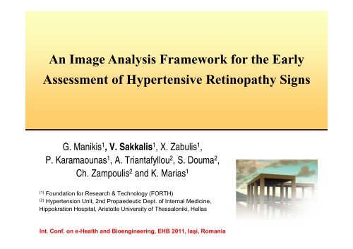
An Image Analysis Framework for the Early<br />
Assessment of Hypertensive Retinopathy Signs<br />
G. Manikis 1 , V. Sakkalis 1 , X. Zabulis 1 ,<br />
P. Karamaounas 1 , A. Triantafyllou 2 , S. Douma 2 ,<br />
Ch. Zampoulis 2 and K. Marias 1<br />
(1)<br />
Foundation for Research & Technology (FORTH)<br />
(2)<br />
Hypertension Unit, 2nd Propaedeutic Dept. of Internal Medicine,<br />
Hippokration Hospital, Aristotle University of Thessaloniki, Hellas<br />
Int. Conf. on e-Health and Bioengineering, EHB 2011, Iaşi, Romania
Introduction<br />
• This study presents a framework for the detection and measurement<br />
of retinal vessels in fundoscopy images.<br />
• The development of advanced fundus cameras along with image processing<br />
techniques offer an accurate, objective, and repeatable re<strong>presentation</strong> of retinal<br />
blood vessels.<br />
• Retinal vessels can be easily visualized with non-invasive techniques providing<br />
valuable information in the diagnosis, classification and surveillance of<br />
retinopathy signs.<br />
• In this study, a method was implemented to segment retinal vessels,<br />
enabling the actual measurement of the vessel diameter, along with a<br />
• Graphical User Interface (GUI) to support automatic and interactive measurement of<br />
vessel diameters at any selected point or region of interest. Editing the vessel<br />
re<strong>presentation</strong> in order to recover from possible segmentation misclassifications.<br />
• The proposed methodology may support vascular risk stratification in<br />
persons with hypertension.
Experimental Data<br />
• Our main aim was to provide a tool which assists<br />
the clinician to handle High Resolution (HR) raw<br />
retinal images. T<strong>here</strong>fore, 10 images of size<br />
2912X2912 were acquired.<br />
• In order to verify the applicability of the proposed<br />
scheme in lower resolutions we evaluated our<br />
methodology on two publicly available datasets;<br />
• DRIVE [1] : 40 retinal images along with manual<br />
segmentations of the vessels with size of 768X584<br />
• STARE [2] : 20 digitized slides to 700X605<br />
• Many of the post-processing steps were applied<br />
only to HR images
Flowchart of the framework<br />
The applied techniques are illustrated<br />
as a pipeline of processes consisting<br />
of two separate modules.<br />
• The first includes the detection and<br />
measurement of vessels in retinal<br />
images.<br />
• The second provides the ability to<br />
validate, edit and represent vessel<br />
information in multiple ways, by<br />
interactively selecting segments of<br />
interest and extracting their statistical<br />
information within spatial regions.<br />
A GUI encapsulates both modules and<br />
increases the automation and usability<br />
of the measurement process.
Pre-Processing 1/2<br />
The green channel of the retinal image was<br />
extracted for use as it provides the greatest<br />
contrast for blood vessels. The acquired<br />
retinal images were transformed from colored<br />
to monochromatic.<br />
HR Image processing was implemented<br />
through distinct blocking. Processing HR<br />
images was a computationally daunting task<br />
w<strong>here</strong> most algorithms fail to achieve because<br />
of memory constraints.<br />
An edge-preserving anisotropic diffusion<br />
filter [3] was applied to smooth the images<br />
within homogeneous regions. Then, the<br />
smoothed image was enhanced using a<br />
contrast limited adaptive histogram<br />
equalization [4].
Pre-Processing 2/2<br />
We then incorporated a multiple scale<br />
filtering technique for vessel enhancement,<br />
based on the eigenvalue analysis of the<br />
Hessian matrix [5] for vessel enhancement.<br />
An iterative thresholding method for<br />
segmenting the blood vessel structure was<br />
then applied for the binarization of the image.<br />
We focused on segmentation techniques that<br />
balance between accuracy and complexity<br />
and Otsu’s thresholding [6] acted fairly well<br />
under those requirements.<br />
Finally, the skeletonization of the segmented<br />
image was required for the characterization of<br />
the morphological structure of the blood<br />
vessel's network.
Evaluation of the Technique in LR Images<br />
• To facilitate the comparison with other well-known retinal vessel segmentation<br />
approaches the performance of the proposed method was evaluated on the DRIVE and<br />
STARE datasets.<br />
• LR images contribute only to the pre-processing approach in order to assess the quality<br />
of the segmented and skeletonized images before entering the post-processing phase.<br />
Performance of vessel segmentation - DRIVE<br />
Method Sensitivity Specificity Accuracy<br />
Human Observer 0.7761 0.9725 0.9473<br />
Supervised Methods Soares [7] 0.7230 0.9762 0.9446<br />
Staal [1] 0.7193 0.9773 0.9441<br />
Niemeijer [8] 0.6793 0.9801 0.9416<br />
Unsupervised Methods Mendonca [9] 0.7344 0.9764 0.9452<br />
Proposed 0.7414 0.9669 0.9371<br />
Vlachos [10] 0.7468 0.9551 0.9285<br />
Jiang [11] 0.6478 0.9625 0.9222<br />
Perez [12] 0.7086 0.9496 0.9181<br />
Chaudhuri [13] 0.2716 0.9794 0.8894<br />
Performance of vessel segmentation - STARE<br />
Method Sensitivity Specificity Accuracy<br />
Human Observer 0.8949 0.9390 0.9354<br />
Supervised Methods Staal [1] 0.6970 0.9810 0.9516<br />
Soares [7] 0.7103 0.9737 0.9480<br />
Unsupervised Methods Mendonca [9] 0.6996 0.9730 0.9440<br />
Perez [12] 0.7506 0.9569 0.9410<br />
Proposed 0.7189 0.9656 0.9318
Post-Processing 1/3<br />
Size-based filtering:<br />
The first stage of the post-processing<br />
procedure is to delete very small isolated<br />
skeleton segments as they typically<br />
correspond to noise artifacts.<br />
Pruning small vessel branches:<br />
The segmentation results obtained from HR<br />
images tend to exhibit a rich structure at<br />
vessel boundaries. In HR images such<br />
structures give rise to spurious skeleton<br />
branches. The effect of filtering the branches<br />
provided a more accurate re<strong>presentation</strong> of<br />
vessels, particularly because vessel<br />
branching points convey important information<br />
in measurements, according to the particular<br />
medical protocol.
Post-Processing 2/3<br />
Vessel width estimation:<br />
• First, a local algorithm is employed to<br />
estimate the vessel width at a skeleton point<br />
p, using both the segmented and the<br />
skeleton images.<br />
• Then, the user provides two input points for<br />
measuring the mean vessel width along a<br />
segment of a vessel.<br />
• The system finally retrieves the skeleton<br />
points of the segment in between the two<br />
points along with their estimated widths and<br />
averages them.<br />
Optical disc detection:<br />
• Optic disk is automatically detected in the acquired image and<br />
estimate of its size is provided. In this way, the application of<br />
medical protocols that are based on measurements around this<br />
disk can be automated.
Post-Processing 3/3<br />
Measuring multiple vessel segments :<br />
The measurement of average width of multiple non-branching<br />
vessels, at a range of distances from the center of the optical<br />
disk is performed. Each vessel segment initiates from the<br />
smaller to the larger circle. Depending on user selection, a<br />
number of statistics can be then estimated.<br />
Statistical Analysis in HR Images:<br />
The ratio of the quantities, CRAE and CRVE<br />
[14], which are determined by measurement<br />
on the arteries and veins detected in the<br />
region of interest respectively, were estimated.<br />
Such measures require the characterization of<br />
vessels, as to if they are veins or arteries and<br />
their mean widths. Hence, a GUI component<br />
is provided to facilitate this characterization by<br />
the medical professional.
User Interface Overview<br />
Semi-automatic pre-processing:<br />
The purpose of implementing this functionality is threefold;<br />
a) user can manually corrects remaining errors in the<br />
segmentation image<br />
b) performs targeted measurements (i.e. focuses on a<br />
particular vessel and ignore its branches)<br />
c) acquire ground truth results by superimposing a basis<br />
segmented image to the original image.<br />
Other utilities:<br />
Saving of data file with measurements, reviewing and<br />
editing of old measurement files.<br />
Image view allowing measurement and skeleton<br />
points calculation using magnification options (visible<br />
image segment in thumbnail view).<br />
Rulers display allowing calibrated grid measurements.
Conclusions<br />
• The presented application employs an image segmentation algorithm,<br />
along with pre-processing and skeletonization techniques in order to<br />
extract a re<strong>presentation</strong> of vessels.<br />
• Additionally, techniques that analyze this re<strong>presentation</strong> and measure<br />
vessel width are introduced and adapted appropriately, in order to<br />
provide measurements according to particular measurement protocols.<br />
• The above functionalities are integrated through a GUI, which assists<br />
the medical professional to perform measurements in an ergonomic<br />
fashion and apply targeted measurements.<br />
• This interface provides also functionalities that allow the clinician to edit<br />
the vessel segmentation result and update the corresponding<br />
measurements, in order to recover from segmentation errors.
Future Work<br />
• Future work will be pursued along two research avenues.<br />
• The first regards the improvement of the segmentation algorithm.<br />
• The second regards the registration of fundoscopy images of the same patient,<br />
acquired across large time intervals.<br />
• The goal is to provide medical professionals with the capability of<br />
automatically comparing vessel measurements, thus offering a<br />
valuable tool in the monitoring, diagnosis, and estimation of the<br />
condition of the cardiovascular system.
References<br />
1. Staal, J.; Abramoff, M.D.; Niemeijer, M.; Viergever, M.A.; van Ginneken, B.; , "Ridge-based vessel segmentation in color images of the<br />
retina," Medical Imaging, IEEE Transactions on , vol.23, no.4, pp.501-509, April 2004.<br />
2. A. Hoover, V. Kouznetsova, and M. Goldbaum, “Locating blood vessels in retinal images by piecewise threshold probing of a matched<br />
filter response,” IEEE Trans. Med. Imag., vol. 19, no. 3, pp. 203–211, Mar. 2000.<br />
3. P. Perona, J. Malik, “Scale-space and edge detection using anisotropic diffusion,” IEEE Trans. PAMI, V. 12, no. 7, pp. 629-639, July<br />
1990.<br />
4. K. Zuiderveld. Contrast limited adaptive histogram equalization. Graphics gems IV, pages 474-485, 1994.<br />
5. A.F. Frangi, W.J. Niessen, K.L. Vincken, M.A. Viergever (1998). Multiscale vessel enhancement filtering. In Medical Image Computing<br />
and Computer-Assisted Intervention - MICCAI'98, W.M. Wells, A. Colchester and S.L. Delp (Eds.), Lecture Notes in Computer Science,<br />
vol. 1496 - Springer Verlag, Berlin, Germany, pp. 130-137.<br />
6. Otsu, N., "A Threshold Selection Method from Gray-Level Histograms," IEEE Transactions on Systems, Man, and Cybernetics, Vol. 9,<br />
No. 1, 1979, pp. 62-66.<br />
7. João V. B. Soares, Jorge J. G. Leandro, Roberto M. Cesar Jr., Herbert F. Jelinek, and Michael J. Cree, “Retinal Vessel Segmentation<br />
Using the 2-D Gabor Wavelet and Supervised Classification,” IEEE Transactions on medical Imaging, vol. 25, no. 9, September 2006.<br />
8. Niemeijer M., Staal J., van Ginneken B, Loog M, and Abràmoff M. D., “Comparative study of retinal vessel segmentation methods on a<br />
new publicly available database,” in Proc. SPIE Med. Imag., M. Fitzpatrick and M. Sonka, Eds., 2004, vol. 5370, pp. 648–656.<br />
9. A. Mendonca and A. Campilho. Segmentation of retinal blood vessels by combining the detection of centerlines and morphological<br />
reconstruction. IEEE Transactions on Medical Imaging, 25(9):1200-1213, September 2006.<br />
10. M. Vlachos, E. Dermatas, Multi-scale retinal vessel segmentation using line tracking, Computerized Medical Imaging and Graphics,<br />
Volume 34, Issue 3, April 2010, Pages 213-227.<br />
11. Xiaoyi Jiang; Mojon, D.; , "Adaptive local thresholding by verification-based multithreshold probing with application to vessel<br />
detection in retinal images," Pattern Analysis and Machine Intelligence, IEEE Transactions on , vol.25, no.1, pp.131-137, Jan. 2003.<br />
12. M.E. Martínez-Pérez, A.D. Hughes, A.V. Stanton, S.A. Thom, A.A. Bharath, and K.H. Parker, "Retinal Blood Vessel Segmentation by<br />
Means of Scale-Space Analysis and Region Growing", in Proc. MICCAI, 1999, pp.90-97.<br />
13. S. Chaudhuri, S. Chatterjee, N. Katz, M. Nelson, and M. Goldbaum,“Detection of blood vessels in retinal images using twodimensional<br />
matched filters,” IEEE Trans. Med. Imag., pp. 263–269, 1989.<br />
14. Hubbard LD, Brothers RJ, King WN, Clegg LX, Klein R, Cooper LS, et al. Methods for evaluation of retinal microvascular abnormalities<br />
associated with hypertension/sclerosis in the Atherosclerosis Risk in Communities Study. Ophthalmology;106(12):2269-80, Dec 1999.
Acknowledgments<br />
• The authors would like to thank the Hypertension Unit of the 2 nd Propaedeutic Dept. of<br />
Internal Medicine, Hippokration Hospital in Thessaloniki for providing the High Resolution<br />
dataset.<br />
• This work was supported in part by the European Commission under the TUMOR (FP7-<br />
ICT-2009.5.4-247754) project.<br />
Vangelis Sakkalis<br />
sakkalis@ics.forth.gr