Elbow Arthroscopy: Basic Technique, Portals and Anatomy - IASM
Elbow Arthroscopy: Basic Technique, Portals and Anatomy - IASM
Elbow Arthroscopy: Basic Technique, Portals and Anatomy - IASM

You also want an ePaper? Increase the reach of your titles
YUMPU automatically turns print PDFs into web optimized ePapers that Google loves.
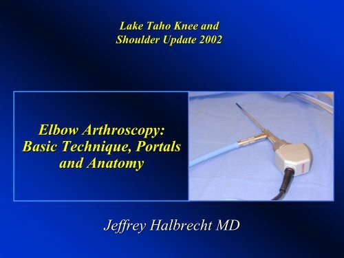
Lake Taho Knee <strong>and</strong><br />
Shoulder Update 2002<br />
<strong>Elbow</strong> <strong>Arthroscopy</strong>:<br />
<strong>Basic</strong> <strong>Technique</strong>, <strong>Portals</strong><br />
<strong>and</strong> <strong>Anatomy</strong><br />
Jeffrey Halbrecht MD
<strong>Elbow</strong> <strong>Arthroscopy</strong><br />
Experience<br />
Uncommonly scoped joint<br />
Based on AAOS Survey:<br />
•Only 7.6% of Orthopaedic Surgeons<br />
perform elbow arthroscopy<br />
•<strong>Elbow</strong> arthroscopy constitutes only<br />
2% of all orthopedic procedures
<strong>Elbow</strong> <strong>Arthroscopy</strong><br />
The most hazardous<br />
joint in terms of risk<br />
to intimately<br />
associated<br />
neurovascular<br />
structures<br />
Poses greater<br />
technical<br />
challenges/neurologic<br />
risks than<br />
knee/shoulder scope
Indications<br />
•Removal of Loose body(ies)<br />
•Olecranon<br />
Impingement<br />
Overload<br />
•OCD<br />
Capitellum<br />
•Debridement<br />
Degenerative<br />
Arthritis<br />
•Synovectomy<br />
•Diagnostic Evaluation<br />
•Capsular Release<br />
•Fx<br />
evaluation/Tx<br />
•Debridement<br />
Lateral<br />
Epicondylitis
Positioning<br />
Supine
Positioning<br />
Prone
Positioning<br />
Lateral Decubitus
<strong>Portals</strong> <strong>Anatomy</strong><br />
Familiarity more critical<br />
than w/ any other<br />
arthroscopic procedure<br />
•Anterior<br />
•Posterior
Anterior <strong>Portals</strong><br />
• Assoc w/ greatest potential<br />
risk to nv structures<br />
• Thorough exam of<br />
anterior compartment<br />
important in all cases<br />
• Lateral<br />
• Medial
Lateral <strong>Portals</strong>
Anterolateral Portal<br />
Initially described by Andrews <strong>and</strong> Carson<br />
(<strong>Arthroscopy</strong>, 1:97-107, 107, 1985)<br />
•3cm distal <strong>and</strong> 1-2cm 1<br />
anterior to lateral epicondyle<br />
•Penetrates Extensor Carpi Radialis Muscle
Anterolateral Portal<br />
Visualization<br />
•Trochlea<br />
•Distal<br />
Humerus<br />
•Coronoid<br />
Process <strong>and</strong><br />
Fossa<br />
•Medial <strong>and</strong><br />
Superior Capsule
Anterolateral Portal<br />
Structures @ Risk<br />
•LABCN<br />
ave. . 7.6mm (0-20mm), in<br />
contact 43%<br />
•Radial nerve is “at extreme risk”<br />
ave. . 4.9mm (2-10mm)<br />
–Recommended placement 3cm from LE results<br />
in entry point that is distal I most pts, below jt in<br />
some pts<br />
•PIN<br />
(fixed @ Arcade of Froshe)<br />
poorly protected, even in<br />
flexion, close to distal<br />
capsular attachment (little<br />
chance of displacement w/ jt<br />
distension)<br />
•No longer used by some, if used,<br />
established from “inside-out”,<br />
proximal to radial head
Proximal Lateral Portal<br />
Described by Strothers, Day <strong>and</strong> Regan<br />
(<strong>Arthroscopy</strong>, Vol. 11, No4, 449-457, 457, Aug 1995)<br />
•1-2cm proximal to the lateral epicondyle, , pierces Brachioradialis<br />
<strong>and</strong> distal Brachialis to reach lateral elbow capsule, lying directly<br />
on anterior surface of the humerus<br />
•Improved margin of safety, supplants earlier AL portal
Proximal Lateral Portal<br />
Visualization<br />
•Anterior/Lateral<br />
aspect of Radial head<br />
<strong>and</strong> capitellum<br />
•Medial aspect of<br />
elbow including:<br />
•Coronoid<br />
Process/Fossa<br />
•Distal<br />
Humerus<br />
•Trochlea<br />
•Medial Capsule<br />
0449F09_.95
Proximal Lateral Portal<br />
Structures @ Risk<br />
•LABCN<br />
branch<br />
ave 6.1mm (0-<br />
14mm), in contact<br />
29%<br />
•Radial nerve 2x<br />
distance vs AL<br />
portal (ave(<br />
9.9mm<br />
in flexion)
Medial <strong>Portals</strong>
Anteromedial Portal<br />
•2cm distal, 2cm ant to medial epicondyle<br />
•Passes through common flexor origin, then<br />
beneath or penetrates brachialis to enter capsule
Anteromedial Portal<br />
Visualization<br />
Affords excellent<br />
view of both<br />
radiocapitellar<br />
<strong>and</strong> ulnohumeral<br />
joints, coronoid<br />
fossa, capitellum<br />
<strong>and</strong> superior<br />
capsule<br />
Radial<br />
Head<br />
Coronoid<br />
Process<br />
Capitellum<br />
Trochlea
Anteromedial Portal<br />
Structures @ Risk<br />
•MABCN<br />
Branch<br />
ave 1mm (0-5mm),<br />
in contact 71%<br />
•Median<br />
Nerve ave<br />
7mm (5-13mm) w/<br />
flexion<br />
•Brachial Artery<br />
ave 15mm (8-<br />
20mm)
Proximal Medial Portal<br />
Described by Poehling<br />
(<strong>Arthroscopy</strong>, 5:222-224, 224, 1989)<br />
•2cm proximal to medial<br />
epicondyle, , directly anterior<br />
to intermuscular septum<br />
•Contact maintained w/<br />
distal humerus to avoid med<br />
nerve risk<br />
•scope lies deep to<br />
brachialis muscle
Proximal Medial Portal<br />
Visualization<br />
•Similar to AM portal<br />
•Excellent view, esp<br />
distal part of jt<br />
•Radiocapitellar<br />
jt<br />
•Ulnohumeral<br />
jt<br />
•70 Deg scope<br />
proximal capsule<br />
visualization prn
Proximal Medial Portal<br />
Structures @ Risk<br />
•MABCN<br />
very close, ave.<br />
2.3mm (0-9mm), in contact<br />
56%<br />
•Median Nerve ave. . 12.4mm<br />
(7-20mm)<br />
•Brachial Artery ave. . 18mm<br />
•Ulnar Nerve av4e 12mm (7-<br />
18mm)<br />
Important to confirm location,<br />
stability of Ulnar nerve before<br />
establishing any medial<br />
portals
Proximal Medial Portal<br />
Recommended that<br />
proximal medial<br />
portal be first portal<br />
used in most cases of<br />
elbow arthroscopy<br />
•Safer than<br />
anteromedial portal<br />
•Excellent view<br />
AM<br />
PM
Posterior <strong>Portals</strong><br />
All are relatively<br />
safe w/ distance to<br />
closest cutaneous<br />
or major nerve 15-<br />
20mm<br />
–Mid or Direct<br />
Lateral (“Soft(<br />
spot”)<br />
–Central Posterior<br />
–Posterolateral
Mid-Lateral Portal<br />
AKA “Soft spot” portal<br />
•Located within triangle<br />
formed by olecranon,<br />
lateral epicondyle <strong>and</strong><br />
radial head<br />
•Pierces<br />
Anconeus muscle<br />
•Initial joint insufflation<br />
•Only portal to provide<br />
easy access to posterior<br />
capitellum <strong>and</strong> radioulnar<br />
joint
Mid-Lateral Portal<br />
Visualization<br />
•Posterolateral<br />
radial head<br />
•Capitellum<br />
•Trochlea<br />
notch<br />
•Olecranon
Mid-Lateral Portal<br />
Structures @<br />
Risk<br />
Articular<br />
Cartilage
Central Posterior Portal<br />
•3cm<br />
proximal to<br />
Olecranon tip<br />
•Passes<br />
through<br />
triceps tendon<br />
at its medial<br />
margin
Central Posterior Portal<br />
Visualization<br />
•Olecranon<br />
Tip<br />
Olecranon<br />
Tip<br />
•Olecranon<br />
Fossa<br />
•Posteromedi<br />
Olecranon<br />
Fossa<br />
al Gutter
Central Posterior Portal<br />
Structures @<br />
Risk<br />
Ulnar Nerve<br />
ave 19.1mm<br />
(15-25mm)
Posterolateral Portal<br />
3 cm proximal<br />
to olecranon<br />
tip, adjacent to<br />
lateral edge of<br />
triceps tendon<br />
Passes through<br />
triceps muscle
Posterolateral Portal<br />
Visualization<br />
•Olecranon<br />
Fossa, , Tip<br />
•Posterior<br />
trochlea
Posterolateral Portal<br />
Structures @<br />
Risk<br />
PABCN<br />
branch
<strong>Technique</strong> - Video
Complications<br />
• Relatively higher incidence compared to<br />
other scope procedures (3-14%)<br />
• Most significant is nerve injury (0-14%)<br />
• Radial/PIN most common<br />
(Median/Ulnar<br />
Ulnar/AIN/MABC)<br />
(Rodeo etal JBJS A 1993;75:917-26)<br />
• Other reported complications include:<br />
• Compartment syndrome<br />
• Septic Arthritis<br />
• Superficial infection<br />
• Persistent Drainage
Complications
Complications<br />
473 cases<br />
11.8% complication rate<br />
0.8% (N=4) “serious” – Infection<br />
11% (N=50) “minor”<br />
•Prolonged drainage/superficial infection @ portal site (N=33)<br />
•Persistent minor contracture
Avoiding Complications<br />
• Most serious complications are reported assoc.<br />
w/ advanced procedures, which ought to be<br />
performed only by experienced surgeons<br />
• Most minor complications however, are probably<br />
avoidable by<br />
– Familiarity w/ normal anatomy/portals<br />
– Attention to technique:<br />
• Incise skin only w/ #11 blade<br />
• Blunt dissection to/through capsule<br />
• Maintain portal sites (avoid repeated re-entry)<br />
entry)<br />
• Beware use of pump<br />
• Limit procedure duration
THANK YOU