

Screening tests - IARC
Screening tests - IARC
Screening tests - IARC

You also want an ePaper? Increase the reach of your titles
YUMPU automatically turns print PDFs into web optimized ePapers that Google loves.
Chapter 2<br />
<strong>Screening</strong> <strong>tests</strong><br />
Cervical cytology<br />
Cytological testing involves collection<br />
of exfoliated cells from the cervix and<br />
microscopic examination of these cells<br />
after staining. The concept of utilizing<br />
exfoliative cytology to identify women<br />
with invasive cervical cancer was introduced<br />
by Papanicolaou and Babes in<br />
the 1920s (Papanicolaou, 1928;<br />
Papanicolaou & Traut, 1941). Subsequently,<br />
Papanicolaou refined the technique<br />
and demonstrated that conventional<br />
cytology could also be used to<br />
identify precancerous lesions of the<br />
cervix (Papanicolaou, 1954). The shift<br />
in emphasis from using cytology as a<br />
way to identify cases of invasive cervical<br />
cancer to using it to identify women<br />
with high-grade precursor lesions who<br />
are at risk for subsequently developing<br />
invasive cervical cancer was highly significant,<br />
as it meant that cervical cytology<br />
could be used to actually prevent<br />
the development of cervical cancer<br />
rather than simply identify cases at an<br />
early stage. In the 1960s, cervical cytology<br />
began to be widely used in many<br />
developed countries as a technique for<br />
cervical cancer prevention. Although<br />
the method was introduced over a half<br />
century ago, cytology-based screening<br />
programmes continue to be the mainstay<br />
of cervical cancer prevention.<br />
Cytological terminology<br />
Papanicolaou classes<br />
The terminology developed by<br />
Papanicolaou separated cervical<br />
cytological findings into five categories<br />
or classes (Table 14) (Papanicolaou,<br />
1954). At the time the classification was<br />
developed, there was only limited<br />
understanding of the relationship<br />
between cervical cancer precursor<br />
lesions and invasive cancers. Moreover,<br />
invasive cervical cancer was common<br />
and cervical cytology was initially<br />
viewed as a way of detecting earlystage,<br />
easily treated cancers.Therefore,<br />
the Papanicolaou classification system<br />
focused on how closely the exfoliated<br />
cells resembled those from an invasive<br />
cancer. Although the Papanicolaou<br />
classification was modified many times<br />
over the years, the problems inherent in<br />
this classification remain. For example,<br />
although is clear how Class I and Class<br />
V translate into known histological entities,<br />
Classes II, III or IV correlate less<br />
clearly with standard histopathological<br />
lesions. For example, should a carcinoma<br />
in situ be classified as Class IV<br />
and all grades of dysplasia as Class III,<br />
or does mild dysplasia correspond to<br />
Class II? There was also no consensus<br />
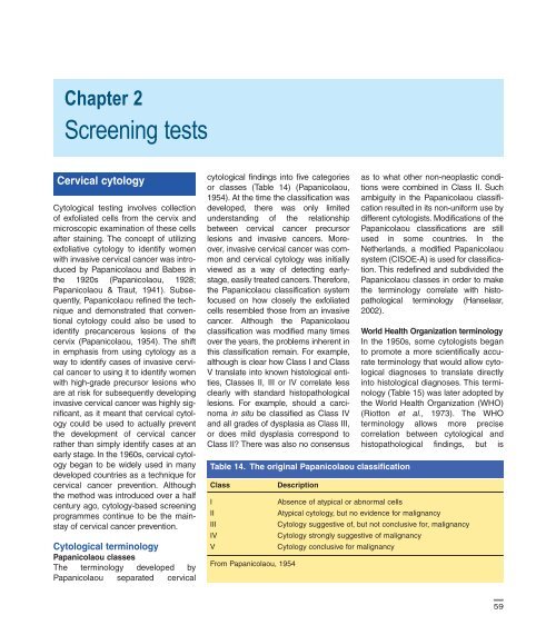
Table 14. The original Papanicolaou classification<br />
Class<br />
I<br />
II<br />
III<br />
IV<br />
V<br />
From Papanicolaou, 1954<br />
Description<br />
as to what other non-neoplastic conditions<br />
were combined in Class II. Such<br />
ambiguity in the Papanicolaou classification<br />
resulted in its non-uniform use by<br />
different cytologists. Modifications of the<br />
Papanicolaou classifications are still<br />
used in some countries. In the<br />
Netherlands, a modified Papanicolaou<br />
system (CISOE-A) is used for classification.<br />
This redefined and subdivided the<br />
Papanicolaou classes in order to make<br />
the terminology correlate with histopathological<br />
terminology (Hanselaar,<br />
2002).<br />
World Health Organization terminology<br />
In the 1950s, some cytologists began<br />
to promote a more scientifically accurate<br />
terminology that would allow cytological<br />
diagnoses to translate directly<br />
into histological diagnoses. This terminology<br />
(Table 15) was later adopted by<br />
the World Health Organization (WHO)<br />
(Riotton et al., 1973). The WHO<br />
terminology allows more precise<br />
correlation between cytological and<br />
histopathological findings, but is<br />
Absence of atypical or abnormal cells<br />
Atypical cytology, but no evidence for malignancy<br />
Cytology suggestive of, but not conclusive for, malignancy<br />
Cytology strongly suggestive of malignancy<br />
Cytology conclusive for malignancy<br />
59
<strong>IARC</strong> Handbooks of Cancer Prevention Volume 10: Cervix cancer screening<br />
Table 15. Comparison of different terminologies used for cytologic<br />
reporting<br />
Papanicolaou World Health CIN Bethesda System<br />
class system Organization<br />
Class I<br />
Class II<br />
difficult to use since it includes a number<br />
of different entities. These are mild<br />
dysplasia, moderate dysplasia, severe<br />
dysplasia, epidermoid carcinoma in<br />
situ, epidermoid carcinoma in situ with<br />
minimal stromal invasion, invasive epidermoid<br />
microcarcinoma and invasive<br />
epidermoid carcinoma. Studies have<br />
shown high rates of intra-observer and<br />
inter-observer variation with cervical<br />
cytology in general (Yobs et al., 1987;<br />
Klinkhamer et al., 1988; Selvaggi,<br />
1999; Stoler & Schiffman, 2001).<br />
Classification systems that utilize more<br />
diagnostic categories have inherently<br />
higher rates of variability than do classification<br />
systems with fewer diagnostic<br />
categories (Yobs et al., 1987; Selvaggi,<br />
1999; Stoler & Schiffman, 2001; Kundel<br />
& Polansky, 2003). Other limitations of<br />
the WHO terminology are that it does<br />
not adequately deal with non-neoplastic<br />
conditions nor with specimen adequacy.<br />
Despite its limitations, many<br />
cytologists around the world continue to<br />
utilize the WHO terminology.<br />
Within normal limits<br />
Benign cellular changes<br />
ASC<br />
Mild dysplasia CIN1 Low-grade SIL<br />
Class III Moderate dysplasia CIN2<br />
Severe dysplasia<br />
CIN3 High-grade SIL<br />
Class IV Carcinoma in situ CIN3<br />
Class V Microinvasive carcinoma Invasive Invasive carcinoma<br />
Invasive carcinoma carcinoma<br />
Abbreviations: CIN, Cervical intraepithelial neoplasia; ASC, Atypical squamous cells;<br />
SIL, Squamous intraepithelial lesions<br />
From Papanicolau (1954), Riotton et al. (1973), Richart (1968, 1973), Solomon et al.<br />
(2002)<br />
Cervical intraepithelial neoplasia (CIN)<br />
terminology<br />
As a result of advances in understanding<br />
of the pathogenesis of cervical<br />
cancer, the cervical intraepithelial neoplasia<br />
(CIN) terminology was<br />
introduced in the late 1960s (Richart,<br />
1968, 1973). The CIN concept emphasized<br />
that dysplasia and carcinoma in<br />
situ represent different stages of the<br />
same biological process, rather than<br />
separate entities. It had a major impact<br />
on how precancerous lesions were<br />
treated, since all types of cervical<br />
cancer precursor were considered to<br />
form a biological and clinical continuum.<br />
In the CIN terminology, mild<br />
dysplasia is classified as CIN 1,<br />
moderate dysplasia as CIN 2 and<br />
severe dysplasia and carcinoma in situ<br />
are grouped together and classified as<br />
CIN 3 (Table 15). The CIN terminology<br />
is still widely used in many countries<br />
for reporting both histological and<br />
cytological diagnoses.<br />
The Bethesda System terminology<br />
By the late 1980s, advances in our<br />
understanding of the role of human<br />
papillomavirus (HPV) in the pathogenesis<br />
of cervical cancer needed to be<br />
incorporated into cytological terminology.<br />
Moreover, it was recognized that<br />
clinicians were often confused by the<br />
non-standard terminologies used to<br />
report cytological results and that this<br />
had a potential adverse impact on<br />
clinical care.Therefore, in 1988, the US<br />
National Institutes of Health held a<br />
conference in Bethesda, Maryland, to<br />
develop a new terminology that would<br />
ensure better standardization and<br />
accommodate current concepts of the<br />
pathogenesis of cervical disease,<br />
so that cytological findings could be<br />
transmitted to clinicians as accurately<br />
and concisely as possible. The<br />
terminology that resulted is known as the<br />
Bethesda System. In 1991 the Bethesda<br />
System was slightly modified on the<br />
basis of experience obtained during the<br />
first three years of its use and it was<br />
further modified in 2001 to take into<br />
account the results of new research and<br />
over a decade of experience with the terminology<br />
(Luff, 1992; Solomon et al.,<br />
2002).<br />
The Bethesda system is viewed<br />
with caution in the United Kingdom,<br />
which retains its own British Society for<br />
Clinical Cytology (BSCC) ‘dyskaryosis’<br />
terminology (British Society for Clinical<br />
Cytology, 1997). This can largely be<br />
mapped to the Bethesda system for<br />
comparison of data in a research setting,<br />
except for the borderline category,<br />
which may include koilocytes. Due<br />
largely to the robust nature of the<br />
‘severe dyskaryosis’ category, fear of<br />
increasing the overtreatment inherent<br />
in cervical screening and the difficulty<br />
of achieving inter- and intra-observer<br />
agreement on ‘low-grade’ reports, the<br />
United Kingdom continues to use this<br />
terminology.<br />
There are three distinct parts to<br />
each Bethesda System report: a state-<br />
60
<strong>Screening</strong> <strong>tests</strong><br />
ment of the specimen adequacy, a<br />
general categorization and a descriptive<br />
diagnosis (Table 16). These categories<br />
assist clinicians by providing<br />
answers to three basic questions: (1)<br />
Do I need to repeat the cervical cytology?<br />
(2) Was the cervical cytology<br />
normal? (3) If the specimen was not<br />
completely normal, what specifically<br />
was wrong?<br />
Because cervical cytology is considered<br />
a screening, rather than diagnostic,<br />
test, the 2001 Bethesda<br />
System reports cytological findings as<br />
an ‘interpretation’ or ‘result’ rather than<br />
as a ‘diagnosis’. This stresses the fact<br />
that cytological findings usually need<br />
to be interpreted in the light of clinical<br />
findings, and that the test is designed<br />
to reflect the underlying disease state<br />
but does not always do so.<br />
In this Handbook, the Bethesda<br />
System (SIL) terminology is used for<br />
cytological interpretation of screening<br />
<strong>tests</strong> unless otherwise reported.<br />
Specimen adequacy<br />
The 2001 Bethesda System requires<br />
that every cervical cytology specimen<br />
be assessed with respect to its adequacy<br />
(Solomon et al., 2002).<br />
Specimens are classified into one of<br />
two categories: ‘satisfactory for evaluation’<br />
or ‘unsatisfactory for evaluation’.<br />
This represents a departure from the<br />
1991 Bethesda System, which also<br />
included a third category for specimen<br />
adequacy that was called ‘satisfactory<br />
for evaluation but limited by…..’ or<br />
SBLB. This ‘satisfactory but limited<br />
by…’ category was most frequently<br />
used when a specimen lacked either<br />
endocervical cells or squamous metaplasia<br />
from the transformation zone but<br />
was in all other aspects ‘satisfactory’.<br />
With the 2001 Bethesda System,<br />
cytology specimens previously classified<br />
as ‘SBLB’ are classified as ‘satisfactory<br />
for evaluation’ and a quality<br />
indicator comment is made indicating<br />
what limiting features are present.<br />
A ‘satisfactory for evaluation’ specimen<br />
must be appropriately labelled. To<br />
ensure proper identification, the<br />
woman’s name or identifying number<br />
should be written on, or affixed to, the<br />
slide before it is sent to the cytology<br />
laboratory. Cytology laboratories<br />
should not accept unlabelled slides<br />
and should return them to the submitting<br />
clinician. It is also critical that<br />
the smear-taker provide pertinent clinical<br />
information to the laboratory that<br />
will evaluate the specimen, including<br />
the woman’s age, date of last menstrual<br />
period, previous history of<br />
abnormal cervical cytology specimens<br />
or treatment for cervical disease, and<br />
the source of the specimen (e.g.,<br />
vaginal or cervix). The minimal cellular<br />
requirements for a specimen to be<br />
considered ‘satisfactory for evaluation’<br />
in the 2001 Bethesda System<br />
vary depending on whether the specimen<br />
is a conventional cytology specimen<br />
or a liquid-based cytology specimen.<br />
For classification of conventional<br />
cytology specimens as ‘satisfactory for<br />
evaluation’, an estimated 8000 to<br />
12 000 well visualized squamous cells<br />
need to be present. For liquid-based<br />
cytology specimens, an estimated<br />
5000 cells need to be present (Figure<br />
29). Although the selection of these<br />
cut-offs is fairly arbitrary, the limit of<br />
5000 cells for a liquid-based cytology<br />
specimen to be classified as ‘satisfactory<br />
for evaluation’ is based on a cellcounting<br />
study in which referent<br />
samples were diluted to produce<br />
preparations with defined numbers of<br />
squamous cells (Studeman et al.,<br />
2003). A clear demarcation in<br />
sensitivity was observed using the<br />
SurePath procedure (see below)<br />
between specimens with less than<br />
5000 squamous cells and those with<br />
5000 cells or more: the sensitivity for a<br />
reference diagnosis case of low-grade<br />
squamous intraepithelial lesion (LSIL)<br />
increased from 73% for specimens<br />
with less than 5000 squamous cells to<br />
98% for preparations with over 5000<br />
cells.<br />
There is much controversy over the<br />
importance of identifying a transformation<br />
zone component (e.g., squamous<br />
metaplastic cells) or endocervical cells<br />
in a cervical cytology preparation.<br />
Because the majority of high-grade<br />
precursor lesions arise within the<br />
transformation zone, it was widely<br />
believed until recently that specimens<br />
lacking a transformation zone component<br />
(TZC) or endocervical cells (EC)<br />
should be considered somewhat less<br />
than ‘satisfactory for evaluation’. This<br />
view is supported by several studies<br />
that have shown the prevalence of SIL<br />
to be higher among cytology specimens<br />
that contain TZC/EC than<br />
among those that do not (Vooijs et al.,<br />
1985; Mitchell & Medley, 1992;<br />
Szarewski et al., 1993; Mintzer et al.,<br />
1999). However, other studies have<br />
failed to confirm this association and,<br />
perhaps more importantly, several retrospective<br />
longitudinal cohort studies<br />
have found that women lacking<br />
TZC/EC are no more likely on followup<br />
to be diagnosed with squamous<br />
lesions than are women whose specimens<br />
contain TZC/EC (Mitchell &<br />
Medley, 1991; Mitchell, 2001). One retrospective<br />
case–control study of true<br />
Figure 29 Liquid-based cytology:<br />
superficial and intermediate squamous<br />
cells and a cluster of columnar<br />
endocervical cells (obj. 5x)<br />
61
<strong>IARC</strong> Handbooks of Cancer Prevention Volume 10: Cervix cancer screening<br />
positive and false negative cervical<br />
cytology specimens from women with<br />
CIN 3 found no difference in true positive<br />
rates between cases with or without<br />
TZC/EC (O’Sullivan et al., 1998). A<br />
prospective study of women with normal<br />
cytology at entry found that<br />
although specimens containing EC at<br />
the subsequent test were at significantly<br />
higher risk of both low- and<br />
high-grade squamous intraepithelial<br />
lesions than those without EC, the<br />
presence or absence of EC at entry<br />
had no significant effect (Mitchell,<br />
2001). In another compelling study on<br />
the lack of importance of EC, all negative<br />
cervical cytology specimens<br />
obtained in the Netherlands between<br />
1990 and 1991 were matched with<br />
results of subsequent cytological and<br />
histological examinations (Bos et al.,<br />
2001). There was no significant difference<br />
in the number of women subsequently<br />
diagnosed with CIN between<br />
women whose initial cytology specimens<br />
contained EC and those that did<br />
not. Moreover, the proportions of<br />
women diagnosed with cervical cancer<br />
were the same in both groups. It is also<br />
important to recognize that EC are less<br />
frequently found in cervical cytology<br />
specimens from women using oral<br />
contraceptives, who are pregnant or<br />
who are postmenopausal (Davey et al.,<br />
2002). It has therefore been argued<br />
that specimens lacking EC or a TZC<br />
should not be considered unsatisfactory<br />
and may not need to be repeated<br />
(Davey et al., 2002; Birdsong, 2001;<br />
Bos et al., 2001). The 2001 Bethesda<br />
System recommends that reports<br />
should state whether or not EC or a<br />
TZC are present. Specimens lacking<br />
endocervical cells or squamous metaplastic<br />
cells should be classified as<br />
‘satisfactory for evaluation’ and the<br />
quality indicator comment should indicate<br />
that these components are not<br />
present. The numeric criterion for stating<br />
that such a component is present is<br />
10 well preserved endocervical or<br />
squamous metaplastic cells. Specimens<br />
in which inflammation, blood or<br />
poor preservation cause 50–75% of<br />
the epithelial cells to be obscured<br />
should be classified as ‘satisfactory for<br />
evaluation’, but a quality indicator comment<br />
made indicating that there are<br />
partially obscuring factors.<br />
A specimen is classified as ‘unsatisfactory<br />
for evaluation’ when either the<br />
minimal number of epithelial cells<br />
required for interpretation is not present<br />
or blood, inflammation or poor<br />
preservation obscures more than 75%<br />
of the epithelial cell component (Figure<br />
30). Cases which the laboratory cannot<br />
process, such as those received<br />
unlabelled, are also classified as<br />
‘unsatisfactory for evaluation’ and no<br />
interpretation is rendered.<br />
General categorization<br />
The ‘general categorization’ is included<br />
as an optional component of the<br />
Bethesda System to allow clinicians to<br />
readily determine whether any degree<br />
of abnormality is present. With the<br />
2001 Bethesda System, all cytology<br />
specimens are classified into one of<br />
three general categories.These include<br />
‘negative for intraepithelial lesion or<br />
malignancy’, ‘epithelial cell abnormalities’<br />
and ‘other’. These categories are<br />
mutually exclusive and specimens<br />
should be categorized according to the<br />
most significant findings.<br />
‘Negative for intraepithelial lesion<br />
or malignancy’ includes all specimens<br />
in which no intraepithelial lesion or<br />
malignancy is identified. This includes<br />
cases with common infections such as<br />
Trichomonas vaginalis, fungal organisms<br />
such as Candida species,<br />
Actinomyces or herpes simplex virus,<br />
a shift in bacterial flora consistent with<br />
bacterial vaginosis, reparative/reactive<br />
changes, changes associated with<br />
intrauterine devices, radiation reactions<br />
or atrophic changes.<br />
The category ‘epithelial cell abnormalities’<br />
includes both squamous and<br />
glandular cell abnormalities. This category<br />
is used whenever there are<br />
epithelial cell abnormalities, except for<br />
benign reactive or reparative changes.<br />
The 2001 Bethesda System introduced<br />
a new general categorization,<br />
‘other’. This category is used whenever<br />
there are no morphological abnormalities<br />
in the cells per se, but there are<br />
findings indicative that the woman is at<br />
some increased risk. An example is<br />
when benign-appearing endometrial<br />
cells are identified in a woman 40<br />
years of age or older.<br />
Squamous cell abnormalities<br />
Atypical squamous cells (ASC):<br />
Epithelial cell abnormalities are subdivided<br />
into four categories (Table 16).<br />
‘Atypical squamous cells’ (ASC) is<br />
used when cytological findings are<br />
considered suggestive but not diagnostic<br />
of a squamous intraepithelial<br />
lesion (SIL) (Figure 31). The term ASC<br />
was retained in the 2001 Bethesda<br />
System because of the wide recognition<br />
that these cells imply a significant<br />
risk for an underlying high-grade cervical<br />
intraepithelial lesion (SIL). In various<br />
studies, the prevalence of CIN 2 or<br />
3 in women with ASC has varied<br />
between 10% and 20% (Wright et al.,<br />
2002a). The ASC category roughly<br />
correlates with the ‘borderline<br />
dyskaryosis’ category used in the<br />
Figure 30 Unsatisfactory smear<br />
because of inflammation. Cell cluster<br />
difficult to analyse. Repeat after local<br />
treatment (obj. 10x)<br />
62
<strong>Screening</strong> <strong>tests</strong><br />
Table 16. The 2001 Bethesda system<br />
Specimen adequacy<br />
Satisfactory for evaluation (note presence/absence of endocervical transformation<br />
zone component)<br />
Unsatisfactory for evaluation (specify reason)<br />
- Specimen rejected/not processed (specify reason)<br />
- Specimen processed and examined, but unsatisfactory for evaluation of epithelial<br />
abnormality because of (specify reason)<br />
General categorization (optional)<br />
Negative for intraepithelial lesion or malignancy<br />
Epithelial cell abnormality<br />
Other<br />
Interpretation/result<br />
Negative for intraepithelial lesion or malignancy<br />
Organisms<br />
Trichomonas vaginalis<br />
Fungal organisms morphologically consistent with Candida species<br />
Shift in flora suggestive of bacterial vaginosis<br />
Bacteria morphologically consistent with Actinomyces species<br />
Cellular changes consistent with herpes simplex virus<br />
Other non-neoplastic findings (Optional to report; list not comprehensive)<br />
Reactive cellular changes associated with inflammation (includes typical<br />
repair), radiation, intrauterine contraceptive device<br />
Glandular cells status posthysterectomy<br />
Atrophy<br />
Epithelial cell abnormalities<br />
Squamous cell<br />
Atypical squamous cell (ASC)<br />
of undetermined significance (ASCUS)<br />
cannot exclude HSIL (ASC-H)<br />
Low-grade squamous intraepithelial lesion (LSIL)<br />
High-grade squamous intraepithelial lesion (HSIL) (can use modifiers to<br />
separate into CIN 2 and CIN 3)<br />
Squamous-cell carcinoma<br />
Glandular cell<br />
Atypical glandular cells (AGC) (specify endocervical, endometrial or not<br />
otherwise specified)<br />
Atypical glandular cells, favour neoplastic (specify endocervical or not<br />
otherwise specified)<br />
Endocervical adenocarcinoma in situ (AIS)<br />
Adenocarcinoma<br />
Other (List not comprehensive)<br />
Endometrial cells in a woman ≥ 40 years of age<br />
From Solomon et al. (2002)<br />
United Kingdom. However, neither the<br />
WHO terminology nor the CIN terminology<br />
incorporates a category similar<br />
to ASC. The 2001 Bethesda System<br />
also clearly separates ASC from reactive/reparative<br />
changes and an interpretation<br />
of ASC should not be made<br />
whenever a cytopathologist identifies<br />
minor cytological abnormalities. The<br />
term ASC should be used only when<br />
the cytological findings are suggestive,<br />
but not diagnostic, of SIL. Currently,<br />
approximately 4–5% of all cervical<br />
cytology specimens are classified as<br />
ASC in the USA (Jones & Davey, 2000).<br />
The ‘atypical squamous cell’ category<br />
is formally subdivided into two<br />
subcategories: ‘atypical squamous<br />
cells – of undetermined significance’<br />
(ASCUS or ASC-US) and ‘atypical<br />
squamous cells – cannot exclude a<br />
high-grade SIL’ (ASC-H). This subdivision<br />
was felt to be important because<br />
Figure 31 Parakeratotic cell (arrow),<br />
with an eosinophilic cytoplasm denser<br />
than normal superficial cells and a relatively<br />
regular but enlarged nucleus:<br />
ASCUS (rule out LSIL) (obj. 10x)<br />
women with ASC-H (Figure 32) are at<br />
considerably higher risk for having CIN<br />
2 or 3 and of being high-risk HPV<br />
DNA-positive than are women with<br />
ASCUS (Genest et al., 1998; Sherman<br />
et al., 1999, 2001; Selvaggi, 2003).<br />
Information from the US National<br />
Cancer Institute ASCUS–LSIL Triage<br />
Study (ALTS) clinical trial indicates that<br />
the risk that a woman with ASC-H has<br />
CIN 2 or 3 is over twice that of a<br />
woman with ASCUS (Table 17)<br />
(Sherman et al., 2001). Moreover, the<br />
prevalence of high-risk HPV DNA-<br />
63
<strong>IARC</strong> Handbooks of Cancer Prevention Volume 10: Cervix cancer screening<br />
Table 17. Prevalence of high-risk HPV DNA and CIN 2 and CIN 3 in women<br />
with ASCUS and ASC-H in the ASCUS-LSIL triage study (ALTS)*<br />
Cytology No. % high-risk HPV % biopsy- % biopsyresult<br />
DNA-positive confirmed CIN 2+ confirmed CIN 3+<br />
ASCUS 764 63.2% 11.6% 4.7%<br />
ASC-H 116 85.6% 40.5% 24.1%<br />
HSIL 213 98.7% 59.2% 37.6%<br />
* Study provides the results for liquid-based cytology specimens that were tested for<br />
high-risk types of HPV using Hybrid Capture 2<br />
From Sherman et al. (2001)<br />
to as ‘koilocytosis’, a term derived from<br />
the Greek koilos, meaning hollow.<br />
The classical studies of Reagan<br />
and others identified the key cytological<br />
features of CIN 1 (Table 18)<br />
(Reagan & Hamomic, 1956). The cells<br />
are of the superficial or intermediatecell<br />
type. They are classically<br />
described as having nuclei 4–6 times<br />
the size of a normal intermediate-cell<br />
nucleus (Figure 33). However, nuclei<br />
may vary in size and, in many cases of<br />
LSIL that are characterized by marked<br />
HPV cytopathic effects, are only twice<br />
the size of a normal intermediate-cell<br />
nucleus. The nuclei are usually hyperchromatic,<br />
and multinucleation is common.<br />
The chromatin is finely granular<br />
and uniformly distributed. The cells typically<br />
occur as individual cells or as<br />
sheets of cells with well defined cell<br />
borders.<br />
High-grade squamous intraepithelial<br />
lesion: Because the Bethesda<br />
System combines moderate and<br />
severe dysplasia together with carcinoma<br />
in situ in the HSIL category,<br />
there is wide variation in the cytological<br />
appearance of HSIL. When applying<br />
the 2001 Bethesda System, many<br />
cytopathologists utilize the option of<br />
subdividing HSIL into CIN 2 and CIN 3<br />
lesions. As the severity of the lesion<br />
increases, the degree of differentiation<br />
and the amount of cytoplasm<br />
decreases, the nuclear:cytoplasmic<br />
ratio increases, and the degree of<br />
Figure 32 Inflammatory smear with<br />
parabasal squamous cells with<br />
enlarged nuclei: ASC-H (ellipse) (obj.<br />
20x)<br />
positivity among women with ASC-H is<br />
almost as high as that of women with a<br />
high-grade squamous intraepithelial<br />
lesion (HSIL) cytological result.<br />
Therefore the recommended management<br />
of women with ASCUS and ASC-<br />
H differs (Wright et al., 2002a).<br />
Low-grade squamous intraepithelial<br />
lesion: The LSIL category in the<br />
Bethesda System includes both HPV<br />
effects and CIN 1 (i.e. mild<br />
dysplasia). Most cytologists consider<br />
the cytopathic effects of HPV, including<br />
multinucleation, perinuclear halos<br />
and nuclear atypia with irregular<br />
nuclear outlines and hyperchromasia,<br />
to overlap the cytological features of<br />
CIN 1. These features are referred<br />
Table 18. Cytological features of squamous intraepithelial lesions<br />
Bethesda system LSIL HSIL<br />
CIN terminology CIN 1 CIN 2 CIN 3<br />
WHO terminology Mild Moderate Severe Carcinoma in situ<br />
dysplasia dysplasia dysplasia<br />
Cell type Superficial Parabasal Basal Basal, spindle,<br />
or intermediate<br />
pleomorphic<br />
Cell arrangement Singly or sheets Singly or Singly or Singly or sheets<br />
sheets sheets or syncytia<br />
Number abnormal + ++ +++ ++++<br />
Koilocytosis +++ + +/– +/–<br />
Nuclear size +++ ++ + +<br />
Hyperchromasia + ++ +++ ++++<br />
Nuclear:cytoplasmic + ++ +++ ++++<br />
ratio<br />
From Reagan & Hamomic, 1956<br />
64
<strong>Screening</strong> <strong>tests</strong><br />
Figure 33 LSIL: typical eosinophilic<br />
and basophilic koilocytes associated<br />
with some parakeratosis and binucleated<br />
cells (obj. 20x)<br />
nuclear atypia also increases. HSIL of<br />
the moderate dysplasia type typically<br />
contains cells similar to those seen in<br />
LSIL, as well as atypical immature<br />
cells of the parabasal type (Figure 34).<br />
The nuclei of these cells are more<br />
hyperchromatic and irregular than typically<br />
seen in LSIL. In severe dysplasia,<br />
the overall size of the cells is reduced<br />
compared to mild and moderate<br />
dysplasia, but because the cells<br />
demonstrate minimal differentiation,<br />
the nuclear:cytoplasmic ratio is greatly<br />
increased. In severe dysplasia, there<br />
are usually considerably greater numbers<br />
of neoplastic cells that are typically<br />
found individually. Carcinoma in<br />
situ can be of the small-cell type, of the<br />
large-cell non-keratinizing type or of<br />
the large-cell keratinizing (pleomorphic)<br />
type. Although separation of carcinoma<br />
in situ into these three different<br />
cytological types has little clinical significance,<br />
all three have quite different<br />
cytological appearances. Small-cell<br />
lesions consist of small basal-type<br />
cells similar to those seen in severe<br />
dysplasia but which demonstrate even<br />
less cytoplasm and higher nuclear:cytoplasmic<br />
ratios (Figure 35).<br />
Because of their small size, these cells<br />
can easily be overlooked during routine<br />
screening and such cases account<br />
for a disproportionate percentage of<br />
false negative cytological results. The<br />
cells of large-cell non-keratinizing<br />
lesions typically form syncytial-like cell<br />
sheets in which individual cell membranes<br />
are difficult to identify. These<br />
cells have enlarged, hyperchromatic<br />
nuclei and minimal amounts of cytoplasm.<br />
The keratinizing large-cell type<br />
of carcinoma in situ is composed of<br />
pleomorphic, highly atypical cells,<br />
many of which have thick keratinized<br />
cytoplasm. These cells are often spindled<br />
or tadpole-shaped and have<br />
extremely dense nuclear chromatin<br />
(Figure 36).<br />
Invasive squamous-cell carcinoma:<br />
Cytologically, squamous-cell carcinomas<br />
of the cervix are subdivided into<br />
keratinizing and non-keratinizing types.<br />
Non-keratinizing carcinomas (Figure<br />
37) typically have large numbers of<br />
malignant cells that form loose cell<br />
sheets and syncytial arrangements.<br />
The cells have enlarged nuclei with<br />
coarsely clumped chromatin, prominent<br />
macronucleoli and focal chromatin<br />
clearing. A key cytological feature<br />
is the presence of a ‘dirty’ background<br />
containing blood and necrotic<br />
material. This is often referred to as a<br />
tumour diathesis. This characteristic<br />
background is usually less prominent<br />
in liquid-based cytology specimens.<br />
Cervical smears from women with<br />
keratinizing carcinomas contain malignant<br />
cells of a variety of shapes and<br />
sizes (Figure 38). Some of the cells are<br />
pleomorphic or tadpole-shaped with<br />
nuclei that are irregular in shape and<br />
Figure 34 Parabasal cells arranged in<br />
a pile with nuclear enlargement, irregular<br />
nuclear outlines and coarse chromatin.<br />
HSIL (moderate dysplasia) (obj.<br />
20x)<br />
Figure 35 HSIL (severe dysplasia):<br />
inflammatory smear containing many<br />
parabasal cells with enlarged nuclei<br />
with irregular chromatin (black arrow).<br />
Some cells with a mildly eosinophilic<br />
cytoplasm (ellipse) (obj. 20x)<br />
Figure 36 HSIL (severe dysplasia):<br />
basal cells with enlarged nuclei and<br />
irregular or very dense and opaque<br />
chromatin (arrow), accompanied by an<br />
atypical mature cell (obj. 40x)<br />
65
<strong>IARC</strong> Handbooks of Cancer Prevention Volume 10: Cervix cancer screening<br />
Figure 37 Invasive squamous cell<br />
carcinoma<br />
One cluster of pleomorphic and poorly<br />
differentiated malignant cells and one<br />
isolated cell of abnormal shape<br />
(arrow). Inflammation, blood and<br />
necrosis in the background (obj. 20x)<br />
Figure 38 Invasive squamous cell<br />
carcinoma<br />
Pleomorphic malignant cells, isolated<br />
or in clusters, sometimes keratinized<br />
or necrotic with bizarre cell shapes<br />
(arrow). Inflammation, blood and<br />
necrosis in the background (obj. 10x)<br />
quite hyperchromatic. Unlike non-keratinizing<br />
squamous-cell carcinoma,<br />
keratinizing squamous-cell carcinomas<br />
usually do not have a ‘dirty’ background<br />
or evidence of tumour diathesis.<br />
Glandular cell abnormalities<br />
Glandular cell abnormalities are categorized<br />
into four categories: atypical<br />
Figure 39 Smear from the transformation<br />
zone and endocervix<br />
Sheets of atypical glandular cells<br />
(AGC) with enlarged nuclei with similar<br />
chromatin pattern in all cells (A and<br />
B: obj. 20x)<br />
Figure 40 Endocervical adenocarcinoma<br />
in situ (AIS)<br />
Atypical columnar endocervical cells,<br />
with enlarged, elongated and hyperchromatic<br />
nuclei. Typical feathering<br />
and palisading. (obj. 20x)<br />
glandular cells (AGC), atypical glandular<br />
cells – favour neoplasia, adenocarcinoma<br />
in situ and adenocarcinoma.<br />
Whenever possible, atypical glandular<br />
cells are categorized as to whether they<br />
are endocervical or endometrial in origin.<br />
Atypical glandular cells (AGC):<br />
Glandular cytological abnormalities<br />
are considerably less common than<br />
squamous abnormalities and most<br />
cytologists tend to be less comfortable<br />
recognizing and diagnosing them. In<br />
addition, the criteria used to differentiate<br />
reactive endocervical changes<br />
from neoplasia are less well established<br />
than those used for squamous<br />
lesions. Cytologists even have difficulty<br />
in differentiating atypical endocervical<br />
cells from cases of CIN 2 or CIN 3 that<br />
have extended into endocervical<br />
crypts. This accounts for the high<br />
prevalence of squamous abnormalities<br />
(approximately 30%) detected in<br />
women referred to colposcopy for AGC<br />
(Eddy et al., 1997; Veljovich et al.,<br />
1998; Ronnett et al., 1999; Jones &<br />
Davey, 2000; Krane et al., 2004).<br />
The cytological features of atypical<br />
glandular cells vary depending on the<br />
degree of the underlying histopathological<br />
abnormality and whether or not<br />
the cells are endocervical or endometrial<br />
in origin. Atypical glandular cells of<br />
endocervical origin frequently form<br />
dense two- or three-dimensional<br />
aggregates that have minor degrees of<br />
nuclear overlapping. In some cases,<br />
the chromatin is somewhat granular<br />
and nuclear feathering can be seen at<br />
the periphery of the cellular aggregates<br />
(Figure 39). In cases interpreted<br />
as atypical glandular cells – favour<br />
neoplasia, there is more marked cytological<br />
abnormality and typically a<br />
greater number of abnormal cells.<br />
Adenocarcinoma in situ: In cases<br />
of adenocarcinoma in situ, there are<br />
usually a larger number of atypical<br />
glandular cells that form crowded cellular<br />
clusters (Figure 40). The sheets<br />
are usually three-dimensional. The<br />
cells within these sheets occasionally<br />
form rosettes and have extensive<br />
feathering of the cells at the periphery.<br />
Individual endocervical cells are highly<br />
atypical with enlarged round, oval or<br />
elongated nuclei that vary in size from<br />
cell to cell. In most cases, the chromatin<br />
is coarsely clumped and multiple<br />
mitoses are seen.<br />
66
<strong>Screening</strong> <strong>tests</strong><br />
Adenocarcinoma: Invasive adenocarcinomas<br />
should be subclassified<br />
into the endocervical or endometrial<br />
type whenever possible. The cytological<br />
diagnosis of invasive adenocarcinoma<br />
is relatively straightforward.<br />
Adenocarcinoma cells from either an<br />
endocervical or an endometrial primary<br />
type have enlarged nuclei, high<br />
nuclear:cytoplasmic ratios, coarsely<br />
clumped chromatin and prominent<br />
nucleoli (Figure 41). They can occur<br />
singly or in clusters.<br />
Other terminologies<br />
Although the 2001 Bethesda System<br />
classification is applied in many countries,<br />
other classification systems are<br />
also widely used. As mentioned<br />
previously, many countries prefer to<br />
subclassify high-grade intraepithelial<br />
lesions into at least two categories.<br />
This is the approach used in the United<br />
Kingdom, where squamous intraepithelial<br />
abnormalities are divided into<br />
five categories (borderline changes;<br />
mild, moderate, severe dyskaryosis<br />
and severe dyskaryosis or possibly<br />
invasive cancer) (British Society for<br />
Clinical Cytology, 1997).<br />
Figure 41 Histologically proven invasive<br />
adenocarcinoma<br />
More or less cohesive mallignant<br />
columnar cells next to a less atypical<br />
cell group (obj. 40x)<br />
Conventional cervical cytology<br />
The importance of proper specimen<br />
collection cannot be overemphasized.<br />
Although no formal studies have<br />
demonstrated that educating clinicians<br />
on the optimal technique of obtaining<br />
cervical cytology samples improves<br />
specimen quality, there is considerable<br />
anecdotal evidence that this is important<br />
(Krieger et al., 1998). One half to<br />
two thirds of false negative cervical<br />
cytology results are attributable to<br />
either poor patient conditions at the<br />
time the cervical specimen is collected<br />
or the manner in which it is collected<br />
(Morell et al., 1982; Gay et al., 1985;<br />
Vooijs et al., 1985; Agency for Health<br />
Care Policy and Research, 1999).<br />
Therefore it is important that clinicians<br />
and nurses obtaining specimens be<br />
adequately trained in specimen collection<br />
and that they avoid situations that<br />
may reduce the performance of the<br />
test (McGoogan et al., 1998). This is<br />
especially important in low-resource<br />
settings, where women may undergo<br />
screening only once or twice in their<br />
lifetime.<br />
Preparing the woman<br />
Whenever possible, appointments for a<br />
cervical cytology examination should<br />
be scheduled approximately two<br />
weeks after the first day of the last<br />
menstrual period. Patients should be<br />
instructed to avoid sexual intercourse<br />
and douching for 24 to 48 hours before<br />
having the cytology specimen collected.<br />
In addition, women should not<br />
use any intravaginal products or medicine<br />
for several days before the smear<br />
is taken. Women using an intravaginal<br />
estrogen product should discontinue<br />
its use several days before the examination.<br />
Circumstances that may interfere<br />
with the interpretation of a cervical<br />
cytology test include active menstruation,<br />
significant cervical or vulvovaginal<br />
infections and a timing less than eight<br />
weeks post-partum. When a woman is<br />
actively menstruating, blood and cellular<br />
debris from the endometrium tend<br />
to obscure the cells on the smear, particularly<br />
during the first few days.<br />
Similarly, a cytology specimen should<br />
not be obtained when an abnormal<br />
vaginal or cervical discharge is<br />
observed. Women with a discharge<br />
should be evaluated for cervicitis and<br />
vaginitis using appropriate <strong>tests</strong> and<br />
be treated before the cytology specimen<br />
is taken, otherwise the specimen<br />
may be compromised by the inflammatory<br />
exudates or mildly reactive cells<br />
may be misinterpreted as a significant<br />
cytological abnormality.<br />
There is controversy as to the ideal<br />
timing of post-partum smears. Smears<br />
obtained less than eight weeks postpartum<br />
are often difficult to interpret<br />
because of marked inflammation and<br />
reparative changes, so a high rate of<br />
mild cytological abnormalities may be<br />
diagnosed. Another factor that can<br />
adversely affect the interpretation of<br />
cervical cytology specimens is severe<br />
atrophy.<br />
Although one should strive to collect<br />
specimens under ideal conditions,<br />
failure to comply with suggested<br />
screening intervals presents a greater<br />
risk to women. For previously noncompliant<br />
women, particularly those at<br />
risk for cervical neoplasia, a smear<br />
obtained under less than ideal conditions<br />
is preferable to no smear at all.<br />
Equipment<br />
To collect a conventional cervical cytological<br />
specimen, the equipment<br />
required is a speculum, a light source,<br />
a collection device, a glass slide and<br />
fixative. Since most cervical cancer<br />
precursors and invasive cancers occur<br />
in the transformation zone, the use of<br />
specially designed devices that sample<br />
this area is recommended. The most<br />
common is a wooden or plastic spatula<br />
that conforms to the curvature of the<br />
portio. It is critical that the endocervical<br />
canal be sampled in order to obtain<br />
67
<strong>IARC</strong> Handbooks of Cancer Prevention Volume 10: Cervix cancer screening<br />
reasonable sensitivity (Martin-Hirsch<br />
et al., 1999) and many spatula-type<br />
devices have extended tips designed<br />
to collect cells from this area. Either a<br />
moistened cotton swab or a brush-type<br />
endocervical sampler device (e.g.,<br />
cytobrush) can be used to collect a<br />
second sample directly from the endocervical<br />
canal after the portio has been<br />
sampled (Koonings et al., 1992;<br />
Kohlberger et al., 1999). Recently<br />
developed collection devices that sample<br />
the endocervix and exocervix<br />
simultaneously do not provide a significantly<br />
lower false negative rate than<br />
the combination of spatula and a conical<br />
cervical brush (Szarewski et al.,<br />
1993).<br />
There is no consensus as to<br />
whether a single-slide technique, with<br />
both samples of the ectocervix and<br />
endocervix placed on the same slide,<br />
or a technique in which the two samples<br />
are put on two separate slides is<br />
preferable. Comparative studies of the<br />
two techniques have reported similar<br />
results (Saitas et al., 1995;<br />
Quackenbush, 1999). The single-slide<br />
approach has the advantage of reducing<br />
screening time and laboratory<br />
workload and it decreases the storage<br />
space required for archiving slides.<br />
When a single-slide technique is utilized,<br />
there also is no consensus on<br />
whether the specimens from the ectocervix<br />
and endocervix should be mixed<br />
together on the slide or kept separate<br />
as in the V (vagina) C (ectocervix) E<br />
(endocervix) technique.<br />
Collecting the sample<br />
A conventional cytology specimen is<br />
typically obtained using a spatula and<br />
conical cervical brush. The slide must<br />
first be labelled with the woman's<br />
name or number. Laboratories should<br />
have a written protocol specifying what<br />
is considered adequate labelling and<br />
should not accept inadequately<br />
labelled specimens. The person collecting<br />
the specimen should ensure<br />
that a test requisition is accurately and<br />
legibly filled out before collecting the<br />
specimen. The information most commonly<br />
requested by laboratories<br />
includes:<br />
• Woman's name and indication if<br />
there has been a name change in<br />
the last five years. Some laboratories<br />
also use unique patient identifier<br />
numbers<br />
• Date of birth or age<br />
• Menstrual status (date of last menstrual<br />
period, whether the woman<br />
is pregnant, post-partum, on hormone<br />
replacement therapy, or has<br />
had a hysterectomy)<br />
• Previous history of abnormal cervical<br />
cytology, or treatment for CIN or<br />
cancer<br />
• Whether the clinician considers the<br />
woman to be at high risk for developing<br />
CIN or cancer. Possible risk<br />
factors include smoking, infection<br />
with HIV, lack of previous screening<br />
and multiple partners.<br />
• Specimen source – vaginal or cervical<br />
Good visualization of the cervix is<br />
important for obtaining an adequate<br />
specimen. Cervical cytology specimens<br />
are generally collected with the<br />
woman in the dorsolithotomy position.<br />
A sterilized or single-use bivalve<br />
speculum of appropriate size is<br />
inserted into the vagina in such a manner<br />
as to allow complete visualization<br />
of the cervical os and as much of the<br />
transformation zone as possible. The<br />
cervix should not be contaminated with<br />
lubricant or water-soluble gel that may<br />
obscure the smear. Therefore the<br />
smear must be obtained before any<br />
bimanual examination. Gentle removal<br />
of excess mucus and discharge from<br />
the cervix with a large cotton-tipped<br />
applicator can produce a better-quality<br />
smear (Kotaska & Matisic, 2003), but<br />
vigorous cleansing may remove many<br />
of the most easily exfoliated cells.<br />
Saline should not be used to help clear<br />
debris from the surface of the cervix. It<br />
is also preferable not to apply 3–5%<br />
acetic acid to the cervix before taking<br />
the cytology specimen, as this can<br />
reduce the cellularity of the smear and<br />
produce poor staining (Griffiths et al.,<br />
1989; Cronje et al., 1997).<br />
Before the specimen is collected,<br />
the cervix should be carefully<br />
inspected with the naked eye for<br />
grossly visible masses or ulcerations<br />
that may indicate an invasive cervical<br />
cancer. If a grossly visible lesion is<br />
identified, the woman should be<br />
referred for further confirmation. In<br />
many cases, the lesion can be directly<br />
sampled and the cellular sample<br />
obtained can be submitted separately<br />
for cytological assessment. The procedure<br />
for collecting cells from the cervix<br />
varies depending on the type of device<br />
used and the number of slides to be<br />
prepared. If a spatula and conical cervical<br />
brush are utilized, the first step is<br />
to place the spatula firmly against the<br />
ectocervix with the long projection<br />
extending into the endocervical canal.<br />
The spatula is then rotated several<br />
times 360° around the portio and<br />
removed. It is important to ensure that<br />
the entire squamocolumnar junction is<br />
sampled, since this is the site where<br />
most CIN lesions develop. In most<br />
women, the spatula will come into contact<br />
with the squamocolumnar junction<br />
if the pointed end is placed in the os,<br />
but in young women with a large<br />
ectopy, the spatula may need to be<br />
moved laterally to sample a peripherally<br />
positioned squamocolumnar junction.<br />
When rotating the spatula, it is<br />
easy to miss part of the cervix; this can<br />
be alleviated by directly visualizing the<br />
cervix while sampling. Transfer is best<br />
performed by using the spatula to thinly<br />
68
<strong>Screening</strong> <strong>tests</strong><br />
spread the cells onto the glass slide. It<br />
is important to ensure that as much cellular<br />
material as possible is transferred<br />
from both sides of the spatula.<br />
The endocervical canal is then<br />
sampled, using a conical cervical<br />
brush, which is placed in the endocervical<br />
canal so that the last few bristles<br />
remain visible and then gently rotated<br />
90° to 180° once. One such rotation<br />
will adequately sample the endocervical<br />
canal and generally does not produce<br />
bleeding. Material from both<br />
sides of the spatula should be spread<br />
onto the slide.<br />
If collection devices that simultaneously<br />
sample both the endocervix and<br />
the ectocervix are used, the manufacturers'<br />
directions should be followed for<br />
each type of device.<br />
Cell fixation must be performed<br />
within a few seconds of specimen collection<br />
in order to prevent air-drying,<br />
which obscures cellular detail and hinders<br />
interpretation (Somrak et al.,<br />
1990). Immersing the slide in alcohol<br />
or spraying it with a specially formulated<br />
spray fixative can prevent air-drying.<br />
With immersion fixation, the slide<br />
is either immersed in alcohol and<br />
transferred to the laboratory in the<br />
container of alcohol or allowed to fix for<br />
20 to 30 minutes in the alcohol,<br />
removed and allowed to air-dry.<br />
Various different spray fixatives are<br />
available. Only spray fixatives specifically<br />
designed for cytological specimens<br />
should be used and the manufacturer's<br />
instructions for a given product<br />
must be followed. The fixative<br />
should be liberally applied such that<br />
the slide appears moist over its entire<br />
surface. In order to prevent<br />
disruption of the cellular layer on the<br />
slide, the container of spray fixative<br />
should generally be held 15–25 cm<br />
from the slide during application.<br />
Performance of conventional cytology<br />
Despite the proven effectiveness of<br />
cervical cytological screening in reducing<br />
the incidence of cervical cancer,<br />
over the last decade the accuracy of<br />
cervical cytology has been questioned.<br />
Two factors need to be considered<br />
when assessing the accuracy of any<br />
screening or diagnostic test. One is<br />
whether the test is specific in detecting<br />
a given condition; the other is the sensitivity<br />
of the test for detecting the condition.<br />
Several large meta-analyses<br />
have indicated that both the sensitivity<br />
and specificity of cervical cytology are<br />
lower than previously thought (Fahey<br />
et al., 1995; McCrory et al., 1999;<br />
Nanda et al., 2000). [The Working<br />
Group considered the estimates of<br />
cytology test performance obtained<br />
through these meta-analyses to be of<br />
concern, given current cytology practices.<br />
In particular, it felt that it is very<br />
unlikely that specificities as low as<br />
60–70% would be observed in a modern<br />
cytological screening practice.]<br />
Table 19 presents the sensitivities and<br />
specificities of conventional cervical<br />
cytology observed in a number of<br />
recent large cervical cancer screening<br />
studies. Even within the confines of<br />
research studies, a wide range of performance<br />
has been reported.<br />
Liquid-based cervical cytology<br />
Liquid-based cytology (LBC) was introduced<br />
in the mid-1990s as a way to<br />
improve the performance of the test.<br />
Rather than having the clinician prepare<br />
the cytological specimen at the<br />
bedside by spreading the exfoliated<br />
cells onto a glass slide, the cells are<br />
transferred to a liquid preservative<br />
solution that is transported to the laboratory,<br />
where the slide is prepared.<br />
Table 19. Performance of conventional cytology in various large research studies<br />
Author Country Ages Study Sensitivity (%) Specificity (%) Histological<br />
size<br />
cut-off<br />
Cuzick et al. (1999a) United Kingdom 34+ 2988 86 98 CIN 2+<br />
Hutchinson et al. (1999) Costa Rica 18+ 8636 55 98 CIN 2+<br />
Ratnam et al. (2000) Canada 18–69 2098 56 62 CIN 2+<br />
Denny et al. (2000a)* South Africa 35–65 2944 70 85 CIN 2+<br />
Denny et al. (2002)* South Africa 35–65 2754 40 96 CIN 1+<br />
Cuzick et al. (2003) United Kingdom 30–60 11 085 77 96 CIN 1+<br />
Petry et al. (2003) Germany 30+ 8466 44 98 CIN 2+<br />
Salmerón et al. (2003) Mexico 15–85 7868 59 98 CIN 1+<br />
Sankaranarayan et al. (2004b) India 25–65 10 591 65 92 CIN 2+<br />
Cytological cut-off for referral for all studies is ASC or greater except for those studies marked by asterisk, where a cut-off of LSIL or<br />
greater was used.<br />
Sensitivity and specificity are estimated cross-sectionally (see Chapter 4)<br />
69
<strong>IARC</strong> Handbooks of Cancer Prevention Volume 10: Cervix cancer screening<br />
A number of different LBC techniques<br />
are in use worldwide. These<br />
include ThinPrep®, SurePath,<br />
Cytoscreen, Cyteasy®, Labonord<br />
Easy Prep, Cytoslide, SpinThin and<br />
PapSpin. The first two of these are<br />
approved for use in the USA by the<br />
Food and Drug Administration (FDA)<br />
and are the most widely used methods<br />
worldwide. They are therefore the best<br />
characterized in terms of performance.<br />
With the ThinPrep method, clumps of<br />
cells and mucus are broken up by<br />
mechanical agitation and then the<br />
liquid preservative solution is filtered<br />
through a membrane filter with a pore<br />
size specifically designed to trap<br />
epithelial cells while allowing contaminating<br />
red blood cells and inflammatory<br />
cells to pass through. The epithelial<br />
cells collected on the membrane<br />
filter are then transferred onto a glass<br />
slide and stained. This produces a<br />
relatively thin, monolayer-type preparation.<br />
The ThinPrep-2000 processor<br />
allows one specimen to be processed<br />
at a time, whereas the newer<br />
ThinPrep-3000 processor is more fully<br />
automated and allows up to 80 samples<br />
to be processed at a time. In contrast,<br />
with the SurePath method,<br />
clumps of cells and mucus are broken<br />
up by aspiration through a syringe. The<br />
cell suspension is then layered on top<br />
of a density gradient and the red blood<br />
cells and inflammatory cells are separated<br />
from the epithelial cells by density<br />
gradient centrifugation. The resulting<br />
cell pellet containing predominantly<br />
epithelial cells is then inserted into a<br />
robotic workstation, where it is resuspended<br />
and transferred to a glass<br />
microscope slide. The SurePath<br />
method allows up to 48 samples to be<br />
processed at a time.<br />
LBC is purported to have a number<br />
of advantages over conventional cervical<br />
cytology. These include a more representative<br />
transfer of cells from the<br />
collection device to the glass slide, a<br />
reduction in the number of unsatisfactory<br />
cytology specimens, the availability<br />
of residual cellular material for subsequent<br />
molecular testing or for making<br />
additional glass slides, and possibly<br />
increased detection of HSIL.<br />
Performance of liquid-based cytology<br />
methods<br />
Numerous studies have evaluated the<br />
comparative performance of the two<br />
most commonly used LBC methods<br />
(ThinPrep and SurePath) and conventional<br />
cytology with respect to test positivity,<br />
their sensitivity and specificity for<br />
identification of CIN, the time required<br />
for evaluation of the specimens, and<br />
specimen adequacy. Although there is<br />
reasonable agreement that LBC<br />
improves specimen adequacy and<br />
reduces screening time compared to<br />
conventional cytology, there is considerable<br />
controversy surrounding the relative<br />
sensitivity and specificity of the<br />
two approaches, largely due to a lack<br />
of well designed comparative studies.<br />
Most comparative studies have utilized<br />
one of two types of study design:<br />
split-sample studies and historical control<br />
studies. Split-sample studies collect<br />
cells from the cervix using a single<br />
collection device and a conventional<br />
cervical cytology specimen is prepared<br />
first. Residual cells remaining on the<br />
device are then transferred to a liquidbased<br />
cytology preservative. Therefore<br />
each woman acts as her own control<br />
and detection rates in conventional<br />
and LBC specimens are compared.<br />
The other widely used study design,<br />
known as ‘direct to vial’, compares the<br />
performance of LBC collected in the<br />
routine manner (direct transfer to the<br />
preservative solution) during a given<br />
time period with historic control data<br />
obtained using conventional cytology.<br />
Both study designs have significant<br />
limitations. With split-sample studies, it<br />
is difficult to ensure that the two cytology<br />
specimens are comparable. Since<br />
the conventional cytology slide is prepared<br />
first and the LBC specimen is<br />
prepared second, this design would<br />
seem to lead inherently to bias against<br />
LBC. Therefore it has been argued that<br />
split-sample studies do not demonstrate<br />
the full benefit that could be<br />
obtained when LBC is utilized in routine<br />
clinical practice. Studies utilizing<br />
historical controls avoid the need to<br />
prepare several cytology specimens<br />
from a single woman, but introduce<br />
other potential biases, including the<br />
comparability of the populations being<br />
compared.<br />
Other significant limitations found<br />
in many of the studies evaluating LBC<br />
include failure to compare test performance<br />
with a reference standard of<br />
‘blinded’ colposcopy/biopsy and a<br />
study population of women followed up<br />
for a prior cytological abnormality<br />
rather than women undergoing routine<br />
screening. A review of new cervical<br />
cytology methods conducted in 2001<br />
for the US Preventive Services Task<br />
Force and the Agency for Healthcare<br />
Research and Quality found that out of<br />
962 potentially relevant studies, not<br />
one met their predefined inclusion criteria<br />
(Hartmann et al., 2001). This was<br />
commonly due to lack of an adequate<br />
reference standard, but most studies<br />
were excluded for more than one reason.<br />
At the time the review was conducted,<br />
only one study, from Costa<br />
Rica, had applied a definitive clinical<br />
reference standard to a random sample<br />
of women with normal screening<br />
test results and allowed the relative<br />
sensitivity and specificity for LBC and<br />
conventional cervical cytology to be<br />
calculated (Hutchinson et al., 1999).<br />
The Costa Rica study was a split-sample<br />
study rather than a direct-to-vial<br />
study. The other studies that were<br />
reviewed used various types of clinical<br />
reference standard, including a<br />
combination of histological follow-up<br />
and conventional cytology follow-up<br />
with incomplete data, a consensus<br />
expert panel diagnosis of the index<br />
specimens, histological follow-up or<br />
70
<strong>Screening</strong> <strong>tests</strong><br />
consensus expert panel diagnosis in<br />
cases of missing follow-up, and histological<br />
follow-up of HSIL combined<br />
with a balanced follow-up diagnosis of<br />
all other available follow-up data. The<br />
most common reference standard<br />
used in studies of LBC performance<br />
has been the expert panel review of<br />
selected cytology specimens.<br />
Unfortunately, with expert panel<br />
review, the screening test findings are<br />
not related to the true disease status of<br />
the cervix, making determination of<br />
false negatives and false positives, and<br />
hence sensitivity and specificity,<br />
impossible. Cervical biopsy diagnoses<br />
obtained as part of routine follow-up of<br />
women with abnormal cervical cytology<br />
results are another commonly<br />
used reference standard in studies of<br />
LBC. However, unless the pathologist<br />
is blinded to the original cytological<br />
findings, it is quite possible that the<br />
interpretation of cervical biopsy specimens<br />
will be biased. Large, randomized<br />
controlled clinical trials comparing<br />
the performance of LBC and conventional<br />
cytology need to be conducted<br />
by laboratories in which the techniques<br />
are well established. Although the<br />
results of no such studies are yet available,<br />
one large randomized trial is currently<br />
under way in the Netherlands<br />
(M.A. Arbyn, personal communication).<br />
Several systematic, evidencebased<br />
reviews of the published literature<br />
on LBC have been published<br />
(Nanda et al., 2000; Payne et al., 2000;<br />
Bernstein et al., 2001; Hartmann et al.,<br />
2001; Sulik et al., 2001; Abulafia et al.,<br />
2003; Klinkhamer et al., 2003; Arbyn et<br />
al., 2004a). These reviews are based<br />
on test positivity ratios or detection<br />
rates, i.e., relative sensitivities and<br />
specificities of histologically confirmed<br />
lesions. They have come to somewhat<br />
conflicting conclusions (Table 22). It is<br />
important to note that the comparative<br />
utility of LBC relative to conventional<br />
cervical cytology will vary from one<br />
setting to another. The National Health<br />
Service of the United Kingdom<br />
recently agreed to introduce LBC<br />
throughout the country, in view of the<br />
reduction of inadequate specimens<br />
from 9% with conventional cervical<br />
cytology to 1–2% with LBC (National<br />
Institute for Clinical Excellence, 2003).<br />
Table 20 presents data from a<br />
number of ‘direct-to-vial’ studies.<br />
Although there is considerable<br />
variation between the studies in the<br />
prevalence of HSIL identified using<br />
either conventional cytology or LBC, on<br />
average the use of LBC increased the<br />
rate of detection of HSIL in these stud-<br />
Table 20. Comparison of identification of SIL using conventional cytology with LBC in representative "direct-tovial"<br />
studies<br />
Reference LBC Population Conventional Liquid-based cytology Increase<br />
test No. LSIL HSIL No. LSIL HSIL in HSIL<br />
Bolick & Hellman (1998) TP <strong>Screening</strong> 39 408 0.8% 0.3% 10 694 2.3% 0.8% 173%<br />
Dupree et al. (1998) TP <strong>Screening</strong> 22 323 0.9% 0.2% 19 351 1.4% 0.3% 50%<br />
Papillo et al. (1998) TP <strong>Screening</strong> 18 569 0.9% 0.5% 8541 1.6% 0.7% 55%<br />
Carpenter & Davey (1999) TP High-risk 5000 4.4% 1.9% 2727 6.9% 2.4% 26%<br />
Diaz-Rosario & Kabawat TP <strong>Screening</strong> 74 756 1.6% 0.26% 56 339 2.7% 0.52% 102%<br />
(1999)<br />
Guidos & Selvaggi (1999) TP <strong>Screening</strong> 5423 1.0% 0.3% 9583 3.6% 1.0% 233%<br />
Vassilakos et al. (1999) SP <strong>Screening</strong> 88 569 1.6% 0.4% 111 358 2.5% 0.7% 79%<br />
Hatch (2000) TP High-risk 16 260 2.9% 1.5% 7934 6.1% 3.2% 116%<br />
Tench (2000) SP <strong>Screening</strong> 10 367 0.6% 0.5% 2231 1.0% 0.7% 46%<br />
Weintraub & Morabia (2000) TP <strong>Screening</strong> 126 619 0.5% 0.1% 39 455 1.8% 0.5% 400%<br />
Obwegeser & Brack (2001) TP <strong>Screening</strong> 1002 3.7% 1.8% 997 4.7% 1.6% – 11%<br />
Baker (2002) TP <strong>Screening</strong> 4872 2.8% 0.7% 3286 4.1% 1.0% 43%<br />
Cheung et al. (2003) TP <strong>Screening</strong> 191 581 1.0% 0.25% 190 667 1.7% 0.24% – 4%<br />
Moss et al. (2003) TP <strong>Screening</strong> 67 856 2.3% 1.4% 34 128 2.6% 1.7% 21%<br />
SP <strong>Screening</strong> 43 280 2.3% 1.4% 47 642 2.3% 1.2% –14%<br />
Colgan et al. (2004) SP <strong>Screening</strong> 445 225 1.4% 0.40% 445 011 1.8% 0.35% –<br />
Abbreviations: TP, ThinPrep; SP, SurePath<br />
71
<strong>IARC</strong> Handbooks of Cancer Prevention Volume 10: Cervix cancer screening<br />
Table 21. Comparison of specimen adequacy in conventional cytology with LBC in "direct-to-vial" studies<br />
Reference LBC Population Conventional Liquid-based cytology<br />
test No. Limited Unsatisf. No. “Limited’ Unsatisf.<br />
(%) (%) (%) (%)<br />
Bolick and Hellman (1998) TP <strong>Screening</strong> 39 408 17.8 1.0 10 694 11.6 0.3<br />
Dupree et al. (1998) TP <strong>Screening</strong> 22 323 2.0 19 351 3.8<br />
Diaz-Rosario and Kabawat TP <strong>Screening</strong> 74 756 22.0 0.2 56 339 18.7 0.7<br />
(1999)<br />
Carpenter and Davey (1999) TP High-risk 5000 19.4 0.6 2727 10.5 0.3<br />
Guidos and Selvaggi (1999) TP <strong>Screening</strong> 5423 21.4 1.2 9583 0.7 0.5<br />
Vassilakos et al. (1999) SP <strong>Screening</strong> 88 569 4.7 1.5 111 358 1.2 0.2<br />
Tench (2000) SP <strong>Screening</strong> 10 367 31.0 2.9 2231 15.8 0.4<br />
Weintraub and Morabia (2000) TP <strong>Screening</strong> 130 050 27.8 0.3 39 790 8.1 0.2<br />
Obwegeser and Brack (2001) TP <strong>Screening</strong> 1002 2.5 0 997 5.5 1.4<br />
Baker (2002) TP <strong>Screening</strong> 4872 18.2 0.7 3286 9.1 0.8<br />
Cheung et al. (2003) TP <strong>Screening</strong> 191 581 2.6 0.48 190 667 0.5 0.32<br />
Moss et al. (2003) TP <strong>Screening</strong> 74 584 9.7 34 813 2.0<br />
SP <strong>Screening</strong> 47 632 9.1 21 456 0.9<br />
ies. The wide variety of study populations<br />
makes comparisons difficult. This<br />
is because estimates of performance<br />
are influenced by outlying results of a<br />
few studies. In a comprehensive formal<br />
meta-analysis of all published ‘direct-tovial’<br />
studies that adjusted for outlying<br />
results, Arbyn et al. (2004a) found a<br />
pooled ratio for detection rate of HSIL in<br />
ThinPrep specimens versus conventional<br />
cytology of 1.72 (95% CI<br />
1.42–2.08) and for SurePath specimens<br />
versus conventional cytology of 1.47<br />
(95% CI 1.14–1.89). It is important to<br />
bear in mind the limitations to interpretation<br />
of these studies, as described<br />
above, and that the actual number of<br />
additional cases classified as HSIL<br />
using LBC is quite small—only about<br />
three cases per 1000 women screened.<br />
Specimen adequacy<br />
The effect of LBC on specimen adequacy<br />
rates has been evaluated in a<br />
number of the ‘direct-to-vial’ studies<br />
(Table 21). Both the ThinPrep and<br />
SurePath methods appear to produce<br />
fewer specimens classified as either ‘limited<br />
by obscuring factors’ such as blood,<br />
inflammation or poor preservation than<br />
does conventional cytology. In addition,<br />
in many studies both methods have<br />
reduced the number of specimens classified<br />
as ‘unsatisfactory for evaluation’. In<br />
a recent pilot study in the United<br />
Kingdom (Moss et al., 2003), the use of<br />
ThinPrep reduced the ‘inadequate’ rate<br />
from 9.7% to 2.0%.The use of SurePath<br />
reduced the ‘inadequate’ rate from 9.1%<br />
to 0.9%. For all study sites combined,<br />
there was an 82.7% reduction (rate ratio<br />
0.173, 95% CI 0.17–0.19). The reduction<br />
was significant in each of three age<br />
groups: 20–34, 35–49 and 50–64 years.<br />
In a meta-analysis of the comparative<br />
performance of LBC, Arbyn et al.<br />
(2004a) estimated the ratio of the inadequacy<br />
rate versus conventional cytology<br />
of ThinPrep in ‘direct-to-vial’ studies to<br />
be 0.70 (95% CI 0.39–1.27) and of<br />
SurePath to be 0.13 (95% CI<br />
0.07–0.26).<br />
Specimen interpretation time<br />
A few studies have evaluated the<br />
impact of LBC on specimen interpretation<br />
time. LBC seems to be associated<br />
with shorter interpretation times than<br />
required for conventional cytology<br />
specimens. Payne et al. (2000), in their<br />
systematic review for the United<br />
Kingdom National Health Service, provided<br />
estimates of three minutes for<br />
LBC compared with 4–6 minutes for<br />
conventional cytology. This is not surprising<br />
given that the total surface area<br />
that needs to be screened is considerably<br />
less for both ThinPrep and<br />
SurePath than for conventional cytology<br />
specimens. The need for continuous<br />
adjustment to focus is also<br />
reduced using LBC, since the cells<br />
tend to be in the same plane of focus.<br />
With conventional cytology specimens,<br />
the screener needs to continually<br />
adjust the focus to evaluate clusters of<br />
cells. Payne et al. (2000) reported,<br />
however, that cytologists in Edinburgh<br />
found screening monolayers to require<br />
more intense concentration than<br />
screening conventional cytology specimens,<br />
making it more tiring. In part,<br />
this reflects the fact that occasionally<br />
only one or two HSIL cells are present<br />
72
<strong>Screening</strong> <strong>tests</strong><br />
Table 22. Systematic reviews of comparative performance of LBC and conventional cytology<br />
Author Subgroup Indicator Key conclusions<br />
Nanda et al. (2000) ThinPrep Histologically confirmed lesion Higher sensitivity of LBC, but only three<br />
studies were evaluated<br />
Payne et al. (2000) Test positivity or histologically Some evidence that LBC offers an<br />
confirmed lesion<br />
improvement in sensitivity<br />
Bernstein et al. (2001) ThinPrep Test positivity ThinPrep is as good as, or superior to,<br />
conventional cytology for diagnosing CIN<br />
Hartmann et al. (2001) All studies Histologically confirmed lesion Current evidence is inadequate to gauge<br />
whether LBC is "better" than<br />
conventional cytology<br />
Sulik et al. (2001) Histologically confirmed lesion LBC demonstrated higher sensitivity<br />
(90%; 95% CI 77–96%) than<br />
conventional cytology (79%; 95% CI:<br />
59–91%) for CIN 2 or more severe<br />
Abulafia et al. (2003) ThinPrep only Test positivity ThinPrep tends to be more sensitive than<br />
conventional smears in detecting CIN<br />
Klinkhamer et al. (2003) All studies Histologically confirmed lesion Indications that SurePath has lower<br />
sensitivity than conventional for ASC<br />
or greater<br />
No definitive statement can be made for<br />
detection of LSIL or higher or HSIL or<br />
higher for SurePath because of<br />
conflicting results<br />
Indications that ThinPrep has higher<br />
sensitivity than conventional for ASC<br />
or greater<br />
Likely that ThinPrep has higher sensitivity<br />
than conventional for LSIL or higher<br />
Likely that ThinPrep has higher positivity<br />
rate and greater absolute sensitivity than<br />
conventional for HSIL<br />
Arbyn et al. (2004a) Split-sample studies Test positivity More LSIL in LBC than in conventional<br />
cytology<br />
Positivity rates for ASC and HSIL not<br />
statistically different<br />
Direct-to-vial studies<br />
More LSIL detected by LBC<br />
80% (95% CI 52–112%) ThinPrep; 54%<br />
(95% CI 25–90%) SurePath<br />
More HSIL detected by LBC<br />
72% (95% CI 42–108%) ThinPrep; 47%<br />
(95% CI 14–89%) SurePath<br />
Positivity rates for ASC were the same<br />
There was no reduction in positive<br />
predictive value for CIN 2 and CIN 3 of<br />
LBC versus conventional cytology<br />
73
<strong>IARC</strong> Handbooks of Cancer Prevention Volume 10: Cervix cancer screening<br />
in an LBC specimen, necessitating<br />
careful scrutiny of every individual cell.<br />
Availability of residual cellular material<br />
for molecular testing<br />
One of the major benefits of LBC in<br />
many settings is the availability of<br />
residual cellular material for molecular<br />
testing for agents such as Chlamydia<br />
trachomatis or HPV. In the USA, the<br />
2001 Consensus Guidelines for the<br />
Management of Women with Cytologic<br />
Abnormalities considered HPV DNA<br />
testing of residual LBC fluid to be the<br />
preferred approach to managing<br />
women with ASCUS cytological results<br />
(Wright et al., 2002a), on the grounds<br />
that such reflex HPV DNA testing<br />
offers the advantage that women do<br />
not need to return to the office or clinic<br />
for an additional clinical examination.<br />
In addition, the 40–60% of women who<br />
are high-risk HPV DNA-negative will<br />
be spared a colposcopic examination<br />
and can be rapidly assured that they<br />
do not have a significant cervical<br />
lesion. A comprehensive study of<br />
triage methods for women with<br />
ASCUS indicated that reflex HPV DNA<br />
testing provides the same or greater<br />
life expectancy benefits and is more<br />
cost-effective than either a programme<br />
of repeat cytology or immediate colposcopy<br />
(Kim et al., 2002).<br />
Quality assurance and quality<br />
control issues<br />
An advantage of cervical cytology over<br />
screening methods such as visual<br />
screening is that even though quality<br />
assurance and quality control programmes<br />
can be developed for both,<br />
the availability of archival glass slides<br />
facilities such programmes. Various<br />
definitions for quality control and quality<br />
assurance are used by laboratories.<br />
In general, quality control can be<br />
thought of in terms of the actual<br />
assessments that are done to ensure<br />
high quality and quality assurance can<br />
be thought of in terms of the entire<br />
process of maintaining minimum standards<br />
and continually striving for excellence.<br />
Quality assurance should be a<br />
coordinated effort that is designed to<br />
control, detect and prevent the occurrence<br />
of errors and hopefully to<br />
improve patient care. In general, there<br />
are three stages to the process of<br />
quality control (Bozzo, 1991):<br />
• Setting standards for what one<br />
wishes to control and defining the<br />
benchmarks;<br />
• Developing a mechanism for<br />
assessing what one wishes to control;<br />
• Defining the response to be taken<br />
when deficiencies are identified.<br />
For cervical cytology screening,<br />
quality assurance programmes can<br />
include a number of types of activity<br />
and should take into account countryand<br />
location-specific needs. What may<br />
be considered acceptable or even<br />
mandatory in one setting may serve<br />
simply to limit the availability of screening<br />
in other settings. It is critical,<br />
however, that any cervical cytology<br />
laboratory or programme have an<br />
established quality assurance<br />
programme. In general, it is preferable<br />
for cytology services to be centralized<br />
as much as possible, to facilitate<br />
quality assurance. The use of computerized<br />
data collection systems that can<br />
integrate cytological findings, histological<br />
findings and follow-up information<br />
is highly desirable (Miller et al.,<br />
2000).<br />
Preanalytic quality control<br />
Preanalytic quality control measures<br />
include the records that laboratories<br />
should maintain relating to specimen<br />
receipt, preparation of specimens,<br />
staining of specimens and upkeep of<br />
equipment and microscopes, as well<br />
as records of personnel and their training<br />
and education.<br />
Training<br />
Training of both the cytotechnicians<br />
who perform the initial screening in the<br />
laboratory and the pathologists who<br />
provide the final interpretation is critical<br />
to obtaining optimal performance of a<br />
cervical cytology programme. Cytopathologists<br />
should either receive formal<br />
training in an established academic<br />
programme or be trained in an<br />
established national centre for cervical<br />
cytology for at least six months (Miller<br />
et al., 2000). This training should typically<br />
include not only the interpretation<br />
of cervical cytology specimens, but<br />
also cervical histopathology. A cytopathologist<br />
who will run a laboratory is<br />
generally selected for leadership<br />
potential and ability to organize, run<br />
and manage a successful cytopathology<br />
laboratory, and will require training<br />
in laboratory management skills.<br />
In most developed countries,<br />
cytotechnicians undergo 1–2 years of<br />
formal didactic training in order to<br />
develop a high level of competence in<br />
evaluating all types of cytological specimens,<br />
including gynaecological cytology.<br />
However, in some countries consideration<br />
is now being given to intensive<br />
six-month training programmes<br />
focusing only on gynaecological cytology.<br />
In addition, cytotechnicians should<br />
periodically participate in competencebased<br />
education programmes. Unfortunately,<br />
cytotechnology training programmes<br />
are not available in many<br />
developing countries and extended formal<br />
training programmes are not an<br />
option. In these settings, cytotechnicians<br />
are often ‘bench-trained’, being<br />
tutored by a person with some level of<br />
training in interpreting gynaecological<br />
cytology specimens. Training in this<br />
manner should be avoided unless the<br />
laboratory where it occurs processes<br />
at least 15 000 specimens annually<br />
and training should last for at least six<br />
months (Miller et al., 2000). Although<br />
there is little evidence that cytotechnicians<br />
who are trained in such a ‘hands-<br />
74
<strong>Screening</strong> <strong>tests</strong><br />
on’ fashion perform less well than<br />
those who receive formal training, the<br />
variability in training inherent in this<br />
approach is a cause for concern.<br />
Whenever possible, cytotechnicians<br />
should receive formal, structured,<br />
competence-based training in interpreting<br />
cervical cytology specimens.<br />
The International Federation of<br />
Cytology has an international qualification<br />
for cytotechnicians, which can be<br />
used to ensure that competence has<br />
been obtained.<br />
Workload limits – maximum and minimum<br />
It is now widely accepted that, because<br />
of the repetitive nature of screening<br />
cytology specimens, there should be<br />
workload limits on the number of specimens<br />
that a cytotechnician can screen<br />
in any given period. In the USA, federal<br />
regulations require that anyone performing<br />
primary screening of cervical<br />
cytology specimens should evaluate no<br />
more than 100 cytology specimens per<br />
24-hour period and in not less than<br />
eight hours (Federal Register, 1992). In<br />
addition, every laboratory must establish<br />
individual workload limits for each<br />
cytotechnician, based on their experience<br />
and skill. This must be<br />
reassessed every six months using laboratory-defined<br />
performance standards.<br />
In many European countries,<br />
this workload limit is considered too<br />
high and other limits are used. In the<br />
United Kingdom, for example, time limits<br />
rather than slide limits are used.<br />
Cytotechnicians are restricted to<br />
screening for only four hours per day,<br />
regardless of whether they are screening<br />
conventional or LBC specimens.<br />
Since LBC specimens can be screened<br />
more rapidly than conventional cytology<br />
specimens, this means that greater<br />
numbers of LBC specimens can be<br />
screened by each cytotechnician. A<br />
recent consensus panel recommended<br />
that a daily workload limit of 60 cases<br />
was preferable (Miller et al., 2000).<br />
It is also important that a laboratory<br />
process a minimum number of specimens<br />
per year in order to maintain an<br />
adequate level of competence (Krieger<br />
et al., 1998). In reviews of US laboratories<br />
by the College of American<br />
Pathologists, screening error rates<br />
were found to be greatest in laboratories<br />
processing less than 5000 specimens<br />
per year and having no dedicated<br />
screening cytotechnicians<br />
(College of American Pathologists,<br />
1997). In the United Kingdom, laboratories<br />
are now required to process<br />
at least 15 000 specimens per year.<br />
Evaluation of a minimum annual number<br />
of specimens is also to be considered<br />
desirable in low-resource settings.<br />
The Peruvian Society of Cytopathology<br />
does not certify laboratories<br />
that process under 5000 specimens<br />
annually (Salvetto & Sandiford, 2004).<br />
A recent World Health Organization<br />
consensus panel recommended that<br />
each laboratory should process at<br />
least 20 000 specimens yearly in order<br />
to maintain acceptable skills (Miller et<br />
al., 2000).<br />
Review of abnormal cases<br />
It is generally accepted that a pathologist<br />
should review all specimens<br />
deemed by the screening cytotechnician<br />
to have any degree of cytological<br />
abnormality (American Society of<br />
Cytopathology, 2001). Identification of<br />
discordant cases provides an element<br />
of quality control for the screening<br />
process and allows identification of<br />
specific cytotechnicians and specific<br />
areas of cytology requiring additional<br />
education. It is important for quality<br />
monitoring that all reviews be documented.<br />
Rescreening of negative cases<br />
Some form of rescreening of specimens<br />
initially considered negative is<br />
important for quality control. In the<br />
USA, federal regulations stipulate that<br />
at least 10% of all samples interpreted<br />
as negative by each cytotechnician<br />
must be reassessed by either a pathologist<br />
or a supervising cytotechnologist<br />
before the result is reported (Federal<br />
Register, 1992). This regulation is controversial<br />
for a variety of reasons. One<br />
is the level of discrepancy that is considered<br />
significant. It has been argued<br />
that negative specimens classified as<br />
atypical (e.g., ASC) upon review<br />
should not be considered errors,<br />
because of the inherent subjectivity of<br />
this diagnosis (Krieger et al., 1998).<br />
Another problem with performing 10%<br />
rescreening of negative cases is that<br />
significant lesions are quite uncommon<br />
in the reviews. Given an underlying<br />
rate of SIL of only 2–3% in the<br />
screened population and assuming<br />
that even a poorly performing cytotechnician<br />
will be able to identify 75% of<br />
specimens containing SIL, large numbers<br />
of specimens must be rescreened<br />
to determine which cytotechnicians or<br />
laboratories are performing poorly. This<br />
lack of statistical power greatly hampers<br />
its use as a quality control measure<br />
(Hutchinson, 1996).<br />
The technique of rapid rescreening<br />
of all negative specimens has been the<br />
subject of a number of studies and<br />
appears to present an attractive alternative<br />
to the 10% rescreening<br />
approach. Using this technique all, or<br />
most, of the specimens classified as<br />
negative by a laboratory undergo a<br />
second, more rapid evaluation by a different<br />
screener. This is the approach to<br />
rescreening adopted in the United<br />
Kingdom by the National Health<br />
Service (NHSCSP, 2000). Another<br />
approach is referred to as ‘prescreening’,<br />
in which all specimens undergo a<br />
rapid review before the intensive<br />
screening. In a recent meta-analysis of<br />
published data, Arbyn et al. (2003)<br />
found that the pooled estimated sensitivity<br />
of rapid prescreening for HSIL or<br />
more severe lesions was 86% and that<br />
the technique showed diagnostic properties<br />
that support its use as a quality<br />
75
<strong>IARC</strong> Handbooks of Cancer Prevention Volume 10: Cervix cancer screening<br />
control measure. The same group previously<br />
demonstrated that rapid prescreening<br />
was superior to 10% random<br />
rescreening in identifying cases<br />
that were missed (Arbyn & Schenck,<br />
2000).<br />
Cytology–histology correlations and<br />
clinical follow-up<br />
If a laboratory has access to histological<br />
specimens obtained at the time of<br />
colposcopy for an abnormal cytological<br />
finding, it should compare all premalignant<br />
and malignant cytological results<br />
with the histopathological observations.<br />
This allows the laboratory to<br />
refine its cytological criteria. If histological<br />
specimens are not available, the<br />
laboratory may attempt to obtain referral<br />
and follow-up information. It is<br />
important that the laboratory obtain follow-up<br />
information on women with<br />
HSIL to ensure that they have not been<br />
lost to follow-up.<br />
Measuring the performance of the<br />
laboratory<br />
Laboratories need to carefully and<br />
continuously monitor their performance<br />
as a whole, as well as that of<br />
individual cytotechnicians. Information<br />
that can be useful for a given laboratory<br />
includes the percentages of specimens<br />
classified as having a given<br />
result (e.g., ASC, LSIL, HSIL, etc.), the<br />
rate of unsatisfactory specimens, the<br />
ASC:LSIL ratio, the laboratory turnaround<br />
time, etc. One of the most<br />
important measures is screening sensitivity<br />
(Krieger et al., 1998; NHSCSP,<br />
2000; American Society of Cytopathology,<br />
2001). However, it is very<br />
difficult to determine the sensitivity of<br />
screening in a real-world laboratory<br />
setting. One approach that has been<br />
proposed to estimate screening sensitivity<br />
in a laboratory is to calculate the<br />
‘false negative proportion’, which is<br />
essentially the number of false negative<br />
LSIL or greater specimens identified<br />
through a 100% rapid rescreen<br />
programme divided by the total number<br />
of LSIL or greater specimens identified<br />
through regular screening and<br />
the rapid rescreen process combined<br />
(Krieger et al., 1998).<br />
Proficiency testing<br />
Proficiency testing programmes provide<br />
laboratories, cytopathologists and<br />
individual cytotechnicians with sets of<br />
stained cytology specimens on which<br />
the interpretation has been agreed to<br />
according to a set procedure. The slide<br />
sets are then evaluated by the person<br />
being tested and their interpretation is<br />
compared with that of the panel or with<br />
their peers (Coleman & Evans, 1999;<br />
NHSCSP, 2000). This allows the performance<br />
of both whole laboratories<br />
and individual cytotechnicians or<br />
cytopathologists to be compared<br />
against others in an unbiased manner.<br />
Periodic retesting should be conducted<br />
every 6–12 months (Miller et al., 2000).<br />
Individuals who perform poorly on proficiency<br />
testing should receive additional<br />
training to improve their skills<br />
and any who continue to perform<br />
poorly after retraining should be reassigned<br />
to non-screening tasks.<br />
Recent evidence suggests that<br />
performance on proficiency testing<br />
provides some evidence of the realworld<br />
performance of cytotechnicians<br />
(Keenlyside et al., 1999). A recent<br />
report from Peru and Nicaragua has<br />
shown that proficiency testing can be<br />
implemented successfully in developing<br />
countries (Salvetto & Sandiford,<br />
2004).<br />
Visual inspection<br />
The use of visual inspection methods<br />
to screen for cervical neoplasia began<br />
with the use of Schiller’s test in the<br />
1930s (Schiller, 1933). In the 1980s,<br />
the idea of looking at the cervix with<br />
the naked eye for early detection of<br />
disease (known as ‘down-staging’) in<br />
low-resource settings was promoted<br />
(Stjernswärd, 1987). Over the last ten<br />
years, the use of dilute (3–5%) acetic<br />
acid applied to the cervix before<br />
inspection (visual inspection with<br />
acetic acid, VIA) has been investigated.<br />
More recently, the application of<br />
Lugol’s solution has been used and is<br />
referred to as visual inspection with<br />
Lugol’s iodine (VILI).<br />
Visual <strong>tests</strong> are inherently subjective.<br />
Published studies of the test performance<br />
characteristics vary with<br />
regard to important methodological<br />
aspects that result in biases and other<br />
difficulties in generalizing the findings<br />
to other populations. For example,<br />
studies may use different definitions of<br />
test positivity. Differences in training of<br />
personnel and in the light sources<br />
used also generate variability in test<br />
performance characteristics across different<br />
study settings. Varying abilities<br />
of colposcopists to detect lesions and<br />
of pathologists to interpret histology<br />
accurately also affect the assessment<br />
of test performance.<br />
Colposcopy with directed biopsy is<br />
the usual reference standard by which<br />
the performance of visual <strong>tests</strong> is<br />
assessed, but biases may impair the<br />
validity of the assessment. Verification<br />
bias arises if colposcopy is not applied<br />
equally to all women because of the<br />
study design. Blinding between those<br />
performing the visual test under evaluation<br />
and those performing the reference<br />
colposcopy is crucial to avoid<br />
information or expectation bias. Test<br />
performance for detection of CIN 2 or<br />
worse lesions and potential biases of<br />
all studies reviewed are summarized<br />
below.<br />
Unaided visual inspection (VI)<br />
Visual inspection (VI) (also called<br />
‘down-staging’ or ‘unaided visual<br />
inspection’) consists of a clinical examination<br />
of the cervix using only a<br />
speculum and a light source. Test positivity<br />
is defined by the presence or<br />
76
<strong>Screening</strong> <strong>tests</strong><br />
absence of specific characteristics, usually<br />
with low and high thresholds of positivity<br />
(Table 23). Only one of the six<br />
published studies reporting test characteristics<br />
of VI (Table 24) did not suffer<br />
from obvious verification bias (see<br />
Glossary and Chapter 4) (Basu et al.,<br />
2002); it found sensitivity to be low<br />
(< 50%) irrespective of the threshold<br />
used to define test positivity. The other<br />
five studies used cytology as the reference<br />
standard. In all studies, the highthreshold<br />
definition of test positivity<br />
(corresponding to a 5–10% positivity<br />
rate) was associated with rather low<br />
sensitivity (30–60%). Gains in sensitivity<br />
using the low-threshold definition of test<br />
positivity led to concomitant decreases<br />
in specificity.Thus it is clear that VI lacks<br />
sufficient sensitivity for use as a primary<br />
screening test.<br />
Visual inspection using acetic<br />
acid (VIA)<br />
VIA involves naked-eye inspection of the<br />
cervix one minute after application of a<br />
3–5% solution of acetic acid using a cotton<br />
swab or a spray. Test positivity is<br />
based on the appearance of acetowhite<br />
areas in the transformation zone, close<br />
to the squamocolumnar junction or the<br />
os.The cervix is examined using a bright<br />
light source such as a torch or halogen<br />
focus lamp. VIA is also known as direct<br />
visual inspection (DVI), acetic acid test<br />
(AAT) and cervicoscopy.<br />
Dilute acetic acid causes what is<br />
thought to be a reversible coagulation<br />
of intracellular proteins, resulting in<br />
noticeable opacity and a decrease in<br />
the usual reddish hue imparted by the<br />
subepithelial vasculature. This effect,<br />
called acetowhitening, is not specific to<br />
cervical neoplasia and may also occur<br />
in immature squamous metaplasia and<br />
in inflamed, regenerating cervical<br />
epithelium. The degree of opacity due<br />
to the acetowhite reaction varies<br />
according to the thickness of the neoplastic<br />
change present in the epithelium<br />
and thus according to the grade of<br />
intraepithelial neoplasia.<br />
The most common features<br />
observed using VIA are summarized in<br />
Table 25. VIA results are reported<br />
using negative and positive categories.<br />
VIA-positive cervices are illustrated in<br />
Figure 42.<br />
In 17 published studies, test positivity<br />
rates ranged from 3% to 53% (Table<br />
26). Seven studies were designed to<br />
minimize verification bias. In two other<br />
studies (Denny et al., 2000a, 2002),<br />
only women negative by cytology, VIA,<br />
HPV DNA testing and cervicography<br />
were not subjected to colposcopy,<br />
reducing susceptibility to bias. Seven of<br />
these nine studies (Londhe et al., 1997;<br />
University of Zimbabwe/JHPIEGO<br />
Cervical Cancer Project, 1999; Denny<br />
et al., 2000a; Belinson et al., 2001;<br />
Denny et al., 2002; Cronjé et al., 2003;<br />
Sankaranarayanan et al., 2004a),<br />
accounting for more than 95% of the<br />
total sample size, reported sensitivities<br />
of approximately 75%.<br />
One study used a gold standard for<br />
enhanced disease ascertainment that<br />
was based on directed biopsy of any<br />
abnormal area(s), four-quadrant biopsies<br />
and endocervical curettage (ECC)<br />
in all women (Belinson et al., 2001).The<br />
other study that had enhanced design<br />
features was based on 55 000 women<br />
enrolled at 11 sites in six West African<br />
countries and India (Sankaranarayanan<br />
et al., 2004a). Each site followed a common<br />
testing protocol that included VIA,<br />
VILI and colposcopy with directed<br />
biopsy, as required, performed by separate<br />
individuals. Although similar training<br />
methods and test result definitions<br />
were used, there was substantial variation<br />
in the reported positivity rates<br />
(7–27%), sensitivity (56–94%) and<br />
specificity (74–94%).<br />
Numerous studies have shown VIA<br />
to have sensitivity similar to that of cervical<br />
cytology for identifying women<br />
with HSIL, but much lower specificity<br />
(Table 27). Only two studies compared<br />
the accuracy of VIA and HPV DNA<br />
testing (Table 28); these showed the<br />
two <strong>tests</strong> to have similar accuracy. The<br />
reproducibility of VIA has been documented<br />
to be equivalent to that of histology,<br />
cytology and colposcopy<br />
(Sellors et al., 2002). In the multicentre<br />
study in Africa and India, the agreement<br />
between master trainers and<br />
local providers using 36 cervical photographs<br />
was fair (raw agreement,<br />
64.5%; kappa, 0.38) (Sankaranarayanan<br />
et al., 2004a).<br />
Table 23. Test definition for visual inspection<br />
Test definition<br />
Normal<br />
Positive (low threshold)<br />
Positive (high threshold)<br />
Characteristics<br />
Normal-looking cervix, nabothian cysts<br />
Cervicitis, erosion, polyp, wart, unhealthy cervix,<br />
reddish-looking cervix<br />
Low-threshold features plus bleeding on touch, bleeding<br />
erosion, hypertrophied elongated cervix, growth, ulcer<br />
Visual inspection using acetic<br />
acid with low-level magnification<br />
(VIAM)<br />
VIAM is VIA with low-level magnification<br />
(2–4 x), using a hand-held device<br />
to inspect the cervix one minute after<br />
application of acetic acid. Table 29 presents<br />
test results from four studies<br />
comparing VIA and VIAM. None of<br />
these studies documented any significant<br />
difference in test performance<br />
characteristics between VIAM and VIA.<br />
77
<strong>IARC</strong> Handbooks of Cancer Prevention Volume 10: Cervix cancer screening<br />
Table 24. Studies of visual inspection a<br />
Study Sample Population (age, Provider Reference Positivity Sensitivity SpecificityComments<br />
size recruitment, diagnosis rate (%) (%) (%)<br />
location)<br />
Singh et al. b (1992) 44 970 Cytology/ (H) 11 63 89 Verification<br />
Opportunistic histology (L) 69 bias<br />
India<br />
Bhargava et al. b (1993) 3608 Midwife Cytology/ (H) 5 25 96 Verification<br />
Opportunistic histology (L) 65 92 37 bias<br />
India<br />
Sujathon et al. (1995) 3602 30+ Cyto- Cytology (L) 63 89 50 Not<br />
Opportunistic and pathologist designed for<br />
referred<br />
accuracy<br />
India<br />
estimation<br />
Nene et al. c (1996) 2135 35–60 Health Cytology/ (H) 6 60 94 Verification<br />
Community-based worker histology (L) 57 90 43 bias<br />
Wesley et al. (1997) 2843 30+ Health Cytology/ (H) 6 29 94 Verification<br />
Opportunistic worker histology (L) 45 66 55 bias<br />
India<br />
Basu et al. (2002) 6399 Community-based Health Colposcopy/ (H) 7 32 93<br />
India worker histology (L) 25 49 76<br />
a Some test characteristics of the table are not exactly those reported in corresponding publications. Estimates have been computed<br />
when they were not provided or have been corrected to achieve comparability between studies. This correction was performed to take<br />
into account differences in study design or analysis, due to various factors: different threshold of test positivity, different disease definition,<br />
only a subset of the population used for estimation of characteristics, improper computation method, etc.<br />
b Detection of any lesions<br />
c Detection of cancer<br />
Test positivity was defined at high-threshold (H) and at low-threshold (L), the sensitivity was estimated with the threshold CIN2–3 unless<br />
otherwise specified<br />
The study with no verification bias is highlighted<br />
A correlation study, with a sample size<br />
of 2080 previously screened women<br />
and a positivity rate of approximately<br />
5% for VIAM, reported poor associations<br />
between VIAM and HPV test positivity<br />
and between VIAM and cytology<br />
test results (Rodriguez et al., 2004).<br />
Visual inspection using Lugol’s<br />
iodine (VILI)<br />
The use of Lugol’s solution to aid<br />
inspection of the cervix with the naked<br />
eye was described in 1933 by Schiller,<br />
but fell into disuse as cytological<br />
testing became available (Schiller, 1933;<br />
Wright, 2003). Several decades later,<br />
research on visual inspection methods<br />
led to the observation that nurses and<br />
midwives recognized non-staining areas<br />
on the cervix after application of Lugol’s<br />
solution more readily than acetowhite<br />
areas (Sankaranarayanan & Wesley,<br />
2003), which led to renewed interest in<br />
this technique (referred to in the past as<br />
‘Lugol’s iodine test’ and ‘Schiller’s iodine<br />
test’).<br />
Lugol’s iodine stains glycogen<br />
stored in cervical epithelial cells. Mature<br />
squamous epithelium stores more<br />
glycogen than either columnar epithelium<br />
or immature squamous metaplastic<br />
epithelium. The application of iodine<br />
solution to the cervix thus results in<br />
black or dark brown staining of mature<br />
squamous epithelium. Columnar epithelium<br />
does not stain and retains its reddish<br />
hue. Areas of immature metaplasia<br />
stain a very light brownish hue, if at all.<br />
Neoplastic squamous epithelium<br />
78
<strong>Screening</strong> <strong>tests</strong><br />
Table 25. Test definition of visual inspection with acetic acid<br />
Denomination Possible thresholds Characteristics<br />
A B C<br />
Normal<br />
Indeterminate<br />
Ill-definite lesion<br />
Definite lesion<br />
Suspicious cancer<br />
negative<br />
positive<br />
negative<br />
positive<br />
positive negative<br />
Normal looking cervix: no white lesion, smooth, uniform, featureless<br />
Atypical cervix: ectopion, polyp, cervicitis, inflammation, Nabothian cysts<br />
Severe inflammation or cervicitis so that cervix cannot be adequately<br />
assessed for acetowhite lesion<br />
Pale white lesion (acetowhite lesion), poorly circumscribed and faintly<br />
acetowhite<br />
Focal, small punctuated areas of acetowhitening usually involving the<br />
transformation zone<br />
Dense white lesion with sharp border; one border abutting the squamocolumnar<br />
junction<br />
Cervical ulcer or growth cauliflower-like growth or ulcer<br />
Fungation mass<br />
contains little or no glycogen and does<br />
not stain with Lugol’s iodine, taking a<br />
bright mustard or saffron yellow colour.<br />
Atrophic epithelium stains partially with<br />
Lugol’s iodine, which makes interpretation<br />
difficult in postmenopausal women.<br />
A condylomatous lesion may not stain or<br />
only partially stain with Lugol’s iodine.<br />
Areas of leukoplakia (hyperkeratosis)<br />
and areas partially denuded of squamous<br />
epithelium do not stain with iodine<br />
and remain colourless in a surrounding<br />
black or dark brown background. Results<br />
of VILI are categorized in Table 30.<br />
Images of VILI-positive cervices are<br />
shown in Figure 43.<br />
The single published report of VILI<br />
test characteristics (Sankaranarayanan<br />
et al., 2004a) involved 54 981 women<br />
aged 25–65 years. The reference standard<br />
was colposcopy-directed biopsy.<br />
VIA and VILI were performed independently<br />
by blinded individuals in order to<br />
minimize information (expectation)<br />
bias. In this setting, VILI was more<br />
sensitive than VIA and equally specific<br />
(Figure 44). The reproducibility of<br />
Figure 42 Example of VIA-positive lesions<br />
79
<strong>IARC</strong> Handbooks of Cancer Prevention Volume 10: Cervix cancer screening<br />
Table 26. Studies of visual inspection with acetic acid a<br />
Study Sample Population Provider Reference diagnosis Positivity Sensitivity Specificity Comments<br />
size (age, recruitment, rate (%) (%) (%)<br />
location)<br />
Slawson et al. (1992) 2690 15–45 Clinician Colposcopy/histology 3 29 97 Not designed for<br />
Family practice for Pap+ or VIA+ test accuracy<br />
USA estimation<br />
Cecchini et al. (1993) 2036 17–83 Smear- Colposcopy/histology 25 88 75 Not designed for<br />
Opportunistic taker for Pap+, VIA+ or test accuracy<br />
Italy or cervicography+ estimation<br />
Megevand et al. (1996) 2426 20–83 Nurse Colposcopy/histology 3 65 98 Not designed for<br />
Opportunistic for Pap+ or VIA+ test accurary<br />
South Africa estimation<br />
Londhe et al. (1997) 372 Opportunistic Clinician Colposcopy 53 78 49 Only 74% of the<br />
India enrolled patient<br />
underwent<br />
colposcopy<br />
Sankaranarayanan et al. 2935 20+ Cytotech. Colposcopy/histology 10 87 91 Not designed for<br />
(1998) Opportunistic for Pap+ or VIA+ test accuracy<br />
India estimation<br />
University of Zimbabwe/ 2148 25–55 Midwife Colposcopy/histology 40 77 64<br />
JHPIEGO (1999) Opportunistic<br />
Zimbabwe<br />
Sankaranarayanan et al. 1268 22–70 Nurse Colposcopy/histology 36 95 68 Not designed for<br />
(1999) Opportunistic for Pap+ or VIA+ test accuracy<br />
India estimation<br />
Denny et al. (2000) 2944 Opportunistic Nurse Colposcopy/histology 18 67 83 Minimal verifica-<br />
South Africa for Pap+, VIA+, HPV+ tion bias<br />
or cervicography+<br />
Cronjé et al. (2001) 6298 Mean age: 34 Nurse Histology for VIA+ 18 50 84 Unbiased esti-<br />
Opportunistic and 20% of VIA- mation not<br />
South Africa possible from the<br />
published data<br />
80
<strong>Screening</strong> <strong>tests</strong><br />
Table 26 (contd)<br />
Study Sample Population Provider Reference diagnosis Positivity Sensitivity Specificity Comments<br />
size (age, recruitment, rate (%) (%) (%)<br />
location)<br />
Belinson et al. (2001) 1997 35–45 Gynaecol. 4 quadrant histology 28 71 74<br />
Opportunistic + ECC<br />
China<br />
Singh et al. (2001) 402 Mean age: 37 Gynaecol. Colposcopy/histology 42 87 82<br />
Referred<br />
Symptomatic<br />
India<br />
Denny et al. (2002) 2754 35–65 Nurse Colposcopy/histology 25 70 79 Minimal verification<br />
Opportunistic for Pap+, HPV-, VIA+ bias<br />
South Africa or cervicography+<br />
Rodriguez-Reyes et al. 376 19–45 Histology 48 92 59<br />
(2002) Opportunistic<br />
Mexico<br />
Cronjé et al. (2003) 1093 21–65 Nurse Histology 53 79 49<br />
Opportunistic<br />
South Africa<br />
Ngelangel et al. (2003) 3316 25–65<br />
Opportunistic<br />
Philippine<br />
Nurse Colposcopy/histology 10 37 91<br />
Tayyeb et al. (2003) 501 30–60 Colposcopy/histology 31 94 78 Verification bias<br />
Opportunistic for Pap+ or VIA+<br />
Pakistan<br />
Sankaranarayanan et al. 54 981 25–65 Midwife Colposcopy/histology 16 77 86<br />
(2004a) Opportunistic [R b :7–27] [R b :56–94] [R b :74–94]<br />
India, Africa<br />
(11 studies)<br />
a Some test characteristics of the table are not those reported in the corresponding publications. Estimates have been computed when they were not provided<br />
or have been corrected to achieve comparability between studies. This correction was performed to take into account differences in study design or analysis,<br />
due to various factors: different threshold of test positivity, different disease definition, only a subset of the population used for estimation of characteristics,<br />
improper computation method, etc.<br />
b R stands for the range within the studies reported<br />
Outcome threshold CIN 2–3<br />
The studies with no verification bias are highlighted<br />
81
<strong>IARC</strong> Handbooks of Cancer Prevention Volume 10: Cervix cancer screening<br />
Table 27. Comparison of VIA and cytology accuracy in published studies a<br />
Study Sample Positivity rate (%) Sensitivity (%) Specificity (%)<br />
size VIA Cytology VIA Cytology VIA Cytology<br />
Slawson et al. (1992) 2690 3 6 29 87 97 95<br />
Cecchini et al. (1993) 2036 25 4 88 63 75 96<br />
Megevand et al. (1996) 2426 3 13 65 100 98 88<br />
Londhe et al. (1997) 372 53 6 78 22 49 95<br />
Sankaranarayanan et al. 2935 10 10 87 86 91 91<br />
(1998)<br />
University of Zimbabwe/ 2148 40 13 77 44 64 91<br />
JHPIEGO (1999)#<br />
Sankaranarayanan et al. 1268 36 16 95 62 68 87<br />
(1999)<br />
Denny et al. (2000) 2944 18 15 67 80 83 87<br />
Cronjé et al. (2001) # 6298 18 2 50 19 84 99<br />
Singh et al. (2001) 402 42 42 87 81 82 79<br />
Denny et al. (2002) # 2754 25 70 57 79 96<br />
Ngelangel et al. (2003) 3316 (VIA)<br />
3195 10 2 37 14 91 98<br />
(Cytology)<br />
Tayyeb et al. (2003) 501 31 16 94 47 78 89<br />
Cronje et al. (2003) # 1093 53 9 79 53 49 95<br />
Sankaranarayanan et al. b 22 663 17 9 72 65 84 92<br />
(2004b) [R c : 38–81] [R c : 86–99]<br />
a Some test characteristics of the table are not the ones reported in corresponding publications. Estimates have been computed when<br />
they were not provided or have been corrected to achieve comparability between studies. This correction was performed to take into<br />
account differences in study design or analysis, due to various factors: different threshold of test positivity, different disease definition,<br />
only a subset of the population used for estimation of characteristics, improper computation method, etc.<br />
b Subset of five Indian studies from Sankaranarayanan et al.(2004a)<br />
c R stands for the range within the studies reported<br />
Cytology threshold: ASCUS+, unless otherwise indicated (# , LSIL+)<br />
Outcome threshold: CIN 2–3<br />
Figure 43 Example of VILI-positive lesions<br />
82
<strong>Screening</strong> <strong>tests</strong><br />
Table 28. Comparison of VIA and HPV testing accuracy in published studies<br />
Study Sample size VIA HPV testing<br />
Positivity Sensitivity Specificity Positivity Sensitivity Specificity<br />
rate (%) (%) (%) rate (%) (%) (%)<br />
Denny et al. (2000) 2944 18 67 83 16 73 86<br />
Sankaranarayanan et al. 18 085 11 65 89 7 65 94<br />
(2004c) a [R b : 54–79] [R b : 89–90] [R b : 6–9] [R b : 45–81] [R b : 92–95]<br />
Outcome threshold: CIN 2–3<br />
a Subset of five Indian studies from Sankaranarayanan et al. (2004a)<br />
b R stands for the range within the studies reported<br />
Table 29. Comparison of accuracy of visual inspection with acetic acid, with or without magnification (VIA and<br />
VIAM), in published studies<br />
Study Sample size VIA VIAM<br />
Positivity Sensitivity Specificity Device Positivity Sensitivity Specificity<br />
rate (%) (%) (%) rate (%) (%) (%)<br />
Denny et al. (2000) 2944 18 67 83 x 2.5 18 67 83<br />
(hand-held<br />
device)<br />
Denny et al. (2002) 2754 25 70 79 x 4.5 27 74 77<br />
Aviscope<br />
Ngelangel et al. (2003) 3316 (VIA) 10 37 91 Speculo- 11 34 90<br />
3447 (VIAM) scope<br />
(6 x 16<br />
magnification)<br />
Sankaranarayanan et al. 16 900 14<br />
(2004d) (3 studies) [R a: 11–19] [R a : 56–71] [R a : 82–90] x 4 magnifi- 14 64 87<br />
cation [R a : 11–18] [R a : 61–71] [R a : 83–90]<br />
Hand-lens<br />
(2 studies)<br />
and Aviscope<br />
(1 study)<br />
Outcome threshold: CIN 2–3<br />
a<br />
R stands for the range within the studies reported<br />
VILI appears to be greater than that of<br />
VIA.<br />
Quality control for visual<br />
inspection <strong>tests</strong><br />
The substantial variability in test performance<br />
characteristics of visual<br />
inspection <strong>tests</strong> reflects, at least in<br />
part, the subjective nature of visual<br />
inspection. Definitions of test result<br />
categories should be standardized to<br />
improve reproducibility. Due to the subjective<br />
nature of visual inspection<br />
methods, it is difficult to maintain the<br />
quality of assessment among trained<br />
staff. Adequate training, routine<br />
process measurements (e.g., test positivity<br />
rates, histological confirmation<br />
rates) and on-site supervision are<br />
critical to support high-quality visual<br />
inspection-based screening services.<br />
Although reliable methods of correlating<br />
daily competence with proficiency<br />
testing results have not been<br />
83
<strong>IARC</strong> Handbooks of Cancer Prevention Volume 10: Cervix cancer screening<br />
Table 30. Categorization of visual inspection with Lugol’s iodine (VILI) test results<br />
VILI-negative<br />
Patterns include the following:<br />
• A normal pattern of dark brown or black staining of the squamous epithelium and no change in<br />
colour of the columnar epithelium, or<br />
• patchy, indistinct, ill-defined, colourless or partially brown areas, or<br />
• pale areas of no or partial iodine uptake present on polyps, or<br />
• a leopard-skin appearance (associated with T. vaginalis infection), or<br />
• pepper-like, non-iodine uptake areas seen in the squamous epithelium, far away from the<br />
squamocolumnar junction, or satellite, thin, yellow, non-iodine uptake areas with angular, or<br />
digitating margins, resembling geographical areas seen far away from the squamocolumnar<br />
junction<br />
VILI-positive<br />
VILI-positive,<br />
invasive cancer<br />
Presence of a dense, thick, bright, mustard-yellow or saffron-yellow iodine non-uptake area seen in<br />
the transformation zone close to or abutting the squamocolumnar junction or, in the absence of a<br />
visible squamocolumnar junction, close to the os, or when the entire cervix turns bright yellow<br />
Frank, nodular, irregular, ulceroproliferative growth visible on the cervix, which turns densely yellow<br />
on application of iodine<br />
Adapted from Sankaranarayanan & Wesley (2003)<br />
Figure 44 Plot of sensitivity and specificity for VIA and VILI for each of the studies.<br />
The size of the bubble reflects the precision of the estimates. The bigger the<br />
bubble, the lower the variance of the sensitivity and specificity and the higher the precision<br />
due to a larger study sample size. The bubbles with thick borders<br />
represent the pooled estimates. (Adapted from Sankaranarayanan et al., 2004a)<br />
Note: bubble size = k/(d se 2 + d sp 2 ), where k is a constant, d is the difference between the<br />
lower and upper confidence limits, se is sensitivity and sp is specificity.<br />
established for cytological screening<br />
programmes (Vooijs et al., 1998),<br />
visual inspection-based screening programmes<br />
may utilize periodic assessments<br />
of practitioners’ skills using<br />
collections of VIA and VILI cervical<br />
photographs.<br />
There is no consensus on the number<br />
of visual inspections that should be<br />
performed correctly by an individual<br />
before he/she is deemed competent,<br />
nor on the minimum daily rate that is<br />
required to maintain skills. The periodic<br />
computation of test positivity rates for<br />
visual inspections performed by each<br />
individual may be useful for monitoring<br />
visual inspection. However, because<br />
there is no permanent record when<br />
visual assessments are made by VIA<br />
or VILI, unless a photographic image is<br />
taken for subsequent review by a<br />
supervisor (Sellors et al., 2002; Wright,<br />
2003), these positivity rates are not<br />
verifiable by audit. Visual inspection<br />
84
<strong>Screening</strong> <strong>tests</strong><br />
methods coupled with immediate<br />
cryosurgical treatment for test-positive<br />
cases (‘see and treat’) have also been<br />
suggested for cervical screening in<br />
low-resource settings (Denny et al.,<br />
2002, Gaffikin et al., 2003). Because<br />
biopsies to exclude invasive carcinoma<br />
are not performed before ablative<br />
treatment, the ‘see and treat’<br />
approaches carry the risk of undertreatment<br />
of invasive carcinoma and<br />
reduced opportunities to diagnose<br />
potentially curable invasive disease. It<br />
has been argued that, outside of<br />
research settings, it may not be feasible<br />
to monitor the safety or confirm the<br />
effectiveness of ‘see and treat’ programmes<br />
(Suba et al., 2004).<br />
Advantages and potential hazards<br />
of visual inspection methods<br />
Concerns about personal modesty and<br />
discomfort caused by the vaginal<br />
speculum are common to all screening<br />
techniques. Local irritation of tissues<br />
and allergic reactions to iodine or vinegar<br />
are rare. In both VIA and VILI,<br />
staining of the epithelium is temporary,<br />
although iodine staining lasts longer<br />
(up to 45 minutes) than acetowhitening<br />
(Sankaranarayanan et al., 2004a).<br />
Application of visual inspection methods<br />
should probably be restricted to<br />
women under the age of 50 years. With<br />
increasing age, the squamocolumnar<br />
junction migrates inward from the readily<br />
visible portion of the ectocervix towards<br />
the endocervical canal, so lesions probably<br />
become more difficult to identify with<br />
visual methods in older women. In addition,<br />
the accuracy of visual inspection<br />
may be highly dependent on the underlying<br />
prevalence of sexually transmitted<br />
diseases, which may increase the level<br />
of inflammation and render visual<br />
inspection difficult to assess.<br />
Visual inspection-based <strong>tests</strong> are<br />
simple, safe and well accepted. They<br />
require a very low level of infrastructure<br />
and can be performed by a wide range of<br />
personnel, such as doctors, nurses, midwives,<br />
paramedical workers and trained<br />
non-medical personnel, after a short<br />
period of training (1–3 weeks). In addition,<br />
results are available without delay,<br />
allowing immediate referral for confirmatory<br />
testing. However, if immediate treatment<br />
is performed, high rates of<br />
overtreatment may result, given the relatively<br />
low test specificity of VIA and VILI.<br />
Colposcopy<br />
Colposcopy is a procedure that allows<br />
illuminated stereoscopic and magnified<br />
(typically 6–40 x) viewing of the<br />
cervix and the vagina. For colposcopy,<br />
the woman is placed in the lithotomy<br />
position, with the cervix exposed with a<br />
bivalve speculum in place, and various<br />
solutions (normal saline, 3–5% dilute<br />
acetic acid and Lugol’s iodine) are<br />
applied to the cervical epithelium in<br />
sequence. A green filter is rarely used<br />
except when the subepithelial vascular<br />
pattern is examined (Jordan, 1985;<br />
Sellors & Sankaranarayanan, 2003).<br />
The aim is to examine the transformation<br />
zone, an area bounded laterally by<br />
the original squamocolumnar junction,<br />
in which metaplastic squamous epithelium<br />
develops, the medial or internal<br />
border being defined by the new<br />
squamocolumnar junction. This latter<br />
junction defines the upper limit of the<br />
squamous metaplastic process, which<br />
in certain conditions may become<br />
abnormal. When such abnormal areas<br />
develop within this zone, they are<br />
graded according to morphological features,<br />
namely, acetowhiteness, margins,<br />
blood vessels and iodine uptake.<br />
Hinselmann (1925) first described<br />
colposcopy. The modern colposcope is a<br />
binocular microscope with a variableintensity<br />
light source providing a stereoscopic<br />
view of the cervix, with a field of<br />
view and depth of focus that vary<br />
inversely with the magnification selected.<br />
Provision of a high-quality colposcopic<br />
service requires the availability<br />
of a trained colposcopist and access to<br />
a competent histopathologist able to<br />
perform assessment of removed<br />
biopsy material. The findings from<br />
video colposcopy seem to agree with<br />
those obtained with traditional optical<br />
colposcopy (Ferris et al., 2000b).<br />
Colposcopic findings<br />
Terminology to describe the morphological<br />
findings in a standard fashion<br />
has evolved over the years (Dexeus et<br />
al., 1977; Jordan, 1985; Sellors &<br />
Sankaranarayanan, 2003; Walker et<br />
al., 2003) (Table 31). Many of the qualitative<br />
descriptions have been quantified<br />
as to the degree of abnormality<br />
and have been combined into a scoring<br />
system (Table 32) that is used by<br />
many colposcopists to grade abnormal<br />
squamous epithelial areas (Reid &<br />
Scalzi, 1985; Reid, 1993). The uncommon<br />
glandular epithelial lesions tend to<br />
be more difficult to diagnose and<br />
appear as strikingly dense acetowhite<br />
or milky-white areas compared to the<br />
surrounding villi of columnar epithelium.<br />
Microinvasive and frankly invasive<br />
squamous cancers are densely<br />
acetowhite with markedly atypical<br />
blood vessels (bizarre, irregular<br />
branching and gross fluctuations in<br />
calibre and course). The surface configuration<br />
gradually changes from<br />
small protuberances, excrescences or<br />
microconvolutions in microinvasive<br />
cancer to frankly invasive cancer with<br />
strikingly raised edges, irregular surface<br />
contour, and strikingly bizarre<br />
blood vessels that bleed spontaneously<br />
or on touch.<br />
Histological confirmation<br />
Biopsies are obtained under colposcopic<br />
visualization from the locations<br />
with the most severe changes, in order<br />
to histologically confirm the degree of<br />
severity of the neoplastic process.<br />
Since it is essential to rule out the<br />
presence of cancer, it is standard<br />
practice in some settings to obtain a<br />
85
<strong>IARC</strong> Handbooks of Cancer Prevention Volume 10: Cervix cancer screening<br />
Table 31. International terminology for colposcopy from the International<br />
Federation for Cervical Pathology and Colposcopy<br />
I<br />
II<br />
III<br />
IV<br />
V<br />
Normal colposcopic findings<br />
Original squamous epithelium<br />
Columnar epithelium<br />
Transformation zone<br />
Abnormal colposcopic findings<br />
Flat acetowhite epithelium<br />
Dense acetowhite epithelium a<br />
Fine mosaic a<br />
Fine punctuation<br />
Coarse punctuation a<br />
Iodine partial positivity<br />
Iodine negativity a<br />
Atypical vessels a<br />
Colposcopic features suggestive of invasive cancer<br />
Unsatisfactory colposcopy<br />
Squamocolumnar junction not visible<br />
Severe inflammation, severe atrophy, trauma<br />
Cervix not visible<br />
Miscellaneous findings<br />
Condylomata<br />
Keratosis<br />
Erosion<br />
Inflammation<br />
Atrophy<br />
Deciduosis<br />
Polyps<br />
From Walker et al. (2003)<br />
a Major changes<br />
histological sample from the endocervical<br />
canal if the new squamocolumnar<br />
junction (and thus the entire transformation<br />
zone) cannot be examined.<br />
Debate continues as to whether histological<br />
sampling of the endocervical<br />
canal should be performed routinely in<br />
all women undergoing colposcopic<br />
examination or only in circumstances<br />
such as when the new squamocolumnar<br />
junction cannot be seen or the colposcopic<br />
examination is deemed to be<br />
unsatisfactory. It is also suggested to<br />
be used when the colposcopic examination<br />
is satisfactory but a cytological<br />
test indicates a higher grade of lesion<br />
(Spirtos et al., 1987; Fine et al., 1998;<br />
Pretorius et al., 2004).<br />
The colposcope can also be used<br />
to assess the remainder of the lower<br />
genital tract (vagina, vulva and perianal<br />
skin), especially if no cervical<br />
lesion is found in a woman with abnormal<br />
cytology. Women who are HIVpositive<br />
tend to have multifocal disease<br />
involving the vagina, vulva and perianal<br />
areas, and therefore these<br />
regions need to be examined<br />
(Abercrombie & Korn, 1998).<br />
Primary screening and diagnosis<br />
Colposcopy continues to be used<br />
routinely as part of a standard gynaecological<br />
examination by many clinicians<br />
in some European and Latin<br />
American countries, probably as a<br />
result of the long-standing tradition<br />
rooted in German medical teaching<br />
from the time of Hinselmann in<br />
Hamburg (Jordan, 1985; Dexeus et al.,<br />
2002). When colposcopy has been<br />
evaluated for primary screening, it has<br />
been usually accompanied by simultaneous<br />
cytology (Dexeus et al., 2002).<br />
The rationale behind this combined<br />
testing approach is that it decreases<br />
false negative and false positive rates<br />
associated with cytology alone and<br />
also reduces the need for call-back for<br />
repeat cytology, the colposcope being<br />
used as a guide to collection of the<br />
cytology specimens (Van Niekerk et<br />
al., 1998). Within Germany at least,<br />
there is some reluctance to support the<br />
continued use of colposcopy as a<br />
screening tool to assist in the taking of<br />
cytological specimens, since there is<br />
no evidence that the quality of smears<br />
is improved (Hilgarth & Menton, 1996).<br />
Furthermore, constraints limiting the<br />
application of colposcopy to universal<br />
screening include its high cost relative<br />
to cytology, the availability and accessibility<br />
of adequately trained colposcopists,<br />
and the lower ability of colposcopy<br />
to detect endocervical lesions<br />
(Van Niekerk et al., 1998; Belinson et<br />
al., 2001).<br />
Since colposcopy was introduced<br />
in the 1960s to the English-speaking<br />
world, it has been selectively applied<br />
for diagnosis in women who are<br />
referred because of an abnormal cytological<br />
test. Current indications for colposcopy<br />
are listed in Table 33.<br />
Biases and caveats in the<br />
assessment of colposcopy<br />
Most assessments of the sensitivity<br />
and specificity of colposcopy and<br />
directed biopsy are susceptible to bias.<br />
The colposcopic impression confounds<br />
the reference standard of diagnosis<br />
(histology) since it dictates where the<br />
histological specimen is obtained from,<br />
leading to an inflated estimate of the<br />
accuracy of colposcopy. In contrast to<br />
86
<strong>Screening</strong> <strong>tests</strong><br />
Table 32. Combined colposcopic index, commonly used to score and document abnormal areas seen on colposcopic<br />
examination<br />
Colposcopic sign Zero points One point Two points<br />
Colour Less intense acetowhitening (not Intermediate, shiny, Dull, oyster-white<br />
completely opaque); indistinct,<br />
grey-white shade<br />
semi-transparent acetowhitening.<br />
Acetowhitening beyond the margin<br />
of the transformation zone<br />
Snow-white colour with with intense<br />
surface shine<br />
Lesion margin and surface configuration Feathery, indistinct, or finely scalloped Regularly-shaped Rolled, peeled edges<br />
edges lesion with sharp, Internal margins<br />
Angular, irregularly shaped, geographic straight edges separating lesions<br />
margins<br />
with differing scores,<br />
Satellite lesions with margins well<br />
the more central one<br />
removed from the new squamocolumnar<br />
with the higher<br />
junction<br />
score tending to be<br />
Lesion with a condylomatous or<br />
nearest the new<br />
micropapillary contour<br />
squamocolumnar<br />
junction<br />
Blood vessels Fine punctuation or mosaic pattern Absent vessels (after Coarse punctuation<br />
application of acetic or mosaic pattern<br />
acid<br />
Iodine staining Positive iodine staining (mahogany- Partial iodine uptake Negative for uptake<br />
brown colour) giving a veriegated giving a mustard<br />
Negative iodine staining in an area that pattern yellow appearance<br />
scores 3 or less on the first 3 criteria<br />
in area that is<br />
significant (4 or more<br />
points) by the other<br />
3 criteria<br />
From Reid & Scalzi (1985); Reid (1993)<br />
A score of 0–2 is compatible with CIN 1; 3–5 with CIN 1 or 2; and 6–8 with CIN 2 or 3.<br />
studies in which colposcopy is used for<br />
primary screening (with or without cytology),<br />
studies assessing colposcopy as a<br />
diagnostic procedure are conducted on<br />
women referred with abnormal screening<br />
cytology and having, therefore, a<br />
higher probability and possibly a more<br />
severe spectrum of cervical pathology.<br />
Since women with more pronounced<br />
findings and disease may be selected by<br />
screening, the performance of colposcopy<br />
in a diagnostic capacity may<br />
exceed its accuracy and reproducibility<br />
when it is used as a screening tool. If<br />
possible, all women evaluated with a<br />
test under assessment should have the<br />
reference standard applied to avoid verification<br />
bias and where this is not possible,<br />
statistical correction should be<br />
made. When colposcopic findings are<br />
compared with the pathological diagnosis,<br />
the colposcopist and the pathologist<br />
should be blind to corresponding information<br />
from the other test.<br />
In relation to a large multidisciplinary<br />
study of precancerous lesions in<br />
China, Belinson et al. (2001) observed<br />
that increased use of technology alone<br />
does not guarantee that detection<br />
improves. Important factors are<br />
whether the quality of light used optimizes<br />
perception, the adequacy of<br />
training of the personnel, and the<br />
attributes of the population studied,<br />
such as prevalence of cervical inflammation.<br />
The definition of abnormality<br />
and certainty thresholds used by colposcopists<br />
in a study is important,<br />
since these determine the replicability<br />
87
<strong>IARC</strong> Handbooks of Cancer Prevention Volume 10: Cervix cancer screening<br />
Table 33. Indications for colposcopy<br />
• Positive screening test result suggesting an increased probability of cervical<br />
neoplasia, e.g., cytology a , visual inspection with acetic acid (VIA) and/or Lugol’s<br />
iodine (VILI)<br />
• Suspicious-looking cervix (where cancer cannot be excluded); regardless of the<br />
screening test result<br />
• Presence of clinically apparent leukoplakia since a hyperkeratotic area may obscure<br />
a lesion and preclude adequate cytological sampling of the underlying area<br />
• Presence of external genital warts; regardless of the screening test results (in<br />
some systems) (Howard et al., 2002; Li et al., 2003)<br />
• Women at increased risk of cervical neoplasia b<br />
a Abnormal cytology including ASCUS (with positive oncogenic HPV test), LSIL, HSIL<br />
b Those who are HIV-positive; those with external genital warts<br />
of findings and the test cut-off for what<br />
are the minimal criteria for abnormality.<br />
Studies of diagnostic colposcopy<br />
Two meta-analyses have been performed<br />
on the accuracy of diagnostic<br />
colposcopy applied to women referred<br />
with abnormal cytology. Mitchell et al.<br />
(1998a) performed a systematic review<br />
of 86 articles published between 1960<br />
and 1996, nine of which met the inclusion<br />
criteria and eight were eligible for<br />
meta-analysis. At the cut-off level of<br />
normal versus abnormal on colposcopy,<br />
the average weighted sensitivity,<br />
specificity and area under the<br />
receiver operating characteristic<br />
(ROC) curve of histological CIN 2 or<br />
more were 96%, 48% and 80%,<br />
respectively. At the cut-off level of normal<br />
and LSIL versus HSIL and cancer<br />
on colposcopy, the corresponding<br />
results were 85%, 69%, and 82%. This<br />
suggests that, independent of prevalence<br />
and compared with low-grade<br />
lesions, high-grade lesions and cancer<br />
are diagnosed with higher sensitivity.<br />
Olaniyan (2002) reviewed publications<br />
from 1966 to 2000 and the results of<br />
his meta-analysis, based on eight<br />
studies, seven of which were included<br />
in the previous meta-analysis, were<br />
similar.<br />
A recent study of the diagnostic<br />
accuracy of colposcopy in China<br />
(Belinson et al., 2001) included<br />
methodological features intended to<br />
reduce selection bias and to assess<br />
the degree to which colposcopically<br />
directed biopsy is confounded with the<br />
colposcopic impression and the reference<br />
standard. In this study, vaginal<br />
and cervical specimens from 8497<br />
women (aged 27 to 56 years) were<br />
screened for 13 oncogenic types of<br />
HPV (Hybrid Capture 2 assay) and by<br />
liquid-based cytology (AutoCyte,<br />
TriPath, Burlington, NC) (Pretorius et<br />
al., 2004). Colposcopy was performed<br />
on 3063 women who had an abnormality<br />
on screening and a directed<br />
biopsy was obtained from any abnormality.<br />
If colposcopy showed no lesion<br />
in a quadrant of the transformation<br />
zone, a biopsy was obtained in the<br />
original squamocolumnar junction in<br />
that quadrant. An ECC was then performed<br />
after biopsies had been<br />
obtained. Based on all of the women<br />
who had colposcopy (including 11 with<br />
unsatisfactory colposcopy), the sensitivity<br />
and specificity of colposcopy for<br />
detection of CIN 2 or worse lesions<br />
were 62.4% (234/375; 95% CI<br />
57.3–67.3%) and 93.7% (7612/8122;<br />
95% CI 93.2–94.2%), respectively<br />
(Pretorius et al., 2004).<br />
Among the women with satisfactory<br />
colposcopy in the same study,<br />
directed biopsy detected 57.1% of<br />
high-grade lesions and cancers, while<br />
four-quadrant biopsy and ECC<br />
detected 37.4% and 5.5%, respectively.<br />
Among women referred for a<br />
cytological abnormality, directed biopsies<br />
were 4.8 times more likely to show<br />
a high-grade lesion or cancer than<br />
four-quadrant biopsies (26.5% versus<br />
5.5%). The yields of CIN 2 or higher<br />
from four-quadrant biopsies for women<br />
referred because of HSIL, LSIL or<br />
ASCUS with a positive HPV test were<br />
17.6%, 3.6% and 1.7%, respectively.<br />
One of 20 women in whom CIN 2 or<br />
worse was detected only by ECC had<br />
cancer despite satisfactory colposcopy.<br />
A cohort study of 255 colposcopically<br />
negative women with abnormal<br />
cytology and 726 controls with normal<br />
cytology were followed for five years to<br />
assess the probability of false-negative<br />
colposcopy (Milne et al., 1999).<br />
Subsequent neoplasia was found in<br />
19% versus 3% of controls (p <<br />
0.0001).<br />
Studies of screening colposcopy<br />
In a cross-sectional study, 1997<br />
unscreened Chinese women (aged<br />
35–45 years) first were assessed by<br />
VIA performed by a gynaecologist and<br />
then a second gynaecologist (blinded<br />
to the VIA results) performed colposcopy<br />
with directed biopsies being<br />
taken from abnormal areas (Belinson<br />
et al., 2001). All women also had a<br />
biopsy taken from each of the four<br />
quadrants (and all had an ECC) in<br />
order to estimate the performance of<br />
colposcopy in a screening setting.<br />
Sensitivity and specificity of colposcopy<br />
and directed biopsy for high-<br />
88
<strong>Screening</strong> <strong>tests</strong><br />
grade CIN or cancer were 81% (95%<br />
CI 72–89%) and 77% (95% CI<br />
75–78%) compared with the combined<br />
histological findings from the directed,<br />
four-quadrant and ECC specimens.<br />
A similar study in Germany<br />
enrolled 4761 women 18–70 years of<br />
age who were screened by conventional<br />
cytology (obtained under colposcopic<br />
vision), HPV testing of cervicovaginal<br />
samples by PCR and probing<br />
for 13 high-risk types and colposcopy<br />
when they visited one of ten gynaecologists<br />
for standard care (Schneider et<br />
al., 2000). Biopsy and EEC were performed<br />
where appropriate and if colposcopy<br />
was normal, biopsies at 6 and<br />
12 o’clock and ECC were obtained.<br />
The sensitivity and specificity of<br />
screening colposcopy for detecting at<br />
least CIN 2, by histological confirmation,<br />
were 13.3% (95% CI 7.0–20.5)<br />
and 99.3% (95% CI 99.0–99.6),<br />
respectively.<br />
Five studies of the simultaneous<br />
use of colposcopy and cytology to<br />
detect cervical cancer, performed<br />
more than 30 years ago, showed that<br />
the combined sensitivity of the two<br />
methods for cervical cancer varied<br />
from 95.0% to 99.4% (Dexeus et al.,<br />
1977). A recent case series from a<br />
German university using colposcopy<br />
and cytology for primary screening<br />
showed that the sensitivity of colposcopy<br />
for detecting at least CIN 2<br />
was 90.8% (148/163) based on<br />
directed biopsy (Hilgarth & Menton,<br />
1996). A similar study in the USA,<br />
based on 196 women who were<br />
screened opportunistically in a gynaecologist’s<br />
practice, gave estimated<br />
sensitivities of screening cytology, colposcopy<br />
and their combination of 48%,<br />
76% and 91% (Davison & Marty,<br />
1994). The estimated specificities were<br />
100%, 96% and 96%, respectively.<br />
Validity of visual signs<br />
Reid and Scalzi (1985) published a<br />
scoring system which quantified the<br />
degrees of difference within certain<br />
morphological parameters (Table 32).<br />
These included reference to the colour<br />
of the cervical epithelial, blood vessel<br />
structure and the surface configuration<br />
of the epithelium of the transformation<br />
zone, as well as the degree of iodine<br />
staining. However, few major studies<br />
have studied the incorporation of this<br />
scoring system within a colposcopic<br />
management regime. One retrospective<br />
study of 134 women with biopsyproven<br />
lesions using the modified Reid<br />
index score showed that it gave more<br />
accurate prediction of low-grade versus<br />
high-grade disease than when the<br />
1976 International Nomenclature for<br />
Colposcopic Classification was<br />
employed (Carriero et al., 1991).<br />
Prospective research on the predictive<br />
validity of visual signs in 425<br />
women with abnormal cytology<br />
referred to a Canadian colposcopy<br />
clinic has shown that among three<br />
morphological characteristics routinely<br />
evaluated within the abnormal transformation<br />
zone (borders, degree of acetowhitening,<br />
abnormal blood vessels),<br />
performance based on acetowhitening<br />
was as good as all three signs combined<br />
(Shaw et al., 2003). A prospective<br />
study of 2112 women referred to<br />
the Cook County Hospital Dysplasia<br />
Clinic in Chicago did not use standardized<br />
grading criteria, but did show an<br />
association between histology and colposcopic<br />
impression (p < 0.001),<br />
although agreement was poor (kappa,<br />
0.20) (Massad & Collins, 2003).<br />
The size of a lesion (categorized as<br />
the number of quadrants with positive<br />
histology) affects the sensitivity of colposcopy<br />
for detecting at least CIN 2<br />
when the lesion grade on referral cytology<br />
or histology is controlled (Pretorius<br />
et al., 2001). Colposcopy had a sensitivity<br />
of 65% (95% CI 47–79%) if the<br />
lesion involved only one quadrant of<br />
the cervical surface and 100% if more<br />
surface was involved (Belinson et al.,<br />
2001). Shafi et al. (1991) excised the<br />
entire transformation zone by loop<br />
electrosurgical excision procedure<br />
(LEEP) and confirmed an association<br />
between lesion area and histological<br />
grade. A study that estimated lesion<br />
size from cervigrams concluded that<br />
lesion size affects the sensitivity of<br />
cytology (Barton et al., 1989).<br />
Colposcopically inapparent high-grade<br />
lesions, remaining after a directed<br />
biopsy was taken, were evenly distributed<br />
among the four quadrants at 2, 4, 8,<br />
and 10 o’clock (Pretorius et al., 2004).<br />
While most studies of colposcopically<br />
directed biopsy have shown less<br />
than perfect sensitivity for detecting the<br />
presence of a higher-grade lesion<br />
found on a subsequent LEEP specimen<br />
(Howe & Vincenti, 1991; Barker et<br />
al., 2001), the rate of underestimation<br />
among HIV-positive women may be<br />
substantially higher (Del Priore et al.,<br />
1996).<br />
Reproducibility of colposcopy<br />
Observer agreement studies of visual<br />
methods have been conducted using<br />
cervical photographs taken after the<br />
application of dilute acetic acid.<br />
Between three expert colposcopists,<br />
intra-observer and inter-observer<br />
agreement was poor to good when<br />
assessing border characteristics (range<br />
of inter-observer kappa, 0.13–0.41; of<br />
intra-observer kappa, 0.26–0.58) and<br />
the colour of acetowhitening (range of<br />
inter-observer kappa, 0.21–0.47; of<br />
intra-observer kappa, 0.34–0.75).<br />
There was excellent agreement as to<br />
the site of the lesion from which a<br />
biopsy should have been obtained<br />
(raw agreement, 95.3%, 143/150)<br />
(Sellors et al., 1990). Ferris et al.<br />
(2000b) studied the inter-observer<br />
agreement within pairs of colposcopists<br />
using optical and video colposcopes<br />
and found that colposcopic<br />
impression agreement with histopathology<br />
(kappa, 0.60; 95% CI<br />
0.53–0.68), biopsy intent agreement<br />
(79.9%) and biopsy site selection<br />
89
<strong>IARC</strong> Handbooks of Cancer Prevention Volume 10: Cervix cancer screening<br />
agreement by quadrant (A, 78.3%; B,<br />
81.3%; C, 85.3%; D, 82.7%) were not<br />
significantly different (p ≥ 0.3), despite<br />
the use of different colposcopes.<br />
Similar agreement was obtained when<br />
telecolposcopy (using a video colposcope)<br />
was viewed by an expert colposcopist<br />
at a remote location and compared<br />
with the video colposcopy performed<br />
by an expert on-site. The kappa<br />
values for colposcopic impression and<br />
histopathology agreement varied<br />
between 0.16 and 0.31 (p values not<br />
given) and for biopsy intent, kappa was<br />
0.32 (p = 0.002) (Ferris et al. 2002).<br />
Assuming that colposcopists use<br />
the same definitions, reproducibility of<br />
colposcopic assessment depends in<br />
part on colposcopists using similar<br />
‘thresholds of certainty’ for categorizing<br />
findings as to normal versus abnormality<br />
and grade.<br />
Quality control<br />
Like other medical services, colposcopy<br />
services can be audited and<br />
compared with national standards,<br />
such as those established for the<br />
English National Health Service, for<br />
process and outcome (Ferris et al.,<br />
2002). Indicators recommended for<br />
periodic audit include waiting time for<br />
colposcopy by grade of referral smear;<br />
adequacy of communication between<br />
primary and secondary level; frequency<br />
of procedures; agreement<br />
between colposcopic diagnosis, referral<br />
cytology and histology; treatment<br />
method by histological diagnosis; efficacy<br />
of treatment (e.g., whether histological<br />
evidence of CIN is present in<br />
over 90% of women undergoing ‘see<br />
and treat’); and follow-up rates at one<br />
year (Luesley, 1996).<br />
Cervical imaging using colpophotography,<br />
video colpography, and<br />
telecolposcopy has been studied. All<br />
methods give a true representation of<br />
what is seen at colposcopy and have<br />
been recommended for teaching and<br />
audit, as well as for patient care<br />
(Sellors et al., 1990; Etherington et al.,<br />
1997; Milne et al., 1999; Harper et al.,<br />
2000; Li et al., 2003). Harper et al.<br />
(2000) showed that a telescopy network<br />
that allows transmission and<br />
sharing of static colposcopic images<br />
for consultation and teaching purposes<br />
on a regular basis was technically feasible,<br />
acceptable to women and health<br />
care providers living in remote areas,<br />
and gave good inter-observer agreement<br />
between the on-site colposcopists<br />
and the off-site review colposcopists<br />
as to degree of abnormality<br />
(kappa = 0.68; 95% CI 0.54–0.82).<br />
Ferris et al. (2003) showed that network<br />
telecolposcopy using high-speed<br />
telecommunications lines and computer<br />
telecolposcopy using modems<br />
and telephone lines to transmit static<br />
images was superior to cervicography<br />
as measured by the number of confirmed<br />
CIN lesions detected and timeliness<br />
of results. On-site colposcopy<br />
had the highest sensitivity to detect<br />
CIN because of the stereoscopic<br />
vision, the ability to manipulate the<br />
cervix and view the acetowhite reaction<br />
as it occurs, and the ability to<br />
resolve vascular and epithelial features.<br />
Compared with telecolposcopy,<br />
the ability to assess whether a colposcopic<br />
examination is satisfactory<br />
appears to be better with on-site colposcopy<br />
(Sellors et al., 1990).<br />
Documentation of colposcopic<br />
images and data using the latest digital<br />
photographic and information systems<br />
allows not only recording and<br />
comparison of colposcopic findings<br />
with subsequent examinations, but<br />
also the retention of data for audit,<br />
post-treatment follow-up and comparison<br />
of data between units.<br />
Potential side-effects of<br />
colposcopy<br />
A routine colposcopic examination<br />
involves some discomfort due to the<br />
insertion of the vaginal speculum and<br />
more when a tissue specimen is<br />
obtained by punch biopsy.<br />
Psychological morbidity should be<br />
appreciated and counselling considered<br />
(Howard et al., 2002a). Studies<br />
using measures of anxiety such as the<br />
State-Trait Anxiety Inventory have consistently<br />
shown that anxiety scores<br />
before colposcopy are markedly elevated<br />
to levels seen in patients awaiting<br />
surgery, and fall immediately after<br />
colposcopy is completed. The fears<br />
that women have before colposcopy<br />
relate to cancer, fertility, danger to partner,<br />
social stigma and pain or embarrassment<br />
during the procedure. Other<br />
women may have a significant level of<br />
anxiety about the examination<br />
because of a possible history of sexual<br />
abuse. Educational booklets and counselling<br />
are effective in reducing anxiety<br />
(Ferris et al., 2003). Colposcopy service<br />
providers need to be sensitive and<br />
responsive to women’s needs in order<br />
to provide an acceptable service and<br />
to optimize adherence to appointments.<br />
Cervicography<br />
Cervigrams are replicate photographs<br />
of the cervix taken after application of<br />
5% acetic acid, using a camera with a<br />
fixed focal length and internal light<br />
source. The images are projected onto<br />
a screen at a fixed distance to simulate<br />
magnification and are interpreted by a<br />
trained evaluator.<br />
It is now possible to achieve equal<br />
visual resolution with digital cameras,<br />
producing images that can be immediately<br />
downloaded and transmitted for<br />
expert review, the images being evaluated<br />
with computer-generated magnification<br />
as needed. Future efforts related<br />
to cervicography will depend on digital<br />
techniques capable of generating<br />
images as good as those using highquality<br />
film, with the advantages of ‘telemedicine’-based<br />
screening and centralized<br />
image analysis (Wright, 2003).<br />
90
<strong>Screening</strong> <strong>tests</strong><br />
Cervigrams are interpreted using<br />
the categories presented in Table 34.<br />
With these criteria, initial studies<br />
showed poor reproducibility because<br />
of differing distinction of the very subtle<br />
acetowhite lesions that represent<br />
either immature squamous metaplasia<br />
or HPV changes.<br />
In a large inter-observer study<br />
among 3637 women, a comparison of<br />
dichotomous results (positive versus<br />
not) assigned by the initial versus the<br />
Table 34. Cervigram classification a<br />
Not referred for colposcopy:<br />
Negative:<br />
Atypical 1 (A1):<br />
Atypical 2 (A2):<br />
Technically defective:<br />
Referred for colposcopy:<br />
No definite lesion is visible<br />
A lesion inside the transformation zone is visible; based<br />
on the lesion’s site and morphology, the lesion is<br />
presently considered to be of doubtful significance<br />
A lesion outside the transformation zone is visible;<br />
based on the lesion’s site and morphology, the lesion is<br />
presently considered to be of doubtful significance<br />
The cervigram slide is not adequate for evaluation<br />
Positive (all categories below): A lesion is visible and colposcopy is recommended<br />
because of the lesion’s site and morphology, or no definite lesion is visible, but the<br />
appearance warrants colposcopy to exclude significant disease.<br />
Positive 0 (P0):<br />
Positive 1A (P1A):<br />
Positive 1B (P1B):<br />
Positive 2 (P2):<br />
Positive 3 (P3):<br />
Probably normal variant; appearance warrants<br />
colposcopy to exclude significant disease<br />
A lesion extending into the canal, the visible portion of<br />
which is presently considered to be of doubtful<br />
significance<br />
Compatible with a low-grade lesion<br />
Compatible with a high grade lesion<br />
Compatible with cancer<br />
a<br />
As of 1 January 1995, National Testing Laboratories worldwide revised the atypical<br />
category. Previously, atypical 1 referred to trivial lesions outside the transformation<br />
zone and atypical 2 referred to trivial lesions inside the transformation zone.<br />
Modified from Schneider et al. (1999)<br />
second evaluator yielded a kappa statistic<br />
of 0.5, indicating only moderate<br />
agreement beyond that expected by<br />
chance (Schneider et al., 2002).<br />
The deficiencies and inconclusive<br />
results of several small-scale studies<br />
of cervicography were summarized by<br />
Nuovo et al. (1997). In later large-scale<br />
evaluations, summarized in Table 35,<br />
cervicography proved insufficiently<br />
accurate to serve as a stand-alone<br />
screening test. In summary, cervicography<br />
appeared somewhat less accurate<br />
than cytology, primarily because<br />
of inferior specificity. This was a result<br />
of the overcalling of acetowhite epithelial<br />
changes.<br />
The percentage of technically inadequate<br />
cervigrams varies widely by<br />
study; satisfactory results depend on<br />
the experience of the evaluator (De<br />
Sutter et al., 1998). Adjudicated cervigram<br />
reviews and histological re-confirmation<br />
of CIN 2, CIN 3 or cancer did<br />
improve performance over a single<br />
interpretation, but suggested the upper<br />
limit of sensitivity (Schneider et al.,<br />
2002). The sensitivity and specificity<br />
depended on the cut-point of positivity<br />
and targeted disease end-point, but no<br />
choice of cut-point generated excellent<br />
overall accuracy of detection of CIN 3<br />
and cancer.<br />
A major limitation of cervicography<br />
(and possibly, by extension, other static<br />
visual techniques) is the poor sensitivity<br />
among older women, whose<br />
transformation zones are often beyond<br />
the field of vision (Schneider et al.,<br />
1999). Women aged 40–60 years<br />
would be expected to represent a sizeable<br />
proportion of women being<br />
screened in the low-resource settings<br />
where a non-cytological technique<br />
such as cervicography might be particularly<br />
helpful (Wright, 2003). However,<br />
in this group, the targeted precancerous<br />
lesions can be small enough to be<br />
easily missed. In one evaluation, the<br />
sensitivity of cervicography for detection<br />
of CIN 2, CIN 3 and cancer was<br />
only 30.0% among 2196 postmenopausal<br />
women, compared with<br />
54.7% among 6264 pre-menopausal<br />
women, using a positive cut-point, and<br />
findings were similar using an atypical<br />
cut-point (see Table 34 for definitions)<br />
(Schneider et al., 1999).<br />
The relative accuracy of direct<br />
visual inspection compared with distant,<br />
expert review of a static visual<br />
image is not clear. There appears to<br />
be a trade-off between colposcopic<br />
91
<strong>IARC</strong> Handbooks of Cancer Prevention Volume 10: Cervix cancer screening<br />
Table 35. Selected screening studies of cervicography<br />
Study Population Disease target Cervicography Sensitivity Specificity PPV (%) NPV (%)<br />
threshold (%) (%)<br />
Coibion et al. Belgium, n = 4015 > CIN 1, n = 123 Old Atyp. a 86 99 76 99.0<br />
(1994)<br />
Schneider et al. Germany, n = 967 > CIN 2, n = 38 Atyp. or Pos. a 45 91 17 97.6<br />
(1996)<br />
Baldauf et al. France, n = 1351, > CIN 1, n = 168 Positive a 51 96 44 97.1<br />
(1997) mixed screening/<br />
referral population<br />
De Sutter et al. Belgium, n = 5192 > CIN 2, n = 33 Positive a 55 97 11 99.7<br />
(1998)<br />
Schneider et al. Costa Rica, > CIN 2, n =136 Atyp. or Pos. 63 85 6 99.3<br />
(1999) n = 8460 b Positive 49 95 14 99.1<br />
Denny et al. (2000) South Africa, > CIN 2, n = 79 Positive 58 91 58 93.4<br />
n = 2611<br />
Costa et al. (2000) Italy, n = 992 > CIN 2, n = 90c Atyp. or Pos. 76 91 51 97.4<br />
Cronjé et al. (2001) South Africa, > CIN 1, n = 342 Positive 42 79 32 84.8<br />
n = 1747 d<br />
Cronjé et al. (2003) South Africa, > CIN 2, n = 90 Positive (not P0) 49 88 26 95.0<br />
n = 1093<br />
Ferreccio et al. Costa Rica, > CIN 3, n = 110 Atyp. or Pos. 62 85 5 99.4<br />
(2003) n = 8457 including follow-up<br />
a Evaluation not performed according to National Testing Laboratory criteria.<br />
b Population-based screening of a high-risk province, where attempts were made to vary cervicography cut-point and disease endpoint<br />
to explore performance.<br />
c Women with negative colposcopy presumed to be disease-negative.<br />
d Analysis of subgroup of large group of screened women. Subgroup included those with biopsied acetowhite lesions, as well as 1/5<br />
of women with seemingly normal cervix. Predictive values not adjusted for sampling.<br />
Sensitivity and specificity are estimated cross-sectionally (see Chapter 4)<br />
expertise and the loss in visual discrimination<br />
inherent in examining an<br />
image compared to real-time examination.<br />
In one cross-sectional screening<br />
study with limited statistical power<br />
due to small numbers of precancerous<br />
outcomes, cervicography was<br />
apparently more accurate than direct<br />
visual inspection by nurses, due to<br />
increased specificity (Rodriguez et<br />
al., 2004). However, in a statistically<br />
more powerful study, distant review by<br />
experts of digitized, static colposcopy<br />
images was significantly less sensitive<br />
(but more specific) than colposcopy<br />
performed by local gynaecologists<br />
and nurses with varied training<br />
(Ferris & Litaker, 2004).<br />
Since it has been concluded, on<br />
the basis of accumulated data, that<br />
cervicography is inadequate as a<br />
stand-alone screening technique,<br />
research has shifted to evaluation<br />
of combining cervicography with<br />
92
<strong>Screening</strong> <strong>tests</strong><br />
cytology or HPV for screening, and to<br />
a possible role for cervicography in<br />
the triage of women with equivocal<br />
cytology. These topics are considered<br />
below in the section on combined<br />
techniques.<br />
HPV DNA testing<br />
Research on the use of HPV DNA<br />
assays as a potential cervical cancer<br />
screening tool began in the late 1980s,<br />
as a reflection of the emerging evidence<br />
that these viruses played a<br />
causal role in the genesis of cervical<br />
neoplasia (zur Hausen, 1976;<br />
Deligeorgi-Politi et al., 1986). Although<br />
much of that research began with a<br />
focus on viral detection as an end in<br />
itself (reviewed by Schiffman, 1992),<br />
attention soon turned to the potential<br />
clinical utility of HPV testing for identifying<br />
cervical cancer precursors<br />
(Lörincz et al., 1990; Wilbur & Stoler,<br />
1991; Lörincz, 1992). The basic<br />
assumption was that standardized<br />
molecular testing of exfoliated cervical<br />
cells for the putative causal agent of<br />
cervical cancer could have acceptable<br />
diagnostic performance, while being<br />
more reproducible and more easily<br />
adapted for automated, high-volume<br />
testing in clinical practice than conventional<br />
cytological testing. Concerns in<br />
the USA about the quality of smears<br />
processed in cytopathology laboratories<br />
added pressure to study the<br />
potential use of HPV testing as an<br />
adjunct to cytology (Reid et al., 1991;<br />
Reid & Lörincz, 1991), despite some<br />
opposing views (Nuovo & Nuovo,<br />
1991; Beral & Day, 1992). More<br />
recently, cytology has been characterized<br />
not only as a sufficient screening<br />
test, but also as a likely necessary<br />
component of future screening programmes<br />
based on HPV or visual testing,<br />
due to the low relative specificity of<br />
non-cytological methods (Suba &<br />
Raab, 2004).<br />
Techniques to detect the presence<br />
of HPV in cervical cell specimens have<br />
evolved considerably in the last 25<br />
years, from (i) simple scoring of koilocytes<br />
(a type of cytopathic effect taken<br />
to indicate the presence of HPV in the<br />
host epithelial cells) in cervical smears<br />
(Komorowski & Clowry, 1976) to (ii)<br />
immunocytochemical staining (Syrjänen<br />
& Pyrhonen, 1982); non-amplified<br />
nucleic acid hybridization methods,<br />
such as (iii) dot blot (Parkkinen et al.,<br />
1986), (iv) Southern blot (Okagaki et<br />
al., 1983) and (v) filter in-situ hybridization<br />
(Schneider et al., 1985); signalamplified,<br />
immunoassay-based nucleic<br />
acid hybridization techniques such as<br />
(vi) the Hybrid Capture (HC) assay<br />
(Farthing et al., 1994); and (vii) a variety<br />
of type-specific (Dallas et al., 1989)<br />
and general or consensus-primer<br />
(Gregoire et al., 1989; Manos et al.,<br />
1989; Snijders et al., 1990; Roda<br />
Husman et al., 1995; Kleter et al.,<br />
1998; Gravitt et al., 2000) polymerase<br />
chain reaction (PCR) techniques. In<br />
addition, adaptations of the solutionbased,<br />
non-amplified hybridization and<br />
PCR protocols have been used to<br />
detect HPV DNA in histological sections<br />
or smears, to allow confirmation<br />
of the presence of the virus in particular<br />
target cells. Such in situ techniques<br />
(Gupta et al., 1985; Nuovo et al., 1991)<br />
have been useful in molecular pathology<br />
studies, but have found little interest<br />
as potential screening tools for cervical<br />
cancer and its precursors.<br />
Serological assays to detect antibodies<br />
to HPV capsid or functional protein<br />
antigens have also received attention<br />
as investigational tools in epidemiological<br />
and clinical studies (Jochmus-<br />
Kudielka et al., 1989; Galloway, 1992).<br />
However, as with in situ assays, they<br />
have not been considered as candidate<br />
methods for screening cervical<br />
cancer precursors. Serology detects<br />
humoral immune response to HPV<br />
antigens, which may reflect cumulative<br />
exposure to HPV infection acquired in<br />
mucosal sites other than genital, and<br />
thus is not suitable, in principle, as a<br />
screening tool.<br />
Early clinical studies used nonamplified<br />
DNA hybridization methods<br />
(without signal amplification) to gauge<br />
the screening utility of HPV testing to<br />
identify and manage cervical lesions.<br />
Such methods are no longer used,<br />
however, because of their insufficient<br />
sensitivity and specificity for epidemiological<br />
and clinical studies (Franco,<br />
1992; Schiffman & Schatzkin, 1994).<br />
Only the commercially available HC<br />
assay and a few PCR protocols have<br />
been the focus of investigations conducted<br />
in the last 10 years. Although a<br />
number of biotechnology companies<br />
are currently developing HPV DNA<br />
diagnostic systems for clinical use, few<br />
are yet available commercially or have<br />
reached the stage of large-scale clinical<br />
studies. For this reason, this<br />
overview focuses primarily on the HC<br />
assay and on the more popular PCR<br />
protocols that have been used in<br />
screening studies.<br />
Hybrid Capture assay<br />
Most clinical investigations of HPV<br />
testing have used first- or second-generation<br />
Hybrid Capture (HC) systems<br />
(Digene, Inc., Gaithersburg, MD),<br />
the only HPV test currently approved<br />
by the US FDA. The HC system is a<br />
nucleic acid hybridization assay with<br />
signal amplification for the qualitative<br />
detection of DNA of high-risk, cancerassociated<br />
HPV types in cervical specimens.<br />
It cannot determine the specific<br />
HPV type present, since detection is<br />
performed with a combined probe mix.<br />
The first HC assay (HC1) was a tubebased<br />
detection system and probed for<br />
only nine of the high-risk HPV types:<br />
16, 18, 31, 33, 35, 45, 51, 52 and 56.<br />
The second-generation HC system<br />
(HC2) has improved reagents and is<br />
based on a microplate assay lay-out<br />
that targets 13 high-risk HPV types:<br />
16, 18, 31, 33, 35, 39, 45, 51, 52, 56,<br />
93
<strong>IARC</strong> Handbooks of Cancer Prevention Volume 10: Cervix cancer screening<br />
58, 59 and 68. A probe set for a few<br />
non-oncogenic HPV types (6, 11, 42,<br />
43, 44) has been available for both the<br />
HC1 and HC2 assays but its utility has<br />
not been sufficiently investigated in<br />
clinical or epidemiological studies. It is<br />
often designated as probe A, whereas<br />
probes for high-risk HPV types are<br />
referred to as probe B.<br />
HC2 is an entire system that can<br />
be used with a dedicated cervical sampler<br />
kit containing a special cervical<br />
conical brush and a vial with specimen<br />
transport medium (STM). The brush is<br />
designed for optimal collection of cells<br />
from both the ectocervix and endocervix.<br />
The brush is shaped as a cone<br />
(Christmas-tree-like) that fits the cervical<br />
canal and samples the endo- and<br />
ecto-cervix. This brush is inserted gently<br />
into the cervical canal and fully<br />
rotated three times. It is then retrieved<br />
without touching the vaginal wall and<br />
inserted into the collection tube containing<br />
STM. The tip is broken and the<br />
tube is closed. According to the manufacturer,<br />
specimens in STM can be<br />
held at room temperature for up to two<br />
weeks and can be stored for an additional<br />
week at 4°C. If not tested in the<br />
first three weeks after collection, they<br />
can be stored at –20°C for up to three<br />
months.<br />
HC2 is a solution hybridization<br />
assay that uses long synthetic RNA<br />
probes that are complementary to the<br />
DNA sequence of the 13 high-risk HPV<br />
types (or to the probe A types) listed<br />
above. The initial reaction step denatures<br />
the exfoliated cells in STM, thus<br />
releasing host and any existing HPV<br />
DNA molecules to the solution. HPV<br />
DNA molecules then bind (i.e.,<br />
hybridize) with the respective RNA<br />
probe, resulting in the formation of<br />
DNA–RNA hybrids reflecting the composition<br />
of HPV types present in the<br />
mixture. This hybridization step occurs<br />
in solution inside the wells of a<br />
specially treated 96-well plastic<br />
microtitration plate previously coated<br />
with polyclonal IgG antibodies that are<br />
specific for RNA–DNA hybrids, regardless<br />
of sequence homology. Any such<br />
hybrids will then be captured by the<br />
solid-phase-bound antibodies, hence<br />
the name 'hybrid capture'. After washing<br />
steps to remove unbound molecules,<br />
a solution of a conjugate<br />
reagent consisting of the same anti-<br />
RNA–DNA hybrid antibody covalently<br />
linked with the enzyme alkaline phosphatase<br />
is added to the wells.<br />
Conjugate antibody molecules will<br />
then bind to any solid-phase-bound<br />
hybrids. After further washing to<br />
remove unbound molecules, a solution<br />
containing a chemiluminescent dioxetane<br />
substrate is added to the wells.<br />
Cleavage of the substrate by the alkaline<br />
phosphatase releases a luminescent<br />
reaction product into the solution.<br />
The intensity of the light emitted is proportional<br />
to the amount of HPV DNA<br />
originally present in the specimen and<br />
is measured in a luminometer provided<br />
with the system. The reaction signal of<br />
each specimen is expressed on a<br />
scale (relative light units or RLU) relative<br />
to the average reactivity measured<br />
in triplicate wells with a positive control<br />
containing 1.0 pg of HPV16 DNA per<br />
ml. Specimens yielding RLUs greater<br />
than or equal to 1.0 are considered<br />
positive; some studies have assessed<br />
the validity of this cut-point using ROC<br />
curve analysis (Schiffman et al., 2000).<br />
In most clinical settings, the manufacturer<br />
(Digene) certifies the laboratory<br />
that intends to perform HC2 testing,<br />
thus ensuring quality control.<br />
Because the RLU signal is proportional<br />
to the amount of HPV DNA present<br />
in the specimen, the HC2 assay<br />
has occasionally been used to infer<br />
viral load, on a semi-quantitative basis<br />
(Clavel et al., 1998; Sun et al., 2001;<br />
Cuzick et al., 2003). The assay is easy<br />
to perform in clinical practice and<br />
amenable to automation, which makes<br />
it attractive for high-volume screening<br />
use. To this end, a robotic assay workstation<br />
named Rapid Capture<br />
System (Digene) is available, which<br />
performs specimen transfer, all pipetting<br />
operations, incubations, shakings<br />
and washings. However, the denaturation<br />
of specimens in the sample device<br />
tubes still has to be performed by<br />
hand. This automatic station increases<br />
the accuracy of the test and allows a<br />
single user to test 352 specimens<br />
within four hours.<br />
Since it is based on signal, rather<br />
than target amplification (as in the<br />
case of PCR protocols), HC2 is less<br />
prone to cross-specimen contamination,<br />
thus obviating the need for special<br />
laboratory facilities to avoid cross-contamination<br />
(Coutlee et al., 1997). In<br />
practice, only the high-risk probe mix<br />
(probe B) is used for cervical lesion<br />
screening, which reduces the time and<br />
cost to perform the test. At the standard<br />
FDA-approved cut-off of 1 pg/ml<br />
(RLU ≥ 1.0) and even at higher<br />
discriminant levels, there is cross-reactivity<br />
between certain HPV types not<br />
present as targets in the probe B set<br />
(e.g., 53, 66, 67, 73) and the RNA<br />
probes used in that set (Peyton et al.,<br />
1998; Vernon et al., 2000; Howard et<br />
al., 2002b, 2004). Cross-reactivity with<br />
non-cancer-causing types would have<br />
an adverse impact on test specificity in<br />
settings with high prevalence of the<br />
low-risk types. On the other hand,<br />
cross-reactivity with other high-risk<br />
types not represented in the probe B<br />
set may be beneficial for test sensitivity<br />
(Castle et al., 2003).<br />
Polymerase chain reaction<br />
PCR is based on the repetitive replication<br />
of a target sequence of DNA<br />
flanked at each end by a pair of specific<br />
oligonucleotide primers, which initiate<br />
the polymerase-catalysed chain<br />
reaction. Because of the exponential<br />
increase in the amount of target DNA<br />
sequence after a few reaction cycles of<br />
denaturation, annealing and extension,<br />
PCR has very high levels of molecular<br />
94
<strong>Screening</strong> <strong>tests</strong><br />
sensitivity and permits the detection of<br />
less than 10 copies of HPV DNA in a<br />
mixture. Therefore, PCR has a lower<br />
threshold of molecular detection for<br />
HPV DNA than the HC assay. PCR is<br />
based on target amplification with<br />
type-specific or consensus or general<br />
primers. The latter are able to amplify<br />
sequences from several different HPV<br />
types because they target conserved<br />
DNA regions in the HPV genome. The<br />
amplified DNA products can be<br />
revealed by ethidium bromide staining<br />
following agarose or acrylamide gel<br />
electrophoresis, which permits presumptive<br />
verification of the expected<br />
molecular weight of the amplified target,<br />
thus confirming positivity.<br />
Verification can also be done by methods<br />
that further probe the post-amplification<br />
products for their sequence<br />
homology with the target. Dot blot,<br />
Southern blot or line strip hybridization<br />
are used to this end and generally<br />
result in improved molecular sensitivity<br />
and specificity as compared with electrophoresis<br />
and staining (Gravitt &<br />
Manos, 1992; Gravitt et al., 1998).<br />
Finally, use of restriction enzymes to<br />
analyse the fragment length signatures<br />
in combination with probe hybridization<br />
(Bernard et al., 1994) and direct DNA<br />
sequencing provide the highest possible<br />
resolution to distinguish the HPV<br />
types present in a biological specimen.<br />
The very high sensitivity of PCR is<br />
its very limiting factor in terms of clinical<br />
applicability. Molecular threshold<br />
does not correlate directly with clinical<br />
sensitivity and specificity (Snijders et<br />
al., 2003). Because millions of copies<br />
of the DNA target can be produced<br />
from a single molecule, there is a high<br />
probability of contamination of other<br />
specimens and control samples with<br />
HPV sequences in airborne droplets<br />
and aerosolized reaction mixtures. In<br />
fact, cross-contamination was a major<br />
problem in some early applications of<br />
PCR in HPV testing. Extreme care is<br />
needed in PCR testing laboratories.<br />
Several procedures are well established<br />
to minimize the potential for contamination,<br />
the most important of<br />
which is the separation of pre-amplification<br />
and post-amplification areas.<br />
Judicious analysis of sequence<br />
homology among different genes of<br />
distinct HPV types using software that<br />
aligns DNA sequences will reveal<br />
countless segments that could serve<br />
as candidates for PCR primer design.<br />
In fact, many type-specific and consensus<br />
HPV testing PCR protocols<br />
have been published in the last 15<br />
years. However, because of the<br />
requirements for validation, reproducibility,<br />
and general acceptability, relatively<br />
few have become established to<br />
the point of being widely used in clinical<br />
and epidemiological studies.<br />
Primer systems targeting sequences in<br />
the L1, E1, E6 and E7 genes have<br />
been most commonly used. Because<br />
of their well conserved sequences, L1<br />
and E1 have been targeted by the consensus<br />
primer protocols. E6 and E7,<br />
on the other hand, have more<br />
sequence variation among HPV genotypes,<br />
making them less suitable as<br />
targets for amplification of a broad<br />
spectrum of HPV types (Gravitt &<br />
Manos, 1992).<br />
The most widely used PCR protocols<br />
are of the consensus or general<br />
primer (degenerate or non-degenerate)<br />
type, i.e., they can potentially<br />
amplify sequences of multiple HPV<br />
types with one primer set in one reaction<br />
pass. The size of the amplified<br />
product is the same irrespective of the<br />
HPV type present in the starting mixture,<br />
and thus electrophoresis cannot<br />
reveal the actual type present in the<br />
sample. The post-amplification hybridization<br />
or sequencing techniques<br />
described above must be used to identify<br />
the HPV type or types originally<br />
present. Three consensus primer systems<br />
(and their technical variations)<br />
based on L1 sequence detection have<br />
become well established. They can<br />
detect essentially all types of HPV that<br />
infect the mucosal areas of the lower<br />
genital and upper aerodigestive tracts.<br />
Two of these, the MY09/11 (Manos et<br />
al., 1989) and the GP5/6 (Snijders et<br />
al., 1990; Van den Brule et al., 1990)<br />
systems have evolved into variants<br />
with better primer composition and<br />
internal oligonucleotide probing, such<br />
as the PGMY09/11 (Gravitt et al.,<br />
1998, 2000) and the GP5+/6+ (Roda<br />
Husman et al., 1995; Jacobs et al.,<br />
1995, 1997; Van den Brule et al., 2002)<br />
protocols. Over the years, the original<br />
radioactively labelled hybridization<br />
probes have gradually been abandoned<br />
in favour of biotinylated probes<br />
and enzyme immunoassay formats.<br />
The third protocol is designated SPF10<br />
LiPA, for line probe assay based on the<br />
SPF10 primer set (Kleter et al., 1998;<br />
Quint et al., 2001). Although these<br />
three consensus protocols amplify targets<br />
within the L1 gene of HPV, they<br />
do so for segments of considerably different<br />
sizes: 450 base pairs (bp) for<br />
MY09/11, 140 bp for GP5/6, and 65 bp<br />
for SPF10 LiPA. The size of the amplified<br />
product is not a trivial matter.<br />
Although discrimination of sequence<br />
homology is better for longer gene<br />
segments and thus would in theory<br />
permit improved HPV type resolution,<br />
shorter fragments tend to yield better<br />
sensitivity with severely degraded<br />
specimens, such as paraffin-embedded,<br />
archival tumour tissue. Damage is<br />
often pronounced in DNA extracted<br />
from such archival specimens, resulting<br />
in DNA fragments of less than 200<br />
bp. In these circumstances, a protocol<br />
targeting a short fragment, such as<br />
GP5+/6+ or SPF10 LiPA, tends to yield<br />
fewer false negative results (Gravitt &<br />
Manos, 1992).<br />
The newly developed Roche prototype<br />
Microwell plate assay (Roche<br />
MWP) employs an oligonucleotide set<br />
which amplifies a short fragment of the<br />
L1 gene of high-risk HPV types (170<br />
bp, compared with 450 bp with<br />
95
<strong>IARC</strong> Handbooks of Cancer Prevention Volume 10: Cervix cancer screening<br />
PGMY09/11). This amplicon is immobilized<br />
using a pool of capture molecules<br />
bound to the wells of a microtitre well<br />
plate and visualized by colorimetric<br />
detection. The new test was developed<br />
to employ the TaqGold DNA polymerase,<br />
which minimizes the amount<br />
of non-specific amplification and<br />
increases the sensitivity of the test.<br />
Since it amplifies a shorter fragment, it<br />
is considered to be more sensitive than<br />
PGMY09/11 PCR and also suitable for<br />
less well preserved specimens; it has<br />
been reported that these primers<br />
detect about 13% more HPV in cervical<br />
smears than the PGMY primers<br />
(Iftner & Villa, 2003). However,<br />
because these primers were designed<br />
for high-risk types only (HPV 16, 18,<br />
31, 33, 35, 39, 45, 51, 52, 56, 58, 59<br />
and 68), this test is not truly generic,<br />
but rather comparable to the HC2 test.<br />
In addition, the use of PCR assays<br />
aiming at maximum sensitivity for<br />
detection of HPV in a screening setting<br />
irrespective of concomitant disease<br />
may be inappropriate with regard to<br />
clinical usefulness.<br />
The reproducibility and agreement<br />
of HPV testing results among the three<br />
most popular PCR protocols, as well<br />
as between them and the FDAapproved<br />
HC2 assay, for overall HPV<br />
detection have been extensively studied.<br />
While agreement at the overall<br />
positivity level may be considered adequate<br />
in clinical settings, concordance<br />
at the level of type detection leaves<br />
much to be desired (Qu et al., 1997;<br />
Kleter et al., 1998, 1999; Peyton et al.,<br />
1998; Swan et al., 1999; Gravitt et al.,<br />
2000; Castle et al., 2002c; Van Doorn<br />
et al., 2002; Castle et al., 2003).<br />
Biomedlab Co. (Republic of Korea)<br />
has developed an HPV oligonucleotide<br />
microarray-based system for detection<br />
of HPV types that currently allows<br />
detection of 22 HPV types, by immobilizing<br />
HPV type-specific oligonucleotide<br />
probes and a control (betaglobin<br />
probe) on an aldehyde-derivatized<br />
glass slide. Target DNA is subjected<br />
to a standard PCR in the presence<br />
of fluoresceinated nucleotides<br />
(labelled with Cy5 or Cy3) employing<br />
primers for both the beta-globin<br />
(PC03/04) and for the L1 region (modified<br />
GP5/6 primers) of several HPV<br />
types. Randomly labelled PCR products<br />
are then hybridized to specific<br />
oligonucleotides on the chip, which is<br />
afterwards scanned by laser fluorescence.<br />
In the case of multiple infections,<br />
multiple hybridization signals can<br />
be seen. Because signal detection in<br />
microarrays is subject to variation,<br />
additional levels of control would be<br />
desirable. These should include quality<br />
control of the efficiency of the PCR<br />
reaction and the hybridization conditions,<br />
include a measurement of the<br />
homogeneity of the probes on the<br />
chips and allow some sort of quantification.<br />
In addition, the read-out<br />
requires expensive equipment for signal<br />
detection and would need to be<br />
performed with the help of special software<br />
that allows threshold settings.<br />
Sensitivity and specificity of<br />
HPV assays<br />
Dozens of studies have provided data<br />
on the diagnostic performance of HPV<br />
DNA testing methods. However, only<br />
some of them provided direct comparisons<br />
with cytological testing in detecting<br />
high-grade precancerous lesions<br />
and clearly specified the type of population,<br />
i.e., whether it was a primary<br />
screening or secondary triage study.<br />
The vast majority of studies either did<br />
not provide data on cytology or presented<br />
data on mixed series of subjects<br />
and could not be unequivocally<br />
designated as screening or triage settings.<br />
Table 36 summarizes the main<br />
features of selected published studies<br />
that provided data on the comparison<br />
of HPV testing with cytology in primary<br />
screening for cervical cancer and its<br />
precursors; it also gives estimates of<br />
sensitivity and specificity for HPV testing<br />
and for cytology in the same studies.<br />
Data on the performance of HPV<br />
testing in triage studies are presented<br />
later in this chapter. For all studies,<br />
specificity estimates are based on<br />
women free of histologically demonstrable<br />
squamous lesions.<br />
The studies vary considerably in<br />
terms of investigational design, choice<br />
of population and methods, which, as<br />
expected, leads to enormous variability<br />
in the results observed. Most studies<br />
assessed HPV test performance on<br />
the basis of prevalent lesions using<br />
simple cross-sectional designs or retrospective<br />
case series, whereas some<br />
assessed both prevalent and seemingly<br />
short-term incident lesions based<br />
on cross-sectional investigations with<br />
extended follow-up (ASCUS-LSIL<br />
Triage Study (ALTS) Group, 2003a, b).<br />
Lesion definition varied across studies<br />
and included either CIN 1 or CIN 2/3 or<br />
worse lesions, diagnosed by histology<br />
on specimens obtained by colposcopyguided<br />
biopsy. Sometimes the SIL terminology<br />
was used for these histological<br />
diagnoses. In some studies, the<br />
colposcopic result was used if no<br />
biopsy was taken. Some studies used<br />
direct community recruitment, but usually<br />
the study population was clinicbased.<br />
None of the studies was based<br />
on long-term follow-up for more relevant<br />
end-points, such as incidence of CIN 2<br />
or 3 or cancer or mortality from invasive<br />
cervical cancer (see Chapter 4).<br />
For many of the studies, the purpose<br />
was to compare HPV testing with<br />
other screening technologies for cervical<br />
cancer (primarily cytology). None of<br />
the investigations was a randomized<br />
controlled trial; all were based on<br />
concomitant testing for HPV and cytology<br />
alone or with additional <strong>tests</strong>. Such<br />
investigations are known as split-sample<br />
studies because the cervical<br />
specimen, collected in single or multiple<br />
exfoliative procedures using a<br />
swab, a cytobrush or other collection<br />
device, is split into several sub-<br />
96
<strong>Screening</strong> <strong>tests</strong><br />
Table 36. Selected studies that provided data on the comparison of HPV testing with cytological testing a in primary screening for<br />
cervical cancer and its precursor lesions: characteristics and estimates of screening performance<br />
Study, country Study Age HPV test Study features Sensitivity (%) Specificity (%) Comments<br />
size (years) HPV Pap HPV Pap<br />
Cuzick et al. (1995), 2009 20–45 Type-specific Women free of cytological 75 46 96 98<br />
UK PCR (16, 18, abnormalities at<br />
31, 33) enrolment<br />
Cuzick et al. (1999), 2988 34+ HC1, HC2, Women free of cytological 95 79 95 99 HPV indices based on<br />
UK MYO9/11 PCR abnormalities at enrolment HC2 (N = 1703)<br />
Kuhn et al. (2000), 2944 35–65 HC1, HC2 Unscreened population, 88 78 82 97 LSIL in histology<br />
South Africa community recruitment excluded, HPV indices<br />
based on HC2 (N=424)<br />
73 88 LSIL in histology<br />
excluded, HPV indices<br />
based on HC1<br />
(N=2861)<br />
Ratnam et al. (2000), 2098 18–69 HC1, HC2 Multiple screening practices, 68 27 91 96 All ages, bias-adjusted b<br />
Canada 10% random sample of specificity includes<br />
Pap-/HPV- women referred for CIN 1<br />
colposcopy<br />
Schiffman et al. (2000), 8554 18+ HC1, HC2 Population-based, HPV 88 78 89 94 Conventional cytology<br />
Costa Rica positivity not a criterion for and HC2, all ages<br />
colposcopy referral 93 80 HC2, ages 18–30<br />
81 90 HC2, ages 31–40<br />
93 94 HC2, age > 40<br />
Wright et al. (2000), 1356 35–65 HC2 Subset of sample in Kuhn 84 61 83 96 Clinician-collected<br />
South Africa et al. (2000) cervical samples<br />
tested for HPV<br />
Schneider et al. (2000), 4761 18–70 GP5+/6+ Multiple screening practices, 89 20 94 99 Bias-adjusted b<br />
Germany PCR cross-sectional plus 8 months<br />
follow-up testing<br />
Belinson et al. (2001), 1997 35–45 HC2 Unscreened population, 95 87 85 94 ThinPrep cytology<br />
China community recruitment, all<br />
women underwent colposcopy<br />
Blumenthal et al. 2073 25–55 HC2 Completed recruitment (irres- 80 44 61 91 Bias-controlled b<br />
(2001), Zimbabwe pective of HIV sero-status) of<br />
study in Womack et al. (2000), all<br />
women underwent colposcopy<br />
Clavel et al. (2001), 7932 15–76 HC2 Women free of cytological 100 68 87 95 All ages, paired set<br />
France abnormalities at enrolment, with conventional<br />
cross-sectional plus 15 months cytology (N=2281)<br />
follow-up testing, HPV positivity 100 88 86 93 All ages, paired set<br />
alone not a criterion for with ThinPrep cytoimmediate<br />
colposcopy referral logy (N=5651)<br />
97
<strong>IARC</strong> Handbooks of Cancer Prevention Volume 10: Cervix cancer screening<br />
Table 36 (contd)<br />
Study, country Study Age HPV test Study features Sensitivity (%) Specificity (%) Comments<br />
size (years) HPV Pap HPV Pap<br />
100 58 90 96 Age > 30, paired set<br />
with conventional<br />
cytology (N=1550)<br />
100 84 88 95 Age > 30, paired set<br />
with ThinPrep cytology<br />
(N=4121)<br />
Kulasingam et al. 4075 18–50 HC2, Family planning clinic recruit- 91/88 57 73/79 90 All ages, detection of<br />
(2002), UK MYO9/11 ment, ThinPrep cytology, 41% > CIN 3, HPV <strong>tests</strong>:<br />
PCR random sample of Pap-/HPV- HC2/PCR, bias<br />
women referred for colposcopy adjusted b<br />
74/70 46 71/78 89 Age < 30, detection of<br />
≥CIN 2, HPV <strong>tests</strong>:<br />
HC2/PCR, biasadjusted<br />
63/57 36 83/87 96 Age ≥ 30, detection of<br />
≥CIN 2, HPV <strong>tests</strong>:<br />
HC2/PCR, biasadjusted**<br />
Petry et al. (2003), 8466 30+ HC2 Primary screening network of 98 37 95 99 HC2, CIN 2/3+, bias-<br />
Germany and PCR clinics and gynaecological prac- adjusted b<br />
tices: patients attending routine 97 40 95 99 HC2, CIN 3+, biascervical<br />
screening stratified to be adjusted b<br />
representative of all of Germany.<br />
Bias controlled<br />
Salmeron et al. (2003), 7868 15–85 HC2 Women attending opportunistic 93 59 92 98 CIN 2/3+, clinician-<br />
Mexico screening. Self-collected vaginal collected samples<br />
compared with clinician-collected<br />
cervical samples. Those positive<br />
for HPV or by cytology were<br />
referred for colposcopy and biopsy<br />
Sankaranarayanan 18 085 25–65 HC2 Women attending primary 46 37 92 87 Kolkata<br />
et al. (2004), India screening in 3 different sites 69 70 94 99 Mumbai<br />
in India. All subjects investi- 81 72 95 98 Trivandrum<br />
gated by colposcopy and, when<br />
necessary, underwent a biopsy.<br />
Averted verification bias in the<br />
design.<br />
a LSIL threshold (majority of studies) or LSIL or persistent ASCUS (Kulasingam et al., 2002). Unless otherwise indicated, results are based on conventional<br />
cytology. Unless otherwise stated, estimates shown are for CIN 2/3 lesions or worse as disease outcome.<br />
b Verification bias: bias-controlled denotes verification of disease status among all participants and bias-adjusted denotes correction of estimates based on<br />
the verification of disease status in a random sample of test-negative women.<br />
98
<strong>Screening</strong> <strong>tests</strong><br />
samples for testing. Studies varied in<br />
terms of timing of collection, collection<br />
method, or whether or not visual methods<br />
for cervical inspection were used<br />
as adjunct screening techniques.<br />
One advantage of HPV DNA testing<br />
is that it is suitable for self-sampling,<br />
as in many of the studies shown<br />
in Table 36. Self-sampling is likely to<br />
improve compliance, and is particularly<br />
appealing in populations with social or<br />
religious limitations on the acceptability<br />
of vaginal examinations.<br />
A few large randomized controlled<br />
trials of HPV testing in primary cervical<br />
cancer screening are currently in<br />
progress. Of note are the UK "HPV in<br />
Addition to Routine Testing" (HART)<br />
investigation (Cuzick et al., 2003), the<br />
Dutch POBASCAM trial (Bulkmans et<br />
al., 2004), the UK "A Randomized Trial<br />
in <strong>Screening</strong> to Improve Cytology"<br />
(ARTISTIC) (H. Kitchener, personal<br />
communication), the Osmanabad trial<br />
in India (R. Sankaranarayanan, personal<br />
communication), the Italian trial<br />
(G. Ronco, personal communication),<br />
the Canadian Cervical Cancer<br />
<strong>Screening</strong> Trial (CCCaST) (E. Franco,<br />
personal communication) and a trial in<br />
Finland (Nieminen et al., 2003).<br />
The majority of the estimates in<br />
Table 36 must be interpreted with caution<br />
because of selection biases and<br />
other issues that affect computation of<br />
screening performance indices. For<br />
instance, the sensitivity and specificity<br />
estimates of most studies shown in<br />
Table 36 are relative, not absolute,<br />
because they are not based on interval<br />
cancer incidence and are subject to<br />
verification bias (see Chapter 4). The<br />
latter occurs whenever the probability<br />
of disease verification via the gold<br />
standard is dependent on the screening<br />
test result. In general, such studies<br />
used a design in which only women<br />
with one or more positive screening<br />
<strong>tests</strong> were referred for colposcopy and<br />
biopsy, which prevented the unbiased<br />
estimation of absolute sensitivity and<br />
specificity (their estimates should be<br />
considered relative). These studies<br />
relied on the fact that with two or more<br />
<strong>tests</strong>, there were always combinations<br />
of either cytology-negative or HPVnegative<br />
women with verified disease<br />
status available for analysis. However,<br />
the biasing effects of the unequal verification<br />
of disease status can be strong<br />
and may lead to estimates of screening<br />
efficacy that cannot be generalized<br />
for cost considerations and other public<br />
health uses (Franco, 2000). Such<br />
verification bias was averted (by applying<br />
the gold standard of disease verification<br />
to all women) or corrected (by<br />
extrapolating the screening results<br />
from a random fraction of women with<br />
negative screening <strong>tests</strong> to those without<br />
colposcopic verification) in a few<br />
studies, as indicated in Table 36.<br />
An important assumption in dealing<br />
with the issue of verification bias is the<br />
expectation that the gold standard of<br />
colposcopy-guided biopsy provides<br />
perfect ascertainment of disease.<br />
Studies that either avoided or corrected<br />
for the putative bias assumed<br />
that a colposcopy-guided biopsy accurately<br />
reveals the existence of cervical<br />
lesional tissue, which was then used to<br />
ascertain the distribution of diseased<br />
and non-diseased women, allowing<br />
the computation of adjusted estimates<br />
of screening validity. While the<br />
approach is correct for its intended<br />
purpose, i.e., to obtain an improved<br />
estimate of the distribution of disease<br />
conditional on test results, it should be<br />
recognized that a simple colposcopy or<br />
even a colposcopy-guided biopsy cannot<br />
guarantee that a lesion will be<br />
detected. In many test-negative<br />
women, the colposcopist cannot visualize<br />
lesional tissue and may decide<br />
that the colposcopic impression of no<br />
disease alone serves as definitive<br />
diagnosis. However, a lesion could be<br />
hidden in the endocervical canal and<br />
not visible. Although this pitfall could be<br />
minimized by adopting a colposcopy<br />
protocol in which blind biopsies are<br />
collected, it is still possible that a fraction<br />
of the existing lesions will remain<br />
undetected, because of either their<br />
location or size. Therefore, in any<br />
cross-sectional survey of screening<br />
efficacy, the ethically acceptable gold<br />
standard for cervical lesions (colposcopy-guided<br />
biopsies) is an imperfect<br />
one because of inadequate sampling<br />
of the entire cervical tissue that is<br />
at risk for squamous-cell malignancy.<br />
Only a more aggressive diagnostic<br />
approach such as a detailed histological<br />
examination of serial sections from<br />
cone biopsies or from specimens collected<br />
LEEP or by large loop excision<br />
of the transformation zone (LLETZ)<br />
would approach the definition of an<br />
acceptable gold standard of disease,<br />
but such an approach even in a sample<br />
of test-negative women would be<br />
unethical as well as impractical.<br />
Even if tissue sampling could be<br />
done optimally with respect to lesion<br />
site and time of development, one<br />
needs to consider also the misclassification<br />
of lesion outcome status that<br />
exists even with histopathological<br />
ascertainment. Studies that involve<br />
multiple expert pathologists indicate<br />
that the reproducibility in grading<br />
histopathology specimens is not high,<br />
even with large specimens, such as<br />
LEEP-obtained tissue samples.<br />
Therefore, a study that is simply based<br />
on lesion ascertainment by a single<br />
expert pathologist will be more prone<br />
to lesion misclassification than one<br />
employing a panel of readers that<br />
reaches a consensus diagnosis in<br />
every case.<br />
Furthermore, as the design of<br />
screening efficacy studies evolves<br />
from the traditional single-opportunity<br />
sampling, cross-sectional layout to<br />
long-term, repeated sampling investigations<br />
over many years, disease case<br />
definition becomes a more dynamic<br />
process, requiring the juxtapositioning<br />
of screening and diagnostic test results<br />
99
<strong>IARC</strong> Handbooks of Cancer Prevention Volume 10: Cervix cancer screening<br />
obtained from multiple samples<br />
collected over time. This process<br />
involves combining the results from different<br />
diagnostic approaches, which<br />
may be differentially triggered by the<br />
severity of the lesion grade presumed<br />
by the test (HPV or cytology), e.g., colposcopy<br />
with simple biopsy for equivocal<br />
or low-grade lesions, LEEP for<br />
high-grade lesions, etc. Natural history<br />
investigations of HPV and cervical<br />
neoplasia are examples of studies that<br />
have to grapple with this added complexity<br />
by having to differentiate<br />
between prevalent and incident<br />
lesions, progression and regression,<br />
and relating them to screening test<br />
performance. Calculation of sensitivity<br />
and specificity in such studies involves<br />
the combination of diagnostic information<br />
over multiple samples, which<br />
greatly reduces the chance that any<br />
lesions are missed through the pitfalls<br />
described above for a cross-sectional<br />
study relying on colposcopy-guided<br />
biopsies alone. On the other hand, the<br />
repeated sampling layout of these<br />
investigations obviates the need for<br />
invasive diagnostic procedures among<br />
women testing consistently negative for<br />
both HPV and cytology over many visits.<br />
The longitudinal nature of the investigation<br />
ends up providing the test and<br />
diagnostic data that approaches the<br />
true distribution of disease dynamics,<br />
conditional on study duration.<br />
Therefore, correction for verification<br />
bias is not a critical issue in these longitudinal<br />
studies with intensive followup<br />
of test-negative women and<br />
repeated histological sampling of testpositive<br />
cases. However, such studies<br />
do have to contend with the issue of<br />
distinguishing between prevalent and<br />
incident lesions to properly assign the<br />
distribution of disease for the purposes<br />
of gauging screening test efficacy.<br />
As described in Chapter 4, the<br />
ideal estimation of sensitivity assumes<br />
follow-up and clinical surveillance, via<br />
a cancer registry or otherwise, for invasive<br />
cancer that is diagnosed between<br />
the screening <strong>tests</strong>.<br />
Another issue that affects screening<br />
performance of HPV <strong>tests</strong> is the type of<br />
specimen. In theory, clinician-collected<br />
cervical specimens are ideal in terms of<br />
sampling exfoliated cells from the target<br />
tissue. Therefore, clinical correlations<br />
between lesion severity and presence<br />
of HPV should be optimal with such<br />
specimens. However, in public-health<br />
practice, convenience for the patient<br />
and cost-saving considerations have<br />
led some to propose self-sampling as a<br />
viable alternative to collection by a clinician;<br />
the assumption is that the loss in<br />
screening accuracy would not be substantial<br />
to the point of offsetting the benefits<br />
of simplifying specimen collection<br />
(Sellors et al., 2000). Self-sampling of<br />
genital specimens remains an attractive<br />
option in developing countries and in<br />
remote regions where health-care<br />
providers cannot be available at pointof-care<br />
settings. However, issues of<br />
validity, acceptability and training present<br />
obstacles to wider application of<br />
self-sampling.<br />
Costs and potential hazards<br />
The most important obstacles to more<br />
widespread acceptance of HPV testing<br />
in cervical cancer screening are its<br />
high unit cost and the fact that the<br />
technology is not in the public domain,<br />
as it is for cervical cytology. The costeffectiveness<br />
of HPV testing is heavily<br />
dependent on assumptions related to<br />
the intrinsic cost of the test, the infrastructure<br />
available in the setting where<br />
the screening will be implemented, the<br />
length of interval between screening<br />
visits, and the existing expenditures<br />
incurred by quality assurance imposed<br />
by local legislation.<br />
There are no additional physical<br />
hazards associated with the application<br />
of HPV testing technology for the<br />
purpose of cervical cancer screening,<br />
as the specimen used in the test is the<br />
same as that collected for a traditional<br />
cytological test. Only minor discomfort<br />
and very minimal risk are associated<br />
with obtaining exfoliated cervical cell<br />
samples. On the other hand, little is<br />
known about the psychological and<br />
emotional impact of communicating<br />
positive HPV test results to women. As<br />
knowledge about HPV has become<br />
more widespread, there has been a<br />
gradual shift in how the medical and<br />
public-health communities consider<br />
cervical cancer prevention; the perspective<br />
has moved from an oncological<br />
one to a model in which a sexually<br />
transmitted infection is the target<br />
(Franco, 2003). Implementation of testing<br />
for HPV in primary screening for<br />
cervical cancer would lead to a large<br />
proportion of women having to be told<br />
that they harbour a sexually transmitted<br />
viral infection that can ultimately<br />
cause cancer. There is a dearth of<br />
research on the merits and consequences<br />
of conveying this information.<br />
The vast majority of such women will<br />
not be required to change their lifestyle<br />
or to be referred for a more aggressive<br />
diagnostic procedure on the basis of<br />
this information, since their infection<br />
will be found to be transient. Therefore,<br />
it is debatable whether conveying this<br />
information would bring any real benefit<br />
to a screening participant. Practically<br />
nothing is known on the potential negative<br />
impact, including social and legal<br />
implications, of imparting this information.<br />
Also the dynamics of between-gender<br />
transmission of HPV infection are<br />
poorly understood. Such information is<br />
important in screening contexts, e.g. for<br />
health providers to convey meaningful<br />
information on risk to couples.<br />
Another concern with the use of<br />
HPV testing in cancer screening is the<br />
potential for a breakdown in qualitycontrol<br />
safeguards if too many<br />
commercial test suppliers enter the<br />
market without a certain level of<br />
regulatory control of performance standards<br />
by health-care or government<br />
agencies. At present there are only a<br />
100
<strong>Screening</strong> <strong>tests</strong><br />
few commercial suppliers of HPV diagnostic<br />
systems. The two major ones<br />
can afford to keep up strict quality-control<br />
standards in reagent batch production<br />
and performance characteristics<br />
by passing on the costs to the consumers<br />
or their health insurers, private<br />
or public. However, increased competition<br />
resulting in diminishing market<br />
shares and reductions in the cost of<br />
testing might lead test manufacturers<br />
to relax their standards of quality. Such<br />
a scenario could prove disastrous in<br />
many respects, since there are theoretically<br />
many more variables that can<br />
affect the performance of HPV testing<br />
than there are for cytology-based<br />
screening. It is imperative, therefore,<br />
that early performance and proficiency<br />
standards be agreed upon by public<br />
health agencies involved with quality<br />
assurance of cervical cancer screening.<br />
Other emerging techniques<br />
This section describes three new<br />
developments in screening methods:<br />
(1) computer-assisted cytological interpretation<br />
of cervical smears, (2) use of<br />
physical real-time devices and (3)<br />
detection of molecular surrogate markers<br />
of cancer progression.<br />
Computer-assisted reading of<br />
cervical smears<br />
The aim of automation-assisted<br />
screening is to increase the sensitivity<br />
of cytological testing by finding, for<br />
instance, small abnormal squamous<br />
and glandular cells, known to be very<br />
difficult to detect in conventional<br />
screening; it should also increase<br />
specificity by selecting only lesions<br />
corresponding to objective reproducible<br />
criteria. Automated screening<br />
should also increase productivity by<br />
excluding normal slides or part of the<br />
slides from manual screening by<br />
selecting most atypical images from a<br />
slide to be checked by the cytologist,<br />
so as to allow more slides to be<br />
screened without increasing the number<br />
of staff.<br />
The PAPNET system, that is no<br />
longer commercially available, included<br />
neural network software and traditional<br />
imaging technology. It selected 128 of<br />
the most suspect fields in conventional<br />
cervical cytological specimens and<br />
presented these images on a video<br />
review screen. The cytotechnician then<br />
interpreted the images on the screen<br />
and decided whether to carry out manual<br />
screening. Another system that<br />
was introduced in the 1990s was<br />
AutoPap. This computerized scanning<br />
device was originally designed for<br />
algorithmic classification of conventional<br />
cervical cytology specimens, but<br />
was later approved for liquid-based<br />
cytology specimens. The device was<br />
initially approved in the USA by the<br />
FDA as a method to be used for quality<br />
control. In the quality control mode,<br />
only those specimens classified as<br />
normal (‘within normal limits’) were<br />
reviewed through the device.<br />
Subsequently AutoPap was approved<br />
by the FDA for primary screening<br />
(Dunton, 2000). In the primary screening<br />
mode, all slides are processed<br />
through the device and then, on the<br />
basis of an ‘abnormality index’<br />
assigned to the slide by the algorithmic<br />
processing feature, each slide is either<br />
filed without manual review by a<br />
cytotechnician (up to 25% of all specimens)<br />
or reviewed manually in the normal<br />
manner.<br />
Both of these devices have served<br />
as prototypes for newer devices that<br />
are being developed to help automate<br />
the evaluation of cervical cytology<br />
specimens. Recently, the ThinPrep<br />
Imaging System (Cytyc, Boxborough,<br />
MA, USA) has received FDA approval<br />
for use in primary screening of liquidbased<br />
cytology specimens in the USA.<br />
This system uses image analysis and<br />
algorithmic processing to identify a<br />
fixed number of the worst microscopic<br />
fields on a given slide; a motorized<br />
computer-controlled microscope stage<br />
then takes the cytotechnician directly<br />
to these specific microscope fields.<br />
TriPath Imaging has published results<br />
using a similar type of device, referred<br />
to as the Focal Point location-guided<br />
screening device, that is not yet FDAapproved<br />
(Wilbur et al., 2002). This<br />
device is based on the earlier AutoPap<br />
device.<br />
Several studies have reported the<br />
test accuracy of automation-assisted<br />
screening (Kok & Boon, 1996; Wilbur<br />
et al., 1996; Koss et al., 1997;<br />
Michelow et al., 1997; Bartels et al.,<br />
1998; Doorneward et al., 1999; Halford<br />
et al., 1999; PRISMATIC Project<br />
Management Team, 1999; Bergeron et<br />
al., 2000a; Duggan, 2000; Kok et al.,<br />
2000). They show generally a better<br />
test sensitivity with at least the same<br />
specificity as conventional screening.<br />
Most studies were retrospective (quality<br />
control) and/or involved rather small<br />
numbers of smears. One larger<br />
prospective study, conducted by the<br />
PRISMATIC Project Management<br />
Team (1999), including 21 700 smears,<br />
also showed equal sensitivity but better<br />
specificity for automated screening,<br />
as well as higher productivity. Results<br />
of only two randomized prospective<br />
public health trials in a primary screening<br />
setting have been reported. One of<br />
these studies found clearly higher<br />
detection rates of in situ and invasive<br />
carcinoma (Kok & Boon, 1996).<br />
However, the second study, integrated<br />
in the Finnish organized screening programme<br />
and involving several cytological<br />
laboratories, did not clearly confirm<br />
this result (Nieminen et al., 2003)<br />
(Table 37), showing sensitivity and<br />
specificity nearly equal to those of traditional<br />
cytological screening (Table<br />
38).<br />
The few randomized prospective<br />
studies and other performance studies<br />
have shown that automation-assisted<br />
screening may be feasible as a part of<br />
101
<strong>IARC</strong> Handbooks of Cancer Prevention Volume 10: Cervix cancer screening<br />
Table 37. Comparison of histologically verified cervical lesions between the PAPNET ® arm and the conventional<br />
screening arm: number (N) and proportion (per 1000) of screenees, odds ratios (OR), with 95% confidence<br />
intervals (CI) (logistic regression)<br />
Histological PAPNET arm Conventional arm OR Significance<br />
diagnosis Total 65 527 Total 25 767<br />
N per 1000 N per 1000<br />
Invasive cancer 44 0.67 8 0.31 2.16 p < 0.05<br />
In situ carcinoma 79 1.20 18 0.68 1.76 p < 0.05<br />
CIN 3 124 1.89 44 1.70 1.11 NS<br />
NS, not significant<br />
From Kok & Boon (1996)<br />
Histological PAPNET arm Conventional arm OR 95% CI<br />
diagnosis Total 36 225 Total 72 461<br />
N per 1000 N per 1000<br />
Invasive cancer 3 0.08 4 0.06 1.50 0.30–6.80<br />
CIN 3 51 1.4 100 1.4 1.02 0.72–1.42<br />
CIN 2 51 1.4 104 1.4 0.98 0.70–1.36<br />
CIN 1 40 1.1 96 1.3 0.83 0.57–1.20<br />
Normal and other 36 080 996 72 157 996 1.00 Reference<br />
From Nieminen et al. (2003)<br />
Table 38. Specificity of the PAPNET and conventional Pap-smear test with cut-off levels of ASCUS+ and LSIL+<br />
for invasive cancer and for an outcome of CIN 2+ or invasive cancer in primary screening setting<br />
Negative histology Negative Pap smear Specificity %<br />
Cytological threshold: ASCUS+<br />
Outcome: invasive cancer<br />
PAPNET 36 222 33 447 92.3<br />
Conventional 72 453 67 241 92.8<br />
Outcome: CIN2+<br />
PAPNET 36 171 33 447 92.5<br />
Conventional 72 353 67 240 92.9<br />
Cytological threshold: LSIL+<br />
Outcome: invasive cancer<br />
PAPNET 36 222 35 972 99.3<br />
Conventional 72 453 71 890 99.2<br />
Outcome: CIN2+<br />
PAPNET 36 171 35 970 99.4<br />
Conventional 72 353 71 887 99.4<br />
From Nieminen et al. (2003)<br />
102
<strong>Screening</strong> <strong>tests</strong><br />
routine primary screening and with the<br />
devices tested it seems to perform at<br />
least as well as conventional screening<br />
in an organized well functioning programme.<br />
Automation-assisted screening<br />
may improve the results of a suboptimal<br />
screening organization, but<br />
may have no advantage over a well<br />
organized, high-quality screening programme<br />
other than possibly handling<br />
more samples with same quality.<br />
A new generation of automated<br />
devices for use with liquid-based cytology<br />
is now being launched, the performance<br />
of which has not yet been evaluated<br />
in randomized trials<br />
Physical real-time devices<br />
Advantages of physical real-time<br />
devices would be to allow non-invasive<br />
on-spot diagnosis, an ability to acquire<br />
an objective machine-generated result,<br />
applicability in primary health-care settings,<br />
the non-requirement for highly<br />
trained colposcopists, and high acceptability<br />
by women (Soler & Blumenthal,<br />
2000; Basen-Engquist et al., 2003;<br />
Wright, 2003).<br />
Normal and neoplastic cervical<br />
epithelia have different physical and<br />
biochemical properties, yielding distinct<br />
patterns in conductance of electrical<br />
pulses and reflectance of light<br />
waves (Mahadevan et al., 1993;<br />
Ramanujam et al., 1994; Richards-<br />
Kortum et al., 1994; Wright et al.,<br />
2002c). These differences have been<br />
applied in fluorescent spectroscopic<br />
devices which capture electro-physical<br />
signals from the stimulated cervix and<br />
analyse the patterns using algorithms<br />
to discriminate between normal and<br />
neoplastic tissue (Burke et al., 1999;<br />
Follen Mitchell et al., 1999). The<br />
TruScreen (formerly Polarprobe,<br />
Polartechnics Limited, Sydney, Australia)<br />
is a portable device that measures<br />
the response of the cervical surface to<br />
low-voltage electric stimuli and light<br />
waves of four different wavelengths.<br />
The sensor captures the emitted signals<br />
and computer software integrates<br />
the information and provides a diagnosis<br />
of CIN or the absence of CIN<br />
(Mould & Singer, 1997; Singer et al.,<br />
2003). In a study of 651 women in ten<br />
international centres, the relative<br />
sensitivity for histologically confirmed<br />
CIN 2 or worse lesions as diagnosed<br />
by TruScreen was 70%; the corresponding<br />
sensitivity for cytology was<br />
69% and for a combination of<br />
TruScreen and cytology was 93%<br />
(Singer et al., 2003).<br />
Fluorescence spectroscopy is<br />
based on the measurement of autofluorescence<br />
from tissue molecules<br />
such as FAD, NADPH and collagen<br />
that emit light after excitation with lowpower<br />
laser light of certain wavelengths<br />
(Burke et al., 1999; Ferris et<br />
al., 2001a; Follen Mitchell et al., 1999).<br />
Multi-modal spectroscopy integrates<br />
several types of spectroscopy such as<br />
intrinsic fluorescence, diffuse<br />
reflectance and light scattering. These<br />
integrated techniques allow the examination<br />
of both biochemical characteristics<br />
and morphological features<br />
(nuclear size, blood perfusion, cellular<br />
changes), so as to optimize the distinction<br />
between normal and abnormal tissue<br />
(Nordstrom et al., 2001;<br />
Georgakoudi et al., 2002). Spectroscopic<br />
instruments are continually<br />
being improved (Drzek et al., 2003).<br />
The performance of fluorescence<br />
spectroscopic devices has been found<br />
promising in several small trials,<br />
usually conducted by the manufacturer<br />
and most often on selected groups of<br />
women. In a series of 111 women,<br />
accuracy to detect CIN 2 or worse<br />
lesions was higher for multimodal<br />
spectroscopy than for cytology (area<br />
under the ROC curve (AUC): 95% for<br />
spectroscopy and 78% for cytology)<br />
(Ferris et al., 2001a). Larger multicentre<br />
trials are needed to confirm<br />
these preliminary results.<br />
Molecular surrogate markers<br />
Because both HPV DNA testing and<br />
cytological screening yield considerable<br />
numbers of women to be referred<br />
for colposcopy and biopsy, who are<br />
subsequently found not to harbour<br />
high-grade disease, it seems worthwhile<br />
to look for markers which, at one<br />
test occasion, might identify women<br />
susceptible to progression with a high<br />
predictive value.<br />
Biomolecular pathways leading<br />
from HPV infection to the development<br />
of cervical dysplasia and cancer are<br />
becoming well understood (zur<br />
Hausen, 2000, 2002). Continued<br />
expression of the viral early oncogenes<br />
E6 and E7 appears to be an<br />
essential factor in the neoplastic transformation<br />
and maintenance of immortalized<br />
cell lines, apparently by inactivation<br />
of the tumour-suppressor proteins<br />
p53 and pRb, respectively (zur<br />
Hausen, 1994, 2000) (see Chapter 1).<br />
Certain key DNA, RNA or protein<br />
markers arising during the neoplastic<br />
transformation process might be<br />
measured to predict the progressive<br />
character of disease in screening,<br />
diagnosis and prognosis. However, it is<br />
not clear that one common molecular<br />
carcinogenetic pathway is involved and<br />
it is therefore possible that no single<br />
molecular marker will ever on its own<br />
allow distinction between progressive<br />
and non-progressive disease.<br />
Potential markers of progression<br />
include messenger RNA for the E6 or<br />
E7 proteins, HPV DNA sequences<br />
integrated into the human genome,<br />
over-expression of cell cycle regulator<br />
proteins or proliferation protein markers,<br />
and determination of certain<br />
genetic or immunological profiles<br />
(Sotlar et al., 1998; Arias-Pulido et al.,<br />
2002; Bibbo et al., 2002; Kadish et al.,<br />
2002; von Knebel Doeberitz, 2002;<br />
Altiok, 2003; Sherman, 2003;<br />
Solomon, 2003; Wang & Hildesheim,<br />
2003) (see Table 39).<br />
103
<strong>IARC</strong> Handbooks of Cancer Prevention Volume 10: Cervix cancer screening<br />
Table 39. Potential markers for HPV-induced cervical intraepithelial neoplasia or cancer<br />
Marker Change in expression Family Rationale<br />
Protein markers<br />
p16 INK4A Increased Cyclin-dependent kinase (CDK) E7-mediated degradation of the Rb gene<br />
inhibitor<br />
gene yields enhanced transcription of<br />
the gene coding for p16.<br />
p53 Decreased Anti-tumour regulating protein, Viral E6 protein from oncogenic HPV<br />
involved in apoptosis<br />
types binds p53, facilitating its<br />
degradation through the ubiquitation<br />
pathway.<br />
Ki-67 Increased Cell proliferation marker Abnormal cell proliferation beyond basal<br />
cell layers.<br />
PCNA Increased Cell proliferation marker Abnormal cell proliferation beyond<br />
(proliferating cell nuclear<br />
basal cell layers.<br />
antigen)<br />
Cyclin E Increased Protein associating with CDK2 Cyclin E associated with CDK2 drives<br />
cells from G1 to S phase through<br />
phosphorylation of pRb and other targets.<br />
Mcm5, Cdc6 c-myc Increased Proliferation markers Abnormal cell proliferation beyond<br />
basal cell layers.<br />
Telomerase Increased Nucleoprotein consisting of hTR Controls length of telomeres and plays<br />
(RNA) and hTERT (enzyme) a role in cell immortalization<br />
RNA markers<br />
E7 or E6 mRNA Presence Viral mRNA, trancripts of E6 or Presence of mRNA for E6 or E7 indi-<br />
E7 gene<br />
cates active expression of of oncogenes.<br />
Presence of E6 or E7 mRNA in the<br />
absence of L1 HPV DNA might indicate<br />
integration of viral DNA.<br />
DNA markers<br />
Decrease in the ratio Viral DNA sequences Viral integration often occurs at the E2<br />
E2/E6 and E2/E7 viral integrated in host genome gene of the HPV genome. Disruption of<br />
DNA<br />
the E2 gene yields a more intensive<br />
transcription of the oncogenes E6 and<br />
E7. In the episomal state E2 and E6<br />
DNA are present in equal amounts, while<br />
in the integrated form, less or no intact<br />
E2 is present.<br />
Markers of genetic host p53 with arginine at position 72 of p53<br />
– polymorphism of p53 gene should have a higher affinity for the E6<br />
oncogene. Arg/Arg homozygotes should<br />
therefore have a higher risk of cervical<br />
neoplasia than Arg/Pro heterozygotes or<br />
Pro/Pro homozygotes a<br />
a Increased risk for CIN or cervical cancer among Arg/Arg homozygotes is an inconsistent finding in the literature (see Chapter 1).<br />
104
<strong>Screening</strong> <strong>tests</strong><br />
p16<br />
Cyclin-dependent kinases (CDKs),<br />
cyclins and CDK inhibitors are key<br />
molecules that control the cell cycle<br />
and coordinate DNA synthesis, chromosome<br />
separation and cell division<br />
(Morgan, 1997). Viral oncoproteins<br />
interfere directly or indirectly with several<br />
of these CDKs (Cho et al., 2002).<br />
p16INK4A inhibits the CDK4/6 interaction<br />
with cyclin D1, preventing progression<br />
through the G1/S checkpoint of<br />
the cell cycle (Keating et al., 2001).<br />
Accumulation of p16INK4a mRNA and<br />
protein has been reported in response<br />
to inactivation of the retinoblastoma<br />
gene product (pRb) through binding<br />
with viral E7 (Xiong et al., 1993;<br />
Serrano, 1997). However, this overexpressed<br />
p16 is inert since pRb-function<br />
is neutralized by E7 (Medema et<br />
al., 1995; Khleif et al., 1996). Overexpression<br />
of p16 protein is considered<br />
to be a marker for progression<br />
from HPV infection to cervical cancer<br />
(von Knebel-Doeberitz, 2002).<br />
Immuno-detection of p16 using<br />
monoclonal antibodies in histological<br />
material was described by Klaes et al.<br />
(2001). Other studies of p16<br />
immunoreactivity in histological material<br />
with different levels of abnormality<br />
and HPV infection status (Table 40)<br />
have used various types of primary<br />
and secondary antibody and chromogen.<br />
The sensitivity of p16-immunohistochemical<br />
detection of CIN 2 or<br />
worse lesions in histological preparations<br />
varied between 70% and 100%,<br />
while the specificity ranged from 34%<br />
to 100% (Keating et al., 2001; Bibbo et<br />
al., 2003; Murphy et al., 2003; Negri et<br />
al., 2003; Zielinski et al., 2003; Wang et<br />
al., 2004b).<br />
Klaes et al. (2002) showed improved<br />
inter-observer concordance in histological<br />
interpretation of p16-immunostained<br />
material (group kappa = 0.94; 95% CI<br />
0.84–0.99) compared with haematoxylin–eosin<br />
stained material (group<br />
kappa = 0.71; 95% CI 0.65–0.78).<br />
The potential use of p16 immunostaining<br />
in cytological smears, and the<br />
correlation with cytological and histological<br />
results, has been examined<br />
recently (Table 41). p16 immunostaining<br />
has been found to facilitate the<br />
retrieval of dysplastic cervical cells on<br />
a slide (Bibbo et al., 2002, 2003;<br />
Sahebali et al., 2004).<br />
Strong nuclear and cytoplasmic<br />
p16 staining in conventional or liquidbased<br />
cervical smears gave a sensitivity<br />
for detection of HSIL or worse of<br />
about 90–100%; the specificity varied<br />
between 36% and 100% (Klaes et al.,<br />
2001; Saqi et al., 2002; Murphy et al.,<br />
2003; Nassar et al., 2003). Sporadic<br />
immunoreactivity in normal squamous<br />
metaplastic, inflammatory cells and<br />
more systematic staining of endometrial<br />
and tubal metaplastic cells and of<br />
bacteria has been reported (Bibbo et<br />
al., 2002, 2003; Saqi et al., 2002;<br />
Riethdorf et al., 2002).<br />
These accuracy measures have<br />
been computed from very small and<br />
highly selective series and cannot be<br />
considered representative for real<br />
screening or clinical situations, but the<br />
results are promising.<br />
Possible advantages of immunostaining<br />
of protein markers include the<br />
higher reproducibility of microscopic<br />
interpretation, quicker detection of<br />
stained lesions and appropriateness<br />
for automated detection. A disadvantage<br />
is the presence of background<br />
staining and positive staining of<br />
endometrial or tubal cells, requiring the<br />
determination of criteria to define positivity,<br />
balancing the sensitivity against<br />
specificity.<br />
Biochemical detection of p16 protein<br />
in lysates of cervical swab samples using<br />
a sandwich ELISA assay is a potentially<br />
simple approach for resource-poor settings<br />
(Herkert et al., 2004).<br />
Ki-67<br />
Expression of the Ki-67 protein occurs<br />
in proliferating cells and its presence is<br />
normally confined to the basal or<br />
suprabasal epithelial cell layers.<br />
Expression of Ki-67 allows distinction<br />
of atrophic cells (negative for Ki-67)<br />
from neoplastic cells (positive for Ki-<br />
67) in menopausal women (Ejersbo et<br />
al., 1999; Mittal et al., 1999; Bulten et<br />
al., 2000). Expression beyond the inner<br />
third of the cervical epithelium is<br />
observed in case of CIN and cancer<br />
(Bulten et al., 1996; Keating et al.,<br />
2001). Several authors have found a<br />
significant correlation between the<br />
presence or intensity of Ki-67 and the<br />
severity of cytological abnormality in<br />
cytological preparations (Dunton et al.,<br />
1997; Sahebali et al., 2003). Dunton et<br />
al. (1997) found a sensitivity of 89% for<br />
Ki-67 immunostaining in a set of<br />
selected abnormal smears for detection<br />
of histologically confirmed CIN 2+<br />
lesions, whereas the specificity was<br />
65%.<br />
Other proliferation or cell cycle regulating<br />
markers<br />
Several other proteins are overexpressed<br />
in proliferating cells and certain<br />
cell progression regulators have<br />
been proposed as potential markers for<br />
cervical neoplasia, such as proliferating<br />
cell nuclear antigen (PCNA) (Demeter<br />
et al., 1994; Mittal et al., 1993), Mcm5<br />
and Cdc6 (Williams et al., 1998) and<br />
cyclin E (Altiok, 2003).<br />
Proliferation markers are physiologically<br />
present in basal or para-basal<br />
epithelial cells, and are an objective<br />
indicator of neoplasia when observed<br />
beyond the lower cell layers. In cervical<br />
smears lacking architectural information,<br />
the presence of proliferation<br />
markers is less informative and can<br />
easily yield false positive results.<br />
Telomerase<br />
Telomeres are repeated arrays of six<br />
nucleotides (TTAGGG) at the chromosome<br />
ends that protect chromosomes<br />
against degradation and aberrant<br />
fusion or recombination (Collins &<br />
105
<strong>IARC</strong> Handbooks of Cancer Prevention Volume 10: Cervix cancer screening<br />
Table 40. Overview of p16 immunoreactivity in histological material by severity of lesion and by HPV status<br />
Reference Detection system: primary and p16 Lesion, HPV status N % p16+<br />
antibody and chromogen<br />
positivity<br />
Sano et al. -Mouse monoclolnal antibody (JC8) Diffuse HPV 16 60 100.0%<br />
(1998) -Biotinylated horse anti-mouse antibody Other hrHPV+ 28 96.4%<br />
-Chromogen: DAB HPV 6 or 11 34 0.0%<br />
Keating et al. -Clone G175-405 (Pharmigen, Diffuse Normal 24 0.0%<br />
(2001) -Biotinylated goat anti-mouse antibody LSIL 24 37.5%<br />
-Chromogen: DAB HSIL 37 70.3%<br />
hrHPV+ and CIN 40 70.0%<br />
Klaes et al. -Clone E6H4 (MTM Lab., Heidelberg) Diffuse Normal, hrHPV– 32 0.0%<br />
(2001) -Biotinylated horse anti-mouse antibody Normal, hrHPV+ 10 0.0%<br />
-Chromogen: aminoethylcarbazole Inflammation, hrHPV– 30 0.0%<br />
with hydrogen peroxide in acetate buffer Inflammation, hrHPV+ 18 0.0%<br />
Reserve cell hyperplasia, 13 0.0%<br />
hrHPV-<br />
Reserve cell hyperplasia, 8 12.5%<br />
hrHPV+<br />
CIN1, hrHPV– 32 46.9%<br />
CIN1, hrHPV+ 15 86.7%<br />
CIN2, hrHPV– 14 100.0%<br />
CIN2, hrHPV+ 18 100.0%<br />
CIN3, hrHPV– 9 100.0%<br />
CIN3, hrHPV+ 51 100.0%<br />
Invasive cancer, hrHPV– 5 100.0%<br />
Invasive cancer, hrHPV+ 55 96.4%<br />
Bibbo et al. -Clone E6H4 (MTM Lab., Heidelberg) Focal Normal 3 0.0%<br />
(2002) -Mouse non-avidin-biotin Envision+ or diffuse CIN 1 19 73.7%<br />
polymer (Dako) CIN 2 11 90.9%<br />
-Chromogen: DAB CIN 3 14 100.0%<br />
Bibbo et al. -Clone E6H4 (MTM Lab, Heidelberg) Focal Chronic cervicitis 5 0.0%<br />
(2003) -Mouse non-avidin-biotin Envision+ or diffuse Squamous metaplasia 2 0.0%<br />
polymer (Dako) CIN 1 5 40.0%<br />
-Chromogen: DAB CIN 2 4 100%<br />
CIN 3 11 100%<br />
Murphy et al. -Clone G175-405 (Pharmingen, >10% posi- Normal 21 0.0%<br />
(2003) San Diego) tive staining<br />
-Biotinylated universal antibody, avidin– cGIN 5 100.0%<br />
biotin complex (Vector Laboratories, CIN 1 38 92.1%<br />
Burlingame) CIN 2 33 72.7%<br />
-Chromogen: DAB CIN 3 46 91.3%<br />
Squamous-cell carcinoma 8 100.0%<br />
Adenocarcinoma 2 100.0%<br />
Negri et al. -Clone E6H4 (MTM Lab., Heidelberg) Diffuse Reactive cells 15 0.0%<br />
(2003) -Avidin–biotin kit (Lab. Vision Corp., Endocervical glandular 4 0.0%<br />
Fremont)<br />
atypia<br />
-Chromogen: aminoethylcarbazole Adenocarcinoma in situ 8 100.0%<br />
Adenocarcinoma 18 94.4%<br />
Zielinksi et al. -Clone E6H4 (MTM Lab., Heidelberg) Diffuse Adenocarcinoma, hrHPV– 5 20.0%<br />
(2003) & strong<br />
-Biotinylated rabbit anti-mouse antibody Adenocarcinoma, hrHPV+ 20 95.0%<br />
-Chromogen: DAB or aminoethylcarbazole Adenocarcinoma endom., 15 0.0%<br />
hrHPV-<br />
Abbreviations: DAB, 3,3’-diaminobenzidine; hr, high risk; cGIN, cervical glandular intraepithelial neoplasia<br />
106
<strong>Screening</strong> <strong>tests</strong><br />
Table 41. Overview of p16 immunoreactivity in cervical smears material by severity of cytological abnormality<br />
Reference Detection system: primary Preparation Cytological lesion/histological N % p16+<br />
antibody and chromogen<br />
lesion in corresponding biopsy<br />
Klaes et al. (2001) -Clone E6H4 (MTM Lab., Conventional Pap I/II 36 0.0%<br />
Heidelberg smears Pap IIID+ (LSIL+)<br />
-Biotinylated horse anti-mouse<br />
antibody 7 100.0%<br />
-Chromogen: aminoethylcarbazole<br />
Bibbo et al. (2001) -Clone E6H4 (MTM Lab., ThinPrep Within normal limits 2 0.0%<br />
Heidelberg) LBC LSIL<br />
-Mouse non-avidin-biotin HSIL 26 96.2%<br />
Envision+ polymer (Dako)<br />
Chromogen: DAB<br />
Saqi et al. (2002) -p16 antibody (Neomarkers, SurePath Within normal limits 25 4.0%<br />
Fremont) LBC AGUS 5 60.0%<br />
-Envision + system (Dako) LSIL 30 80.0%<br />
HSIL 10 90.0%<br />
Squamous-cell carcinoma 1 100%<br />
Adenocarcinoma 2 100.0%<br />
Bibbo et al. (2003) -Clone E6H4 (MTM Lab., ThinPrep Chronic cervicitis 5 0.0%<br />
Heidelberg) LBC Squamous metaplasia 2 0.0%<br />
- Mouse non-avidin-biotin CIN 1 5 40.0%<br />
Envision+ polymer (Dako) CIN 2 6 83.3%<br />
- Chromogen: DAB CIN 3 12 100.0%<br />
Murphy et al. -Clone G175-405 ThinPrep Normal 12 0.0%<br />
(2003) Pharmingen, San Diego LBC cGIN 1 100.0%<br />
-Biotinylated universal CIN 1–3 20 100.0%<br />
antibody, avidin-biotin<br />
complex (Vector Laboratories,<br />
Burlingame)<br />
-Chromogen: DAB<br />
Nassar et al. -Monoclonal antibody Surepath Not neoplastic 10 50.0%<br />
(2003) (Neomarkers) LBC Benign cell changes 9 11.1%<br />
Mouse non-avidin-biotin ASCUS 14 14.3%<br />
Envision+ polymer (Dako) LSIL 4 50.0%<br />
HSIL 1 100.0%<br />
Negri et al. (2003) -Clone E6H4 (MTM Lab, ThinPrep AGUS 10 100.0%<br />
Heidelberg)<br />
-Avidin-biotin kit (Lab Vision<br />
Corp., Fremont)<br />
-Chromogen: aminoethylcarbazole<br />
Nieh et al. (2003) -Clone E6H4 (MTM Lab., ASCUS Reactive 21 0.0%<br />
Heidelberg) Pap smears CIN 1 24 8.3%<br />
-Mouse non-avidin-biotin CIN 2/3 17 94.1%<br />
Envision+ polymer (Dako) Squamous carcinoma 2 100.0%<br />
-Chromogen: DAB Adenocarcinoma in situ 2 100.0%<br />
Abbreviations: DAB, 3,3’-diaminobenzidine; LBC, liquid-based cytology; cGIN, cervical glandular intraepithelial neoplasia; AGUS, atypical<br />
glandular cells of undetermined significance<br />
107
<strong>IARC</strong> Handbooks of Cancer Prevention Volume 10: Cervix cancer screening<br />
Mitchell, 2002). They become progressively<br />
shorter as cells multiply, resulting<br />
in chromosomal instability and<br />
senescence when a critical short<br />
length is reached (Counter et al.,<br />
1992). The enzyme telomerase is a<br />
ribo-nucleoprotein composed of an<br />
RNA part (hRT) and a catalytic part<br />
(hTERT), which controls telomere<br />
length and is believed to play a role in<br />
immortalization of cells (Mathon &<br />
Lloyd, 2001; Blasco, 2002). Its activity<br />
is increased in CIN and cancer. The<br />
intensity of telomerase activity is<br />
reported to be correlated with the<br />
severity of the abnormality in biopsies<br />
and in cervical scrapings, but reliable<br />
detection of hTR, hTERT and telomerase<br />
activity is still limited by analytical<br />
deficiencies (Oh et al., 2001;<br />
Jarboe et al., 2002; Fu et al., 2003).<br />
Detection of viral oncogene transcripts<br />
Viral mRNA can be detected using<br />
(nested) real-time PCR or nucleic acid<br />
sequence-based amplification assay<br />
(NASBA) (Smits et al., 1995; Sotlar et<br />
al., 1998; Deiman et al., 2002).<br />
Presence of viral mRNA transcripts<br />
coding for the E6 and E7 proteins from<br />
high-risk HPV might be a more specific<br />
predictor of progressive infection than<br />
simple presence of HPV DNA<br />
(Nakagawa et al., 2000; Cuschieri et<br />
al., 2004). A commercial kit exists<br />
(PreTect HPV-Proofer, NorChip AS,<br />
Klokkarstua, Norway) which detects<br />
E6 mRNA from HPV16 and E7 mRNA<br />
from HPV types 18, 31, 33 and 45.<br />
The presence of E6 and E7 mRNA<br />
and absence of viral L1 DNA (negative<br />
test result on consensus PCR) indicate<br />
integration of viral DNA in the human<br />
genome, yielding enhanced transcription<br />
of the E6–E7 sequence. Molden et<br />
al. (2004) found that rates of HPV-<br />
Proofer positivity and presence of HPV<br />
DNA (measured with GP5+/6+ consensus<br />
PCR and type-specific PCR)<br />
increased with the severity of cytological<br />
or histological cervical abnormality.<br />
Nevertheless, lower proportions of<br />
mRNA-positive results were observed<br />
in normal cases, ASCUS and LSIL<br />
(see Table 42).<br />
Viral DNA integration markers<br />
Testing for HPV integration appears to<br />
increase the predictive value that an<br />
HPV-positive sample is derived from<br />
tissue containing progressive CIN or<br />
cervical cancer (Klaes et al., 1999).<br />
Viral integration often occurs at the E2<br />
gene of the HPV genome. Disruption<br />
of the E2 gene is believed to result in<br />
more intensive transcription of the<br />
oncogenes E6 and E7. In the episomal<br />
state, E2 and E6 DNA are present in<br />
equal amounts, while in the integrated<br />
form, less intact E2 is present (zur<br />
Hausen, 2002). A decrease in E2/E6<br />
DNA ratio assessed with real-time<br />
PCR is another potential progression<br />
marker. However, other authors have<br />
reported exclusively episomal HPV<br />
DNA in tumours (Cullen et al., 1991;<br />
Pirami et al., 1997).<br />
Micro-array technology<br />
It is believed that profiles of multiple<br />
host–virus interaction factors will<br />
reveal possibilities for accurate individualized<br />
risk assessment and prognosis<br />
prediction. By the use of DNA microarray<br />
technology or DNA chips, expression<br />
of many genes can be analysed at<br />
once using only a small amount of<br />
sample (Hughes & Shoemaker, 2001).<br />
The first step consists in extraction of<br />
mRNA from a tissue sample. Using<br />
reverse transcriptase, complementary<br />
DNA (cDNA) is synthesized, which is<br />
labelled with a fluorescent molecule.<br />
This labelled cDNA is subsequently<br />
divided over a slide or membrane<br />
where hundreds or thousands of<br />
known target DNA sequences are<br />
fixed. Hybridization of the labelled<br />
cDNA with target DNA is detected as a<br />
coloured light signal at a particular<br />
locus on the array, which indicates<br />
expression of a particular gene.<br />
Post-translational changes also<br />
play an important role in pathogenesis,<br />
and can be studied using protein arrays<br />
or proteomics techniques (Wulfkuhle et<br />
al., 2003; Lee et al., 2004).<br />
Table 42. Positivity rate for HPV DNA and HPV mRNA in a series of 4136 women presenting at an outpatient<br />
gynaecological service, Oslo, Norway<br />
Normal ASCUS LSIL CIN 2 CIN 3 Squamous cancer<br />
N 3950 57 20 5 12 1<br />
HPV mRNA+ 95 12 6 2 9 1<br />
2.4% 21.1% 30.0% 40.0% 75.0% 100.0%<br />
HPV DNA+ 368 27 15 2 10 1<br />
9.3% 47.4% 75.0% 40.0% 83.3% 100.0%<br />
p-value
<strong>Screening</strong> <strong>tests</strong><br />
General comments<br />
Research on molecular markers has so<br />
far been largely restricted to correlation<br />
studies documenting the presence or<br />
absence or the intensity of the considered<br />
marker in cytological or histological<br />
material from selected women.<br />
These test accuracy measures can be<br />
assessed for detection of CIN 2+ but<br />
are not representative for real screening,<br />
triage or follow-up settings.<br />
Potential advantages from the use<br />
of molecular markers in future clinical<br />
practice include: triage of women with<br />
minor cytological abnormalities<br />
(ASCUS and LSIL) with higher specificity<br />
than HPV DNA detection;<br />
improvement of the accuracy of histology<br />
as gold standard for screening test<br />
assessment, by more accurate and<br />
reproducible classification of histological<br />
squamous and glandular cervical<br />
lesions and clearer distinction between<br />
cervical and endometrial glandular<br />
lesions; selection of best treatment<br />
procedures; prognosis prediction; and<br />
last but not least, more accurate primary<br />
screening for cervical progressive<br />
cancer precursors.<br />
Combinations of different<br />
modalities<br />
As the previous sections have demonstrated,<br />
no single currently available<br />
screening test for cervical cancer provides<br />
an optimal trade-off between<br />
sensitivity and specificity. Because various<br />
screening techniques are available,<br />
applying them in combinations<br />
might be advantageous. Although<br />
combinations of <strong>tests</strong> necessarily<br />
require extra resources, the added<br />
testing accuracy might increase the<br />
detection of treatable disease and<br />
allow lengthened screening intervals.<br />
Research is in progress to find the<br />
combinations that are most complementary,<br />
to determine how these <strong>tests</strong><br />
should be combined, and to clarify how<br />
to interpret and manage the increased<br />
complexity of results. The results so far<br />
are promising but far from complete.<br />
<strong>Screening</strong> with more than one<br />
technique<br />
The main classes of available screening<br />
techniques are cytology, visual<br />
inspection and HPV DNA testing. It is<br />
possible to consider combinations of<br />
two techniques within a class (e.g.,<br />
conventional and liquid-based cytology),<br />
but most interest has focused on<br />
combining two techniques of different<br />
classes in the hope of gaining benefit<br />
from complementarity.Thus, researchers<br />
have examined cytology plus visual<br />
techniques, cytology plus HPV DNA<br />
testing and, to a limited degree, HPV<br />
DNA testing plus visual techniques.<br />
Because of the practical limitations of<br />
resources, there has been only occasional<br />
interest in combining more than<br />
two techniques (Reid et al., 1991).<br />
Whenever two screening techniques<br />
are combined, with abnormal<br />
results from either test taken to constitute<br />
an overall positive result, the sensitivity<br />
will be higher than that of either<br />
test alone (Franco & Ferenczy, 1999).<br />
However, the key question is whether<br />
the increase in sensitivity is sufficiently<br />
greater than random to merit consideration.<br />
Increased sensitivity will typically<br />
lead to an offsetting decrease in specificity<br />
and the trade-off must be examined<br />
to determine the overall effect of<br />
the combination on screening accuracy.<br />
Various statistical methods for<br />
evaluating the added value of adding a<br />
second test have been suggested, but<br />
none has been fully accepted. The<br />
best statistical methods generate<br />
roughly equivalent conclusions<br />
(Macaskill et al., 2002; Ferreccio et al.,<br />
2003), although the interpretations<br />
depend on varying regional standards<br />
of acceptable safety and cost.<br />
The studies on combination of different<br />
modalities have been run with<br />
designs that provide estimates of sensitivity<br />
and specificity that are not<br />
totally comparable. Most of the designs<br />
were cross-sectional without correction<br />
for verification bias (see Chapter 4).<br />
Therefore, the results and conclusions<br />
depend on the number of <strong>tests</strong><br />
applied. If two <strong>tests</strong> only are considered,<br />
the cross-sectional sensitivity<br />
estimate for the test combination is<br />
bound to be 100%. The same applies,<br />
albeit not as a logical consequence, if<br />
too few women with dual negative <strong>tests</strong><br />
are subjected to a commonly accepted<br />
reference standard such as colposcopic<br />
examination with guided biopsy.<br />
Colposcopy itself is not sufficiently<br />
sensitive to rule out missed disease<br />
and therefore its own errors must be<br />
recognized when considering results<br />
that depend upon it. In general, the<br />
sensitivity estimate of any combination<br />
of two <strong>tests</strong> is smaller if more <strong>tests</strong> are<br />
used for detection of disease and<br />
those who tested negative on every<br />
test are not subjected to the reference<br />
standard (colposcopy). Only one study<br />
(Sherman et al., 2003b) has been<br />
based on interval cancer incidence,<br />
the ideal to estimate the true sensitivity<br />
(see Chapter 4). In the absence of data<br />
on the expected incidence, the risk in<br />
screen positives versus that in screen<br />
negatives was used as an indicator of<br />
sensitivity.<br />
Cytology plus HPV testing<br />
The residual cytology specimen from<br />
liquid-based cytology or a co-collected<br />
specimen can be tested for oncogenic<br />
HPV types. There is much evidence<br />
that screening of women with both<br />
cytology and HPV DNA <strong>tests</strong><br />
increases sensitivity for detection of<br />
prevalent CIN 3 or cancer sufficiently<br />
to permit longer screening intervals<br />
than with cytology alone. After consideration<br />
of the accumulated evidence<br />
regarding increased sensitivity,<br />
decreased specificity and the possibility<br />
of lengthened screening intervals<br />
using the combination, the US FDA<br />
109
<strong>IARC</strong> Handbooks of Cancer Prevention Volume 10: Cervix cancer screening<br />
approved HC2 for HPV DNA testing as<br />
an adjunct to cytological testing for<br />
women aged 30 years and older. The<br />
supporting evidence has been summarized<br />
by Franco (2003), with additional<br />
recent support (Cuzick et al.,<br />
2003; Ferreccio et al., 2003). However,<br />
in two other recent studies (de<br />
Cremoux et al., 2003; Coste et al.,<br />
2003), HC2 performed worse than in<br />
other published studies and conventional<br />
cytology performed considerably<br />
better than is usually reported. Several<br />
of the supportive studies are summarized<br />
in Table 43. The increase in sensitivity<br />
from adding HPV testing was<br />
generally greater than the decrease in<br />
specificity and, in some studies, cytology<br />
actually added little to the performance<br />
of HPV testing.<br />
In practice, the introduction of HPV<br />
testing into routine screening in combination<br />
with cytology produces multiple<br />
risk strata ranging from very low to<br />
very high absolute risk (positive predictive<br />
value) of prevalent or incipient CIN<br />
3 or cancer. A woman with HSIL cytology<br />
(especially with a positive oncogenic<br />
HPV test) has a high risk of<br />
underlying CIN 3. In contrast, negative<br />
cytology (whether conventional or liquid-based)<br />
with a negative HPV test is<br />
associated with a risk of CIN 3 within<br />
two years that approaches zero<br />
(Ferreccio et al., 2003). These<br />
extremes of risk stratification are easily<br />
managed. However, strategies to manage<br />
the very large numbers of women<br />
who are HPV-positive and cytologynegative<br />
need to be developed and<br />
evaluated. Repeating viral and cytological<br />
<strong>tests</strong> between six and twelve<br />
months has been proposed (Wright et<br />
al., 2004) as an interim measure until<br />
more data are available to develop<br />
truly evidence-based guidelines.<br />
The negative predictive value of<br />
adding an HPV test to cytology is its<br />
major utility. HPV infection is so common<br />
that a positive test conveys only a<br />
moderate positive predictive value for<br />
prevalent or incipient CIN 3 and cancer<br />
(Sherman et al., 2003b). However,<br />
because persistent infection with oncogenic<br />
types of HPV is the necessary<br />
cause of virtually all cases of cervical<br />
cancer, a negative test for oncogenic<br />
HPV has unusually high negative predictive<br />
value, or reassurance, in the<br />
context of negative or even mildly<br />
abnormal cytology. The uncommon<br />
combination of a negative HPV test<br />
and an HSIL cytological result merits<br />
further evaluation because of its rarity<br />
and the possibility that one of the two<br />
results is in error.<br />
Cytology plus visual techniques<br />
The interest in combining cytology with<br />
some kind of visual inspection is natural,<br />
since cytology and colposcopy<br />
have comprised the strategy responsible<br />
for a half century of successful<br />
screening for cervical cancer.<br />
Population screening using cytology<br />
and colposcopy concurrently has been<br />
explored (Olatunbosun et al., 1991),<br />
but is much too expensive and<br />
demanding of limited expertise for use<br />
in most regions. The search for an easier<br />
alternative to colposcopy has led to<br />
studies of cytology and cervicography<br />
and of cytology and direct visual<br />
inspection. Two representative studies<br />
to examine these combinations are<br />
summarized in Table 44. Cervicography<br />
is no longer available commercially,<br />
but it is worth noting that studies<br />
of cervicography as an adjunct have<br />
suggested that a visual technique<br />
might complement conventional cytology.<br />
The two kinds of technique tend to<br />
detect different groups of women with<br />
CIN 2 or 3 without an obviously unacceptable<br />
loss in specificity (Ferreccio<br />
et al., 2003), resulting in overall<br />
increased accuracy. The cost-effectiveness<br />
of combining cytology with some<br />
kind of visual inspection (Shastri et al.,<br />
2004) should be compared with alternative<br />
strategies and evaluated more<br />
fully and formally on a regional basis.<br />
Real-time cervical scanning based<br />
optico-electrical devices might improve<br />
the sensitivity of cytology (Singer et al.,<br />
2003), but the influence on specificity<br />
of adding such new techniques is not<br />
yet clear.<br />
HPV plus visual inspection<br />
In many developing countries,<br />
approaches that do not rely on an<br />
extensive infrastructure of highly<br />
trained personnel must be considered.<br />
Because first-rate cytological screening<br />
programmes are difficult to create<br />
and maintain, there is interest in establishing<br />
programmes that rely on more<br />
easily performed and standardized<br />
techniques. It may be feasible to combine<br />
an HPV test for primary screening<br />
with triage modalities other than cytology,<br />
such as direct visual assessment<br />
by non-physician health-care providers.<br />
The use of HPV testing plus<br />
visual inspection is best considered as<br />
a sequential strategy, to maximize sensitivity<br />
with acceptable specificity (see<br />
below).<br />
Combining two techniques from<br />
the same class<br />
The combination of two cytological<br />
techniques can be seen as a logical<br />
extension of re-screening of slides or<br />
of computer-assisted screening, as<br />
discussed in the first section of this<br />
chapter. The combination of conventional<br />
and liquid-based cytology is not<br />
particularly complementary (Ferreccio<br />
et al., 2003) and holds little promise<br />
because of the expense of conducting<br />
the two <strong>tests</strong> for each woman<br />
screened. Similarly, there is probably<br />
no reason to combine two visual<br />
techniques (e.g., cervicography and<br />
direct inspection) because of the correlated<br />
nature of the results and limitations<br />
of the techniques (Shastri et al.,<br />
2004). HPV testing with multiple techniques<br />
could reveal some testing<br />
errors, but not enough to justify the<br />
high cost.<br />
110
<strong>Screening</strong> <strong>tests</strong><br />
Table 43. Combined use of cytology and HPV DNA testing for screening<br />
Study No. of Case Colposcopy No. of No. of CIN Test Cut-points Sens. Spec. PPV NPV<br />
<strong>tests</strong> identification for negatives women CIN threshold % % % %<br />
Ratnam et al. 2 P 8 2098 30 CIN2/3 Cytol. ASCUS 40 92 11 98.4<br />
(2000) HC1, HC2 10,1 pg/ml 68 91 15 99.1<br />
Cytol.+HC Either + 76 86 12 99.3<br />
Wright et al. 4 P No 1356 56 CIN2/3 Cytol. ASCUS 68 88 18 96.9<br />
(2000) HC2 1 pg/ml 84 85 17 98.3<br />
Cytol.+HC2 Either + 86 76 13 98.6<br />
Belinson 5 P No 1997 86 CIN2/3 LBC ASCUS 94 78 16 99.7<br />
et al. (2001) HC2 1 pg/ml 95 85 23 99.8<br />
LBC+HC2 Either + 92 93 39 99.6<br />
Salmeron 3 P No 7732 93 CIN3 Cytol. ASCUS 59 98 36 99.5<br />
et al. (2003) HC2 1 pg/ml 93 92 15 99.9<br />
Cytol.+HC2 Either + 98 91 12 99.9<br />
Petry et al. 2 P 5 7908 37 CIN3 Cytol. ASCUS 46 98 10 99.7<br />
(2003) HC2 1 pg/ml 97 95 9 100<br />
Cytol.+HC2 Either + 100 95 8 100<br />
Sherman 2 P No 20810 171 CIN3 Cytol. ASCUS 34 97 10 98.6<br />
et al. (2003a) HC2 1 pg/ml 64 86 7 99.1<br />
Cytol.+HC2 Either + 72 85 7 99.2<br />
Ferreccio 4 P+I No 8551 110 CIN3 Cytol. ASCUS 63 94 12 99.5<br />
et al. (2003) LBC ASCUS 86 88 9 99.8<br />
PCR +/– 85 88 9 99.8<br />
Cytol.+PCR Either + 91 88 9 99.9<br />
LBC+PCR Either + 91 88 9 99.9<br />
Cuzick et al. 2 P+I 5 10358 90 CIN2/3 Cytol. Borderline 77 96 16<br />
(2003) Mild 70 99 34<br />
HC2 1 pg/ml 97 93 13<br />
2 pg/ml 96 94 15<br />
Cytol.+HC2 Mild/2 pg 100 94 14<br />
P, detected at screening; I, interval cancers; Cytol, conventional cytology; LBC liquid-based cytology; PCR, polymerase chain reaction; HC, hybrid capture;<br />
PPV, positive predictive value; NPV, negative predictive value<br />
111
<strong>IARC</strong> Handbooks of Cancer Prevention Volume 10: Cervix cancer screening<br />
Table 44. Combined use of cytology and visual-based methods for screening: two representative studies<br />
Study No. of No. of CIN Test Cut-points Sensit. (%) Specif. (%) PPV (%) NPV (%)<br />
cases CIN<br />
Ferreccio 8551 110 CIN3 Cytol. ASCUS 63 94 12 99.5<br />
et al. (2003) LBC ASCUS 86 88 9 99.8<br />
Cervicog. A 62 85 5 99.4<br />
Cytol. + Cervicog. LSIL or P 75 91 10 99.6<br />
LBC + Cervicog. ASCUS or P 93 84 7 99.9<br />
Shastri et 4039 57 CIN2/3 Cytol. LSIL 57 99 38 99.4<br />
al. (2004) VIA P 60 88 7 99.3<br />
VILI P 75 84 7 99.6<br />
Cytol. + VIA LSIL or P 83 87 9 99.7<br />
Cytol. + VILI LSIL or P 89 83 7 99.8<br />
Abbreviations: Cytol. = conventional cytology; LBC = liquid-based cytology; A = equivocal cervigram; P = positive cervigram; PPV,<br />
positive predictive value; NPV, negative predictive value; VIA, visual inspection with acetic acid; VILI, visual inspection with Lugol’s<br />
iodine<br />
Sequential <strong>tests</strong> (triage)<br />
The classical scheme of secondary<br />
cancer prevention consists of three<br />
steps: sensitive screening of asymptomatic<br />
individuals to identify those at<br />
risk of disease, specific diagnosis of<br />
the disease state and treatment of<br />
those with cancer or a cancer precursor<br />
(see Figure 45). Triage is an additional<br />
step interposed between screening<br />
and diagnosis to further stratify<br />
individuals with positive primary<br />
screening results according to their<br />
risk for the disease state. In other<br />
words, a second test is performed only<br />
if the first test is neither completely normal<br />
nor definitely indicative of need for<br />
treatment. In this respect, triage is conceptually<br />
related to the consideration<br />
of residual error in stepwise prediction<br />
models.<br />
The utility of a triage test in the<br />
context of a cervical cancer screening<br />
programme will depend not only on the<br />
performance characteristics of the test<br />
itself in relation to the primary test, but<br />
also on the target screening population,<br />
the prevalence of disease, the<br />
cost of follow-up, the available<br />
resources (logistic and monetary) and<br />
patient compliance (Solomon, 2003).<br />
Triage is of most value when the<br />
screening test lacks specificity and/or<br />
the diagnostic procedure is expensive<br />
or a limited resource. An efficient triage<br />
test should reduce overtreatment,<br />
patient anxiety and inconvenience, as<br />
well as overall management costs,<br />
usually by reducing the number of<br />
diagnostic procedures performed—all<br />
without excessively sacrificing sensitivity<br />
for detection of disease. However,<br />
the sequential use of imperfect <strong>tests</strong><br />
tends invariably to reduce sensitivity<br />
somewhat.<br />
Triage of cytology<br />
In cervical cancer screening, the primary<br />
test has traditionally been a programme<br />
of repeated cytological <strong>tests</strong>,<br />
which generally succeeds because of<br />
the typically long natural history of<br />
HPV persistence leading to cervical<br />
carcinogenesis. A single cytological<br />
test is not sufficiently sensitive to serve<br />
as an adequate screening test. In an<br />
effort to maximize sensitivity and negative<br />
predictive value, a test finding of<br />
ASCUS or above is used as the<br />
threshold for referral for additional follow-up<br />
in the USA. ASCUS is a common<br />
cytological interpretation, applied<br />
in approximately 5% of screening cytological<br />
<strong>tests</strong>. Similarly, in the United<br />
Kingdom, approximately 4% of all<br />
smears show borderline or mildly<br />
dyskaryotic changes. The threshold of<br />
test positivity at equivocal cytology<br />
substantially increases sensitivity for<br />
identifying histological CIN 2/3 (Kinney<br />
et al., 1998), but at the cost of<br />
repeated cytology or referring millions<br />
of women for colposcopy and biopsy.<br />
Many of these women are not infected<br />
with oncogenic HPV types, and some<br />
90% do not have prevalent CIN 2 or<br />
CIN 3 and are not destined to develop<br />
it in the immediate future. In this setting<br />
of lower specificity, a triage test that<br />
further stratifies women according to<br />
cancer risk is appealing.<br />
A multicentre, randomized clinical<br />
trial was conducted by the US National<br />
Cancer Institute to compare different<br />
strategies for managing the 2–3 million<br />
women with ASCUS and 1.25 million<br />
women with LSIL cytological results in<br />
the USA each year (Schiffman &<br />
Adrianza, 2000). About 40–50% of<br />
women with ASCUS are HPV-positive<br />
(Manos et al., 1999; Solomon et al.,<br />
2001), the actual proportion depending<br />
on the patient population and the<br />
112
<strong>Screening</strong> <strong>tests</strong><br />
cytomorphological threshold utilized.<br />
Importantly, virtually all of the occult<br />
CIN 2 or 3 associated with ASCUS is<br />
found in the HPV-positive fraction.<br />
Therefore, in the context of ASCUS<br />
cytology, triage by HPV testing can<br />
save approximately 50% of women<br />
from unnecessary colposcopy without<br />
compromising sensitivity.<br />
This result has been supported in a<br />
recent meta-analysis of 15 comparable<br />
studies of HPV DNA testing as an<br />
alternative to repeat cytology in<br />
women who had equivocal results on a<br />
previous cytological test (Arbyn et al.,<br />
2004b). The pooled sensitivity and<br />
specificity to detect histologically confirmed<br />
CIN 2 or worse were 84.4%<br />
(95% CI 77.6–91.1%) and 72.9% (95%<br />
CI 62.5–83.3%), respectively, for overall<br />
HPV testing. Restriction to the nine<br />
studies (Table 45) where the HC2<br />
assay was used yielded a pooled sensitivity<br />
of 94.8% (95% CI 92.7–96.9%)<br />
and a pooled specificity of 67.3% (95%<br />
CI 58.2–76.4%).<br />
Consensus management guidelines<br />
for follow-up of an ASCUS cytology<br />
result, based on the accumulated<br />
evidence, were developed for the USA<br />
under the sponsorship of the American<br />
Society for Colposcopy and Cervical<br />
Pathology (ASCCP). Acceptable<br />
options following an ASCUS cytological<br />
interpretation include repeat cytology,<br />
immediate colposcopy or HPV<br />
testing (Wright et al., 2002c; American<br />
College of Obstetricians and Gynecologists,<br />
2003).<br />
Triage by HPV DNA testing of<br />
women with ASCUS is now very common<br />
in the USA, where, if initial liquidbased<br />
cytology is used, ‘reflex’ HPV<br />
testing (see Glossary) is considered<br />
the preferred triage approach, as it<br />
obviates the need for a repeat visit<br />
(Wright et al., 2002c). HPV testing has<br />
also been recommended to be introduced<br />
in the United Kingdom for borderline<br />
cytological cases on a pilot<br />
basis (Cuzick et al., 1999a, b) and the<br />
HART study in women over the age of<br />
30 years has confirmed the validity of<br />
this approach (Cuzick et al., 2003).<br />
With conventional cytology smears,<br />
there is no residual sample available<br />
for HPV testing, but in the USA, an<br />
additional specimen is now often cocollected<br />
to be used for triage if an<br />
ASCUS interpretation is obtained (otherwise<br />
the co-collected specimen is<br />
discarded). When oncogenic HPV is<br />
detected in conjunction with an<br />
ASCUS cytological interpretation, the<br />
tendency at present is to report both<br />
findings, rather than to upgrade the<br />
cytological interpretation to SIL (Levi et<br />
al., 2003).<br />
The ASCUS-LSIL Triage Study<br />
(ALTS) and other studies have shown<br />
that cytologically identified LSIL, when<br />
interpreted stringently (as in, for example,<br />
France, Sweden and the USA) is<br />
so highly associated with HPV that an<br />
HPV triage test (as a sequential test) is<br />
not useful, due to low specificity<br />
(ASCUS-LSIL Triage Study (ALTS)<br />
Group, 2000, 2003a; Arbyn et al.,<br />
2002; Scott et al., 2002). In a metaanalysis<br />
of studies based on HC2 for<br />
detection of CIN 3 (Arbyn et al., 2002),<br />
the estimates of relative sensitivity,<br />
specificity, positive predictive value and<br />
negative predictive value were 95.7%<br />
(95% CI 91.2–100), 32.9% (95% CI<br />
17.8–48.0), 32.4% (95% CI 13.4–51.3)<br />
and 98.8% (95% CI 97.1–100),<br />
respectively. Cuzick et al. (2003) suggested<br />
that HPV testing might be useful<br />
for triage of mild dyskaryosis based<br />
on high negative predictive value,<br />
despite the high HPV prevalence associated<br />
with this cytological interpretation.<br />
Atypical glandular endocervical<br />
cells (AGC), the glandular counterpart<br />
of ASC, is a much less common cytological<br />
interpretation with a higher risk<br />
for underlying precancerous lesions or<br />
cancer than ASCUS. Current ASCCP<br />
guidelines recommend colposcopic<br />
evaluation with endocervical sampling<br />
for all women with AGC or ‘atypical<br />
endocervical cells’ (Wright et al.,<br />
2002c). One study has suggested the<br />
possible utility of HPV triage following<br />
an AGC result (Ronnett et al., 1999).<br />
Finally, colposcopic referral is recommended<br />
for another relatively uncommon<br />
equivocal interpretation, ASC-H,<br />
because of the high risk of underlying<br />
CIN 2 or 3 (Wright et al., 2002c).<br />
International variation in cytological<br />
terminology, compounded by the use<br />
of different morphological criteria for<br />
similarly termed diagnoses, might<br />
imply that results could not be generalized<br />
between countries (Scott et al.,<br />
2002). However, use of an atlas of<br />
cytology images, with known HPV status<br />
and disease outcome, should allow<br />
the performance of HPV triage to be<br />
transferred between classification systems<br />
and screening programmes without<br />
the need for costly repetition of trials<br />
(Solomon, 2003).<br />
In regions where expert colposcopic<br />
services are limited or expensive,<br />
the possibility of triage with<br />
another visual technique is attractive.<br />
However, several evaluations of triage<br />
by cervicography or visual inspection<br />
after cytological testing (Costa et al.,<br />
2000; Denny et al., 2000b; Mould et al.,<br />
2000; Blumenthal et al., 2001; Ferris et<br />
al., 2001b) have indicated lower accuracy<br />
than HPV DNA testing, with inadequate<br />
sensitivity.<br />
HPV first, then triage by cytology or<br />
visual inspection<br />
The combination of HPV as an adjunct<br />
to cytology may be an interim strategy<br />
in an evolution that ultimately leads to<br />
primary screening by HPV with triage<br />
by cytology. In fact, HPV testing followed<br />
by cytology is a rational<br />
approach for older women, given the<br />
higher sensitivity of HPV testing and<br />
the greater specificity of cytology<br />
(Sasieni & Cuzick, 2002).<br />
The performance of cytology as a<br />
triage test might be very different from<br />
113
<strong>IARC</strong> Handbooks of Cancer Prevention Volume 10: Cervix cancer screening<br />
Table 45. Triage of ASCUS cytology by HPV DNA testing (Hybrid Capture 2) for detection of histologically confirmed<br />
CIN 2+<br />
Study Sensitivity Specificity PPV NPV Test positivity Prevalence of CIN 2+<br />
Manos et al. (1999) 0.892 0.641 0.151 0.988 0.395 0.067<br />
Bergeron et al. (2000b) 0.833 0.616 0.208 0.968 0.432 0.108<br />
Fait et al. (2000) 0.857 0.971 0.906 0.954 0.235 0.248<br />
Lin et al. (2000) 1.000 0.745 0.692 1.000 0.527 0.365<br />
Shlay et al. (2000) 0.933 0.739 0.230 0.993 0.313 0.077<br />
Morin et al. (2001) 0.895 0.742 0.162 0.992 0.292 0.053<br />
Rebello et al. (2001) 0.857 0.759 0.581 0.932 0.413 0.280<br />
Solomon et al. (2001) 0.959 0.484 0.196 0.989 0.568 0.116<br />
Zielinski et al. (2001) 0.917 0.687 0.149 0.993 0.347 0.056<br />
PPV, positive predictive value; NPV, negative predictive value<br />
Modified from Arbyn et al. (2004)<br />
its characteristics as a screening test<br />
(Solomon, 2003). If used as a triage<br />
test, there would be a dramatic reduction<br />
in the number of <strong>tests</strong> overall and<br />
a marked increase in the yield of positive<br />
results, altering the ratio of negative<br />
to abnormal specimens. It is<br />
unclear how this would affect the sensitivity<br />
and specificity of cytological<br />
testing.<br />
HPV then visual inspection<br />
It might be possible to combine an<br />
inexpensive, rapid HPV test of the<br />
kinds now under development with<br />
simplified visual inspection to produce<br />
screening strategies with good characteristics.<br />
Evidence supporting this possibility<br />
comes from a very few studies<br />
where HPV testing and cervicography<br />
were both analysed (Ferreccio et al.,<br />
2003; Jeronimo et al., 2003).<br />
<strong>Screening</strong>, triage and even treatment<br />
services could be combined in the<br />
same visit and thereby reduce loss to<br />
follow-up in areas remote from health<br />
clinics. Simple visual assessment categories<br />
(e.g., normal versus lesion<br />
treatable by cryotherapy versus lesion<br />
requiring a gynaecologist) could be<br />
calibrated to maximize reliability and<br />
sensitivity if screening was done only<br />
once or twice in a woman’s lifetime.<br />
The use of this approach depends<br />
upon the development of a widely<br />
applicable inexpensive and rapid HPV<br />
test. For such a combined approach,<br />
revised (more sensitive) criteria might<br />
be needed for visual assessment, to<br />
prevent a serious loss of overall sensitivity<br />
(Denny et al., 2000b). A disadvantage<br />
would be the loss of the limited<br />
ability of cervical cytology to detect<br />
non-cervical neoplasia, particularly<br />
endometrial cancer, in a strategy<br />
restricted to HPV and visual inspection.<br />
Triage following visual inspection<br />
Some investigators have considered<br />
two-stage cervical cancer screening in<br />
which visual inspection would be followed<br />
by a second test (Denny et al.,<br />
2000b). However, in a simple application<br />
requiring both results to be positive<br />
before treatment, the overall sensitivity<br />
would be limited by the numbers<br />
of cases missed by either test, regardless<br />
of the order in which they are<br />
applied.<br />
Follow-up of positive test<br />
results<br />
The conventional confirmatory test following<br />
an abnormal primary screening<br />
result has been colposcopically<br />
directed biopsy. Certain procedures<br />
could appear in several of the categories<br />
depicted in Figure 45. Thus colposcopy<br />
is used in some settings, particularly<br />
in Europe, as an adjunctive<br />
screening test, but it can also be categorized<br />
as a triage modality.<br />
Although colposcopically directed<br />
biopsy has been used as the gold<br />
standard for diagnosis, recent findings<br />
suggest that it misses about a quarter<br />
or more of prevalent CIN 3 (ASCUS-<br />
LSIL Triage Study (ALTS) Group,<br />
2003b). This implies that women with<br />
an apparently negative diagnosis on<br />
colposcopy remain at increased cancer<br />
risk, possibly requiring more than<br />
resumption of routine screening (Viikki<br />
et al., 2000). Moreover, the three-stage<br />
strategy of screening, requiring a<br />
return for histological diagnosis and a<br />
third visit for treatment, leads in many<br />
regions to unacceptable loss to followup.<br />
Consequently, there is reason to<br />
explore other ways of combining the<br />
first test, triage, diagnostic test and<br />
management. Women diagnosed with<br />
less than CIN 2 by colposcopy are at<br />
approximately 10% risk of CIN 2 or<br />
CIN 3 within two years; this risk is similar<br />
regardless of whether the colposcopically<br />
directed biopsy result was<br />
‘negative’ or ‘CIN 1’ (Cox et al., 2003).<br />
There is insufficient evidence regard-<br />
114
<strong>Screening</strong> <strong>tests</strong><br />
SCREENING<br />
Cytology<br />
LBC<br />
Smear<br />
****<br />
HPV DNA<br />
****<br />
VIA<br />
Cervicography<br />
Colposcopy<br />
Triage<br />
Repeat<br />
cytology<br />
****<br />
HPV DNA<br />
****<br />
Colposcopy<br />
DIAGNOSIS<br />
Colposcopically<br />
directed<br />
biopsy and<br />
histological<br />
diagnosis<br />
Diagnosis<br />
and risk<br />
clarification<br />
Molecular<br />
markers of<br />
risk ?<br />
TREATMENT<br />
Cryotherapy<br />
***<br />
Laser therapy<br />
****<br />
LEEP<br />
****<br />
Cold-knife<br />
conization<br />
Posttreatment<br />
monitoring<br />
Cytology<br />
****<br />
HPV DNA<br />
***<br />
Colposcopy<br />
Figure 45 Sequence of cervical cancer screening and prevention<br />
The classical steps of cancer screening and prevention are screening, diagnosis and treatment, in bold capitals. Triage and Diagnosis<br />
and Risk Clarification are steps to clarify the risk of respective subpopulations (adapted from Solomon, 2003).<br />
ing the optimal management of women<br />
diagnosed with less than CIN 2 by colposcopically<br />
directed biopsy. In ALTS,<br />
various re-triage strategies combining<br />
follow-up cytology and HPV testing<br />
were compared (Guido et al., 2003). A<br />
single HPV test at 12 months gave the<br />
best trade-off of sensitivity and referral<br />
percentage. As an alternative, semiannual<br />
cytological sampling appeared<br />
to be useful. Further studies are needed<br />
to find assays or strategies that more<br />
efficiently identify women with occult<br />
CIN 3 and permit the majority of women<br />
to safely return to routine screening.<br />
More sensitive screening and<br />
triage strategies that translate into<br />
increased detection of ‘early’ and often<br />
very small CIN 3 lesions may lead to<br />
earlier treatment, but the impact on<br />
cancer outcomes has not been established.<br />
Many small high-grade lesions<br />
might regress, and others could be<br />
detected later, when larger but still<br />
intraepithelial (Sherman et al., 2002).<br />
Very early detection leads to a greater<br />
likelihood of overtreatment of lesions,<br />
particularly CIN 2, that might otherwise<br />
regress. Identifying markers of risk of<br />
progression to cancer is a priority in<br />
order to reduce unnecessary treatment<br />
and attendant complications and<br />
costs associated with treating all cases<br />
of CIN 2 or 3. Novel approaches were<br />
considered in the previous section of<br />
this chapter.<br />
115


