

Invasive breast carcinoma - IARC
Invasive breast carcinoma - IARC
Invasive breast carcinoma - IARC

Create successful ePaper yourself
Turn your PDF publications into a flip-book with our unique Google optimized e-Paper software.
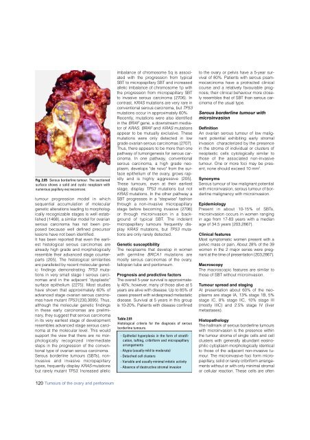
Fig. 2.05 Serous borderline tumour. The sectioned<br />
surface shows a solid and cystic neoplasm with<br />
numerous papillary excrescences.<br />
tumour pro g ression model in which<br />
sequential accumulation of molecular<br />
genetic alterations leading to morphologically<br />
recognizable stages is well established<br />
{1468}, a similar model for ovarian<br />
s e rous <strong>carcinoma</strong> has not been proposed<br />
because well defined precursor<br />
lesions have not been identified.<br />
It has been reported that even the earliest<br />
histological serous <strong>carcinoma</strong>s are<br />
already high grade and morphologically<br />
resemble their advanced stage counterparts<br />
{205}. The histological similarities<br />
are paralleled by recent molecular genetic<br />
findings demonstrating TP53 mutations<br />
in very small stage I serous <strong>carcinoma</strong>s<br />
and in the adjacent "dysplastic"<br />
surface epithelium {2275}. Most studies<br />
have shown that approximately 60% of<br />
advanced stage ovarian serous <strong>carcinoma</strong>s<br />
have mutant TP53 {230,3095}. Thus,<br />
although the molecular genetic findings<br />
in these early <strong>carcinoma</strong>s are preliminary,<br />
they suggest that serous <strong>carcinoma</strong><br />
in its very earliest stage of development<br />
resembles advanced stage serous <strong>carcinoma</strong><br />
at the molecular level. This would<br />
support the view that there are no morphologically<br />
recognized interm e d i a t e<br />
steps in the progression of the conventional<br />
type of ovarian serous <strong>carcinoma</strong>.<br />
S e rous borderline tumours (SBTs), noninvasive<br />
and invasive micro p a p i l l a ry<br />
types, frequently display K R A S m u t a t i o n s<br />
but rarely mutant T P 5 3. Increased allelic<br />
imbalance of chromosome 5q is associated<br />
with the pro g ression from typical<br />
SBT to micro p a p i l l a ry SBT and incre a s e d<br />
allelic imbalance of chromosome 1p with<br />
the pro g ression from micro p a p i l l a ry SBT<br />
to invasive serous <strong>carcinoma</strong> {2706}. In<br />
contrast, K R A S mutations are very rare in<br />
conventional serous <strong>carcinoma</strong>, but T P 5 3<br />
mutations occur in approximately 60%.<br />
Recently, mutations were also identified<br />
in the BRAF gene, a downstream mediator<br />
of KRAS. BRAF and KRAS mutations<br />
appear to be mutually exclusive. These<br />
mutations were only detected in low<br />
grade ovarian serous <strong>carcinoma</strong>s {2707}.<br />
Thus, there appears to be more than one<br />
pathway of tumorigenesis for serous <strong>carcinoma</strong>.<br />
In one pathway, conventional<br />
serous <strong>carcinoma</strong>, a high grade neoplasm,<br />
develops "de novo" from the surface<br />
epithelium of the ovary, grows rapidly<br />
and is highly aggressive {205}.<br />
These tumours, even at their earliest<br />
stage, display TP53 mutations but not<br />
KRAS mutations. In the other pathway a<br />
SBT progresses in a "stepwise" fashion<br />
t h rough a non-invasive micro p a p i l l a ry<br />
stage before becoming invasive {2706}<br />
or through microinvasion in a backg<br />
round of typical SBT. The indolent<br />
m i c ro p a p i l l a ry tumours frequently display<br />
KRAS mutations, but TP53 mutations<br />
are only rarely detected.<br />
Genetic susceptibility<br />
The neoplasms that develop in women<br />
with germline B R C A 1 mutations are<br />
mostly serous <strong>carcinoma</strong>s of the ovary,<br />
fallopian tube and peritoneum.<br />
Prognosis and predictive factors<br />
The overall 5-year survival is appro x i m a t e-<br />
ly 40%; however, many of those alive at 5<br />
years are alive with disease. Up to 85% of<br />
cases present with widespread metastatic<br />
disease. Survival at 5 years in this gro u p<br />
is 10-20%. Patients with disease confined<br />
Table 2.01<br />
Histological criteria for the diagnosis of serous<br />
borderline tumours.<br />
- Epithelial hyperplasia in the form of stratification,<br />
tufting, cribriform and micropapillary<br />
arrangements<br />
- Atypia (usually mild to moderate)<br />
- Detached cell clusters<br />
- Variable and usually minimal mitotic activity<br />
- Absence of destructive stromal invasion<br />
to the ovary or pelvis have a 5-year survival<br />
of 80%. Patients with serous psamm<br />
o c a rcinoma have a protracted clinical<br />
course and a relatively favourable pro g-<br />
nosis; their clinical behaviour more closely<br />
resembles that of SBT than serous <strong>carcinoma</strong><br />
of the usual type.<br />
Serous borderline tumour with<br />
microinvasion<br />
Definition<br />
An ovarian serous tumour of low malignant<br />
potential exhibiting early stromal<br />
invasion characterized by the presence<br />
in the stroma of individual or clusters of<br />
neoplastic cells cytologically similar to<br />
those of the associated non-invasive<br />
tumour. One or more foci may be present;<br />
none should exceed 10 mm 2 .<br />
Synonyms<br />
Serous tumour of low malignant potential<br />
with microinvasion, serous tumour of borderline<br />
malignancy with microinvasion.<br />
Epidemiology<br />
P resent in about 10-15% of SBTs ,<br />
microinvasion occurs in women ranging<br />
in age from 17-83 years with a median<br />
age of 34.5 years {203,2867}.<br />
Clinical features<br />
Most symptomatic women present with a<br />
pelvic mass or pain. About 28% of the 39<br />
women in the 2 major series were pre g-<br />
nant at the time of presentation {203,2867}.<br />
Macroscopy<br />
The macroscopic features are similar to<br />
those of SBT without microinvasion.<br />
Tumour spread and staging<br />
At presentation about 60% of the neoplasms<br />
are stage IA, 13% stage 1B, 5%<br />
stage IC, 8% stage IIC, 10% stage III<br />
(mostly IIIC) and 2.5% stage IV (liver<br />
metastases).<br />
Histopathology<br />
The hallmark of serous borderline tumours<br />
with microinvasion is the presence within<br />
the tumour stroma of single cells and cell<br />
clusters with generally abundant eosinophilic<br />
cytoplasm morphologically identical<br />
to those of the adjacent non-invasive tum<br />
o u r. The microinvasive foci form microp<br />
a p i l l a ry, solid or rarely cribriform arrangements<br />
without or with only minimal stro m a l<br />
or cellular reaction. These cells are often<br />
120 Tumours of the ovary and peritoneum


