

Thesis-PDF - IAP/TU Wien
Thesis-PDF - IAP/TU Wien
Thesis-PDF - IAP/TU Wien

You also want an ePaper? Increase the reach of your titles
YUMPU automatically turns print PDFs into web optimized ePapers that Google loves.
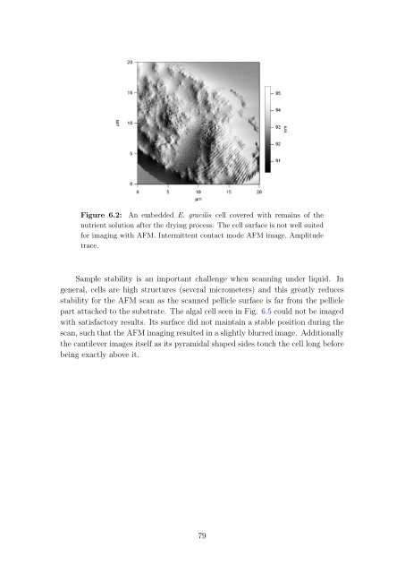
Figure 6.2: An embedded E. gracilis cell covered with remains of the<br />
nutrient solution after the drying process. The cell surface is not well suited<br />
for imaging with AFM. Intermittent contact mode AFM image, Amplitude<br />
trace.<br />
Sample stability is an important challenge when scanning under liquid. In<br />
general, cells are high structures (several micrometers) and this greatly reduces<br />
stability for the AFM scan as the scanned pellicle surface is far from the pellicle<br />
part attached to the substrate. The algal cell seen in Fig. 6.5 could not be imaged<br />
with satisfactory results. Its surface did not maintain a stable position during the<br />
scan, such that the AFM imaging resulted in a slightly blurred image. Additionally<br />
the cantilever images itself as its pyramidal shaped sides touch the cell long before<br />
being exactly above it.<br />
79


