
Create successful ePaper yourself
Turn your PDF publications into a flip-book with our unique Google optimized e-Paper software.
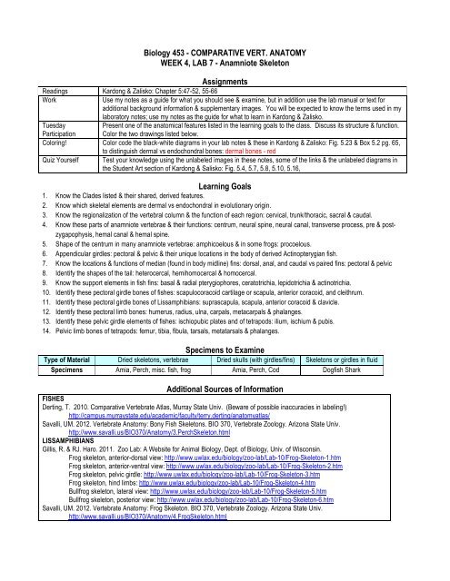
Biology 453 - COMPARATIVE VERT. ANATOMY<br />
WEEK 4, LAB 7 - <strong>Anamniote</strong> Skeleton<br />
Assignments<br />
Readings Kardong & Zalisko: Chapter 5:47-52, 55-66<br />
Work<br />
Use my notes as a guide for what you should see & examine, but in addition use the lab manual or text for<br />
additional background information & supplementary images. You will be expected to know the terms used in my<br />
laboratory notes; use my notes as the guide for what to learn in Kardong & Zalisko.<br />
Tuesday<br />
Present one of the anatomical features listed in the learning goals to the class. Discuss its structure & function.<br />
Participation Color the two drawings listed below.<br />
Coloring! Color code the black-white diagrams in your lab notes & these in Kardong & Zalisko: Fig. 5.23 & Box 5.2 pg. 65,<br />
to distinguish dermal vs endochondral bones: dermal bones - red<br />
Quiz Yourself Test your knowledge using the unlabeled images in these notes, some of the links & the unlabeled diagrams in<br />
the Student Art section of Kardong & Salisko: Fig. 5.4, 5.7, 5.8, 5.10, 5.16,<br />
Learning Goals<br />
1. Know the Clades listed & their shared, derived features.<br />
2. Know which skeletal elements are dermal vs endochondral in evolutionary origin.<br />
3. Know the regionalization of the vertebral column & the function of each region: cervical, trunk/thoracic, sacral & caudal.<br />
4. Know these parts of anamniote vertebrae & their functions: centrum, neural spine, neural canal, transverse process, pre & postzygapophysis,<br />
hemal canal & hemal spine.<br />
5. Shape of the centrum in many anamniote vertebrae: amphicoelous & in some frogs: procoelous.<br />
6. Appendicular girdles: pectoral & pelvic & their unique locations in the body of derived Actinopterygian fish.<br />
7. Know the locations & functions of median (found in body midline) fins: dorsal, anal, and caudal vs paired fins: pectoral & pelvic<br />
8. Identify the shapes of the tail: heterocercal, hemihomocercal & homocercal.<br />
9. Know the support elements in fish fins: basal & radial pterygiophores, ceratotrichia, lepidotrichia & actinotrichia.<br />
10. Identify these pectoral girdle bones of fishes: scapulocoracoid cartilage or scapula, anterior coracoid, and cleithrum.<br />
11. Identify these pectoral girdle bones of Lissamphibians: suprascapula, scapula, anterior coracoid & clavicle.<br />
12. Identify these pectoral limb bones: humerus, radius, ulna, carpals, metacarpals & phalanges.<br />
13. Identify these pelvic girdle elements of fishes: ischiopubic plates and of tetrapods: ilium, ischium & pubis.<br />
14. Pelvic limb bones of tetrapods: femur, tibia, fibula, tarsals, metatarsals & phalanges.<br />
Specimens to Examine<br />
Type of Material Dried skeletons, vertebrae Dried skulls (with girdles/fins) <strong>Skeletons</strong> or girdles in fluid<br />
Specimens Amia, Perch, misc. fish, frog Amia, Perch, Cod Dogfish Shark<br />
Additional Sources of Information<br />
FISHES<br />
Derting, T. 2010. Comparative Vertebrate Atlas, Murray State Univ. (Beware of possible inaccuracies in labeling!)<br />
http://campus.murraystate.edu/academic/faculty/terry.derting/anatomyatlas/<br />
Savalli, UM. 2012. Vertebrate Anatomy: Bony Fish <strong>Skeletons</strong>. BIO 370, Vertebrate Zoology. Arizona State Univ.<br />
http://www.savalli.us/BIO370/Anatomy/3.PerchSkeleton.html<br />
LISSAMPHIBIANS<br />
Gillis, R. & RJ. Haro. 2011. Zoo Lab: A Website for Animal Biology, Dept. of Biology, Univ. of Wisconsin.<br />
Frog skeleton, anterior-dorsal view: http://www.uwlax.edu/biology/zoo-lab/Lab-10/Frog-Skeleton-1.htm<br />
Frog skeleton, anterior-ventral view: http://www.uwlax.edu/biology/zoo-lab/Lab-10/Frog-Skeleton-2.htm<br />
Frog skeleton, pelvic girdle: http://www.uwlax.edu/biology/zoo-lab/Lab-10/Frog-Skeleton-3.htm<br />
Frog skeleton, hind limbs: http://www.uwlax.edu/biology/zoo-lab/Lab-10/Frog-Skeleton-4.htm<br />
Bullfrog skeleton, lateral view: http://www.uwlax.edu/biology/zoo-lab/Lab-10/Frog-Skeleton-5.htm<br />
Bullfrog skeleton, posterior view: http://www.uwlax.edu/biology/zoo-lab/Lab-10/Frog-Skeleton-6.htm<br />
Savalli, UM. 2012. Vertebrate Anatomy: Frog Skeleton. BIO 370, Vertebrate Zoology. Arizona State Univ.<br />
http://www.savalli.us/BIO370/Anatomy/4.FrogSkeleton.html
Major Clades & Shared Derived Traits in <strong>Anamniote</strong> Skulls<br />
(Features in living taxa, observed in lab)<br />
---------------------------------------------------- Vertebrata ---------------------------------------------------------------------------------<br />
------------------------------------------------ Gnathostomata ---------------------------------------------------<br />
--------------------------------------- Osteichthyes-------------------------------<br />
-------- Tetrapoda ----<br />
Cyclostomata Chondrichthyes Actinopterygii Actinopterygii Lissamphibia<br />
(Lamprey) (Sharks) (Bowfin) (Teleostii) (Frog)<br />
Paedomorphic:<br />
Endochondral<br />
skeleton fails to<br />
ossify<br />
No dermal skeleton<br />
Hemihomocercal<br />
tail<br />
Homocercal tail<br />
Tetrapoda:<br />
1 cervical vert.<br />
1 sacral vert.<br />
zygapophyses<br />
ilium<br />
lost<br />
lepidotrichia<br />
digits<br />
Lepidotrichia<br />
Cleithrum, clavicle<br />
Paired pectoral & pelvic fins<br />
Pectoral girdle: Scapulocoracoid<br />
Pelvic girdle: Ischiopubic<br />
Heterocercal tail<br />
Vertebrae
INTRODUCTION<br />
Dermal bones are derived from dermal scales that sank deep & joined the internal skeleton. These bones form as connective<br />
tissue membranes that later ossify. Endochondral or replacement "elements" begin as cartilage & may ossify or remain cartilaginous<br />
in the adult.<br />
The axial skeleton elements are on the midline or mid-sagittal axis of the body. The axial skeleton includes the notochord,<br />
vertebrae, ribs & median fins. The notochord is a centrally located, gel-filled rod that supports the body of a developing embryo. In<br />
most vertebrates, vertebrae replace it, partly or completely. The vertebral body or centrum is the large solid disk that takes the<br />
compressive forces during body movement. Some centra are biconcave (amphicoelous) or flat (acoelous). In fishes, amphicoelous<br />
vertebrae are tightly held together with sheathing of connective tissue. Other centra have rounded cavities on the anterior (procoelous)<br />
or posterior (opisthocoelous) surface with a corresponding rounded opposite side that articulates with adjacent vertebrae. The<br />
vertebrae enclose the spinal cord with neural (or vertebral) arches. Caudal vertebrae have hemal arches that enclose caudal arteries<br />
& veins in the tail. All vertebrae have several areas of muscle attachment via neural spines, transverse processes & hemal spines.<br />
Tetrapod vertebrae have zygapophyses (zygo= yoke) to support the body & keep the trunk from sagging. The zygapophyses<br />
allow vertebrae to articulate with each other, and limit the total range & direction of body movement. The pre-zygapophyses are on the<br />
anterior side of vertebra & have articulating facets that face upward. The post-zygapophyses are on the posterior side & have<br />
articulating facets that face downward. Post-zygapophyses fit on top of pre-zygapophyses.<br />
Fishes have 2 regions in vertebral column: trunk vertebrae have ribs and caudal vertebrae form the tail. <strong>Anamniote</strong> tetrapods<br />
have 4 regions in vertebral column. They have 1 cervical or neck vertebra, called the atlas. This vertebra has large facets (prezygapophyses)<br />
that articulate with the paired occipital condyles seen in living amphibians. Tetrapods have a single sacral vertebra<br />
that attaches to the pelvic girdle. In frogs, the caudal vertebrae are fused into a rod-like urostyle.<br />
A sternum is present only in tetrapods, and even then may be missing or very small in a few species. The sternum acts as a<br />
brace to attach to support the rib cage or an additional brace for the pectoral girdle. Fishes have unpaired, median fins located along the<br />
mid-sagittal axis. These include dorsal, anal & caudal fins. Fish may have 1 or more dorsal fins. Caudal fins vary in design.<br />
Heterocercal caudal fins are present in the Chondrichthyes and some primitive Actinopterygians. In these tails, the caudal vertebrae<br />
extend into the dorsal & larger lobe of the caudal fin. Some Actinopterygians have a hemi-homocercal design where the vertebrae just<br />
turn dorsally at the very tail tip & do not extend far into the tail fin. The tail fin doesn’t appear to be asymmetrical, but the asymmetry can<br />
be seen in the skeleton & in the body profile. Most living Actinopterygians have a homocercal caudal fin that forms both a symmetrical<br />
profile & symmetrical caudal fin.<br />
The appendicular skeleton refers to the elements that form the paired, laterally placed appendages for the paired front & hind<br />
fins or limbs. The pectoral girdle of fishes is a combination of dermal & endochondral elements. The dermal pectoral girdle bones of<br />
ray-finned fish form an arc just behind the opercular bones of the skull. These dermal bones include the cleithrum, clavicle & other<br />
bones that attach the pectoral girdle to the skull. The sharks lack these dermal bones as they lack all dermatocranium in the skull as<br />
well. Tetrapods lose many dermal bones when the pectoral girdle detaches from the skull. Thus in tetrapods, the head moves<br />
independently of the legs. Some tetrapods retain the clavicles as paired bones that extend from the sternum towards the humerus. One<br />
new dermal pectoral girdle bone, the interclavicle, appears in Sarcopterygians & some tetrapods. This is an unpaired bone that may be<br />
in the midline between the paired clavicles.<br />
The endochondral elements of the pectoral girdle are called scapulocoracoid or coracoscapula, if they don’t ossify. In<br />
derived Actinopterygians (e.g. Perch) these ossify into a ventrally positioned anterior coracoid (procoracoid) & a more dorsally<br />
positioned scapula. The tetrapods may add a suprascapula dorsal to the scapula. Amniotes gained a new element, the posterior<br />
coracoid (metacoracoid). In<br />
Fish pelvic girdles are suspended in muscle: ischiopubic or pubioischiatic plates. In derived Actinopterygians this girdle &<br />
its associated fins are anterior in the body. This position may improve the braking ability of those fins. In some ray-finned fish, the pelvic<br />
girdle attaches to the pectoral girdle but is still ventral to the pectoral fins. All tetrapods (with legs) have 3 bones that form the pelvic<br />
girdle. The pubic bone or pubis is directed forward & down & remains cartilaginous in many anamniotes. The ischium faces<br />
downward & posteriorly. The ilium is the new bone in the tetrapod pelvis. It is oriented dorsally & attaches the pelvic girdle to the sacral<br />
vertebra.<br />
The median & paired fins of fishes contain the same types of support elements. The most proximal support elements are<br />
called basal pterygiophores & they are typically larger and fewer in number than the more distal radial pterygiophores. Different<br />
materials in fish support the fin membranes. Ceratotrichia are made of keratin & support the fins of sharks, skates & rays.<br />
Lepidotrichia are tiny, overlapping dermal scales in Actinopterygian fins. Actinotrichia are bony spines that may be present at the<br />
anterior edge of a fin or replace lepidotrichia throughout an entire fin.<br />
The paired limbs of tetrapods (with limbs) comprise a standard pattern with 1 bone, followed by a pair of bones then many<br />
bone in series. The forelimb has a humerus; radius & ulna; carpals; metacarpals; phalanges. The hind limb has a femur; tibia & fibula;<br />
tarsals; metatarsals; phalanges.
Axial Skeleton Summary<br />
Divisions of the Vertebral Column in Vertebrates Observed in Lab<br />
Chondrichthyes Actinopterygii Lissamphibia Lepidosauria Archosauria, Aves & Mammalia<br />
1 Cervical (neck)<br />
(atlas only)<br />
> 1 Cervical (neck)<br />
(atlas, axis & more)<br />
1 Cervical (neck)<br />
(atlas, axis & more)<br />
Trunk – has ribs Trunk – has ribs Trunk – has short ribs<br />
in salamander<br />
Trunk – has ribs<br />
Thoracic – has ribs<br />
Lumbar<br />
1 Sacral > 1 Sarcral > 1 Sarcral<br />
Caudal Caudal Caudal (tail) Caudal (tail) Caudal (tail)<br />
Structures in Most Vertebrae<br />
Cervical Trunk or Thoracic Lumbar<br />
Sacral<br />
Caudal<br />
(tetrapods only)<br />
(some amniotes)<br />
(tetrapods)<br />
Neural Spine – reduced Neural Spine Neural Spine Neural Spine Neural Spine<br />
or absent<br />
Neural Arch Neural Arch Neural Arch Neural Arch Neural Arch<br />
Neural Canal Neural Canal Neural Canal Neural Canal Neural Canal<br />
Centrum Centrum may show rib<br />
Centrum<br />
Centrum – may fuse to<br />
Centrum<br />
articulation facets<br />
each other if > 1<br />
Birds have a synsacrum<br />
made of thoracic, lumbar,<br />
sacral & caudal vertebrae<br />
that fuse together<br />
Most: transverse<br />
processes are small &<br />
articulate with short ribs<br />
Transverse processes<br />
articulate with long<br />
ribs (in most)<br />
Long transverse processes<br />
for muscle attachment, but<br />
no ribs<br />
Transverse processes large<br />
& sutured to ilium of pelvis<br />
Transverse processes<br />
large in some or may be<br />
reduced or absent<br />
Birds & Mammals:<br />
Transverse processes &<br />
ribs fuse, forming small<br />
transverse foramina for<br />
blood vessels<br />
Most tetrapods: ribs<br />
insert onto sternum<br />
Sternum in birds is<br />
large & has a keel<br />
Archosauria: alligators have<br />
gastralia (abdominal ribs)<br />
Pygostyle: last caudal<br />
vert. in Aves<br />
Coccyx: reduced<br />
caudal vert. in human<br />
Hemal Canal<br />
holds arteries & veins<br />
Hemal Arch<br />
reduced or lost if caudal<br />
vertebrae are tiny<br />
Hemal Spine may be<br />
long, short or absent<br />
Specializations in Some Tetrapod Vertebrae<br />
Atlas – 1 st cervical<br />
Axis – 2 nd cervical<br />
Pre-zygapophysis Post- zygapophysis<br />
Present in all Tetrapods<br />
Present in Amniotes<br />
Large neural canal Long neural spine On anterior side On posterior side<br />
Transverse processes small Facets face up & in Facets face down & outward<br />
Centrum reduced Odontoid process on anterior of centrum<br />
Articulates with occipital<br />
condyle(s)<br />
Articulates with atlas. Odontoid process<br />
fits inside neural canal of atlas<br />
Snakes have unique extra articulations between vertebrae to<br />
stop rotation: zygosphene & zygantrum<br />
Shapes of Vertebral Centrum in Vertebrates Observed in Lab<br />
Chondrichthyes Actinopterygii Lissamphibia Lepidosauria Aves<br />
Mammalia<br />
or Archosauria<br />
Amphicoelous Amphicoelous Amphicoelous - Necturus Acoelous<br />
Procoelous - frog Procoelous Heterocoelous -<br />
cervicals<br />
Opisthocoelous –<br />
cervicals in some<br />
large ungulates
Appendicular Skeleton Summary<br />
Common Structures of ALL Paired or Median Fins of Fish<br />
Pterygiophores Lepidotrichia Ceratotrichia Actinotrichia<br />
Chondrichthyes Cartilaginous - Made of keratin Spines<br />
Actinopterygii Bone typically Derived from tiny dermal scales - Spines<br />
Dermal Bones<br />
Endochondral<br />
Bones<br />
Structures of Pectoral Girdle to Find on Our Specimens:<br />
Chondrichthyes Actinopterygii Lissamphibia<br />
(frog)<br />
Lepidosauria<br />
(lizards)<br />
Archosauria<br />
(alligator & birds)<br />
Mammalia<br />
Cleithrum - - - -<br />
Absent<br />
- Clavicle Clavicle Clavicles fused to form Clavicle<br />
furcula in birds; (reduced in some)<br />
Clavicle absent in alligator<br />
- - Interclavicle Interclavicle in alligators;<br />
-<br />
present in some birds,<br />
Scapulocoracoid<br />
attached to furcula<br />
- Suprascapula Suprascapula - -<br />
Scapula Scapula Scapula Scapula Scapula has spine<br />
& acromion<br />
process<br />
Anterior<br />
Coracoid<br />
Anterior<br />
Coracoid<br />
Anterior<br />
Coracoid<br />
- - Posterior<br />
Coracoid (the<br />
2 coracoids<br />
can’t be<br />
distinguished)<br />
Posterior Coracoid<br />
(Bird “coracoid” has a bit<br />
of anterior coracoid)<br />
-<br />
Posterior Coracoid<br />
(coracoid process<br />
on scapula)<br />
Cartilages or Bones of the Pectoral Fin or Limb<br />
Chondrichthyes Actinopterygii Lissamphibia (Frog) Aves Most Amniotes<br />
Humerus Humerus Humerus<br />
Basal Pterygiophores<br />
Radioulna (fused) Radius (medial) Radius (medial)<br />
Radial Pterygiophores Radial Pterygiophores<br />
Ulna (lateral)<br />
Ulna (lateral)<br />
Carpals Carpometacarpus Carpals<br />
Metacarpals<br />
Metacarpals<br />
Phalanges Phalanges - reduced Phalanges<br />
Endochondral<br />
Bones<br />
Structures of Pelvic Girdle to Find on Our Specimens:<br />
Chondrichthyes Actinopterygii Lissamphibia Most Amniota Marsupial (Opossum)<br />
Ilium – attaches to Ilium – attaches to Ilium – attaches to sacral<br />
sacral vertebrae sacral vertebrae<br />
vertebrae<br />
Ischiopubic<br />
cartilage<br />
Ischiopubic bones Ischium Ischium Ischium<br />
Pubis (cartilaginous) Pubis Pubis<br />
Epipubic<br />
Cartilages or Bones of the Pelvic Fin or Limb<br />
Chondrichthyes Actinopterygii Lissamphibia (Frog) Aves Most Amniotes<br />
Femur Femur Femur<br />
Basal Pterygiophores<br />
Tibiofibula (fused) Tibiotarsus (medial) Tibia (medial)<br />
Radial Pterygiophores Radial Pterygiophores<br />
Fibula (reduced)<br />
Fibula (lateral)<br />
Tarsals (2 elongated) Tarsometatarsus Tarsals<br />
Metatarsals<br />
Metatarsals<br />
Phalanges Phalanges Phalanges
Chondrichthyes: Dogfish Sharks (Squalus)<br />
Dogfish Shark, Squalus, Wax casts, fluid preserved or lucite blocks with vertebral columns<br />
Types of vertebrae: trunk & caudal.<br />
Structures on all vertebrae: neural canal & centrum.<br />
Additional structure on caudal vertebrae: hemal canal.<br />
Centrum design: amphicoelous.<br />
Trunk<br />
Caudal<br />
Rachel Simon was a former biology undergraduate & then peer TA for this class, as well as an outstanding artist.<br />
Enjoy her diagram!
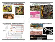
Chondrichthyes: Dogfish Sharks (Squalus)<br />
Complete skeletons in fluid & preserved or wax casts of pectoral & pelvic girdles & fins<br />
Types of vertebrae: trunk & caudal<br />
Ribs: small; only on trunk region<br />
Tail: heterocercal<br />
Median Fins: Dorsal & caudal fin. Caudal fin is a heterocercal design.<br />
Pectoral Girdle: scapulocoracoid is 1 piece of U shaped cartilage<br />
Pelvic Girdle: ischiopubic = pubioischiatic cartilage<br />
Male sharks have claspers that extend from the pelvic girdle. Find them.<br />
Structures in all fins: basal & radial pterygiophores; ceratotrichia (tiny, brown, horny, i.e. keratinized).<br />
King & Custace 1982. Color Atlas of Vertebrate Anatomy.<br />
Pectoral Girdle<br />
Female Pelvic Girdle<br />
Ceratotrichia Radial Pterygoiphores<br />
Basal Pterygiophores<br />
Scapulocoracoid<br />
Male Pelvic Girdle<br />
Claspers are used to aid sperm transfer into female’s cloaca.
Actinopterygii: Bowfin (Amia)<br />
Amia skulls & skeletons with pectoral girdles & fins attached.<br />
Two types of vertebrae: trunk & caudal. Ribs: large, attached to trunk vertebrae.<br />
Structures on all vertebrae: neural spine, neural (vertebral) canal & centrum (vertebral body).<br />
Additional structures on caudal vertebrae: hemal canal & hemal spines.<br />
Median Fins: dorsal, anal, caudal. Tail shape: hemihomocercal.<br />
Pectoral Girdle: small, undifferentiated scapulocoracoid (cartilaginous), large cleithrum, just behind opercular bones.<br />
Pectoral Fins: radial pterygiophores, lepidotrichia in the fin membrane<br />
Hemi-homocercal tail Single dorsal fin All fins have lepidotrichia (soft rays) only<br />
Caudal fin Anal fin Pelvic fins (Abdominal) Pectoral fins<br />
Pectoral Girdle & Fins<br />
Amia Pectoral Girdle: Left lateral view<br />
Cleithrum<br />
Anterior<br />
Medial view of Pectoral Girdle & Fin<br />
scapulocoracoid<br />
scapulocoracoid cartilaginous<br />
Pterygiophores & Lepidotrichia in Pectoral Fin<br />
Pelvic Girdle in Bowfin (anterior to left)<br />
The pelvic girdle in Amia “floats” in abdominal muscle & does<br />
not attach to the rest of the skeleton.<br />
Ischiopubic plates
Actinopterygii: Perch (Teleost) <strong>Skeletons</strong><br />
Perch Skeleton: Label the fins as well as some pterygiophores, actinotrichia & lepidotrichia.<br />
Pterygoiphores:<br />
support fins and are below skin<br />
Actinotrichia:<br />
form “spines” in some fins<br />
Rigid, sharp rods<br />
Lepidotrichia:<br />
form “soft rays” in fins<br />
Tiny, jointed, dermal bony elements form<br />
soft, flexible fin rays.
Actinopterygii: Fish Vertebrae, Pectoral & Pelvic Girdles<br />
Types of vertebrae: trunk & caudal.<br />
Structures on all vertebrae: neural spine, neural (vertebral) canal & centrum (vertebral body).<br />
Ribs: large, attached to trunk vertebrae<br />
Additional structures on caudal vertebrae: hemal canal & hemal spines.<br />
Centrum design: amphicoelous<br />
Trunk<br />
Caudal<br />
Actinopterygii: Perch Skulls with Fins Attached:<br />
Median Fins dorsal fins, anal fins, & caudal fins. Caudal shape: homocercal.<br />
Actinotrichia (spines) in 1 st dorsal fin.<br />
Pectoral Girdle: scapula, anterior coracoid & large cleithrum behind the opercular bones.<br />
Pelvic Girdle: anterior placement of ischiopubic plates.<br />
Paired Fins: lepidotrichia in the fin membrane (pterygiophores may be too small to see)<br />
Perch Pectoral Girdle with cleithrum, scapula & coracoid.<br />
Pectoral girdle & pectoral fin (Right, lateral view)<br />
PT - post-temporal<br />
SCL - supracleithrum<br />
CL - cleithrum<br />
PCL - post-cleithrum<br />
SCA - scapula<br />
CO - coracoid<br />
RA - radial pterygiophores<br />
FR - fin rays (lepidotrichia)<br />
Dineen & Stokley, 1956<br />
Pelvic girdle missing<br />
Perch Pelvic Girdle (ventral view)<br />
Ischiopubic plates<br />
Pelvic fins<br />
Lower jaw<br />
Hyoid & branchiostegal rays<br />
ischiopubic<br />
plates<br />
Perch pelvic girdle & pelvic fins.
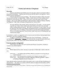
Lissamphibia: Frog Skeleton<br />
Partially & fully articulated <strong>Skeletons</strong><br />
Types of vertebrae: 1 cervical = atlas, trunk, 1 sacral & the urostyle - formed by fusion of caudal vertebrae.<br />
Parts of vertebrae: large transverse processes (The ribs have fused onto the transverses processes.). You can recognize the shape of<br />
their centra by looking at the ventral side to see the concave anterior surface & convex posterior surface.<br />
Sternum: 2 pieces (cartilage & bone). You are not responsible for the names of each part of the sternum.<br />
Pectoral Girdle: suprascapula, scapula, anterior coracoid & clavicle. Pelvic Girdle: long ilium, ischium & partly cartilaginous pubis.<br />
Forelimb: humerus, fused radioulna, carpals, metacarpal & phalange. Hind limb: femur, tibiofibula, tarsal, metatarsal & phalange.
Lissamphibia: Frog Skeleton continued<br />
Vertebral Column, dorsal view<br />
Atlas Sacral Vert. Urostyle<br />
Vertebral Column, ventral view<br />
Goliath Frog Vertebrae, dorsal view<br />
Goliath Frog Vertebrae, ventral view<br />
Pectoral Girdle, Ventral View (straightened)<br />
Pectoral Girdle, ventral view<br />
Sternum Scapula Clavicle Sternum<br />
Suprascapula<br />
Anterior coracoid<br />
Pelvic Girdle<br />
ilium<br />
Pelvic Girdle<br />
ischium<br />
Pubis<br />
Front Limb<br />
Hind Limbs<br />
femur<br />
humerus<br />
tibiofibula<br />
metacarpals<br />
phalanges<br />
carpals<br />
radioulna<br />
tarsals<br />
phalanges<br />
metatarsals