Sudden Death in Addison's Disease - Healthcare Bulletin
Sudden Death in Addison's Disease - Healthcare Bulletin
Sudden Death in Addison's Disease - Healthcare Bulletin

You also want an ePaper? Increase the reach of your titles
YUMPU automatically turns print PDFs into web optimized ePapers that Google loves.
ENDOCRINE DISEASES | CASE STUDY<br />
<strong>Sudden</strong> <strong>Death</strong> <strong>in</strong> Addison’s <strong>Disease</strong>:<br />
Lead Poison<strong>in</strong>g-like Gum Appearance<br />
Boonsak Hanterdsith, M.D. Division of Forensic Medic<strong>in</strong>e, Lamphun Hospital,<br />
& Pongsak Mahanupab, M.D. Department of Pathology, Faculty of Medic<strong>in</strong>e, Chiang Mai University, Thailand<br />
Received: 1/5/2011, Reviewed 20/5/2011, Accepted 6/6/2011<br />
Key words: <strong>Sudden</strong> death, Addison’s disease, Lead poison<strong>in</strong>g-like gum appearance, Autopsy<br />
DOI: 10.5083/ejcm.20424884.32<br />
ABSTRACT<br />
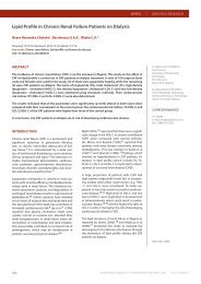
Fatal Addison’s disease is rarely found <strong>in</strong> forensic cases. We report the sudden and unexpla<strong>in</strong>ed death<br />
of a 51-year-old woman on arrival at the emergency room. Previous cl<strong>in</strong>ical history revealed frequent<br />
hypotension, hyponatremia and persistent hyperpigmented sk<strong>in</strong> consistent with Addison’s disease.<br />
However, the diagnosis could only be made dur<strong>in</strong>g autopsy. The adrenal gland was completely<br />
absent. Postmortem blood cortisol was very low (0.86 µg/dL). The thyroid gland showed Hashimoto<br />
thyroiditis. The probable cause of Addison’s disease <strong>in</strong> this case was autoimmune adrenalitis.<br />
INTRODUCTION<br />
Chronic adrenocortical <strong>in</strong>sufficiency is an uncommon<br />
disorder result<strong>in</strong>g from progressive destruction<br />
of the adrenal cortex (1) . Cl<strong>in</strong>ical features<br />
<strong>in</strong>clude progressive anaemia, bronze sk<strong>in</strong> pigmentation,<br />
severe weakness, hypotension, nausea, vomit<strong>in</strong>g,<br />
anorexia, weight loss and hypoglycemia (1-4) .<br />
Hyponatremia is observed <strong>in</strong> about 80% of acute<br />
cases whereas less than half present with hyperkalemia<br />
(5) . These cl<strong>in</strong>ical features were first reported as<br />
Addison’s disease <strong>in</strong> “On the Constitutional and Local<br />
Effects of <strong>Disease</strong> of the Supra-renal Capsules” <strong>in</strong><br />
the London Medical Gazette, 1849 by Thomas Addison<br />
(2) .<br />
Nowadays autoimmune disease is the most common<br />
cause of Addison’s disease <strong>in</strong> western countries<br />
(2, 4, 6) . The rema<strong>in</strong><strong>in</strong>g causes are tuberculosis,<br />
adrenomyeloneuropathy, systemic fungal <strong>in</strong>fection,<br />
AIDS, metastatic carc<strong>in</strong>oma and isolated glucocorticoid<br />
deficiency (2, 4) . The prognosis is excellent (7) ,<br />
but mortality rate was more than 2-fold higher<br />
compared with the normal population <strong>in</strong> a 14 year<br />
period (8) . We report a fatal case of autoimmune<br />
Addison’s disease that was diagnosed only dur<strong>in</strong>g<br />
autopsy.<br />
CASE REPORT<br />
A 51-year-old woman was found unconsciousness<br />
at home. Her relative called the Emergency<br />
Medical Service (EMS) for help. EMS personnel<br />
transported her to the emergency room of the<br />
prov<strong>in</strong>cial hospital. The patient was found apnoeic<br />
and pulseless.<br />
Initial cardiac rhythm showed asystole. F<strong>in</strong>gertip<br />
dextrose-strip exam<strong>in</strong>ation showed 20 mg<br />
of glucose. Cardiopulmonary resuscitation was<br />
performed for an hour but she could not be resuscitated.<br />
<strong>Death</strong> was pronounced at 12.00 AM<br />
on 2 November 2009. The cause of death could<br />
not be explored by external exam<strong>in</strong>ations. Her<br />
relative <strong>in</strong>formed the doctor that the patient<br />
had suffered from toothache for 2 days before<br />
her death.<br />
Accord<strong>in</strong>g to her medical history, she had had<br />
chronic anemia with <strong>in</strong>termittent significant<br />
hyponatremia (serum sodium = 125 mmol/dl)<br />
s<strong>in</strong>ce 12 February 2007. Causes of anemia were<br />
iron deficiency (serum iron = 84 microgram per<br />
dl; total iron b<strong>in</strong>d<strong>in</strong>g capacity = 321 microgram<br />
per dl; serum saturated transferr<strong>in</strong> = 26 percent)<br />
and primary hypothyroid disease. The thyroid<br />
function test was recorded <strong>in</strong> the OPD card on<br />
17 October 2007 as follows: TSH level was 21.04<br />
(normal range is 0.2-3.2 microIU/ml), T3 was<br />
0.813 (normal range is 0.8-2.1 ng/ml). Complete<br />
blood count showed anemia (Hb 7.6-9.1) <strong>in</strong> all of<br />
her follow-up visits.<br />
Her hyperpigmented sk<strong>in</strong> was mentioned <strong>in</strong> the<br />
OPD card two times <strong>in</strong> 2007; however, there was<br />
no further <strong>in</strong>vestigation. She sometimes compla<strong>in</strong>ed<br />
about malaise. Her blood pressure was<br />
<strong>in</strong>termittent, ris<strong>in</strong>g and fall<strong>in</strong>g between 90/60<br />
and 120/80 mmHg (frequent hypotension). The<br />
body weight was stable at 40-42 kg for 1.5 year.<br />
Ma<strong>in</strong> treatments were oral iron tablets, folic and<br />
vitam<strong>in</strong> B complex supplement for treatment of<br />
iron deficiency anemia. The last follow-up visit<br />
was on 3 July 2008.<br />
CORRESPONDENCE<br />
Boonsak Hanterdsith<br />
Division of Forensic Medic<strong>in</strong>e,<br />
Lamphun Hospital,<br />
Lamphun, 51000.<br />
Tel. +66-0-8678-12869<br />
+66-0-5356-9100<br />
ext. 1998-9<br />
Fax: +66-0-5356-9191<br />
E-mail: trapezius60@yahoo.com<br />
FINANCIAL SUPPORT:<br />
No fund or f<strong>in</strong>ancial support<br />
ISSN 2042-4884<br />
38 EUROPEAN JOURNAL OF CARDIOVASCULAR MEDICINE VOL I ISSUE III
SUDDEN DEATH IN ADDISON’S DISEASE: LEAD POISONING-LIKE GUM APPEARANCE<br />
The autopsy was performed on 3 November 2009 (21 hours after<br />
the pronounced death). The body was of a middle aged, poor nourished<br />
female, 153 cm <strong>in</strong> length with short black hair. Axillary and<br />
pubic hair was completely absent. Her sk<strong>in</strong> looked generally dark<br />
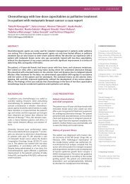
especially on the palmar creases, all jo<strong>in</strong>t areas, and lips. The upper<br />
gum showed bluish-black patches, which looked like a lead l<strong>in</strong>e <strong>in</strong><br />
lead poison<strong>in</strong>g (Figure 1).<br />
Figure 1: Hyperpigmentation of the gum mimick<strong>in</strong>g the<br />
lead l<strong>in</strong>e <strong>in</strong> lead poison<strong>in</strong>g<br />
The toxicological analysis was negative. Lead <strong>in</strong> the blood sample<br />
was negative. Postmortem blood cortisol was 0.86 µg/dL (range<br />
5-25 AM, 2.5-12.5 PM).<br />
Unfortunately, anti-adrenal and anti-thyroid antibodies could not<br />
be analysed <strong>in</strong> Thailand. Bra<strong>in</strong>, thyroid gland, heart, lung and kidney<br />
tissues were submitted for microscopic exam<strong>in</strong>ation: The cardiac<br />
muscles showed hypertrophy with a small focal area of fibrosis.<br />
Some focal haemorrhages were present <strong>in</strong> the subepicardium and<br />
the myocardium. There were random contraction band necroses.<br />
The thyroid gland showed Hashimoto thyroditis. There was pulmonary<br />
oedema with some focal hemorrhages <strong>in</strong> the lungs. The bra<strong>in</strong><br />
tissue was unremarkable.<br />
DISCUSSION<br />
Fatal Addison’s disease is rarely found <strong>in</strong> forensic practice, especially<br />
<strong>in</strong> Northern Thailand. <strong>Sudden</strong> death from Addison’s disease<br />
has been reported (9-12) but mostly <strong>in</strong> Caucasians. Several studies<br />
showed that Addison’s disease could only be diagnosed dur<strong>in</strong>g autopsy<br />
(9-12) . The most specific sign of primary adrenal <strong>in</strong>sufficiency<br />
is hyperpigmentation of the sk<strong>in</strong> and mucosal surfaces which is due<br />
to the high plasma corticotroph<strong>in</strong> concentrations that occur as a result<br />
of a decrease of cortisol feedback (4) . Malaise, hyperpigmented<br />
sk<strong>in</strong>, hypotension and hyponatremia <strong>in</strong> our case were clues for diagnosis<br />
of chronic primary adrenal <strong>in</strong>sufficiency. However, the dark<br />
l<strong>in</strong>e on the gum may be mistaken as the lead l<strong>in</strong>e <strong>in</strong> lead poison<strong>in</strong>g.<br />
Eur J Cardiovasc Med © <strong>Healthcare</strong> Bullet<strong>in</strong> 2011<br />
There was no wound or <strong>in</strong>jection mark on the sk<strong>in</strong>. The <strong>in</strong>ternal exam<strong>in</strong>ation<br />
showed no evidence of vital organs <strong>in</strong>jury. The bra<strong>in</strong> had<br />
no pathological lesion. The pituitary fossa had no abnormal mass.<br />
The thyroid gland was normal shape and weighed 15 g. The airways<br />
showed no edema or foreign body obstruction. Both lungs showed<br />
mild edema with left upper lung consolidation. There was no pulmonary<br />
thromboembolism. The right and the left lung weighed<br />
370 g and 470 g, respectively. There were some petechiae on the<br />
anterior external surface of the heart. The left anterior descend<strong>in</strong>g<br />
coronary artery showed 10% stenosis. The right ma<strong>in</strong> coronary artery<br />
was widely patent. The left ventricular free wall thickness was<br />
12 mm. Neither valvular abnormality nor congenital anomaly was<br />
observed. The heart weighed 290 g and had a normal shape.<br />
There was no evidence of peritonitis. The liver, spleen, small bowel,<br />
large bowel and pancreas had no significant gross pathologic abnormality.<br />
Both kidneys showed a diffuse micronodular surface.<br />
No evidence of acute pyelonephritis was detected. The adrenal<br />
gland was searched for <strong>in</strong> the suprarenal areas and <strong>in</strong> other areas<br />
<strong>in</strong>clud<strong>in</strong>g chest wall, but could not be identified grossly. There was<br />
no fibrosis at the suprarenal areas. The retroperitoneal region had<br />
no blood collection. The pelvic organs showed no significant gross<br />
lesion. There were 10 millilitres of light green mucous-mixed liquid<br />
<strong>in</strong> the stomach. The mucosa of the stomach showed generalised<br />
gastritis. The femoral blood, heart blood and gastric contents were<br />
submitted for toxicological analysis at the Regional Medical Science<br />
Center, Chiang Mai prov<strong>in</strong>ce.<br />
Corticotrop<strong>in</strong> stimulation is the most commonly used test for the<br />
diagnosis of primary adrenal <strong>in</strong>sufficiency (4) , but it cannot be performed<br />
postmortem. However, a very low level of plasma cortisol (3<br />
or less µg/dL) confirmed adrenal <strong>in</strong>sufficiency (4) . A previous study<br />
showed that serum cortisol rema<strong>in</strong>s constant dur<strong>in</strong>g the early postmortem<br />
period (13) . This supports that the very low level of postmortem<br />
blood cortisol is due to severe adrenal <strong>in</strong>sufficiency <strong>in</strong> this<br />
case. The absence of the adrenal gland <strong>in</strong> our case may be caused<br />
by severe atrophy.<br />
However, the detection of blood cortisol <strong>in</strong>dicates some rema<strong>in</strong><strong>in</strong>g<br />
cortisol secret<strong>in</strong>g tissue. Autoimmune adrenalitis is the ma<strong>in</strong> cause<br />
of Addison’s disease and may occur alone or as a component of<br />
type I or II autoimmune polyglandular syndrome (2, 4, 6) . The Hashimoto<br />
thyroiditis of this case <strong>in</strong>dicated that the autoimmune disease<br />
was the probable cause of adrenocortical <strong>in</strong>sufficiency.<br />
Autoantibodies aga<strong>in</strong>st 21-hydroxylase, one of the enzymes <strong>in</strong><br />
steroid biosynthesis <strong>in</strong>side the adrenal glands, can be found <strong>in</strong> approximately<br />
80% of the Addisonian persons. These autoantibodies<br />
thus clearly correlate with the disease (14) and are useful for its diagnosis.<br />
Approximately 21% of persons positive for adrenal cortex<br />
autoantibodies (ACA) developed overt Addison’s disease with<strong>in</strong><br />
5.2 years, while negative ACA persons ma<strong>in</strong>ta<strong>in</strong>ed normal adrenal<br />
function dur<strong>in</strong>g the observation period (15) . ACA is also an additional<br />
marker to predict Addison’s disease. In conclusion, cl<strong>in</strong>icians<br />
should not overlook hyperpigmentation of the sk<strong>in</strong> comb<strong>in</strong>ed with<br />
other significant cl<strong>in</strong>ical signs and basic laboratory tests for the correct<br />
diagnose of adrenal <strong>in</strong>sufficiency which is potentially fatal if<br />
not recognised and promptly treated.<br />
EUROPEAN JOURNAL OF CARDIOVASCULAR MEDICINE<br />
VOL I ISSUE III<br />
39
HEALTHCARE BULLETIN | ENDOCRINE DISEASES<br />
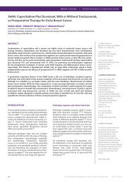
Figure 2: Photomicrograph of the thyroid gland tissue of<br />
the deceased (X 20 magnification): The thyroid parenchyma<br />
conta<strong>in</strong>s a dense lymphocytic <strong>in</strong>filtration with germ<strong>in</strong>al centers<br />
(Hashimoto thyroiditis).<br />
Eur J Cardiovasc Med © <strong>Healthcare</strong> Bullet<strong>in</strong> 2011<br />
8<br />
9<br />
10<br />
11<br />
12<br />
13<br />
14<br />
15<br />
Bergthorsdottir R, Zachrisson ML, Oden A, Johannsson G.<br />
Premature mortality <strong>in</strong> patients with Addison’s disease:<br />
A population-based study. JCEM 2006;91(12):4849-53.<br />
Brosnan CM, Gow<strong>in</strong>g NFC. Addison’s disease. BMJ 1996;312:1085-7.<br />
Sabri AMA, Smith N, Busuttil. <strong>Sudden</strong> death due to auto-immune<br />
Addison’s disease <strong>in</strong> a 12-year-old girl. Int J Legal Med 1997;<br />
110:278-80.<br />
Burke MP, Opesk<strong>in</strong> K. Adrenocortical <strong>in</strong>suffiency. Am J Forensic Med<br />
Pathol 1999;20(1):60-5.<br />
Moallem M, Nader N, Auckley D. A 22-year-old woman with fever,<br />
jaw pa<strong>in</strong>, and shock. CHEST 2007;132:1077-9.<br />
Coe JI. Postmortem chemistry of blood, cerebrosp<strong>in</strong>al fluid and<br />
vitreous humor. In Tedeschi CG, Eckert WC, Tedeschi LG, editors.<br />
Forensic medic<strong>in</strong>e. Philadelphia: saunders Co., 1977:1030-60.<br />
Myhre AG, Undlien DE, Lovas K, et al. Autoimmune adrenocortical<br />
failure <strong>in</strong> Norway autoantibodies and human leukocyte antigen class<br />
II associations related to cl<strong>in</strong>ical features. J Cl<strong>in</strong> Endocr<strong>in</strong>ol Metab<br />
2002;87(2):618-23.<br />
Betterle C, Volpato M, Smith BR, et al. Adrenal cortex and steroid<br />
21-hydroxylase autoantibodies <strong>in</strong> adult patients with organ-specific<br />
autoimmune disease: Markers of low progression to cl<strong>in</strong>ical Addison’s<br />
disease. J Cl<strong>in</strong> Endocr<strong>in</strong>ol Metab 1997;82:932-8.<br />
REFERENCES<br />
1<br />
2<br />
3<br />
4<br />
5<br />
6<br />
7<br />
Maitra A. The endocr<strong>in</strong>e system. In: Kumar V, Abbas AK, Fausto N,<br />
Mitchell RN, editors. Robb<strong>in</strong>s basic Pathology. 8th ed. Philadelphia:<br />
W.B.Saunders Company, 2007: 793-5.<br />
Addison’s disease [onl<strong>in</strong>e]. 2010. Available at:<br />
http://www.whonamedit.com/doctor.cfm/68.html.<br />
Accessed April 5, 2010.<br />
Williams GH, Dluhy RG. Disorders of the Adrenal cortex. In: Fauci AS,<br />
Braunwald E, Kasper DL, et al, editors. Harrison’s pr<strong>in</strong>ciples of <strong>in</strong>ternal<br />
medic<strong>in</strong>e. 17th ed. USA: McGraw-Hill Companies, 2008:2262-4.<br />
Olekers W. Adrenal <strong>in</strong>sufficiency. N Eng J Med 1996;335:1206-12.<br />
Arlt W. The approach to adult with newly diagnosed adrenal<br />
<strong>in</strong>sufficiency. J Cl<strong>in</strong> Endocr<strong>in</strong>ol Metab 2009;94(4):1059-67.<br />
Kong MF, Jeffcoate W. Eighty-six cases of Addison’s disease.<br />
Cl<strong>in</strong> Encocr<strong>in</strong>o 1995;43(1):130-1.<br />
Erichsen MM, Lovas K, Fougner KJ, et al. Normal overall mortality<br />
rate <strong>in</strong> Addison’s disease, but young patients are at risk of premature<br />
death. EJE 2009;160:233-7.<br />
40 EUROPEAN JOURNAL OF CARDIOVASCULAR MEDICINE VOL I ISSUE III