Relationship Between Femoral Anteversion and Findings in ... - Healio
Relationship Between Femoral Anteversion and Findings in ... - Healio
Relationship Between Femoral Anteversion and Findings in ... - Healio

You also want an ePaper? Increase the reach of your titles
YUMPU automatically turns print PDFs into web optimized ePapers that Google loves.
n Feature Article<br />
<strong>Relationship</strong> <strong>Between</strong> <strong>Femoral</strong> <strong>Anteversion</strong><br />
<strong>and</strong> <strong>F<strong>in</strong>d<strong>in</strong>gs</strong> <strong>in</strong> Hips With Femoroacetabular<br />
Imp<strong>in</strong>gement<br />
Le<strong>and</strong>ro Ejnisman, MD; Marc J. Philippon, MD; Pisit Lertwanich, MD; Andrew T. Pennock, MD;<br />
Mackenzie M. Herzog, BA; Karen K. Briggs, MPH; Charles P. Ho, MD, PhD<br />
abstract<br />
Full article available onl<strong>in</strong>e at <strong>Healio</strong>.com/Orthopedics. Search: 20130222-17<br />
The purpose of this study was to <strong>in</strong>vestigate the relationship between femoral neck version<br />
<strong>and</strong> pre- <strong>and</strong> <strong>in</strong>traoperative f<strong>in</strong>d<strong>in</strong>gs <strong>in</strong> hips with femoroacetabular imp<strong>in</strong>gement<br />
(FAI). The authors retrospectively reviewed prospectively collected data on 188 patients<br />
(204 hips) who underwent hip arthroscopy for FAI <strong>and</strong> labral pathology. <strong>Femoral</strong> version<br />
was measured on magnetic resonance imag<strong>in</strong>g by a fellowship-tra<strong>in</strong>ed musculoskeletal<br />
radiologist. The study group comprised 100 men <strong>and</strong> 88 women with a mean<br />
age of 35 years (range, 18 to 62 years). Mean femoral version was 9° (range, 210° to<br />
27°). No relationship was found between femoral version <strong>and</strong> patient demographics<br />
(ie, age, sex, weight, height, <strong>and</strong> body mass <strong>in</strong>dex). A significant correlation was found<br />
between version <strong>and</strong> degrees of external rotation (r520.208; P5.027) <strong>and</strong> <strong>in</strong>ternal<br />
rotation (r50.231; P5.002) on physical exam<strong>in</strong>ation. Patients with femoral version less<br />
than 5° had significantly <strong>in</strong>creased external rotation (P5.027). Intraoperative f<strong>in</strong>d<strong>in</strong>gs<br />
demonstrated that femoral version greater than 15° was related to larger labral tears<br />
that averaged approximately 38 mm <strong>in</strong> size, whereas patients with anteversion less<br />
than 5° had tear sizes measur<strong>in</strong>g 30 mm <strong>and</strong> patients with angles between 5° <strong>and</strong> 15°<br />
had tear sizes averag<strong>in</strong>g 34 mm (P5.008). Hips with femoral version greater than 15°<br />
were 2.2 times more likely (95% confidence <strong>in</strong>terval, 1.2 to 4.1) to have labral tears that<br />
extended beyond the 3 o’clock position, denot<strong>in</strong>g more anterior tears. Hips <strong>in</strong> which a<br />
psoas release was performed had higher version angles (8° vs 11°; P5.023).<br />
The authors are from Steadman Philippon Research Institute (LE, MJP, MMH, KKB, CPH); <strong>and</strong> The<br />
Steadman Cl<strong>in</strong>ic (MJP), Vail, Colorado; the Department of Surgery (MJP), Faculty of Health Sciences<br />
Hamilton, McMasters University, Ontario, Canada; the Department of Orthopaedic Surgery (PL), Faculty<br />
of Medic<strong>in</strong>e Siriraj Hospital, Mahidol University, Bangkok, Thail<strong>and</strong>; <strong>and</strong> the Department of Orthopaedic<br />
Surgery (ATP), Rady Children’s Hospital <strong>and</strong> Health Center, University of California, San Diego, California.<br />
Dr Ejnisman received a scholarship that provided grants from the Instituto Brasil de Tecnologias da<br />
Saúde. Dr Philippon is a board member/owner/officer/committee appo<strong>in</strong>tment of Arthrocare; receives royalties<br />
from Smith & Nephew, Arthrocare, DonJoy, <strong>and</strong> Bledsoe; is a paid consultant for Smith & Nephew;<br />
<strong>and</strong> has stock or stock options <strong>in</strong> Smith & Nephew. Drs Philippon, Pennock, <strong>and</strong> Ho <strong>and</strong> Mss Herzog <strong>and</strong><br />
Briggs receive research or <strong>in</strong>stitutional support from Smith & Nephew, Ossur, Arthrex, <strong>and</strong> Siemens.<br />
Correspondence should be addressed to: Marc J. Philippon, MD, Steadman Philippon Research Institute,<br />
Attn: Center for Outcomes-based Orthopaedic Research, 181 W Meadow Dr, Ste 1000, Vail, CO 81657<br />
(drphilippon@sprivail.org).<br />
doi: 10.3928/01477447-20130222-17<br />
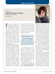
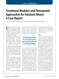
Figure: Arthroscopic image of a labral tear <strong>in</strong> the<br />
anterior position extend<strong>in</strong>g past the 3-o’clock position<br />
(A) compared with a tear <strong>in</strong> the 12- to 2-o’clock<br />
position (B). Abbreviations: ACT, acetabulum; L,<br />
labrum; Lab, labrum.<br />
A<br />
B<br />
MARCH 2013 | Volume 36 • Number 3<br />
e293
n Feature Article<br />
Over the past 2 decades, <strong>in</strong>terest<br />
has <strong>in</strong>creased <strong>in</strong> anatomic<br />
variations <strong>in</strong> the proximal femur<br />
<strong>and</strong> acetabulum as they perta<strong>in</strong> to femoroacetabular<br />
imp<strong>in</strong>gement (FAI), labral<br />
pathology, <strong>and</strong> hip osteoarthritis. It is<br />
now recognized that decreased femoral<br />
head-neck offset is the primary cause of<br />
cam-type FAI. 1,2 In addition, acetabular<br />
overcoverage, which can either be focal<br />
from acetabular retroversion or global retroversion<br />
as seen <strong>in</strong> coxa profunda or protrusio<br />
acetabuli, is the pr<strong>in</strong>cipal cause of<br />
p<strong>in</strong>cer-type FAI. 3-5 Regardless of whether<br />
a patient has cam imp<strong>in</strong>gement, p<strong>in</strong>cer<br />
imp<strong>in</strong>gement, or both, it is now believed<br />
that these variations are risk factors for<br />
early hip osteoarthritis. Many cases of hip<br />
osteoarthritis previously described as idiopathic<br />
may <strong>in</strong> fact be due to these subtle<br />
bony abnormalities. 1,6<br />
Although much attention has been<br />
given to variations <strong>in</strong> proximal femoral<br />
<strong>and</strong> acetabular anatomy, less attention has<br />
been focused on the role of femoral version.<br />
Alterations <strong>in</strong> femoral version have<br />
been associated with hip pa<strong>in</strong> <strong>and</strong> osteoarthritis<br />
for more than 30 years. 7-11 It has<br />
been further speculated that excessive<br />
femoral anteversion or femoral retroversion<br />
may also play a role <strong>in</strong> the pathogenesis<br />
<strong>and</strong> treatment of FAI. 1,12<br />
The soft tissues around the hip are<br />
also important <strong>in</strong> the development of hip<br />
pathology. The iliopsoas muscle is the<br />
most important hip flexor. It has a close<br />
anatomic relationship with the acetabular<br />
labrum <strong>and</strong> the anterior capsule. This<br />
muscle has been associated with anterior<br />
hip pa<strong>in</strong> <strong>in</strong> cases of flexor tend<strong>in</strong>itis,<br />
snapp<strong>in</strong>g hips, <strong>and</strong> psoas imp<strong>in</strong>gement. 13<br />
Psoas imp<strong>in</strong>gement has been described as<br />
a cause of labral tears. 14 These tears can<br />
occur <strong>in</strong> a more anterior position (close to<br />
the psoas tendon) <strong>in</strong> patients with no bony<br />
abnormalities. 14<br />
The purpose of this study was threefold:<br />
(1) to describe values for femoral<br />
anteversion measured us<strong>in</strong>g magnetic<br />
resonance imag<strong>in</strong>g (MRI) <strong>in</strong> patients undergo<strong>in</strong>g<br />
hip arthroscopy for FAI; (2) to<br />
report the relationship between physical<br />
exam<strong>in</strong>ation f<strong>in</strong>d<strong>in</strong>gs <strong>and</strong> femoral version<br />
<strong>in</strong> these patients; <strong>and</strong> (3) to report the relationship<br />
between the degree of femoral<br />
anteversion <strong>and</strong> <strong>in</strong>traoperative f<strong>in</strong>d<strong>in</strong>gs<br />
dur<strong>in</strong>g hip arthroscopy. The authors hypothesized<br />
that patients with significant<br />
variations <strong>in</strong> femoral version would have<br />
differ<strong>in</strong>g preoperative exam<strong>in</strong>ation f<strong>in</strong>d<strong>in</strong>gs<br />
<strong>and</strong> <strong>in</strong>traoperative hip pathology.<br />
Materials <strong>and</strong> Methods<br />
<strong>Between</strong> June 2009 <strong>and</strong> May 2010, a<br />
total of 392 hip arthroscopies were performed<br />
at the authors’ <strong>in</strong>stitution. Patients<br />
were <strong>in</strong>cluded <strong>in</strong> this study if they had undergone<br />
hip arthroscopy for the treatment<br />
of FAI with concomitant labral pathology<br />
<strong>and</strong> had their femoral version measured<br />
by MRI preoperatively. Exclusion criteria<br />
<strong>in</strong>cluded prior hip arthroscopy, age<br />
younger than 18 years, <strong>and</strong> hip dysplasia,<br />
def<strong>in</strong>ed as a lateral center-edge angle less<br />
than 20°. 13 This study was approved by<br />
the <strong>in</strong>stitutional review board.<br />
A total of 188 consecutive patients<br />
(204 hips) met the <strong>in</strong>clusion criteria.<br />
Patient demographics, <strong>in</strong>clud<strong>in</strong>g age, sex,<br />
weight, height, <strong>and</strong> body mass <strong>in</strong>dex, were<br />
prospectively collected <strong>and</strong> retrospectively<br />
reviewed. All patients underwent a<br />
detailed physical exam<strong>in</strong>ation. Range of<br />
motion was measured with a goniometer<br />
<strong>in</strong> all planes, <strong>in</strong>clud<strong>in</strong>g abduction, adduction,<br />
flexion, <strong>and</strong> <strong>in</strong>ternal <strong>and</strong> external<br />
rotation. Internal <strong>and</strong> external rotation<br />
measurements were performed with the<br />
patient ly<strong>in</strong>g <strong>in</strong> the prone position on the<br />
exam<strong>in</strong>ation table. The imp<strong>in</strong>gement test<br />
(flexion–adduction–<strong>in</strong>ternal rotation) <strong>and</strong><br />
the flexion–adduction–<strong>in</strong>ternal rotation<br />
distance were also recorded. The flexion–<br />
adduction–<strong>in</strong>ternal rotation distance is<br />
determ<strong>in</strong>ed by plac<strong>in</strong>g 1 leg <strong>in</strong> a figureof-four<br />
position so the ipsilateral ankle is<br />
positioned proximal to the contralateral<br />
knee. The vertical distance between the<br />
genicular l<strong>in</strong>e <strong>and</strong> the exam<strong>in</strong>ation table<br />
was recorded, <strong>and</strong> the difference between<br />
the affected <strong>and</strong> nonaffected side was calculated.<br />
The test was positive when the<br />
difference between extremities was more<br />
than 4 cm. 15<br />
Radiographic views <strong>in</strong>cluded an anteroposterior<br />
pelvic view, a cross-table<br />
lateral view, <strong>and</strong> a false-profile view.<br />
After radiographic evaluation, hips were<br />
classified as hav<strong>in</strong>g either cam, p<strong>in</strong>cer, or<br />
mixed-type (cam <strong>and</strong> p<strong>in</strong>cer) imp<strong>in</strong>gement.<br />
Hips classified as hav<strong>in</strong>g cam imp<strong>in</strong>gement<br />
had an alpha angle greater than<br />
50°. Hips classified as hav<strong>in</strong>g p<strong>in</strong>cer imp<strong>in</strong>gement<br />
had at least 1 of the follow<strong>in</strong>g<br />
radiographic f<strong>in</strong>d<strong>in</strong>gs: a crossover sign,<br />
coxa profunda, or protusio acetabuli. The<br />
alpha angle was measured <strong>in</strong> the crosstable<br />
lateral view as described by Nötzli<br />
et al. 16 The lateral center-edge angle was<br />
measured on the anteroposterior view. 15<br />
The anterior center-edge angle was not<br />
measured.<br />
Magnetic resonance imag<strong>in</strong>g was<br />
obta<strong>in</strong>ed <strong>in</strong> all cases us<strong>in</strong>g a 3-T unit<br />
(Magnetom Verio 3T; Siemens Medical<br />
Systems, Erlangen. Germany). No arthrograms<br />
were performed. Images were<br />
evaluated for the presence of labral tears,<br />
cartilage disorders, ligamentum teres ruptures,<br />
<strong>and</strong> other soft tissue pathologies.<br />
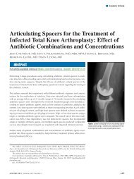
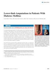
The femoral anteversion was measured<br />
us<strong>in</strong>g the technique described by Tomczak<br />
et al. 17 After obta<strong>in</strong><strong>in</strong>g a scout view of the<br />
knee, an axial slice conta<strong>in</strong><strong>in</strong>g the most<br />
posterior aspect of both femoral condyles<br />
was determ<strong>in</strong>ed, <strong>and</strong> an angle compris<strong>in</strong>g<br />
the horizontal plane <strong>and</strong> a l<strong>in</strong>e conta<strong>in</strong><strong>in</strong>g<br />
the most posterior part of the distal femoral<br />
condyles was determ<strong>in</strong>ed.<br />
A second slice conta<strong>in</strong><strong>in</strong>g the center<br />
of the femoral head <strong>and</strong> the center of the<br />
femoral neck was obta<strong>in</strong>ed <strong>and</strong> considered<br />
to be the femoral neck axis. Then, an angle<br />
compris<strong>in</strong>g the femoral neck <strong>and</strong> the horizontal<br />
plane was measured. The femoral<br />
neck anteversion angle was considered to<br />
be the difference between the femoral neck<br />
angle <strong>and</strong> the posterior distal condyle angle<br />
(Figure 1). Positive angles were considered<br />
anteversion <strong>and</strong> negative angles were cone294<br />
ORTHOPEDICS | <strong>Healio</strong>.com/Orthopedics
<strong>Femoral</strong> <strong>Anteversion</strong> <strong>and</strong> FAI | Ejnisman et al<br />
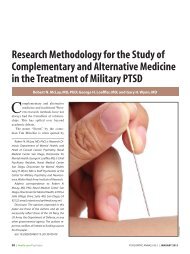
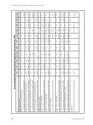
1A 1B 1C<br />
Figure 1: Scout view magnetic resonance image of the right knee show<strong>in</strong>g the angle between the most posterior part of the femoral condyles (l<strong>in</strong>e A) <strong>and</strong> the<br />
horizontal plane (l<strong>in</strong>e B) (A). Plane conta<strong>in</strong><strong>in</strong>g the center of the femoral head <strong>and</strong> the center of the femoral neck (l<strong>in</strong>e C) (B). Slice <strong>in</strong> the plane <strong>in</strong> Figure 1B show<strong>in</strong>g<br />
the angle compris<strong>in</strong>g the center of the femoral neck <strong>and</strong> head (l<strong>in</strong>e D) <strong>and</strong> the horizontal plane (l<strong>in</strong>e B). In this example, the femoral neck version is 9° (angle<br />
<strong>in</strong> Figure 1C2angle <strong>in</strong> Figure 1A) (C).<br />
sidered retroversion. All MRI data were<br />
analyzed by a fellowship-tra<strong>in</strong>ed musculoskeletal<br />
radiologist (C.P.H.) with more<br />
than 10 years of cl<strong>in</strong>ical practice. Magnetic<br />
resonance imag<strong>in</strong>g was obta<strong>in</strong>ed for the <strong>in</strong>jured<br />
hip only.<br />
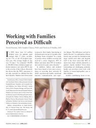
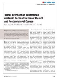
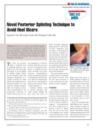
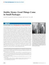
Figure 2: Arthroscopic image show<strong>in</strong>g psoas release<br />
performed <strong>in</strong> the central compartment through a<br />
small capsular w<strong>in</strong>dow us<strong>in</strong>g an arthroscopic knife.<br />
2<br />
Figure 3: Arthroscopic image of <strong>in</strong>tense synovitis <strong>in</strong><br />
the anterior capsule <strong>in</strong> a patient who required psoas<br />
release. Abbreviations: FH, femoral head; L, labrum.<br />
Surgical Technique<br />
All hip arthroscopies were performed<br />
by the senior author (M.J.P.) with patients<br />
<strong>in</strong> the modified sup<strong>in</strong>e position on a fracture<br />
table us<strong>in</strong>g anterolateral <strong>and</strong> midanterior<br />
portals, as previously described. 18,19<br />
Intraoperative data <strong>in</strong>cluded the presence,<br />
location, <strong>and</strong> size of a labral tear,<br />
the presence <strong>and</strong> location of cartilage lesions,<br />
which were graded accord<strong>in</strong>g to the<br />
Outerbridge classification, 20 the presence<br />
of ligamentum teres pathology, <strong>and</strong> the<br />
necessity for either an arthroscopic psoas<br />
release or a capsular plication. Patients<br />
with grade IV cartilage lesions underwent<br />
a microfracture procedure.<br />
All ligamentum teres tears, both partial<br />
<strong>and</strong> complete, were treated with thermal<br />
<strong>and</strong> mechanical debridement. If a psoas<br />
release was necessary, it was performed <strong>in</strong><br />
the central compartment through a small<br />
capsular w<strong>in</strong>dow us<strong>in</strong>g an arthroscopic<br />
knife (Figure 2). This was a partial release,<br />
cutt<strong>in</strong>g only the tend<strong>in</strong>ous part of<br />
the muscle–tendon unit of the iliopsoas<br />
muscle. Indications for a psoas release <strong>in</strong>cluded<br />
<strong>in</strong>ternal snapp<strong>in</strong>g hip (asymptomatic<br />
or symptomatic), flexor tend<strong>in</strong>itis, <strong>and</strong><br />
<strong>in</strong>tense synovitis <strong>in</strong> the anterior capsule<br />
dur<strong>in</strong>g hip arthroscopy (Figure 3). Flexor<br />
tend<strong>in</strong>itis was diagnosed by physical exam<strong>in</strong>ation,<br />
which <strong>in</strong>cluded tenderness<br />
associated with the psoas with palpation<br />
<strong>and</strong> pa<strong>in</strong>ful resisted leg flexion, <strong>in</strong> addition<br />
to signs of edema <strong>in</strong> the flexor tendon<br />
on MRI. Synovitis of the anterior capsule<br />
next to the “psoas U” <strong>in</strong> patients with FAI<br />
was often associated with flexor tend<strong>in</strong>itis<br />
<strong>in</strong> the senior author’s experience.<br />
The senior author performed a capsular<br />
closure <strong>in</strong> the majority of hip arthroscopies<br />
<strong>and</strong> chose to perform a plication <strong>in</strong><br />
cases of capsular laxity. Capsular laxity<br />
was considered <strong>in</strong> cases of global laxity<br />
(knee hyperextension <strong>and</strong> elbow hyperextension),<br />
<strong>in</strong> patients subjectively report<strong>in</strong>g<br />
hip <strong>in</strong>stability, <strong>and</strong> <strong>in</strong> patients where a<br />
patulous capsule was observed dur<strong>in</strong>g hip<br />
arthroscopy. Labral tears were repaired<br />
with suture anchors if possible. The size<br />
<strong>and</strong> location of the labral tears were recorded<br />
us<strong>in</strong>g the clock-face location system.<br />
21 Labral tear size was estimated us<strong>in</strong>g<br />
a burr as a reference size. The same<br />
surgeon measured all tears with the same<br />
device. For the labral tear location analysis,<br />
all data on left hips were mirrored <strong>and</strong><br />
presented as right hips. In cases where the<br />
labral tear was considered unrepairable,<br />
a labral reconstruction us<strong>in</strong>g an iliotibial<br />
autograft was performed. 22<br />
Comparison of cont<strong>in</strong>uous variables<br />
by b<strong>in</strong>ary categorical variables was performed<br />
us<strong>in</strong>g the <strong>in</strong>dependent-samples t<br />
test, <strong>and</strong> comparison of multiple (more<br />
3<br />
MARCH 2013 | Volume 36 • Number 3<br />
e295
n Feature Article<br />
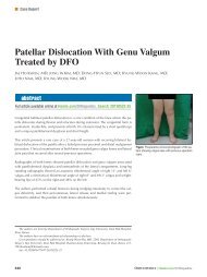
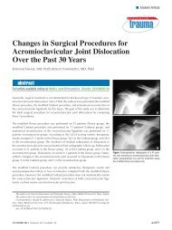
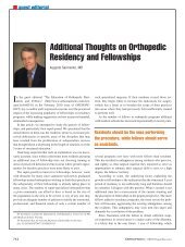
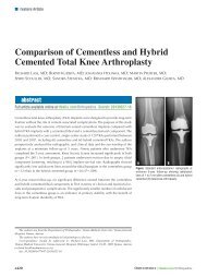
Figure 4: Graph show<strong>in</strong>g a normal femoral neck version distribution with the<br />
peak at 8°. Values less than 0 <strong>in</strong>dicate retroversion. Abbreviation: Std Dev,<br />
st<strong>and</strong>ard deviation.<br />
than 2) categorical variables was performed<br />
us<strong>in</strong>g 1-way analysis of variance.<br />
Comparison of 2 cont<strong>in</strong>uous variables<br />
was performed us<strong>in</strong>g Pearson’s<br />
correlation coefficient. Comparison of<br />
categorical variables was performed us<strong>in</strong>g<br />
Fisher’s exact test for comparisons<br />
of proportions. Statistical analyses were<br />
performed us<strong>in</strong>g SPSS version 11 statistical<br />
software (SPSS Inc, Chicago,<br />
Ill<strong>in</strong>ois). All reported P values are<br />
2-tailed, with an alpha level of .05 <strong>in</strong>dicat<strong>in</strong>g<br />
statistical significance.<br />
Results<br />
The study group comprised 100 men<br />
<strong>and</strong> 88 women with a mean age of 35<br />
years (range, 18 to 62 years). Mean patient<br />
height was 68 <strong>in</strong>ches (range, 61 to 84<br />
<strong>in</strong>ches), mean weight was 165 lb (range,<br />
110 to 318 lb), <strong>and</strong> mean body mass <strong>in</strong>dex<br />
was 24 kg/m 2 (range, 15 to 41 kg/m 2 ).<br />
Mean femoral anteversion angle was<br />
9° (range, 210° to 27°). For statistical<br />
analysis, hips were divided <strong>in</strong>to 3 groups:<br />
group 1 <strong>in</strong>cluded hips with an anteversion<br />
angle of less than 5° (n545; 22%),<br />
group 2 <strong>in</strong>cluded hips with an anteversion<br />
angle between 5° <strong>and</strong> 15° (n5102;<br />
50%), <strong>and</strong> group 3<br />
<strong>in</strong>cluded hips with<br />
an anteversion<br />
angle greater than<br />
15° (n557; 28%).<br />
<strong>Femoral</strong> version<br />
was normally distributed<br />
accord<strong>in</strong>g<br />
to the Kolmogorov-<br />
Smirnov test (Figure<br />
4). No relationship<br />
was found between<br />
patient demographics<br />
(ie, age, sex,<br />
height, weight, <strong>and</strong><br />
4 body mass <strong>in</strong>dex)<br />
<strong>and</strong> femoral version<br />
angle. In the 16 bilateral<br />
patients, no<br />
statistical difference<br />
was found <strong>in</strong> the<br />
femoral neck version between sides. The<br />
first operated side had an average of 8°<br />
of femoral version, <strong>and</strong> the second side<br />
had an average of 6° of femoral version<br />
(P5.134).<br />
The physical exam<strong>in</strong>ation revealed<br />
the follow<strong>in</strong>g mean range of motion for<br />
the affected hip jo<strong>in</strong>t: abduction545°<br />
(range, 18° to 95°), adduction519°<br />
(range, 2° to 35°), flexion5109° (range,<br />
45° to 145°), external rotation540°<br />
(range, 2° to 96°), <strong>and</strong> <strong>in</strong>ternal rotation525°<br />
(range, 3° to 95°). All hips had<br />
a positive imp<strong>in</strong>gement sign. Mean difference<br />
of the FABER distance between<br />
the affected <strong>and</strong> nonaffected sides was<br />
4 cm (range, 0 to 26 cm). Seventy-two<br />
(35%) patients had a difference greater<br />
than 4 cm between sides <strong>and</strong> were considered<br />
to have a positive FABER test.<br />
A significant correlation was found between<br />
version degrees <strong>and</strong> degrees of<br />
external rotation (r520.208; P5.027)<br />
<strong>and</strong> <strong>in</strong>ternal rotation (r50.231; P5.002).<br />
A significant difference existed between<br />
version groups based on external rotation<br />
(P5.027). External rotation for group<br />
1 (average, 45°614°) was significantly<br />
higher than that for groups 2 (average,<br />
38°612°) <strong>and</strong> 3 (average, 36°613°).<br />
Average <strong>in</strong>ternal rotation was 23° for<br />
group 1, 25° for group 2, <strong>and</strong> 30° for<br />
group 3, but this was not statistically different.<br />
After radiographic evaluation, 20%<br />
of hips were diagnosed with p<strong>in</strong>cer imp<strong>in</strong>gement,<br />
5% with cam imp<strong>in</strong>gement,<br />
<strong>and</strong> 75% with mixed-type imp<strong>in</strong>gement.<br />
Average lateral center-edge angle was<br />
36° (range, 20° to 51°). Mean alpha angle<br />
was 70° (range, 41° to 99°). No significant<br />
correlation was found between alpha<br />
angle or center-edge angle <strong>and</strong> version.<br />
Dur<strong>in</strong>g hip arthroscopy, labral tears<br />
were identified <strong>in</strong> 88% of patients. Mean<br />
tear size was 34 mm (range, 10 to 72 mm).<br />
Mean tear size was 30 mm <strong>in</strong> group 1, 34<br />
mm <strong>in</strong> group 2, <strong>and</strong> 38 mm <strong>in</strong> group 3<br />
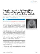
(P5.008). Regard<strong>in</strong>g labral tear position,<br />
<strong>in</strong> hips with more than 15° of femoral<br />
anteversion, 26 tears extended past the<br />
3-o’clock position <strong>and</strong> 33 did not; however,<br />
<strong>in</strong> hips with less than 15° of anteversion,<br />
37 extended past the 3-o’clock<br />
position <strong>and</strong> 103 did not. Hips with anteversion<br />
angles greater than 15° were 2.2<br />
times more likely to have labral tears that<br />
extended past the 3-o’clock position (95%<br />
confidence <strong>in</strong>terval, 1.2 to 4.1), denot<strong>in</strong>g<br />
more anterior tears (Figure 5). Labral repairs<br />
were performed <strong>in</strong> 178 patients <strong>and</strong><br />
labral reconstructions <strong>in</strong> 13 patients.<br />
Outerbridge grade III or IV cartilage lesions<br />
were identified <strong>in</strong> 97 (52%) hips on<br />
the femoral side (23 on the weight-bear<strong>in</strong>g<br />
zone) <strong>and</strong> <strong>in</strong> 56 (30%) hips on the acetabular<br />
side. A microfracture was performed <strong>in</strong><br />
23 (12%) of these cases. The psoas tendon<br />
was arthroscopically released <strong>in</strong> 44 (21%)<br />
patients. Tears of the ligamentum teres,<br />
both full (3 hips) <strong>and</strong> partial (126 hips),<br />
were identified dur<strong>in</strong>g 129 (63%) of the<br />
surgeries (Figure 6). A significant association<br />
was found between femoral version<br />
<strong>and</strong> the need for a psoas release. Mean<br />
femoral version angle was 11° <strong>in</strong> patients<br />
<strong>in</strong> whom a psoas release was performed<br />
<strong>and</strong> 8° <strong>in</strong> patients <strong>in</strong> whom a psoas release<br />
was not performed (P5.023). No asso-<br />
e296<br />
ORTHOPEDICS | <strong>Healio</strong>.com/Orthopedics
<strong>Femoral</strong> <strong>Anteversion</strong> <strong>and</strong> FAI | Ejnisman et al<br />
5A<br />
Figure 5: Arthroscopic image of a labral tear <strong>in</strong> the anterior position extend<strong>in</strong>g past the 3-o’clock position<br />
(A) compared with a tear <strong>in</strong> the 12- to 2-o’clock position (B). Abbreviations: ACT, acetabulum; L, labrum;<br />
Lab, labrum.<br />
5B<br />
Figure 6: Arthroscopic image show<strong>in</strong>g a partial<br />
tear of the ligamentum teres (arrow).<br />
6<br />
ciation was found between the presence of<br />
grade III or IV cartilage lesions or ligamentum<br />
teres tears <strong>and</strong> femoral version.<br />
Discussion<br />
The results of this study revealed that<br />
patients undergo<strong>in</strong>g hip arthroscopy for<br />
FAI had a mean femoral neck version of<br />
9°. Although no relationship was found<br />
between femoral neck version <strong>and</strong> patient<br />
demographics, hip external rotation was<br />
negatively correlated with femoral version<br />
<strong>and</strong> <strong>in</strong>ternal rotation was positively correlated<br />
with femoral version. In addition,<br />
hips with greater anteversion were more<br />
likely to have larger <strong>and</strong> more anterior<br />
labral tears <strong>and</strong> to require a psoas tendon<br />
release at the time of arthroscopy.<br />
<strong>Femoral</strong> neck version is the torsion<br />
of the proximal femoral head with reference<br />
to the distal femur. 23 When discuss<strong>in</strong>g<br />
femoral version, def<strong>in</strong><strong>in</strong>g normal<br />
values is of paramount importance. The<br />
Table shows normal values reported <strong>in</strong><br />
the literature. Values range from 8° 23 to<br />
20°. 7,12,17,24,25,37,51-61 The current study<br />
found a mean femoral anteversion angle<br />
of 9°, which is consistent with the literature.<br />
Therefore, it appears that femoral<br />
version does not differ <strong>in</strong> the FAI population<br />
compared with healthy <strong>in</strong>dividuals.<br />
The current f<strong>in</strong>d<strong>in</strong>gs are also similar to<br />
the value of 9.7° found by Ito et al. 12 A<br />
broad range of values for femoral version<br />
(range, 210° to 27°) has been reported<br />
<strong>in</strong> prior studies, 23-39,51-61 which makes it<br />
challeng<strong>in</strong>g to def<strong>in</strong>e a normal range for<br />
femoral version.<br />
Several methods have been described<br />
to measure femoral version, <strong>in</strong>clud<strong>in</strong>g<br />
physical exam<strong>in</strong>ation, 26,27 radiography, 29,30<br />
ultrasonography, 31-34 two-dimensional<br />
computed tomography (CT), 25,35-37 threedimensional<br />
CT 23,38 <strong>and</strong> MRI. 17,39 Magnetic<br />
resonance imag<strong>in</strong>g has the advantage over<br />
radiography <strong>and</strong> CT of avoid<strong>in</strong>g radiation<br />
exposure. Moreover, MRI is used rout<strong>in</strong>ely<br />
<strong>in</strong> FAI patients to evaluate labral <strong>and</strong> cartilage<br />
pathology. 40,41 Although CT scans<br />
rema<strong>in</strong> the gold st<strong>and</strong>ard for calculat<strong>in</strong>g<br />
femoral version, studies with MRI have<br />
reported high levels of accuracy compared<br />
with CT scans with high <strong>in</strong>ter- <strong>and</strong> <strong>in</strong>traobserver<br />
agreements. 17<br />
In the current study, no relationship<br />
was identified between patient demographics<br />
(ie, age, sex, weight, height, <strong>and</strong> body<br />
mass <strong>in</strong>dex) <strong>and</strong> femoral version. However,<br />
a correlation was found between femoral<br />
version <strong>and</strong> hip external rotation. In addition,<br />
the data showed that external rotation<br />
<strong>in</strong>creased for patients with femoral version<br />
less than 5°. No difference was found between<br />
the other groups. For <strong>in</strong>ternal rotation,<br />
a statistically significant difference<br />
did not exist between the groups, so it was<br />
unclear at what degree of version <strong>in</strong>ternal<br />
rotation is dim<strong>in</strong>ished. This relationship<br />
has been previously described <strong>in</strong> the literature<br />
42,43 <strong>and</strong> is used to estimate femoral<br />
neck version dur<strong>in</strong>g physical exam<strong>in</strong>ation.<br />
This is an important f<strong>in</strong>d<strong>in</strong>g because<br />
symptomatic FAI patients typically have<br />
decreased range of motion, 15,44 so the relation<br />
between hip ROM <strong>and</strong> femoral neck<br />
version could be lost <strong>in</strong> patients with FAI.<br />
However, physical exam<strong>in</strong>ation <strong>in</strong> the current<br />
study was performed while patients<br />
were awake, so it is impossible to estimate<br />
if rotation was limited by pa<strong>in</strong> <strong>and</strong> not just<br />
by bony imp<strong>in</strong>gement.<br />
Dur<strong>in</strong>g hip arthroscopy, patients with<br />
femoral anteversion greater than 15° had<br />
larger labral tears than patients with femoral<br />
anteversion less than 15°. Patients with<br />
higher anteversion also had labral tears located<br />
<strong>in</strong> a more anterior location. In addition,<br />
hips that needed a psoas release had<br />
greater anteversion compared with those<br />
that did not. Psoas imp<strong>in</strong>gement is caused<br />
by a compression or traction force of the<br />
psoas tendon on the anterior capsulolabral<br />
complex. 41 This force may lead to more<br />
anterior labral tears that occur <strong>in</strong> close<br />
proximity to the psoas valley, or psoas<br />
U. 45 The relationship of the psoas muscle<br />
<strong>and</strong> femoral anteversion has been previously<br />
described <strong>in</strong> the literature. Us<strong>in</strong>g a<br />
mathematical model, Fra<strong>in</strong> 46 reported that<br />
when femoral anteversion is marked, the<br />
anterior muscles play a greater role <strong>in</strong> the<br />
ma<strong>in</strong>tenance of equilibrium. Schutte et<br />
al 47 used a computerized model to show<br />
that psoas muscle length is sensitive to<br />
femoral anteversion. Greater antever-<br />
MARCH 2013 | Volume 36 • Number 3<br />
e297
n Feature Article<br />
Study<br />
Table<br />
<strong>Femoral</strong> Neck Version Values <strong>in</strong> the Literature<br />
Study Method<br />
No. of<br />
Femurs<br />
Average6SD<br />
(Range) <strong>Femoral</strong><br />
Neck Version, deg<br />
Current study MRI 204 9 (27 to 210)<br />
Reikerås et al 7 CT 47 1367<br />
Ito et al 12 MRI <strong>in</strong> controls 24 15.764.4<br />
MRI <strong>in</strong> patients with FAI 24 9.764.7<br />
Tomczak et al 17 CT 25 22.269.4<br />
MRI 25 15.7169.3<br />
K<strong>in</strong>gsley & Olmsted 24 Anatomic 630 8 (38 to 220)<br />
Sugano et al 25 3-D CT on specimens 30 19.869.3<br />
Reikerås et al 37 Anatomic 96 10.466.7<br />
Soutter & Bradford 51 Anatomic 154 14.3 (50 to 0)<br />
Parsons 52 Anatomic 300 15.5 (40 to 217)<br />
Durham 53 Anatomic 200 11.9 (35 to 0)<br />
Pick et al 54 Anatomic 152 14 (41 to 218)<br />
Gray 55 N/A N/A (14 to 12)<br />
Dunlap et al 56 Radiographs 200 8.7<br />
Alvik 57 N/A N/A 12<br />
Bauman et al 58<br />
Weighted average<br />
from literature<br />
1436 11.4 (50 to 220)<br />
Wasielewski 59 N/A N/A 15<br />
Koval & Zuckerman 60 N/A N/A 10<br />
Toogood et al 61 Anatomic 375 9.7369.28<br />
(35.9 to 214.63)<br />
Abbreviations: 3-D, 3-dimensional; CT, computed tomography; deg, degrees; MRI, magnetic<br />
resonance imag<strong>in</strong>g; N/A, not available.<br />
Limitations to the current study <strong>in</strong>cluded<br />
its retrospective design <strong>and</strong> the lack of<br />
a control group to compare the values of<br />
femoral anteversion. Nevertheless, a thorough<br />
literature review provided an adequate<br />
historical control group for comparison of<br />
femoral version. Another limitation to the<br />
study was the referral nature of the study<br />
population. The majority of the patients<br />
were seen by other practitioners <strong>and</strong> referred<br />
to the current authors’ <strong>in</strong>stitution for<br />
surgical treatment. This expla<strong>in</strong>s the large<br />
number of patients with FAI <strong>and</strong> the high<br />
percentage of patients with labral tears. To<br />
the authors’ knowledge, this was the first<br />
study to evaluate the association of femoral<br />
anteversion with <strong>in</strong>traoperative f<strong>in</strong>d<strong>in</strong>gs <strong>in</strong><br />
patients with FAI <strong>and</strong> was the largest series<br />
of patients <strong>in</strong> the literature with femoral<br />
version measurements on MRI.<br />
Conclusion<br />
In the current study, patients with FAI<br />
had a mean anteversion angle of 9°, which<br />
is similar to values found <strong>in</strong> the normal<br />
population. No relationship was found between<br />
femoral version <strong>and</strong> patient demographics,<br />
but a correlation was identified<br />
between femoral neck version <strong>and</strong> hip rotation.<br />
Hips with <strong>in</strong>creased femoral version<br />
angles had larger <strong>and</strong> more anterior<br />
labral tears dur<strong>in</strong>g hip arthroscopy.<br />
<strong>Femoral</strong> neck version should be taken <strong>in</strong>to<br />
consideration <strong>in</strong> FAI treatment.<br />
sion angles may lead to an overtightened<br />
psoas. It is unclear how large of an <strong>in</strong>crease<br />
<strong>in</strong> version angle would be necessary<br />
to cause the psoas to require treatment. In<br />
a recent study, <strong>in</strong>creased femoral version<br />
was associated with <strong>in</strong>ferior outcomes<br />
after psoas lengthen<strong>in</strong>g. 48 However, it is<br />
important to note that <strong>in</strong>creased femoral<br />
version was greater than 25° <strong>in</strong> that study.<br />
However, <strong>in</strong> the current study, only 2 patients<br />
had femoral version greater than<br />
25°. Further research is needed to better<br />
elucidate the relationship of the psoas<br />
tendon, femoral version, <strong>and</strong> acetabular<br />
labrum.<br />
<strong>Femoral</strong> version <strong>in</strong>fluenc<strong>in</strong>g hip pathology<br />
is not a novel idea; however,<br />
reports are contradictory regard<strong>in</strong>g<br />
whether higher or lower angles are detrimental<br />
for the hip jo<strong>in</strong>t. In osteoarthritis,<br />
both <strong>in</strong>creased <strong>and</strong> decreased femoral<br />
anteversion have been implicated <strong>in</strong><br />
primary osteoarthritis of the hip. 7-11,49,50<br />
Regard<strong>in</strong>g FAI, decreased femoral version<br />
has been reported as a predispos<strong>in</strong>g<br />
factor <strong>in</strong> the pathomechanics of imp<strong>in</strong>gement.<br />
1,12 The current study demonstrated<br />
that patients with FAI can present with<br />
normal, <strong>in</strong>creased, or decreased femoral<br />
anteversion.<br />
References<br />
1. Ganz R, Parvizi J, Beck M, Leunig M, Notzli<br />
H, Siebenrock KA. Femoroacetabular imp<strong>in</strong>gement:<br />
a cause for osteoarthritis of the hip. Cl<strong>in</strong><br />
Orthop Relat Res. 2003; 417:112-120.<br />
2. Beck M, Kalhor M, Leunig M, Ganz R. Hip<br />
morphology <strong>in</strong>fluences the pattern of damage<br />
to the acetabular cartilage: femoroacetabular<br />
imp<strong>in</strong>gement as a cause of early osteoarthritis<br />
of the hip. J Bone Jo<strong>in</strong>t Surg Br. 2005;<br />
87:1012-1018.<br />
3. Reynolds D, Lucas J, Klaue K. Retroversion<br />
of the acetabulum. A cause of hip pa<strong>in</strong>. J<br />
Bone Jo<strong>in</strong>t Surg Br. 1999; 81:281-288.<br />
4. Jamali AA, Mladenov K, Meyer DC, et al.<br />
Anteroposterior pelvic radiographs to assess<br />
acetabular retroversion: high validity of<br />
the “cross-over-sign”. J Orthop Res. 2007;<br />
25:758-765.<br />
e298<br />
ORTHOPEDICS | <strong>Healio</strong>.com/Orthopedics
<strong>Femoral</strong> <strong>Anteversion</strong> <strong>and</strong> FAI | Ejnisman et al<br />
5. Kalberer F, Sierra RJ, Madan SS, Ganz R,<br />
Leunig M. Ischial sp<strong>in</strong>e projection <strong>in</strong>to the<br />
pelvis: a new sign for acetabular retroversion.<br />
Cl<strong>in</strong> Orthop Relat Res. 2008; 466:677-<br />
683.<br />
6. McCarthy JC, Noble PC, Schuck MR,<br />
Wright J, Lee J. The Otto E. Aufranc Award:<br />
the role of labral lesions to development of<br />
early degenerative hip disease. Cl<strong>in</strong> Orthop<br />
Relat Res. 2001; 393:25-37.<br />
7. Reikerås O, Bjerkreim I, Kolbenstvedt A. <strong>Anteversion</strong><br />
of the acetabulum <strong>and</strong> femoral neck<br />
<strong>in</strong> normals <strong>and</strong> <strong>in</strong> patients with osteoarthritis of<br />
the hip. Acta Orthop Sc<strong>and</strong>. 1983; 54:18-23.<br />
8. Tönnis D, He<strong>in</strong>ecke A. Acetabular <strong>and</strong> femoral<br />
anteversion: relationship with osteoarthritis<br />
of the hip. J Bone Jo<strong>in</strong>t Surg Am. 1999;<br />
81:1747-1770.<br />
9. Giunti A, Moroni A, Olmi R, Rimondi E,<br />
Soldati D, Vicenzi G. The importance of the<br />
angle of anteversion <strong>in</strong> the development of<br />
arthritis of the hip. Ital J Orthop Traumatol.<br />
1985; 11:23-27.<br />
10. Reikerås O, Høiseth A. <strong>Femoral</strong> neck angles<br />
<strong>in</strong> osteoarthritis of the hip. Acta Orthop<br />
Sc<strong>and</strong>. 1982; 53:781-784.<br />
11. Terjesen T, Benum P, Anda S, Svenn<strong>in</strong>gsen<br />
S. Increased femoral anteversion <strong>and</strong> osteoarthritis<br />
of the hip jo<strong>in</strong>t. Acta Orthop Sc<strong>and</strong>.<br />
1982; 53:571-575.<br />
12. Ito K, M<strong>in</strong>ka MA, Leunig M, Werlen S, Ganz<br />
R. Femoroacetabular imp<strong>in</strong>gement <strong>and</strong> the<br />
cam-effect. A MRI-based quantitative anatomical<br />
study of the femoral head-neck offset.<br />
J Bone Jo<strong>in</strong>t Surg Br. 2001; 83:171-176.<br />
13. Philippon MJ, Maxwell RB, Johnston TL,<br />
Schenker M, Briggs KK. Cl<strong>in</strong>ical presentation<br />
of femoroacetabular imp<strong>in</strong>gement. Knee Surg<br />
Sports Traumatol Arthrosc. 2007; 15:1041-<br />
1047.<br />
14. Domb BG, Sh<strong>in</strong>dle MK, McArthur B, Voos JE,<br />
Magennis EM, Kelly BT. Iliopsoas imp<strong>in</strong>gement:<br />
a newly identified cause of labral pathology<br />
<strong>in</strong> the hip. HSS J. 2011; 7:145-150.<br />
15. Wiberg G. Studies on dysplastic acetabula<br />
<strong>and</strong> congenital subluxation of the hip jo<strong>in</strong>t.<br />
Acta Chir Sc<strong>and</strong>. 1939; 83:5-135.<br />
16. Nötzli HP, Wyss TF, Stoeckl<strong>in</strong> CH, Schmid<br />
MR, Treiber K, Hodler J. The contour of the<br />
femoral head-neck junction as a predictor<br />
for the risk of anterior imp<strong>in</strong>gement. J Bone<br />
Jo<strong>in</strong>t Surg Br. 2002; 84:556-560.<br />
17. Tomczak RJ, Guenther KP, Rieber A, Mergo<br />
P, Ros PR, Brambs HJ. MR imag<strong>in</strong>g measurement<br />
of the femoral antetorsional angle<br />
as a new technique: comparison with CT <strong>in</strong><br />
children <strong>and</strong> adults. AJR Am J Roentgenol.<br />
1997; 168:791-794.<br />
18. Philippon MJ, Schenker ML. A new method<br />
for acetabular rim trimm<strong>in</strong>g <strong>and</strong> labral repair.<br />
Cl<strong>in</strong> Sports Med. 2006; 25:293-297.<br />
19. Kelly BT, Weil<strong>and</strong> DE, Schenker ML, Philippon<br />
MJ. Arthroscopic labral repair <strong>in</strong> the hip:<br />
surgical technique <strong>and</strong> review of the literature.<br />
Arthroscopy. 2005; 21:1496-1504.<br />
20. Outerbridge RE. The etiology of chondromalacia<br />
patellae. J Bone Jo<strong>in</strong>t Surg Br. 1961;<br />
43:752-757.<br />
21. Philippon MJ, Stubbs AJ, Schenker ML,<br />
Maxwell RB, Ganz R, Leunig M. Arthroscopic<br />
management of femoroacetabular<br />
imp<strong>in</strong>gement: osteoplasty technique <strong>and</strong><br />
literature review. Am J Sports Med. 2007;<br />
35:1571-1580.<br />
22. Philippon MJ, Briggs KK, Hay CJ, Kuppersmith<br />
DA, Dew<strong>in</strong>g CB, Huang MJ. Arthroscopic<br />
labral reconstruction <strong>in</strong> the hip<br />
us<strong>in</strong>g iliotibial b<strong>and</strong> autograft: technique <strong>and</strong><br />
early outcomes. Arthroscopy. 2010; 26:750-<br />
756.<br />
23. Kim JS, Park TS, Park SB, Kim JS, Kim IY,<br />
Kim SI. Measurement of femoral neck anteversion<br />
<strong>in</strong> 3D. Part 1: 3D imag<strong>in</strong>g method.<br />
Med Biol Eng Comput. 2000; 38:603-<br />
609.<br />
24. K<strong>in</strong>gsley PC, Olmsted KL. A study to determ<strong>in</strong>e<br />
the angle of anteversion of the neck<br />
of the femur. J Bone Jo<strong>in</strong>t Surg Am. 1948;<br />
30:745-751.<br />
25. Sugano N, Noble PC, Kamaric E. A comparison<br />
of alternative methods of measur<strong>in</strong>g femoral<br />
anteversion. J Comput Assist Tomogr.<br />
1998; 22:610-614.<br />
26. Ruwe PA, Gage JR, Ozonoff MB, DeLuca<br />
PA. Cl<strong>in</strong>ical determ<strong>in</strong>ation of femoral anteversion.<br />
A comparison with established techniques.<br />
J Bone Jo<strong>in</strong>t Surg Am. 1992; 74:820-<br />
830.<br />
27. Davids JR, Benfanti P, Blackhurst DW, Allen<br />
BL. Assessment of femoral anteversion <strong>in</strong><br />
children with cerebral palsy: accuracy of the<br />
trochanteric prom<strong>in</strong>ence angle test. J Pediatr<br />
Orthop. 2002; 22:173-178.<br />
28. Hermann KL, Egund N. Measur<strong>in</strong>g anteversion<br />
<strong>in</strong> the femoral neck from rout<strong>in</strong>e radiographs.<br />
Acta Radiol. 1998; 39:410-415.<br />
29. Kuo TY, Skedros JG, Bloebaum RD. Measurement<br />
of femoral anteversion by biplane<br />
radiography <strong>and</strong> computed tomography imag<strong>in</strong>g:<br />
comparison with an anatomic reference.<br />
Invest Radiol. 2003; 38:221-229.<br />
30. Lee DY, Lee CK, Cho TJ. A new method for<br />
measurement of femoral anteversion. A comparative<br />
study with other radiographic methods.<br />
Int Orthop. 1992; 16:277-281.<br />
31. Berman L, Mitchell R, Katz D. Ultrasound<br />
assessment of femoral anteversion. A comparison<br />
with computerised tomography. J<br />
Bone Jo<strong>in</strong>t Surg Br. 1987; 69:268-270.<br />
32. Lausten GS, Jørgensen F, Boesen J. Measurement<br />
of anteversion of the femoral neck.<br />
Ultrasound <strong>and</strong> computerised tomography<br />
compared. J Bone Jo<strong>in</strong>t Surg Br. 1989;<br />
71:237-239.<br />
33. Moulton A, Upadhyay SS. A direct method<br />
of measur<strong>in</strong>g femoral anteversion us<strong>in</strong>g<br />
ultrasound. J Bone Jo<strong>in</strong>t Surg Br. 1982;<br />
64:469-472.<br />
34. Terjesen T, Anda S. <strong>Femoral</strong> anteversion <strong>in</strong><br />
children measured by ultrasound. Acta Orthop<br />
Sc<strong>and</strong>. 1987; 58:403-407.<br />
35. Murphy SB, Simon SR, Kijewski PK,<br />
Wilk<strong>in</strong>son RH, Griscom NT. <strong>Femoral</strong> anteversion.<br />
J Bone Jo<strong>in</strong>t Surg Am. 1987;<br />
69:1169-1176.<br />
36. We<strong>in</strong>er DS, Cook AJ, Hoyt WA, Oravec CE.<br />
Computed tomography <strong>in</strong> the measurement<br />
of femoral anteversion. Orthopedics. 1978;<br />
1:299-306.<br />
37. Reikerås O, Høiseth A, Reigstad A, Fonstelien<br />
E. <strong>Femoral</strong> neck angles: a specimen study<br />
with special regard to bilateral differences.<br />
Acta Orthop Sc<strong>and</strong>. 1982; 53:775-779.<br />
38. Abel MF, Sutherl<strong>and</strong> DH, Wenger DR,<br />
Mubarak SJ. Evaluation of CT scans <strong>and</strong><br />
3-D reformatted images for quantitative assessment<br />
of the hip. J Pediatr Orthop. 1994;<br />
14:48-53.<br />
39. Guenther KP, Tomczak R, Kessler S, Pfeiffer<br />
T, Puhl W. Measurement of femoral anteversion<br />
by magnetic resonance imag<strong>in</strong>gevaluation<br />
of a new technique <strong>in</strong> children<br />
<strong>and</strong> adolescents. Eur J Radiol. 1995; 21:47-<br />
52.<br />
40. Philippon MJ, Schenker ML. Arthroscopy for<br />
the treatment of femoroacetabular imp<strong>in</strong>gement<br />
<strong>in</strong> the athlete. Cl<strong>in</strong> Sports Med. 2006;<br />
25:299-308.<br />
41. Sh<strong>in</strong>dle MK, Voos JE, Nho SJ, Heyworth<br />
BE, Kelly BT. Arthroscopic management of<br />
labral tears <strong>in</strong> the hip. J Bone Jo<strong>in</strong>t Surg Am.<br />
2008; 90:2-19.<br />
42. Staheli LT, Duncan WR, Schaefer E. Growth<br />
alterations <strong>in</strong> the hemiplegic child. A study of<br />
femoral anteversion, neck-shaft angle, hip rotation,<br />
C.E. angle, limb length <strong>and</strong> circumference<br />
<strong>in</strong> 50 hemiplegic children. Cl<strong>in</strong> Orthop<br />
Relat Res. 1968; 60:205-212.<br />
43. Chung CY, Lee KM, Park MS, Lee SH, Choi<br />
IH, Cho TJ. Validity <strong>and</strong> reliability of measur<strong>in</strong>g<br />
femoral anteversion <strong>and</strong> neck-shaft<br />
angle <strong>in</strong> patients with cerebral palsy. J Bone<br />
Jo<strong>in</strong>t Surg Am. 2010; 92:1195-1205.<br />
44. Wyss TF, Clark JM, Weishaupt D, Notzli HP.<br />
Correlation between <strong>in</strong>ternal rotation <strong>and</strong><br />
bony anatomy <strong>in</strong> the hip. Cl<strong>in</strong> Orthop Relat<br />
Res. 2007; 460:152-158.<br />
45. V<strong>and</strong>enbussche E, Saffar<strong>in</strong>i M, Taillieu F,<br />
Mutschler C. The asymmetric profile of the<br />
acetabulum. Cl<strong>in</strong> Orthop Relat Res. 2008;<br />
466:417-423.<br />
46. Fra<strong>in</strong> P. Mechanical effects of femoral anteversion<br />
of the hip. Assessment of the validity<br />
of Pauwels’ theory. Rev Chir Orthop Reparatrice<br />
Appar Mot. 1981; 67:1-9.<br />
47. Schutte LM, Hayden SW, Gage JR. Lengths<br />
of hamstr<strong>in</strong>gs <strong>and</strong> psoas muscles dur<strong>in</strong>g<br />
crouch gait: effects of femoral anteversion. J<br />
Orthop Res. 1997; 15:615-621.<br />
MARCH 2013 | Volume 36 • Number 3<br />
e299
n Feature Article<br />
48. Fabricant PD, Bedi A, De La Torre K, Kelly<br />
BT. Cl<strong>in</strong>ical outcomes after arthroscopic psoas<br />
lengthen<strong>in</strong>g: the effect of femoral version.<br />
Arthroscopy. 2012; 28:965-971.<br />
49. Solomon L. Patterns of osteoarthritis of the<br />
hip. J Bone Jo<strong>in</strong>t Surg Br. 1976; 58:176-183.<br />
50. Tönnis D, He<strong>in</strong>ecke A. Dim<strong>in</strong>ished femoral antetorsion<br />
syndrome: a cause of pa<strong>in</strong> <strong>and</strong> osteoarthritis.<br />
J Pediatr Orthop. 1991; 11:419-431.<br />
51. Soutter R, Bradford EH. Twists <strong>in</strong> normal<br />
<strong>and</strong> congenitally dislocated femora. New<br />
York Med J. 1903; 78:1071-1077.<br />
52. Parsons FG. The characters of the English<br />
thigh-bone. J Anat Physiol. 1914; 48:238-267.<br />
53. Durham HA. <strong>Anteversion</strong> of the femoral<br />
neck <strong>in</strong> the normal femur <strong>and</strong> its relation<br />
to congenital dislocation of the hip. JAMA.<br />
1915; 65:223-224.<br />
54. Pick JS, Stack JK, Anson BJ. Measurements<br />
on the human femur. Lengths, diameters <strong>and</strong><br />
angles. Quarterly Bullet<strong>in</strong> of Northwestern<br />
University Medical School. 1941; 15:281-<br />
290.<br />
55. Gray H. Anatomy of the human body. In:<br />
Warren H, ed. Osteology. Philadelphia, PA:<br />
Lea <strong>and</strong> Febiger; 1942:234.<br />
56. Dunlap K, Sh<strong>and</strong>s AR, Hollister LC, Gaul JS,<br />
Streit HA. A new method for determ<strong>in</strong>ation<br />
of torsion of the femur. J Bone Jo<strong>in</strong>t Surg Am.<br />
1953; 35:289-311.<br />
57. Alvik I. Increased anteversion of the femur<br />
as the only manifestation of dysplasia<br />
of the hip. Cl<strong>in</strong> Orthop Relat REs. 1962;<br />
22:16-20.<br />
58. Bauman PA, S<strong>in</strong>gson R, Hamilton WG.<br />
<strong>Femoral</strong> neck anteversion <strong>in</strong> baller<strong>in</strong>as. Cl<strong>in</strong><br />
Orthop Relat Res. 1994; 302:57-63.<br />
59. Wasielewski RC. The hip. In: Callaghan JJ,<br />
Rosenberg AG, Rubash HE, eds. The Adult<br />
Hip. Philadelphia, PA: Lipp<strong>in</strong>cott Williams<br />
& Wilk<strong>in</strong>s; 2006:51-67.<br />
60. Koval K, Zuckerman J. H<strong>and</strong>book of Fractures.<br />
Philadelphia, PA: Lipp<strong>in</strong>cott Williams<br />
& Wilk<strong>in</strong>s: 2006.<br />
61. Toogood PA, Skalak A, Cooperman DR.<br />
Proximal femoral anatomy <strong>in</strong> the normal<br />
human population. Cl<strong>in</strong> Orthop Relat Res.<br />
2009; 467:876-885.<br />
e300<br />
ORTHOPEDICS | <strong>Healio</strong>.com/Orthopedics