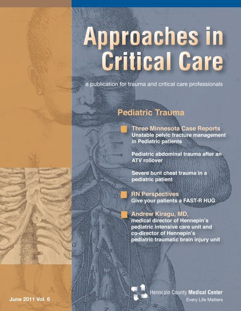
Pediatric Trauma - Hennepin County Medical Center
Pediatric Trauma - Hennepin County Medical Center
Pediatric Trauma - Hennepin County Medical Center

Create successful ePaper yourself
Turn your PDF publications into a flip-book with our unique Google optimized e-Paper software.
Dear Readers:<br />
<strong>Pediatric</strong> trauma is a double whammy. First, the child goes through the experience of<br />
the accident – pain, fear, strange people and places, and frantic parents all playing<br />
major roles. Later, when the child becomes an adult, the memory of the event remains<br />
clear, continuing as one of the defining experiences of their life. My nephew Beau was<br />
11 when he had an ATV accident and was flown to <strong>Hennepin</strong> <strong>County</strong> <strong>Medical</strong> <strong>Center</strong><br />
(HCMC) from a small farming community. At 26 years old, he is still talking about it.<br />
The same is true for the medical personnel who take care of seriously injured children.<br />
Dr. David Plummer, an emergency department physician at HCMC for the past 27<br />
years, made an emergency flight to Dawson, Minnesota, in 1984 that saved the life of<br />
22-month-old Nikki Stanley. She was playing in a school lunchroom when a 300 pound<br />
cafeteria table fell on her, leaving her with a serious head injury.<br />
Dr. Plummer says, “Dr. Gerbig at Johnson Memorial Hospital described a child with a<br />
serious head injury who needed immediate life-saving treatment and who required<br />
specialized medical care during transfer to HCMC. For that reason, I agreed to fly to<br />
Dawson to help.“<br />
When he arrived at Johnson Memorial, she was still unconscious. Nikki was intubated<br />
and received medication to reduce the intracranial pressure from her head injury.<br />
Unfortunately, Nikki went into cardiac arrest. He pupils were fixed and dilated, and she<br />
didnʼt have a pulse. Cardiopulmonary resuscitation and advanced cardiac life support<br />
was immediately performed, and her heart started again.<br />
“I hadnʼt been conscious of any nervousness while taking care of this child, but<br />
somewhere in the back of my mind I was aware this was one of those cases I would<br />
never forget.” After Dr. Plummer and Nikki arrived at HCMC and his role was essentially<br />
over, Dr. Plummer walked away and wept. “I knew Iʼd never forget it.”<br />
The case studies in this issue can only tell a fraction of each familyʼs story, as we<br />
primarily address the medical problems caused by abdominal trauma, pelvic injuries<br />
and chest trauma. In addition, we present a number of other interesting articles related<br />
to the delivery of pediatric critical care, including a short history of pediatrics at HCMC,<br />
the FASTR-HUG method of ICU care and a hopeful article about how one familyʼs<br />
tragedy led to legislation that would save childrenʼs lives.<br />
The theme for our next issue will be Immigrant Health. If you have an interesting case<br />
study you would like to contribute, see the authorʼs guidelines on the Approaches in<br />
Clinic Care website at www.hcmc.approaches. Weʼd love to hear from you.<br />
Sincerely,<br />
Michelle H. Biros, MD, MS<br />
Approaches in Critical Care Editor-in-Chief<br />
Department of Emergency Medicine<br />
<strong>Hennepin</strong> <strong>County</strong> <strong>Medical</strong> <strong>Center</strong><br />
®<br />
Every Life Matters
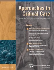
Contents Volume 6 | Approaches in Critical Care | June 2011<br />
Approaches in Critical Care<br />
Editor-in-Chief<br />
Michelle Biros, MD, MS<br />
Managing Editor<br />
Mary Bensman<br />
Graphic Designer<br />
Karen Olson<br />
Public Relations Director<br />
Tom Hayes<br />
Patient Care Director,<br />
Critical Care<br />
Kendall Hicks, RN<br />
Patient Care Director,<br />
Emergency Services<br />
Kelly Spratt<br />
Printer<br />
Sexton Printing<br />
Photographers<br />
Raoul Benavides<br />
HCMC History Museum<br />
Karen Olson<br />
Clinical Reviewers<br />
Andrew Kiragu, MD<br />
Events Calendar Editor<br />
Susan Altmann<br />
Case Reports<br />
2 Unstable Pelvic Fracture Management: a Challenge in <strong>Pediatric</strong> Patients<br />
Benjamin Orozco, MD<br />
5 <strong>Pediatric</strong> Abdominal <strong>Trauma</strong> After an ATV Rollover<br />
Marc Osbourne, MD and Chad Richardson, MD<br />
8 Severe Blunt Chest <strong>Trauma</strong> in a <strong>Pediatric</strong> Patient<br />
Richard Chang, MD and Michelle Biros, MD<br />
10 Critical Care Profile<br />
Andrew Kiragu, MD, medical director of <strong>Hennepin</strong>’s pediatric intensive<br />
care unit and co-medical director of <strong>Hennepin</strong>’s pediatric traumatic brain<br />
injury unit<br />
12 Technology in Critical Care<br />
Radiation Risks from Computed Tomography<br />
Gopal Pujabi, MD<br />
16 RN Perspectives<br />
Give Your Patients a FAST-R HUG<br />
Shayna Hamiel, RN, BSN, CCPN, CCRN<br />
18 Historical Perspectives<br />
History of <strong>Pediatric</strong>s at <strong>Hennepin</strong> <strong>County</strong> <strong>Medical</strong> <strong>Center</strong><br />
Sherri Murphy, RN<br />
22 Calendar of Events<br />
23 News Notes<br />
To submit an article<br />
Contact the managing editor at approaches@hcmed.org. The editors reserve the right to reject the<br />
editorial or scientific materials for publication in Approaches in Critical Care. The views expressed in<br />
this journal do not necessarily represent those of <strong>Hennepin</strong> <strong>County</strong> <strong>Medical</strong> <strong>Center</strong>, or its staff members.<br />
Copyright<br />
Copyright 2011, <strong>Hennepin</strong> <strong>County</strong> <strong>Medical</strong> <strong>Center</strong>. Approaches in Critical Care is published twice per<br />
year by <strong>Hennepin</strong> <strong>County</strong> <strong>Medical</strong> <strong>Center</strong>, 701 Park Avenue, Minneapolis, Minnesota 55415.<br />
Subscriptions<br />
To subscribe, send an email to approaches@hcmed.org with your name and full mailing address.<br />
Approaches in Critical Care | June 2011 | 1
Case Reports<br />
<strong>Pediatric</strong> <strong>Trauma</strong>: Three Case Reports<br />
<strong>Trauma</strong> is the most common cause of<br />
mortality and morbidity in the US pediatric<br />
population. Caring for the injured child<br />
requires special knowledge, precise<br />
management, and scrupulous attention to<br />
details. All clinicians who are responsible<br />
for the care of a pediatric trauma patient,<br />
including pediatricians, emergency<br />
clinicians, pediatric emergency clinicians,<br />
and trauma surgeons, must be familiar with<br />
every tenet of modern trauma care. The<br />
special considerations, characteristics, and<br />
unique needs of injured children must also<br />
be recognized.<br />
In 1962, Peter Kottmeier established the<br />
first pediatric trauma unit at the Kings<br />
<strong>County</strong> Hospital <strong>Center</strong> in Brooklyn. In<br />
1976, the publication of Resources for<br />
Optimal Care of the Injured Patient by the<br />
American College of Surgeons established<br />
requirements that should be met by a<br />
dedicated pediatric trauma center. Since<br />
1985, the National <strong>Pediatric</strong> <strong>Trauma</strong><br />
Registry (NPTR) has collected data<br />
concerning pediatric accidents. According<br />
to the American College of Surgeons, 81<br />
accredited pediatric trauma programs are<br />
currently in the United States.<br />
In fall 2010, <strong>Hennepin</strong> was verified as a<br />
Level 1 <strong>Pediatric</strong> <strong>Trauma</strong> <strong>Center</strong> by the<br />
American College of Surgeons (ACS). This<br />
verification-the highest possible-recognizes<br />
<strong>Hennepin</strong>ʼs distinctive expertise in caring<br />
for critically ill and injured children.<br />
Unstable pelvic fracture<br />
management: a challenge in<br />
pediatric patients<br />
by Benjamin Orozco, MD<br />
Department of Emergency Medicine<br />
<strong>Hennepin</strong> <strong>County</strong> <strong>Medical</strong> <strong>Center</strong><br />
Case presentation<br />
LJ is a 13-year-old male backseat<br />
passenger involved in a high-speed motor<br />
vehicle collision on city streets against a<br />
telephone pole and stone wall. There was<br />
significant vehicular intrusion and four<br />
additional victims, with one on-scene<br />
fatality. Emergency <strong>Medical</strong> Services<br />
(EMS) arrived to find him pinned beneath<br />
the driverʼs seat, unconscious. He awoke<br />
during his two-hour extrication and<br />
intravenous (IV) access was established<br />
and normal saline started. He arrived at<br />
<strong>Hennepin</strong> <strong>County</strong> <strong>Medical</strong> <strong>Center</strong><br />
immobilized on a back board and in a<br />
cervical collar. His initial vitals were: BP<br />
126/55, HR 135, RR 26 and Sp02 100%<br />
on supplemental oxygen.<br />
Emergency Department course<br />
Upon arrival to the emergency department,<br />
the patient remained alert but confused. He<br />
was tachycardic. Additional IV access was<br />
obtained, fluids were continued, and<br />
trauma surgery and orthopedic surgery<br />
were notified of his arrival. His exam was<br />
remarkable for crepitance over the clavicle,<br />
lower abdominal tenderness, pelvic<br />
tenderness and ecchymosis, a deformity of<br />
the right wrist, and a large laceration<br />
overlying his left knee (distal pulses were<br />
present). He was given fentanyl for pain<br />
control and was less alert but still<br />
protecting his airway. The extended<br />
Focused Assessment with Sonography for<br />
<strong>Trauma</strong> (eFAST) examination was<br />
negative, and he was rolled from the back<br />
board. Initial trauma radiography was<br />
performed. His chest X-ray demonstrated a<br />
pulmonary contusion and his pelvis X-ray<br />
showed a diastasis of the symphysis pubis,<br />
right pubic rami fractures, a left sacral alar<br />
fracture with vertical displacement and a<br />
left acetabular fracture (Figure One). X-rays<br />
of his affected extremities revealed a right<br />
wrist fracture and bilateral ankle fractures.<br />
He was transferred to the computed<br />
tomography (CT ) scanner for scans of his<br />
head, cervical spines, chest, abdomen and<br />
pelvis. He was somnolent but able to be<br />
aroused with stimulation. His heart rate<br />
was now 101, BP 111/101 and Sp02 100%.<br />
He was transiently hypotensive while in<br />
radiology and received an additional 500ml<br />
bolus of normal saline. After his imaging<br />
2 | Approaches in Critical Care | June 2011
Case Reports<br />
transferred to the floor on hospital day nine. He was<br />
evaluated with neuropsychological testing, which<br />
revealed mild cognitive deficits. Given his cognitive<br />
and physical deficits, our patient worked heavily with<br />
physical and occupational therapy and was able to<br />
be discharged to home on hospital day 17. He was<br />
followed closely with outpatient therapies. Upon<br />
follow-up in the traumatic brain injury clinic and<br />
orthopedic clinic, he is now walking and attending<br />
school normally.<br />
Figure One. The plain film of the patientʼs pelvis, demonstrating a left<br />
acetabular fracture, left sacral ala fracture, right pubic bone fractures with<br />
pubic symphysis diastasis.<br />
was reviewed, neurosurgery was consulted regarding<br />
scattered areas of subarachnoid hemorrhage (SAH)<br />
and punctate areas of intraparenchymal hemorrhage<br />
(IPH). In addition, active extravasation of contrast,<br />
indicating active hemorrhage, was noted from the<br />
pelvic fracture into the retroperotineal space. At that<br />
time, the orthopedic consultant placed a pelvic wrap<br />
and the patient underwent procedural sedation and<br />
femoral traction pin placement for further reduction of<br />
the vertical shear component of his pelvic injury. His<br />
extremity fractures were then splinted. Prior to<br />
transfer from the stabilization room, known injuries<br />
included the punctate areas of SAH and IPH, right<br />
pulmonary contusion, left acetabular fracture, left<br />
sacral ala fracture, right pubic bone fractures with<br />
pubic symphysis diastasis, and a retroperotineal<br />
hematoma. In addition, he had a right wrist fracture<br />
and bilateral ankle fractures.<br />
Hospital course<br />
Our patient was transferred to the pediatric intensive<br />
care unit (PICU) with a GCS 14, heart rate of 100<br />
and normotensive, in the care of trauma surgery and<br />
pediatrics, with neurosurgery and orthopedics in<br />
consultation. He received two units of blood and FFP<br />
overnight. He had a transient elevation of his hepatic<br />
enzymes and cardiac markers. Repeat head CT<br />
revealed a stable SAH and IPH and neurosurgery<br />
opted for non-operative management. On hospital day<br />
one, he was taken to the operating room with<br />
orthopedics for open reduction and internal fixation<br />
(ORIF) of his left acetabular fracture and sacroiliac<br />
diastasis to stabilize his pelvic ring. A mandible<br />
fracture was discovered upon re-institution of a solid<br />
diet and he underwent ORIF on hospital day seven.<br />
The other fractures were managed with closed<br />
reduction alone. He improved steadily and was<br />
Management of the pediatric poly-trauma patient<br />
with an unstable pelvic fracture<br />
Team-directed care is crucial in prioritizing the life and<br />
limb threatening injuries in the poly-trauma patient.<br />
Pelvic fractures are high-energy mechanism injuries<br />
associated with significant morbidity; 60% are<br />
associated with multi-organ system injury. <strong>Pediatric</strong><br />
pelvic fractures carry a mortality of approximately<br />
3-6%. The management of pelvic fractures can<br />
involve a combination of non-operative, operative, or<br />
interventional radiological techniques. Our patient<br />
was faced with injuries of nearly every organ system;<br />
however, his pelvic fractures were responsible for<br />
significant active hemorrhage upon presentation, and<br />
he required blood transfusion during the night of<br />
presentation, due to this ongoing blood loss. Our<br />
patient illustrates the effectiveness of multidisciplinary<br />
care in the multiple-injured pediatric trauma patient,<br />
with an emphasis on the hemodynamically significant<br />
pelvic fracture. His pelvis X-ray demonstrated diastasis<br />
at the sacroiliac joint with vertical displacement,<br />
suggesting the potential for massive hemorrhage.<br />
<strong>Pediatric</strong> pelvic fracture management differs from<br />
adults in that pediatric patients may have a skeletally<br />
immature or mature pelvis. The skeletally immature<br />
pelvis is open at the triradiate cartilage within the<br />
acetabula, and the mature pelvis is closed with total<br />
ossification of the triradiate cartilage. Fractures of the<br />
immature pelvis are more often isolated iliac wing or<br />
pubic rami fractures. Management of patients with<br />
fractures of the immature pelvis is generally directed<br />
at associated injuries. The mature pelvis, more<br />
similar to adults, has a greater propensity for<br />
acetabular fractures and sacroiliac or pubic symphysis<br />
diastasis necessitating emergent management.<br />
The initial management of a pelvic fracture includes<br />
assessment for an open fracture with external<br />
inspection, vaginal, and rectal examination. Broad<br />
spectrum antibiotics should be given for open<br />
fractures. Urethral injury must be considered and<br />
when suspected, a retrograde urethrogram should be<br />
performed prior to bladder catheterization. Pelvic<br />
stabilization to control hemorrhage is paramount.<br />
Approaches in Critical Care | June 2011 | 3
Case Reports<br />
Multiple classification systems for pelvic fractures<br />
exist, however most crucial is differentiating a stable<br />
vs. unstable pelvic fracture. Stable pelvis fractures<br />
include isolated iliac wing fractures and isolated<br />
fractures of the anterior pelvic ring, such as pubic<br />
rami fractures. Unstable fractures are those that<br />
involve the anterior and/or posterior sacroiliac<br />
ligaments of either or both sacroiliac joints, or vertical<br />
sacral fractures, and may result in pubic symphysis<br />
or sacroiliac diastasis. Significant hemorrhage<br />
generally is seen with unstable fractures and arises<br />
from the venous plexus immediately anterior to the<br />
sacrum, from cancellous bone edges, and from<br />
branches of the internal iliac artery. Measures to<br />
tamponade this hemorrhage include minimizing<br />
pelvic movement, pelvic wrapping, external fixation,<br />
internal packing, angiography with embolization, and<br />
possibly aortic balloon occlusion.<br />
Pelvic wrapping is most effective when there is<br />
sacroiliac or pubic symphysis diastasis without<br />
significant vertical displacement or acetabular fractures.<br />
Commercial pelvic binders may be utilized or a sheet<br />
wrapped low across the pelvis compressing the<br />
greater trochanters of the femur inward (Figure Two).<br />
Wrapping is particularly useful for pre-hospital and<br />
emergency providers and should be applied when<br />
pelvic fracture is suspected. External fixation is best<br />
applied by a skilled orthopedist in cases of significant<br />
hemorrhage; vertical displacement or iliac wing<br />
fractures decrease its effectiveness. Internal packing<br />
of the pelvis and retroperitoneum with surgical<br />
sponges is most often utilized when the patient has<br />
concomitant hemoperitoneum or other indication for<br />
open abdominal surgery and can be done in<br />
conjunction with pelvic wrapping or external fixation.<br />
Angiography with embolization is indicated in patients<br />
with hemodynamically significant bleeding refractory<br />
to more conservative measures and is most effective<br />
when arterial bleeding can be identified. At times<br />
angiography may be prioritized despite hemoperitoneum<br />
or other active bleeding. Aortic balloon occlusion for<br />
pelvic fractures, recently described in adults, is a<br />
method of occluding the abdominal aorta with an<br />
intraluminal balloon as temporizing measure in the<br />
patient dying of hemorrhagic shock and may pose a<br />
future role in pediatric patients.<br />
Our patient had injuries of nearly every organ system,<br />
but his shock was due to ongoing bleeding from a<br />
pelvic fracture. Interventional radiology reviewed the<br />
pelvic CT in the event that the patient should become<br />
unstable and need angiography. In this setting,<br />
despite an acetabula fracture, orthopedics felt the<br />
patient would benefit from external wrapping with<br />
reduction of the vertical displacement of the posterior<br />
pelvis via femoral traction pin under emergency<br />
department procedural sedation. The pediatric team<br />
and trauma surgery managed the continuing<br />
hemodynamic stabilization of the patient while he<br />
was in the PICU pending the operative stabilization<br />
of the pelvis. After a repeat head CT and serial<br />
neurologic examination confirmed stability of his<br />
intracranial hemorrhages, and after receiving a blood<br />
transfusion, he was taken for ORIF of his pelvis on<br />
hospital day one, within 24 hours of presentation. ■<br />
Figure Two. Application of a commercially available pelvic wrap .<br />
References<br />
Hauschild O, Strohm PC, Culemann U, Pohlemann T, Suedkamp<br />
NP, Koestler W, Schmal H. Mortality in patients with pelvic<br />
fractures: results from the German pelvic injury register. J <strong>Trauma</strong>.<br />
2008 Feb;64(2):449-55.<br />
Junkins EP, Furnival RA, Bolte RG. The clinical presentation of<br />
pediatric pelvic fractures. Pediatr Emerg Care. 2001 Feb;17(1):15-8.<br />
Leonard M, Ibrahim M, McKenna P, Boran S, McCormack D.<br />
Paediatric pelvic ring fractures and associated injuries. Injury. 2010<br />
Aug 23.<br />
Martinelli T, Thony F, Declety P, Sengel C, Broux C, Tonetti J, et al.<br />
Intra-Aortic Balloon Occlusion to Salvage Patients With Life-<br />
Threatening Hemorrhagic Shocks From Pelvic Fractures. J<br />
<strong>Trauma</strong>. Feb 18 2010.<br />
Silber JS, Flynn JM. Changing patterns of pediatric pelvic fractures<br />
with skeletal maturation: implications for classification and<br />
management. J Pediatr Orthop. 2002 Jan-Feb;22(1):22-6<br />
Silber JS, Flynn JM, Koffler KM, Dormans JP, Drummond DS.<br />
Analysis of the cause, classification, and associated injuries of<br />
166 consecutive pediatric pelvic fractures. J Pediatr Orthop. 2001<br />
Jul-Aug;21(4):446-50<br />
Spiguel L, Glynn L, Liu D, Statter M. <strong>Pediatric</strong> pelvic fractures: a<br />
marker for injury severity. Am Surg. 2006 Jun;72(6):481-4.<br />
4 | Approaches in Critical Care | June 2011
Case Reports<br />
<strong>Pediatric</strong> abdominal trauma after an<br />
ATV rollover<br />
by Marc Osbourne, MD<br />
Chad Richardson, MD<br />
Department of Surgery<br />
<strong>Hennepin</strong> <strong>County</strong> <strong>Medical</strong> <strong>Center</strong><br />
Case report<br />
A 10-year-old boy taken to a local hospital after an<br />
all-terrain vehicle (ATV) rollover crash was transferred<br />
to <strong>Hennepin</strong> <strong>County</strong> <strong>Medical</strong> <strong>Center</strong> (HCMC) by<br />
helicopter after it was determined that he was<br />
neurologically intact, but had sustained several other<br />
injuries, including an occlusion of the left renal artery.<br />
This case describes a patient with two rare injuries in<br />
an unusual injury pattern. It highlights the complex<br />
surgical and medical management of children with<br />
severe abdominal trauma.<br />
The 10-year-old patient was the passenger on an<br />
ATV driven by his father. Neither the patient nor his<br />
father were wearing a helmet. The ATV struck an<br />
object, resulting in the rollover. The boy was able to<br />
ambulate at the scene, but complained of abdominal<br />
and back pain. Bystanders called 911 and the patient<br />
was initially taken to a local hospital at about 2 p.m.<br />
His airway was intact and he was breathing and<br />
oxygenating well. He was tachycardic, but otherwise<br />
hemodynamically stable. Secondary exam showed a<br />
tender abdomen. Computed tomography (CT) scans<br />
of the head, C spine, chest, abdomen, and pelvis<br />
were obtained. The CT scans revealed several<br />
injuries, including an occlusion of the left renal artery<br />
without perfusion of the left kidney, a hematoma of<br />
the third and fourth portion of the duodenum with<br />
concern for possible perforation, free fluid within the<br />
peritoneum, a fracture of the body of L2, and<br />
fractures of the transverse processes of T12, L2, and<br />
L3. Given these findings, the patient was transferred<br />
via helicopter to HCMC.<br />
The patient arrived at HCMC at 8:30 p.m. He was<br />
mildly tachycardic and normotensive. On secondary<br />
exam, he had a tender and distended abdomen, as<br />
well as tenderness with palpation of the thoracic and<br />
lumbar spine. He had no neurologic deficit. A<br />
Focused Assessment with Sonography for<br />
<strong>Trauma</strong> (FAST) exam was positive for free fluid<br />
around the spleen. His radiographic studies arrived<br />
via electronic transfer to HCMC, where radiologists<br />
were also suspicious for transaction of the duodenum<br />
in the third or fourth portion. Representative images<br />
of his CT are shown in Figures One and Two. All<br />
providers, including trauma and pediatric surgery, did<br />
not feel there were any viable options for renal<br />
salvage. Given the clinical and radiographic findings,<br />
including a possible duodenal transaction, trauma<br />
surgery recommended exploratory laparotomy. This<br />
was discussed with the patientʼs mother.<br />
In the operating room (OR), the patient was found to<br />
have a moderate amount of intra-abdominal<br />
hemorrhage, a small bowel perforation approximately<br />
15 cm from the Ligament of Treitz, a small serosal<br />
tear of the transverse colon, a perforation of the<br />
duodenum at the junction of the third and fourth<br />
portion of the duodenum, and a left Zone II<br />
retroperitoneal hematoma (Figure Three). The Zone<br />
II hematoma was slowly bleeding into the abdomen<br />
via a defect in the posterior peritoneum. These<br />
findings were consistent with severe blunt force<br />
trauma within which the energy was delivered to a<br />
very focal area of the upper abdomen just to the left<br />
of the midline. The Zone II retroperitoneal hematoma<br />
was explored, revealing a small amount of muscular<br />
bleeding, as well as an ischemic left kidney. A<br />
thrombus could be visualized transluminally in his left<br />
renal artery, consistent with an intimal dissection.<br />
Figure One Left. Abdominal CT<br />
showing the ischemic left kidney<br />
and renal artery thrombosis<br />
Figure Two Middle. Abdominal CT<br />
showing the site of the suspected<br />
duodenal transection<br />
Figure Three Right.<br />
Retroperitoneal Zones: Zone<br />
1–Central–bounded by the<br />
diaphragm superiorly and pelvic<br />
inlet inferiorly and the medial border<br />
of the psoas muscles laterally; Zone<br />
2–Lateral–includes the areas lateral<br />
to zone 1 and above the pelvic inlet;<br />
Zone 3–Pelvic–area below the<br />
pelvic inlet<br />
Approaches in Critical Care | June 2011 | 5
Case Reports<br />
Grade<br />
Injury Description<br />
I<br />
II<br />
III<br />
IV<br />
V<br />
Hematoma<br />
Laceration<br />
Hematoma<br />
Laceration<br />
Laceration<br />
Laceration<br />
Hematoma<br />
Laceration<br />
Contusion with microscopic hematuria, urologic studies normal<br />
Non-expanding subcapsular hematoma without parenchymal laceration<br />
Non-expanding peri-renal hematoma confined to renal retroperitoneum<br />
< 1.0 cm parenchymal depth of renal cortex without urinary extravasation<br />
Laceration > 1.0 cm parenchymal depth of renal cortex without collecting system<br />
rupture or urinary extravasation<br />
Parenchymal laceration extending through renal cortex, medulla, and collecting<br />
system with urinary extravasation; injury to main renal artery or vein with<br />
contained hemorrhage<br />
Massive disruption of the duodenopancreatic complex<br />
Devascularization of the duodenum<br />
Table One.<br />
Organ Injury<br />
Scale American<br />
Association for<br />
the Surgery of<br />
<strong>Trauma</strong>: Kidney 3<br />
With these findings, a left nephrectomy was<br />
performed. Given the proximity of the small bowel<br />
and duodenal injury, a partial duodenectomy and<br />
small bowel resection were performed to include the<br />
distal portion of the third portion of the duodenum, the<br />
fourth portion of the duodenum, and the involved<br />
small bowel. This was reconstructed by mobilizing the<br />
remaining third portion of the duodenum and then<br />
creation of a small bowel to duodenal anastomosis<br />
with a partially stapled, partially hand-sewn technique.<br />
An incidental appendectomy was also performed.<br />
The retroperitoneum continued to have a small but<br />
steady amount of bleeding. This was packed. A<br />
temporary abdominal closure was then performed<br />
with a planned second-look laparotomy in 36 hours.<br />
The patient was then transferred to the PICU.<br />
With the assistance of the pediatric intensivists, the<br />
patient received ongoing fluid resuscitation. Postoperatively,<br />
he remained intubated and sedated. The<br />
patient was taken back to the OR on post-operative<br />
day two. The transverse colon serosal tear was<br />
primarily repaired. The bleeding from the retroperitoneum<br />
had stopped. The duodenal small bowel anastomosis<br />
was inspected and found to be intact and well perfused.<br />
His abdomen was definitively closed. His NG tube<br />
was left in place. He was transferred back to the<br />
PICU and was extubated later that day.<br />
This patient did well postoperatively. His nasogastric<br />
tube was removed on hospital day seven. On hospital<br />
day eight, the nasogastric tube was replaced after an<br />
episode of bilious emesis. A CT scan with PO and IV<br />
contrast was obtained. This showed no leak, but<br />
some edema in the third portion of the duodenum, as<br />
well as a fluid collection within the left renal fossa.<br />
The nasogastric tube was removed on hospital day<br />
11. The patient was discharged to home on hospital<br />
day 16. His peak creatinine during his hospital stay<br />
was 0.9.<br />
Discussion<br />
Renal Injury<br />
Nephrectomy for trauma in children is a rare event;<br />
even in high grade injuries. 1 Indications for nephrectomy<br />
include hemodynamic instability despite fluid resuscitation<br />
from ongoing hemorrhage and uncontrolled sepsis.<br />
Nephrectomy for an ischemic kidney from renal artery<br />
thrombosis is recommended if a laparotomy is being<br />
performed for another reason, i.e. hemorrhage,<br />
suspected bowel injury, etc. Childrenʼs National<br />
<strong>Medical</strong> <strong>Center</strong> reviewed its experience with blunt<br />
renal injury in children. 2 This was a single center<br />
retrospective review that included 126 children. Sixty<br />
percent of the patients had a low-grade injury, defined<br />
as American Association for Surgery in <strong>Trauma</strong><br />
(AAST) Grade 1, 2, or 3 level of injury. The remaining<br />
40% suffered an AAST Grade 4 or 5 injuries. Only<br />
four patients (3.2%) required nephrectomy and only<br />
two (1.6%) required immediate surgical intervention.<br />
Childrenʼs National <strong>Medical</strong> <strong>Center</strong> concluded that<br />
initial non-surgical management of high-grade renal<br />
trauma is recommended for hemodynamically stable<br />
children. The AAST renal injury grading definitions are<br />
given in Table One. In a 12-year retrospective series<br />
from Baltimore of 79 patients ages 2-14 with renal<br />
injury, 25% were Grade 4 or 5 injuries. 5 Seven (8.8%)<br />
required nephrectomy, all of whom had Grade 5<br />
injuries. Children who undergo conservative<br />
management of renal injuries appear to have good<br />
renal function in short- and long-term follow- up. 6 In a<br />
series of 16 patients (12 of whom had high-grade<br />
injuries) who were followed for one year post-injury,<br />
all of the children had normal BUN, creatinine, and<br />
blood pressure.<br />
Current recommendations from the American<br />
Academy of <strong>Pediatric</strong>s regarding children with an<br />
absence of a kidney and participation in contact<br />
sports emphasizes the need for clinical judgment and<br />
individual assessment of both the patient and the<br />
contact sport in question. 7 Several studies have<br />
highlighted that the risk of renal loss from contact<br />
sports is rare. 1<br />
6 | Approaches in Critical Care | June 2011
Case Reports<br />
the Surgery<br />
Laceration Massive disruption of the duodenopancreatic complex<br />
of <strong>Trauma</strong>:<br />
V<br />
Duodenum 4 Vascular<br />
Devascularization of the duodenum<br />
Grade Injury Description<br />
I Hematoma Involving single portion of the duodenum<br />
Laceration Partial thickness, non perforation<br />
Hematoma Involving more than one portion of duodenum<br />
Table Two.<br />
II<br />
Organ Injury<br />
Laceration Disruption of 75% of D2 or involving the ampulla or distal common bile duct<br />
Duodenal Injury<br />
Injury to the duodenum is also rare. In the two most<br />
recent series in the pediatric trauma literature, there<br />
were only 52 cases in a 10-year period in the<br />
combined experience of three pediatric Level 1 trauma<br />
centers. 8,9 The retroperitoneal position of the duodenum<br />
and association with other vital structures account for<br />
several challenging features in managing duodenal<br />
injuries. First, signs and symptoms of injury may be<br />
vague or subtle, possibly contributing to a delay in<br />
diagnosis. Second, these injuries are associated with<br />
high-energy mechanisms, given the protection afforded<br />
by the retroperitoneal location. Third, associated<br />
injuries to adjacent structures are common. A blunt<br />
mechanism of injury is the most common cause for<br />
duodenal injury. This is in contrast to adults, in which<br />
penetrating trauma is more common. Non-accidental<br />
trauma, assault and abuse, is, unfortunately, the most<br />
common mechanism of pediatric injury. Of the 52<br />
patients noted above, 19 sustained their injuries from<br />
non-accidental trauma. Motor vehicle collision was the<br />
next most common mechanism of action, accounting<br />
for 15 injuries. Bicycle accidents accounted for 10<br />
injuries. These injuries are more common in boys than<br />
girls. The duodenal organ injury scale from the AAST is<br />
listed in Table Two. A 10-year retrospective chart<br />
review summarizing the experience at two pediatric<br />
Level I trauma centers identified 42 duodenal injuries. 8<br />
A slight majority of the injuries were Grade I or II.<br />
Overall, 25 of the 42 injuries required operative<br />
management with 18 of the patients undergoing<br />
primary repair. The overall average length of stay was<br />
18 days, average length of ICU stay was seven days,<br />
and the complication rate was 33%. Patients who<br />
underwent operative management had an overall<br />
longer length of stay and a longer ICU stay.<br />
ATV Accidents<br />
Injuries associated with all-terrain vehicles (ATVs) have<br />
been closely monitored by the U.S. Consumer Product<br />
Safety Commission for several years. 10 Since 1982,<br />
there have been over 10,000 total deaths and 2,600<br />
pediatric deaths due to ATVs. Forty percent of these<br />
pediatric deaths were in children under the age of 12.<br />
In 2009, there were 33,400 injuries in children related<br />
to ATVs. Additionally in 2009, there were 61 deaths<br />
related to ATVs in children under 16 years old, of which<br />
29 were children under the age of 12. The yearly rate<br />
of ATV accidents has decreased by approximately half<br />
from four years ago. This decrease in the rate of injury<br />
is likely multi-factorial in origin. There has recently been<br />
a change in the definition of ATV by the U.S. Consumer<br />
product Safety Commission, categorizing dirt bikes as<br />
a separate category and not as an ATV. Legislative<br />
changes, as well as public awareness and advocacy<br />
campaigns, have also been important in highlighting<br />
the potential dangers of ATVs. The American Academy<br />
of <strong>Pediatric</strong>s recommends that children under the age<br />
of 12 should not be allowed to ride an ATV. 11 Minnesota<br />
State law prohibits anyone under the age of 11 from<br />
operating an ATV on public lands or trails, though use<br />
on private lands is permissible. 12<br />
Conclusion<br />
Serious injuries to the duodenum and kidney are<br />
relatively rare in children. Injuries to the duodenum may<br />
pose a diagnostic dilemma and may be associated with<br />
other serious injuries. Recent evidence shows that<br />
even high-grade renal injuries can be managed nonoperatively.<br />
Management of these complex injuries<br />
often requires a comprehensive and multi-disciplinary<br />
approach, as described in this case. ■<br />
References<br />
1. Johnson B, Christensen C, Dirusso S, Choudhury M, Franco I. A<br />
need for reevaluation of sports participation recommendations for<br />
children with a solitary kidney. J Urol. 2005 Aug;174(2):686-9<br />
2. Management of high grade renal trauma: 20-year experience at a<br />
pediatric level I trauma center. J Urol. 2007 Jul;178(1):246-50<br />
3. Moore EE, Shackford SR, Pachter HL, et al. Organ injury scaling:<br />
spleen, liver, kidney. J <strong>Trauma</strong>. 1989;29:1664-6<br />
4. Moore E, Cogbill T, et al. Organ injury scaling II: Pancreas,<br />
duodenum, small bowel, colon, and rectum. J <strong>Trauma</strong>. 1990;30:1427-9<br />
5. Rogers CG, Knight V, MacUra KJ, Ziegfeld S, Paidas CN, Mathews<br />
RI. High-grade renal injuries in children--is conservative management<br />
possible? Urology. 2004 Sep;64(3):574-9.<br />
6. Keller MS, Green MC. Comparison of short- and long-term functional<br />
outcome of nonoperatively managed renal injuries in children. J Pediatr<br />
Surg. 2009 Jan;44(1):144-7<br />
7. http://aappolicy.aappublications.org/cgi/content/full/pediatrics;<br />
121/4/841<br />
8. Clendenon JN, Meyers RL, Nance ML, Scaife ER. Management of<br />
duodenal injuries in children. J Pediatr Surg. 2004 Jun;39(6):964-8.<br />
9. Gaines BA, Shultz BS, Morrison K, Ford HR. Duodenal injuries in<br />
children: beware of child abuse. J Pediatr Surg. 2004 Apr;39(4):600-2.<br />
10. www.cpsc.gov/library/foia/foia11/os/atv2009.pdf<br />
11. www.aap.org/advocacy/releases/ATVdeath12610.pdf<br />
12. http://files.dnr.state.mn.us/rlp/regulations/ohv/ohv_regs.pdf<br />
Approaches in Critical Care | June 2011 | 7
Case Reports<br />
Severe Blunt Chest <strong>Trauma</strong> in a<br />
<strong>Pediatric</strong> Patient<br />
by Richard Chang, MD<br />
Michelle Biros, MD, MS<br />
Department of Emergency Medicine<br />
<strong>Hennepin</strong> <strong>County</strong> <strong>Medical</strong> <strong>Center</strong><br />
Case report<br />
CG is a 12-year- old, previously healthy male, who<br />
was brought into the Emergency Department at<br />
<strong>Hennepin</strong> <strong>County</strong> <strong>Medical</strong> <strong>Center</strong> following an<br />
accident while playing with a slingshot. The patient<br />
and a young friend were using a device made of<br />
rubber tubing tied to metal posts on a playground.<br />
They were propelling rocks far into the air when one<br />
particularly large rock struck the patient directly in the<br />
anterior chest and he collapsed.<br />
His friend called for help. When first responders<br />
arrived, the patient was apneic and pulseless.<br />
Cardiopulmonary resuscitation (CPR) was initiated,<br />
he was intubated, and Advanced Cardiac Life<br />
Support (ACLS) medications were administered<br />
through an intraossesous line. His initial rhythm was<br />
asystole and no shocks were given. He was transferred<br />
to the hospital with resuscitation in progress.<br />
Emergency Department Course<br />
The patient arrived to the stabilization room with CPR<br />
ongoing for approximately 40 minutes. A brief exam<br />
was notable for no external signs of chest trauma,<br />
symmetric chest rise with bag-assisted ventilations,<br />
and pupils that were fixed and dilated bilaterally.<br />
Bedside cardiac ultrasound was utilized to exclude<br />
pericardial tamponade and confirm cardiac standstill.<br />
Pacing pads were placed on his chest and external<br />
pacing was attempted, but unsuccessful. Multiple<br />
rounds of intravenous epinephrine, atropine, sodium<br />
bicarbonate, and calcium chloride were given as<br />
CPR continued.<br />
During the course of the resuscitation, the patientʼs<br />
parents arrived to the hospital. His mother, a<br />
cardiology research nurse, quickly rushed to her<br />
sonʼs bedside along with her husband. Emergency<br />
physicians performed internal cardiac pacing, external<br />
defibrillation, and continued ACLS pharmacologic<br />
therapies; however the patient had no return of<br />
spontaneous circulation and was subsequently<br />
pronounced dead in the Emergency Department.<br />
Commotio Cordis<br />
Latin for “agitation of the heart”, commotio cordis is<br />
one of the more common causes of sudden cardiac<br />
deaths in young athletes 1 . It occurs when blunt<br />
trauma to the chest wall disrupts the cardiac rhythm,<br />
resulting in ventricular fibrillation (VF) cardiac arrest.<br />
Although initially described in the 1700s, only<br />
recently has systematic reporting through the<br />
National Commotio Cordis Registry helped to identify<br />
over 200 confirmed cases.<br />
Epidemiologically, commotio cordis occurs most<br />
frequently in young males, usually during athletic<br />
activities. The majority of cases have been with blunt<br />
projectile trauma, commonly a baseball striking the<br />
anterior chest. Cases have also been identified from<br />
hockey pucks, lacrosse balls, and martial arts blows.<br />
The timing of the impact within the cardiac cycle<br />
appears to be the most important factor in the<br />
development of VF 2 . Only impacts during a 20-40<br />
millisecond window on the upslope of the T-wave<br />
(ventricular repolarization) will cause VF 3 .<br />
The diagnosis of commotio cordis is primarily clinical<br />
(i.e. blunt chest trauma followed by collapse).<br />
Electrocardiographic evidence of VF is rarely<br />
obtainable at the time of injury. Further imaging<br />
studies (i.e. echocardiogram, computed tomography)<br />
are often unrevealing, as is autopsy. Commotio<br />
cordis is a distinct entity from other, more severe,<br />
traumatic injuries to the heart, such as cardiac<br />
contusion or myocardial rupture.<br />
The prognosis in commotio cordis is poor, with a<br />
reported survival rate of 25% 4 . Early chest<br />
compressions and defibrillation are critical to<br />
resuscitation. Unfortunately, preventative measures<br />
such as commercially available chest wall protectors<br />
have not been shown to effectively prevent commotio<br />
cordis 5 . Coaching techniques have encouraged young<br />
athletes to avoid direct chest blows, such as turning<br />
away from errant baseball pitches. Softer, less dense<br />
balls have also been introduced in an effort to<br />
decrease the incidence of the devastating condition.<br />
Family Presence During Resuscitations<br />
A death due to an acute traumatic or medical event<br />
is a grief-provoking, unexpected experience that<br />
changes the lives of survivors immediately and<br />
forever. The death usually occurs in an unfamiliar<br />
setting, such as an emergency department, attended<br />
by strangers whose attempts to resuscitate the<br />
patient may appear assaultive (i.e. CPR, intubation<br />
IVs) when viewed by non-medical persons.<br />
Emergency personnel are often reluctant to have<br />
family members present during a resuscitation<br />
because we believe there is insufficient time to<br />
provide an acceptable explanation of what is<br />
occurring, we are afraid that the family will react in<br />
an unpredictable and possibly dangerous manner,<br />
8 | Approaches in Critical Care | June 2011
Case Reports<br />
that they will get in our way, that they may become<br />
a ʻsecondʼ patient if they have a severe physical or<br />
emotional reaction to what they see; and that they<br />
may criticize or question our decisions. None of<br />
these concerns are verified in the literature that<br />
describes family presence during resuscitation.<br />
In our attempts to interrupt the life-threatening<br />
pathology and avert death, we often forget that our<br />
resuscitation is being performed on someone who is<br />
loved and will never again be available to his<br />
survivors. Survivors relate that small details of the<br />
day, the place and the people are remembered<br />
forever and are replayed over and over again. The<br />
mother of our patient recalls a call from the police<br />
that provided no detail, and therefore, did not allow<br />
any mental preparation for what was happening. She<br />
recalls her distress when, arriving before her sonʼs<br />
ambulance, no one at triage seemed aware that he<br />
was arriving. She recalls the charge nurse stating<br />
she could not go into the STAB room, and that, when<br />
she insisted, the charge nurse told her that her son<br />
was receiving CPR. This was the first hint she had of<br />
how serious her sonʼs condition was. Death was<br />
never on her mind.<br />
She is a skilled clinical observer, and recalls<br />
watching the details of the resuscitation effort and<br />
hearing her suggestions acknowledged. She never<br />
felt dismissed in the STAB room. She knows<br />
everything possible was done. She felt empowered<br />
when she was asked to participate in the decision to<br />
discontinue resuscitation; she felt this gave her at<br />
least a little control in this totally uncontrollable event.<br />
She felt it was important to be present, along with her<br />
husband, when the resuscitation was discontinued.<br />
She felt the doctors gave great comfort when they<br />
assured them that their son likely died immediately,<br />
and that he likely did not feel any pain. They continue<br />
to hang on to these few comforting words.<br />
Afterwards, she regretted not staying with her son<br />
jwhen the nurses cleaned him up, and in retrospect,<br />
would have liked to have done this herself. She<br />
appreciated the support of her extended family, who<br />
had arrived during the resuscitation, but looking<br />
back, wishes she had had more time with him alone.<br />
She felt offended that she had little say in the<br />
decision to perform an autopsy on her son.<br />
She and her husband provided a healthy, safe life for<br />
their son. They loved parenting him. They were with<br />
him at his death, and he remains with them everyday.<br />
The insights they have shared with us about their<br />
sonʼs STAB room death can inform us in our<br />
consideration of family presence during a resuscitation.<br />
We, as medical providers, sometimes consider an<br />
unsuccessful resuscitation as a personal failure or a<br />
defeat. We reassure one another that everything that<br />
could be done was done, and everything we did was<br />
the right thing to do. When we think like this, we<br />
ourselves become the center of the case, the victim<br />
of the pathology. We must not forget that our practice<br />
is not about us, but it is about the patient. Family<br />
presence during resuscitations will remind us. ■<br />
Editorʼs note: Our sincere thanks to the mother of our<br />
patient, who met with us several weeks after the<br />
death of her son to share her experiences on that<br />
tragic day. His photo is displayed above with the<br />
familyʼs permission. Without a doubt, we have<br />
learned from her, and our discussions have helped<br />
us understands the real priorities in our practice.<br />
References<br />
1. Maron BJ, Gohman, TE, Kyle SB, et al. Clinical profile and spectrum<br />
of commotio cordis. JAMA 2002; 287: 1142.<br />
2. Madias C, Maron BJ, Weinstock J, et al. Commotio cordis—sudden<br />
cardiac death with chest wall impact. J Cardiovasc Electrophysiol<br />
2007; 18:115.<br />
3. Link MS, Maron BJ, VanderBrink BA, et al. Impact directly over the<br />
cardiac silhouette is necessary to produce ventricular fibrillation in an<br />
experimental model of commotio cordis. J Am Coll<br />
Cardiol 2001; 37:649.<br />
4. Kohl P, Nesbitt AD, Cooper PJ, Lei M. Sudden cardiac death by<br />
Commotio cordis: role of mechano-electric feedback. Cardiovasc Res<br />
2001; 50:280.<br />
5. Evaluation of chest barriers for protection against sudden death due<br />
to commotio cordis. Am J Cardiol 2007; 99:857.<br />
Approaches in Critical Care | June 2011 | 9
Critical Care Profile<br />
Q and A withQ and A with<br />
Andrew Kiragu, MD, FAAP<br />
Andrew Kiragu, MD, FAAP<br />
Dr. Andrew Waititu Kiragu is the medical<br />
director of the pediatric intensive care unit<br />
and the co-medical director of the <strong>Pediatric</strong><br />
<strong>Trauma</strong>tic Brain Injury program at<br />
<strong>Hennepin</strong> <strong>County</strong> <strong>Medical</strong> <strong>Center</strong>. He is<br />
also an assistant professor of pediatrics at<br />
the University of Minnesota <strong>Medical</strong><br />
School. He completed his undergraduate<br />
studies at Dalhousie University in Nova<br />
Scotia, Canada, and subsequently<br />
graduated from Howard University in<br />
Washington, D.C., with an MD degree in<br />
1994. Dr. Kiragu served his residency in<br />
internal medicine and pediatrics at the<br />
University of Minnesota followed by a<br />
fellowship in pediatric critical care at the<br />
University where he was one of the Walter<br />
Ramsey Endowment Fellows. During his<br />
fellowship and as a staff physician, he has<br />
received several awards in recognition of<br />
his commitment to resident and medical<br />
school education. Dr. Kiragu was awarded<br />
a Vikings Foundation grant to study the<br />
inflammatory effects of cardiopulmonary<br />
bypass. He was the co-primary investigator<br />
for a grant from the Robert Wood Johnson<br />
Foundation to establish an Injury-Free<br />
Coalition for Kids site in Minnesota at<br />
<strong>Hennepin</strong>, with a goal of studying and<br />
preventing injury prevention here in the<br />
Twin Cities. Dr. Kiragu is board-certified in<br />
pediatrics and pediatric critical care.<br />
Why did you choose pediatrics?<br />
I have always enjoyed being around kids.<br />
The kids seem to get my sometimes goofy<br />
nature. Kids are the most “fun” patients.<br />
When they are well, they are energetic,<br />
curious, funny, loving human beings. Even<br />
if they are really sick, children are quite<br />
resilient. When they recover from their<br />
illnesses, it makes what we do so<br />
worthwhile. When they donʼt get better,<br />
thatʼs the hardest. You grieve for the lost<br />
potential and the pain that the families feel.<br />
When you work in <strong>Pediatric</strong>s, you take<br />
care of more than just the patient. You also<br />
have to be aware of the needs of family<br />
members and friends. We once had a case<br />
of an injured teenager who had a large<br />
group of classmates come to see him. His<br />
family requested that we explain his<br />
injuries to them and so we gathered all<br />
these kids and some of their parents in a<br />
conference room to talk about their friend.<br />
Now that I have my own family (children<br />
ages 9 and 11), it makes me more<br />
cognizant of how fragile and precious life<br />
is. I am always the one saying, “be careful”<br />
to the kids. When we are with friends and<br />
the kids are doing crazy stuff, everyone is<br />
always looking at me to see my reaction.<br />
How has your interest in the care of<br />
critically injured and ill children shaped<br />
your career?<br />
As the director of the pediatric intensive<br />
care unit, by necessity I have to interact<br />
with a variety of people, my physician<br />
colleagues, <strong>Hennepin</strong> staff and leadership.<br />
This takes me beyond my usual clinical<br />
role as a physician. There are a number of<br />
new initiatives with regard to pediatrics we<br />
have undertaken here at <strong>Hennepin</strong> <strong>County</strong><br />
<strong>Medical</strong> <strong>Center</strong>, and my colleagues know<br />
that I am available to support these efforts.<br />
If I identify an opportunity to advance the<br />
care of children here at <strong>Hennepin</strong>, I can<br />
bring people together to make it happen. I<br />
enjoy doing that.<br />
I am also involved in the Injury Free<br />
Coalition for Kids (IFCK) and serve as the<br />
principal investigator for IFCK-Minneapolis.<br />
Through this organization, research in the<br />
area of injury prevention is being<br />
conducted to identify which injury<br />
prevention measures are most effective.<br />
Injury prevention education methods and<br />
driving behaviors in teens are among<br />
several topics that we have studied. The<br />
Injury Free Coalition for Kids has worked<br />
with other groups, including AAA-<br />
Minnesota to provide the state legislature<br />
with information useful in improving traffic<br />
laws. I am currently on the boards of<br />
10 | Approaches in Critical Care | June 2011
Critical Care Profile<br />
directors of Safe Kids Minnesota and the Brain Injury<br />
Association of Minnesota. These organizations are<br />
also heavily involved in injury prevention efforts<br />
around the state.<br />
What does Minnesota do well when it comes to<br />
trauma care?<br />
There is a long tradition of dedicated Level I trauma<br />
centers in Minnesota. In my opionion, <strong>Hennepin</strong><br />
<strong>County</strong> <strong>Medical</strong> <strong>Center</strong> has played a significant<br />
leadership role in trauma care in Minnesota. We<br />
have developed expertise in caring for patients of all<br />
ages and developed protocols of care that are used<br />
in the management of major trauma. The care that is<br />
provided at <strong>Hennepin</strong> is on the cutting edge of<br />
trauma care. An example is Dr. Gaylan Rockswoldʼs<br />
use of hyperbaric oxygen in the management of<br />
severe traumatic brain injury. <strong>Hennepin</strong> <strong>County</strong><br />
<strong>Medical</strong> <strong>Center</strong> has a strong tradition and culture of<br />
excellent trauma and critical care, which is something<br />
that has developed over decades. When the highway<br />
35W bridge collapsed, this tradition was quite<br />
evident. I remember, a young lady with a severe<br />
head injury who was brought into the emergency<br />
department (ED). Despite how busy the ED was at<br />
that time, her life-threatening injury was evaluated;<br />
she was scanned and was in the operating room in<br />
less than 15 minutes. This kind of efficiency only<br />
comes with years of experience and training.<br />
What sets <strong>Hennepin</strong> <strong>County</strong> <strong>Medical</strong> <strong>Center</strong> apart<br />
when it comes to pediatric trauma care?<br />
<strong>Hennepin</strong> <strong>County</strong> <strong>Medical</strong> <strong>Center</strong> has the resources<br />
available to provide surgical and critical care for<br />
children suffering from all types of traumatic injury.<br />
Most frequently we treat traumatic brain injuries but<br />
we also care for a wide variety of orthopaedic trauma<br />
and internal injuries, including trauma to the spleen,<br />
liver and bowel.<br />
One of the distinguishing factors is the <strong>Pediatric</strong><br />
Brain Injury program, which has been in existence<br />
for the past 21 years. Through this program, multidisciplinary<br />
care is provided to children who have<br />
suffered from a traumatic brain injury.<br />
<strong>Hennepin</strong> was the first acute care hospital in<br />
Minnesota to publish return-to-school guidelines for<br />
children who have experienced a traumatic brain<br />
injury. The <strong>Pediatric</strong> Brain Injury program works<br />
closely with schools to facilitate the return to school<br />
of children with brain injuries. The program also<br />
works closely with both the Brain Injury Association<br />
of Minnesota and the Minnesota Department of<br />
Health as they provide case management and<br />
resources for these children and their families.<br />
We are also involved in several ongoing clinical<br />
research projects, including the use of hypertonic<br />
saline solution in the management of increased<br />
intracranial pressure. Researchers are also reviewing<br />
cases where children have had an emergency<br />
thoracotomy to examine best practices. Upcoming<br />
clinical studies include evaluating sleep patterns in<br />
children with traumatic brain injuries.<br />
“The care that is provided at<br />
<strong>Hennepin</strong> is on the cutting edge<br />
of trauma care.”<br />
What are some things that you anticipate will<br />
happen in your field in the future?<br />
I anticipate even more cooperation between the four<br />
Level 1 trauma centers in our area – <strong>Hennepin</strong>,<br />
Regions, North Memorial and Mayo. We have a lot to<br />
learn from each other and can find ways to make<br />
care better in our state. I also hope for even closer<br />
ties between facilities that care for children. There is<br />
a lot of pediatric care in the Twin Cities area, but it is<br />
disjointed. Some cities have one large childrenʼs<br />
hospital, but that isnʼt the way that things are<br />
developing here.<br />
I think we will see more specialization in critical care.<br />
We can already see this with the growth of cardiac<br />
intensive care and growing interest in the field of<br />
neurointensive care. There will also be more<br />
regionalization and consolidation of critical care<br />
because there are not enough intensivists. Indeed,<br />
many hospitals do not even have an intensivist-run<br />
intensive care unit. There are even fewer pediatric<br />
intensivists nationally.<br />
Technology will improve our ability to monitor<br />
critically ill patients. One area that is growing is in<br />
telemedicine. Given the paucity of intensive care<br />
physicians, technologies that allow physicians to<br />
remotely monitor patients and aid in their management<br />
are key. Also, the emphasis on quality improvement<br />
and ongoing efforts towards patient and family<br />
centered care will continue to affect the way we care<br />
for kids. ■<br />
Approaches in Critical Care | June 2011 | 11
Technology in Critical Care<br />
Technology in Critical Care: Radiation risks from<br />
computed tomography<br />
by Gopal Punjabi, MD<br />
Department of Radiology<br />
<strong>Hennepin</strong> <strong>County</strong> <strong>Medical</strong> <strong>Center</strong><br />
Introduction<br />
The last 40 years have seen dramatic advances in<br />
medical imaging that have revolutionized clinical<br />
medicine. The benefits include more effective<br />
diagnosis, shorter hospital stays, and elimination of<br />
exploratory surgery and rapid diagnosis of lifethreatening<br />
conditions 1 . For example, the trauma<br />
patient can undergo a highly accurate whole-body<br />
computed tomography (CT) scan within a few<br />
minutes that often replaces multiple invasive<br />
examinations, such as angiography and exploratory<br />
laparotomy. This has also lead to an increase in<br />
radiation dose delivered to the patient population.<br />
There has been considerable interest in the medical<br />
literature and in the lay press regarding radiation<br />
dose, often with alarming headlines. In this article,<br />
the biological effects of low-dose ionizing radiation<br />
and methods to reduce radiation risks in CT scanning<br />
will be discussed.<br />
Background<br />
Computed tomography scan is a relatively recent<br />
intervention, developed in the late 1960s by EMI,<br />
Ltd., a music, electronics and leisure company based<br />
in the United Kingdom, funded by profits from the<br />
Beatles' recordings. Sir Godfrey Hounsfield, a British<br />
engineer and Allan Cormack, a physicist born in<br />
South Africa, received the Nobel Prize for this<br />
invention in 1979 2 . The CT scanner uses ionizing<br />
radiation to produce highly detailed images. There<br />
has been rapid advancement in CT technology since<br />
its invention, with current scanners using multidetector<br />
helical technology. This involves an X-ray<br />
beam going through the patient and detected on the<br />
opposite side by multiple detector rows, enabling<br />
sub-second imaging of large portions of human body.<br />
With rapid innovations, the use of CT scans has<br />
dramatically exploded. While in 1980 about 3 million<br />
CT scans were performed, the projection for 2011 is<br />
72 million CT scans. This trend is likely to increase<br />
with an aging population, as well as multiple new<br />
applications of CT, such as CT virtual colonoscopy,<br />
coronary CT angiography, and CT perfusion<br />
scanning.<br />
Radiation dose<br />
Radiation dose describes the amount of energy<br />
absorbed per unit mass at a specific point, and is<br />
expressed in Grays (1 Gy deposits 1 Joule per<br />
kilogram). This does not reflect risk; for instance, a<br />
100 mGy dose to an extremity would not have the<br />
same biological effect as the same dose to the<br />
pelvis. A more useful measurement is the effective<br />
dose, which takes into account the biological<br />
sensitivity of the tissue or organ being irradiated, and<br />
is calculated by multiplying the radiation dose with<br />
tissue and radiation weighing factors 3 . The unit of<br />
effective dose is the Sievert (usually millisieverts<br />
(mSv) are used in diagnostic radiology). The use of<br />
the effective dose facilitates communication with the<br />
patient, and understanding of the likelihood of<br />
potential harm from the radiological exam. The<br />
effective dose is a theoretical number that cannot be<br />
directly measured.<br />
The annual level of naturally occurring background<br />
radiation in the United States is estimated at about<br />
3.1mSv 4 . In 2006, the annual per capita effective<br />
radiation dose from man-made sources was<br />
estimated at about 3.1 mSv; of this, CT accounts for<br />
about 1.47 mSv 5 . The effective dose from an<br />
individual CT scan varies between different body<br />
parts 6 (Table One). There is also tremendous<br />
variation among scanners, depending on the<br />
scanning technique used. For comparison purposes,<br />
the average effective dose from a two-view chest X-<br />
ray is 0.1 mSv, and that from a nuclear cardiac stress<br />
test using technetium 99m labeled sestamibi is about<br />
12.8 mSv.<br />
Procedure<br />
CT Head<br />
CT Chest<br />
CT Abdomen<br />
CT Pelvis<br />
CT Cervical Spine<br />
CT Lumbar Spine<br />
Average Effective Dose<br />
2 mSv<br />
7 mSv<br />
8 mSv<br />
6 mSv<br />
6 mSv<br />
6 mSv<br />
Table One: Radiation doses in common CT exams 6<br />
12 | Approaches in Critical Care | June 2011
Technology in Critical Care<br />
Figure One: Dose report<br />
generated by a CT<br />
scanner, note that the total<br />
DLP from the whole body<br />
CT scan is estimated at<br />
807 mGycm. The effective<br />
dose can be calculated<br />
(by using a conversion<br />
factor of about 0.016)<br />
about 12.9 mSv, or 4<br />
years of natural<br />
background radiation. The<br />
dose savings refer to use<br />
of automated dose<br />
modulation technique.<br />
It is important to remember that radiation doses<br />
cannot be directly measured in a patient. Instead, the<br />
fundamental radiation dose parameter in CT is the<br />
CT dose index, which can be measured in simple<br />
cylindrical phantoms. To estimate dose after<br />
scanning a certain distance, the dose-length product<br />
is used. CT scanners provide automated reports of<br />
dose-length product after every CT scan. Simply put,<br />
this estimates the radiation dose that would be<br />
delivered to a standard phantom with the parameters<br />
used on the scan (Figure One). Standard conversion<br />
factors, which are regularly updated, are then used to<br />
calculate effective dose. As shown by several authors,<br />
there is a significant range of error in these calculations 7 .<br />
Radiation risks<br />
Ionizing radiation has enough energy to strip<br />
electrons from atoms and break chemical bonds.<br />
Radiation effects are in two broad categories: nonstochastic<br />
and stochastic. Non-stochastic effects<br />
appear in exposure to high levels of radiation, and<br />
are proportionate to the level of exposure. These<br />
include radiation burns and radiation sickness. With<br />
CT scans, non-stochastic effects are rare, and<br />
usually related to grossly inappropriate technique. A<br />
recent example is numerous cases of band-like hair<br />
loss following CT perfusion scans for stroke 8 .<br />
With the level of radiation used in CT scans, stochastic<br />
(probablistic) effects are of much more concern.<br />
Increased exposure makes the effect more likely to<br />
occur but does not influence the severity. Cancer is<br />
the primary stochastic effect of radiation. This results<br />
from damage to the DNA that may not have been<br />
successfully repaired. Given the high incidence of<br />
cancer in the general population, and the very low<br />
risk of cancer induction from CT scans, experimentally<br />
proving risks from CT scan would require prolonged<br />
studies involving millions of exposures 9 .<br />
Estimates of the risk of cancer induction by CT are<br />
therefore used. These are based on data from<br />
epidemiologic studies, the largest being the life span<br />
study from survivors of the 1945 atomic bombings in<br />
Japan. A large study of British radiation workers has<br />
shown that the data from Japanese bomb survivors<br />
can be expected to translate well to a Western<br />
population. The National Academies of Science has<br />
published reports on the biological effects of ionizing<br />
radiation (BEIR reports) that form the foundation of<br />
current estimates of radiation risk 10 . The FDA states<br />
that a CT examination with an effective dose of 10<br />
mSv may be associated with an increase in the<br />
possibility of fatal cancer of approximately 1 chance<br />
in 2000 11 .<br />
The BEIR VII report endorses the linear-no-threshold<br />
model of radiation risk, i.e., the risk from high<br />
radiation can be extrapolated to lower radiation<br />
exposure, there is no threshold below which radiation<br />
effects are not observed, and the risks are spread<br />
over the population exposed. While this is the<br />
simplest and safest model, it is not universally<br />
accepted, most notably by the French Academy of<br />
Sciences 12 . It has not been definitely demonstrated<br />
that there is any risk from radiation doses below 100<br />
mSv. The Health Physics Society states that<br />
"Estimation of health risk associated with radiation<br />
doses that are of similar magnitude as those<br />
received from natural sources should be strictly<br />
qualitative and encompass a range of hypothetical<br />
health outcomes, including the possibility of no<br />
adverse health effects at such low levels." 13 .<br />
This debate is not entirely academic, because many<br />
publications and media reports imply that death from<br />
CT induced cancers will significantly rise in the<br />
future. An oft-cited article published in the Archives of<br />
Internal Medicine states that 29,000 cancers could<br />
be related to CT scans performed in the U.S. in<br />
2007 14 . These reports do not adequately take into<br />
account several factors:<br />
Approaches in Critical Care | June 2011 | 13
Technology in Critical Care<br />
Figure Two (a-left):<br />
Normal dose technique<br />
demonstrates a stone in<br />
lower pole of right<br />
kidney. Low dose<br />
technique used on<br />
follow-up scan (b-right)<br />
demonstrated the same<br />
stone with about 20%<br />
the radiation dose from<br />
standard technique.<br />
1. Significant errors in measurement of low-level<br />
radiation doses.<br />
2. Significant errors in the estimation of effective<br />
dose, which is calculated from a regularly<br />
updated conversion factor.<br />
3. Biological plausibility. The biological effects of<br />
radiation decreases as dose decreases, and<br />
DNA repair and elimination of defective cells by<br />
death is a dynamic process.<br />
4. Uncertainty regarding linear-no-threshold model.<br />
5. Non-transferability of the risks and mortality from<br />
radiation. Most medical doses are delivered to a<br />
smaller and more elderly segment of the US<br />
population, but the risks are extrapolated to the<br />
general population.<br />
Radiation risks therefore must be taken in the<br />
appropriate context. If the estimated lifetime number<br />
of deaths from a single CT scan with a dose of 10<br />
mCi is indeed 0.5 per 1000, the estimated lifetime<br />
number of deaths in the same group of individuals<br />
from a lightning strike is 0.0 13 , and from drowning is<br />
0.9. Similarly, the death rate from motor vehicle<br />
accident is 11.9 15 . This does not dissuade most<br />
Americans from driving! Similarly, while the dose<br />
from a head CT is the same as about 20 chest<br />
x-rays, this is still significantly less than a year of<br />
background radiation.<br />
Primum non Nocere<br />
Measurement and quantification of risks from CT<br />
radiation form a fascinating and often controversial<br />
topic, with strong feelings on all sides. Above all, it is<br />
our responsibility as physicians to make sure our<br />
patients are not harmed. With this in mind, any<br />
radiation dose should be assumed to be potentially<br />
harmful, and careful risk-benefit analysis should be<br />
performed. This is particularly important with children,<br />
who are more vulnerable because of longer expected<br />
life spans and more radiation-sensitive body tissues.<br />
At the same time, it would be unconscionable to<br />
deny a patient the benefits from a CT scan that is<br />
appropriately indicated and optimally performed.<br />
What can be done?<br />
Justification: A CT scan should be performed only<br />
when there is the potential of clear benefit. In other<br />
words, the risk of not performing the examination<br />
must exceed the potential risks. A classic situation is<br />
following high-speed trauma, where the risk of not<br />
performing at CT scan includes missing life-threatening<br />
internal organ injury or aortic injury. This clearly exceeds<br />
potential risk of inducing neoplasm later in life.<br />
In most situations, the risk /benefit ratio is more<br />
difficult to evaluate, and individual physician<br />
judgment is paramount. There are many resources<br />
available to assist physicians in making this<br />
judgment. For example, the American College of<br />
Radiology has provided evidence-based guidelines<br />
for appropriate imaging tests 16 . Discussion between<br />
radiologists and physicians, especially physicians-intraining,<br />
clearly helps in the decision-making process<br />
and in tailoring the exam to answer the clinical<br />
question. Decision support systems integrated into<br />
electronic medical systems may also be helpful. It<br />
has been reported that 30% or more of CT scans<br />
currently performed may be unnecessary. This<br />
number must be reduced 17 .<br />
Optimization: Careful attention to technique can<br />
significantly reduce radiation dose. This includes<br />
standardizing protocols, reducing multiple series<br />
within each examination (avoiding "protocol creep"),<br />
implementing dose reduction strategies, and limiting<br />
scanning to part of interest (avoiding "scan creep").<br />
The Society for <strong>Pediatric</strong> Radiology offers very useful<br />
online information regarding tailoring exams in<br />
children, as part of its "image gently" program 18 .<br />
Of late, all CT vendors have introduced numerous<br />
dose reduction techniques. These differ among<br />
vendors, usually involve some form of dose<br />
modulation tailored to the patient body habitus, and<br />
are quite effective in reducing radiation dose while<br />
keeping image quality adequate. In addition, there<br />
are several situations where a low-dose exam with<br />
noisier (grainier, less pleasing) images is appropriate.<br />
In patients with kidney stones, the low-dose<br />
technique takes advantage of inherent contrast<br />
between the stone and surrounding tissues and<br />
produces images of diagnostic quality (Figure Two).<br />
Similarly, low-dose technique may be utilized in<br />
patients with lung nodules for follow-up.<br />
14 | Approaches in Critical Care | June 2011
Technology in Critical Care<br />
practice. At the same time, every CT scan should be<br />
assumed to involve some risks to the patient in terms<br />
of inducing potential cancers. A basic level of<br />
understanding radiation doses and the subsequent<br />
risks is important for all physicians who order these<br />
scans. With careful optimization and appropriate<br />
justification, these risks can–and should–be minimized.<br />
Figure Three. MRI scan shows acute appendicitis, which was surgically<br />
proven. Note gravid uterus in cephalic presentation.<br />
Alternate exams: Whenever feasible, alternate<br />
exams such as ultrasound or magnetic resonance<br />
imaging (MRI) that do not use radiation should be<br />
considered. In children with suspected appendicitis,<br />
ultrasound is often diagnostic. In pregnant women<br />
with suspected appendicitis, ultrasound is often nondiagnostic<br />
and MRI is helpful (Figure Three).<br />
Avoiding repeated scans: Risks from repeated<br />
scans can be assumed to be cumulative. Tracking<br />
and collecting dose information at the patient's level<br />
can be implemented within the electronic medical<br />
record system. Patients who have received<br />
excessive scans should be clearly flagged, and extra<br />
caution should be exercised in these patients.<br />
Communication: In all situations, clear<br />
communication with the patient regarding the reason<br />
for the exam being requested and the risk associated<br />
should be discussed in simple, non-alarmist<br />
language 19 . Statements such as, “We do not know<br />
the exact risks, and we may never know the exact<br />
risk associated with low doses of radiation, but in the<br />
best interests of our patients, we assume there is<br />
some risk” may be appropriate.<br />
It may be useful to express radiation dose in terms of<br />
multiples of annual background exposure, for<br />
example, “The exposure from a chest CT may be<br />
about that of two years of background radiation.” It<br />
may also be useful to express the associated risk of<br />
inducing fatal cancer in appropriate context, for<br />
example, “The lifetime risk of fatal cancer induced by<br />
whole body CT scan is about half that of dying from<br />
drowning,” provided that the uncertainty about these<br />
estimates is clearly stated.<br />
References<br />
1. Managing Radiation Use in <strong>Medical</strong> Imaging: A Multifaceted<br />
Challenge. Hedvig Hricak, David J. Brenner, et al. Radiology, March<br />
2011, 258, 889-905.<br />
2. Sir Godfrey Hounsfield. Ian Isherwood. Radiology, March 2005,<br />
234, 975-976.<br />
3. Radiation Dose in CT. McNitt-Gray, MF. RadioGraphics, November<br />
2002, 22, 1541-1553.<br />
4. National Council on Radiation Protection and Measurements.<br />
Ionizing radiation exposure of the population of the United States:<br />
2006. NCRP report no. 160. Bethesda, Md: National Council on<br />
Radiation Protection and Measurements, 2009.<br />
5. Radiologic and nuclear medicine studies in the United States and<br />
worldwide: Frequency, radiation dose and comparison with other<br />
radiation sources-1950-2007. Fred A Mettler, Mythreyi Bhargavan, et<br />
al. November 2009 Radiology, 253, 520-531.<br />
6. Effective Doses in Radiology and Diagnostic Nuclear Medicine: A<br />
Catalog. Fred A. Mettler Jr, Walter Huda, et al. July 2008 Radiology,<br />
248, 254-263.<br />
7. CT Dosimetry: Comparison of Measurement Techniques and<br />
Devices. John A. Bauhs, Thomas J. Vrieze, et al. January 2008<br />
RadioGraphics, 28, 245-253.<br />
8. After Stroke Scans, Patients Face Serious Health Risks. Walt<br />
Bogdanich, New York Times, July 31, 2010.<br />
9. Estimating cancer risks from low doses of ionizing radiation. Land<br />
CE. Science 12 September 1980: Vol. 209 no. 4462 pp. 1197-1203.<br />
10. U.S. Nuclear Regulatory Commission. Health risks from exposure<br />
to low levels of ionizing radiation: BEIR VII. Washington, DC: National<br />
Academies Press, 2006.<br />
11. What are the Radiation Risks from CT? FDA website, accessed<br />
3/11/2011. http://www.fda.gov/RadiationEmittingProducts/Radiation<br />
EmittingProductsandProcedures/<strong>Medical</strong>Imaging/<strong>Medical</strong>x-rays/ucm<br />
115329.htm.<br />
12. Dose-effect relationships and estimation of the carcinogenic effects<br />
of low doses of ionizing radiation. Aurengo A, Averback D, et al.,<br />
French Academy of Sciences, May 2005, http://www.radscihealth.org/<br />
rsh/Papers/FrenchAcadsFinal07_04_05.pdf, retrieved 3/11/2011.<br />
13. Health Physics Society, 2010. Radiation Risk in Perspective<br />
PS010-2, retrieved 3/11/2011.<br />
14. Projected Cancer Risks From Computed Tomographic Scans<br />
Performed in the United States in 2007. Berrington de Gonzalez A,<br />
Mahesh M, et al. Archives of Internal Medicine, Vol. 169 No. 22, Dec<br />
14/28, 2009.<br />
15. In Defense of Body CT. McCollough CH, Guimaraes L, et al., AJR<br />
2009; 193:28-39.<br />
16. ACR Appropriateness Criteria. ACR website, http://www.acr.org/ac.<br />
Accessed 3/11/2011.<br />
17. Radiation Dose Associated With Common Computed Tomography<br />
Examinations and the Associated Lifetime Attributable Risk of Cancer.<br />
Rebecca Smith-Bindman, ; Jafi Lipson, et al. Archives of Internal<br />
Medicine, Vol. 169 No. 22, Dec 14/28, 2009.<br />
18. Society for <strong>Pediatric</strong> Radiology.<br />
http://www.pedrad.org/associations/5364/ig/. Accessed 3/11/2011.<br />
19. Quality Initiatives Radiation Risk: What You Should Know to Tell<br />
Your Patient. Verdun FR, Bochud F, et al. RadioGraphics, November<br />
2008, 28, 1807-1816.<br />
Conclusions<br />
CT scans offer dramatic advancement in patient<br />
care, and are an essential part of modern medical<br />
Approaches in Critical Care | June 2011 | 15
RN Perspectives<br />
FASTR-HUG can be a<br />
simple way to remember<br />
the esentials of ICU care<br />
that arenʼt always in the<br />
forefront of our priorities<br />
for critically ill patients with<br />
numerous medical needs.<br />
RN Perspectives: Give your patients a “FASTR HUG”<br />
by Shayna Hamiel, RN, BSN, CPN, CCRN<br />
<strong>Hennepin</strong> <strong>County</strong> <strong>Medical</strong> <strong>Center</strong><br />
You may have heard about the acronym<br />
“FAST HUG”, introduced by Jean-Louis<br />
Vincent in relation to the essential aspects<br />
of care for all ICU patients. When patients<br />
are being rushed into the unit in critical<br />
condition, their medical needs are great,<br />
with our minds shifting gear to prioritization<br />
of stabilization and our classic ABCs.<br />
However, there are several aspects of<br />
intensive patient care that need to be<br />
addressed in order to optimize stability and<br />
improve patient outcomes. The FAST HUG<br />
acronym helps guide us through these<br />
important aspects of care that can have<br />
adjunctive effects leading to overall wellbeing.<br />
These aspects need to be addressed<br />
on admission as well as on a daily basis as<br />
we strive towards comprehensive care.<br />
With skin being the largest organ in the<br />
human body, it is becoming more of a<br />
focus among health care workers in all<br />
areas, but has yet to be fully given the<br />
importance that it deserves. Wounds,<br />
pressure ulcers, and skin breakdown of all<br />
types, both acute and chronic, can lengthen<br />
hospital stay and increase morbidity and<br />
mortality. Hospital related skin issues need<br />
to be more of a priority for hospital workers<br />
at all levels, with an emphasis on early<br />
detection and intervention, but more<br />
importantly, prevention. In our practice in<br />
the PICU, we are working on a way to<br />
incorporate skin and prevention into our<br />
daily care of ICU patients, and have added<br />
the “R” to Vincentʼs acronym in order to put<br />
skin into the forefront of our thinking with<br />
each admission to our unit.<br />
F-Feeding<br />
Consider nutrition right from the start.<br />
Malnutrition can complicate all body<br />
systems and worsen outcomes for critically<br />
ill patients. Good nutrition is especially<br />
essential for wound healing and is a part of<br />
the Braden and Braden Q risk assessment<br />
tools for skin breakdown. If you know your<br />
patient is not going to be able to eat<br />
normally for any extended period of time,<br />
don't delay feeding tube placement.<br />
Enteral nutrition is generally preferred over<br />
parenteral nutrition. Although the Braden<br />
scale does not give full points for tube<br />
feedings, many dieticians will argue that<br />
the right formula being fed into the<br />
stomach or jejunum can offer equal or<br />
better nutrition than what the patient might<br />
be getting on a regular diet.<br />
16 | Approaches in Critical Care | June 2011
RN Perspectives<br />
A-Analgesia<br />
Make sure your patient has adequate pain control.<br />
Pain can affect a patientʼs psychological and<br />
physiological well being. Most sedatives do not<br />
provide analgesia, so sedation and analgesia should<br />
go hand in hand for intubated patients. Even if your<br />
patient does not have an obvious source of pain<br />
such as a wound or fracture, remember that all<br />
invasive lines and tubes, as well as immobility, can<br />
create a source of pain or discomfort. Make sure you<br />
are an advocate for your patient's comfort.<br />
S-Sedation<br />
Sedative administration must be individualized. This<br />
is especially important for pediatric patients or those<br />
who are less tolerant of invasive lines and procedures.<br />
Safety must be our priority, and keeping patients<br />
comfortable helps with hemodynamic stability, keeping<br />
lines and airways intact, and allows for adequate rest<br />
for optimal healing.<br />
T-Thromboembolic Prophylaxis<br />
Mortality and morbidity rates increase with venous<br />
thromboembolism, so prophylaxis is imperative. ICU<br />
patients are at a high risk due to immobility and<br />
invasive lines. Although this risk is significantly lower<br />
in children compared to adult patients, femoral<br />
venous lines or the use of birth control in adolescent<br />
females can increase this risk. Make sure your<br />
patient has pneumoboots, and unless contraindicated,<br />
consider subcutaneous anticoagulants. Also ensure<br />
that range of motion exercises are being performed if<br />
tolerated and appropriate.<br />
R-Repositioning<br />
ICU patients have several risk factors for skin<br />
breakdown, including immobility, sheering and friction,<br />
moisture related to incontinence or diaphoresis, poor<br />
tissue perfusion related to oxygenation issues and<br />
vasopressor medications, decreased sensory<br />
perception, and poor nutrition. These risks are<br />
outlined by the Braden Q risk assessment scale,<br />
adapted for pediatrics by Quigley and Curley from<br />
the original Braden scale developed by Braden and<br />
Bergstrom in 1987. Utilizing the tool to assess the<br />
patientʼs level of risk can aid in implementing<br />
appropriate interventions, and minimizing these risk<br />
factors as well as frequent repositioning can help<br />
prevent skin breakdown. All ICU patients need to be<br />
repositioned regularly. If your patient isn't able to<br />
independently make frequent full position changes,<br />
you should be assisting them with repositioning at<br />
least every two hours. All patients need a complete<br />
skin assessment on admission, which would be a<br />
great time to do your first position change. When a<br />
patient is just arriving in the ICU, they may have<br />
already been laying in one position for several hours<br />
or even longer, in the ED or the OR, or even at the<br />
scene prior to arrival at the hospital. If a patient<br />
cannot be fully turned, use positioners for sacral offloading<br />
or micro-turns with slight tilting from one side<br />
to the other. If they already have a sacral or coccyx<br />
wound, minimize the amount of time spent supine. If<br />
your patient is difficult to reposition or has several risk<br />
factors for breakdown, they may benefit from an<br />
alternate surface such as a low air loss mattress or<br />
air bed, but remember that patients on specialty beds<br />
still need to be repositioned regularly. Also ensure that<br />
the patientʼs heels are elevated off the bed regardless<br />
of their position.<br />
H-Head of bed elevation<br />
Elevating the head of the bed decreases risk of reflux<br />
and ventilator associated pneumonia. It also aids in<br />
decreasing intracranial pressures. However,<br />
increasing the head of bed also results in sheering<br />
and friction, so unless ordered higher, keep the head<br />
of the bed to 30 degrees. You can also use<br />
positioners under the patientʼs legs to minimize<br />
friction from sliding down in the bed.<br />
U-Stress ulcer prevention<br />
ICU patients are at high risk for GI ulcers due to<br />
stress, respiratory failure, coagulation abnormalities,<br />
gastric tube placement, and lack of gastric feeds.<br />
Ensure that patients are on H2 blockers if appropriate,<br />
or alternate therapies if gastric bleeding is suspected.<br />
G-Glucose control<br />
Glucose control can help reduce mortality rates in<br />
ICU patients. The actual desired level of glucose<br />
remains controversial, and more data is needed in<br />
pediatrics. However, it is clear that hyperglycemia is<br />
associated with increased morbidity and worse<br />
outcomes. Make sure your patient's blood glucose<br />
levels are being followed either in daily labs or more<br />
frequent finger sticks.<br />
Utilizing the FASTR HUG acronym on admission as<br />
well as in daily patient care can be a simple way to<br />
remember the essentials of ICU care that arenʼt<br />
always in the forefront of our priorities for critically ill<br />
patients with numerous medical needs. Remember to<br />
include full skin inspections on all of your patient<br />
assessments, and that prevention is easier and<br />
faster than treatment. Please join me in giving each<br />
of your patients a FASTR HUG today! ■<br />
References<br />
Vincent JL. Give your patient a fast hug at least once a day. Crit<br />
Care Med. 2005; 33:1225-1229.<br />
Quigley SM, Curley MAQ. Skin integrity in the pediatric population:<br />
preventing and managing -pressure ulcers. JSPN. 1996; 1(1):7-18.<br />
Approaches in Critical Care | June 2011 | 17
Historical Perspectives<br />
<br />
Babies and<br />
nurses at<br />
Minneapolis<br />
General Hospital,<br />
June 26, 1930<br />
Minneapolis<br />
General Hospital<br />
Annex Building,<br />
1960ʼs<br />
<br />
Over 100 Years of <strong>Pediatric</strong> Care<br />
Historical Perspectives: On the Leading Edge of<br />
Medicine for Children<br />
The History of <strong>Pediatric</strong>s at <strong>Hennepin</strong> <strong>County</strong> <strong>Medical</strong> <strong>Center</strong><br />
by Sherrie Murphy, RN<br />
<strong>Hennepin</strong> <strong>County</strong> <strong>Medical</strong> <strong>Center</strong><br />
During the last 100 years, advances in the<br />
specialty of pediatrics have been astounding.<br />
From a half-dozen pediatric practitioners<br />
devoted to the care of children at the<br />
beginning of the 1900s, the ranks of<br />
pediatricians has grown to more than<br />
60,000 in 2010, according to the American<br />
Academy of <strong>Pediatric</strong>s. The infant mortality<br />
rate in 1900 was 200/1000, compared with<br />
the current 6.3/1000. (CHC data, 2010)<br />
Physicians and nursing staff associated<br />
with the founding hospitals of <strong>Hennepin</strong><br />
<strong>County</strong> <strong>Medical</strong> <strong>Center</strong> (HCMC) played<br />
major roles in the development of specialized<br />
care for adults and children in the region.<br />
For example, in 1914 the <strong>Pediatric</strong><br />
Contagion Building was constructed for<br />
children infected with scarlet fever and<br />
diphtheria. In 1920, Minneapolis City<br />
Hospital became Minneapolis General. In<br />
1921, medical residency programs began<br />
at Minneapolis General. In 1940 Sister<br />
Elizabeth Kenny came to the U.S. with a<br />
new treatment for polio. Minneapolis<br />
General was the only hospital anywhere<br />
that allowed her to demonstrate her hot<br />
packing technique. In 1946 Minneapolis<br />
General added 1000 people to the staff<br />
during a polio epidemic. Fifty patients were<br />
admitted daily, and 30 iron lung respirators<br />
were in use at one time.<br />
18 | Approaches in Critical Care | June 2011
Historical Perspectives<br />
<br />
Top left: Milk and formula<br />
laboratory, 1938<br />
<br />
Top right: Lymanhurst Childrensʼ<br />
Ward for Rheumatic Heart<br />
Disease, 1931<br />
<br />
Library time on the boysʼ ward<br />
<br />
Physicians prescribe fresh air on the roof<br />
of the hospital. 1930<br />
Iron Lung respirator<br />
<br />
By the mid-1960s,<br />
Minneapolis General<br />
Hospitalʼs pediatric unit<br />
cared for premature<br />
infants, sick newborns<br />
and children to age 17. This unit also cared for<br />
children with critical trauma and burns. The focus of<br />
pediatrics was changing during this time. Connie<br />
Benson, RN, head nurse of the pediatric unit at the<br />
time, discontinued the restrictive visiting hours and<br />
experimented with playful scrubs and other things<br />
common to modern pediatric practice. “The day I<br />
became head nurse, I ripped down the signs and<br />
opened visiting hours to 24 hours a day for the whole<br />
family. I also made space for parents to stay<br />
overnight,” Benson recounted. “The kids were afraid<br />
of our white uniforms, so I went out and bought polka<br />
dot sheets to make smocks for the staff. The nurses<br />
were afraid to walk out of the break room for fear of<br />
looking like a lollypop until Dr. Raile (Richard Raile,<br />
MD, chief of pediatrics) took them to the cafeteria. It<br />
was the stamp of approval they needed.”<br />
With a census of 60, no private rooms, and admissions<br />
at all levels of acuity, it became clear that a <strong>Pediatric</strong><br />
Intensive Care Unit (PICU) was necessary. Several<br />
local disasters in the 1960s, including the Fridley<br />
tornado and a fire at Benilde High School, made it<br />
painfully clear that there was not enough capacity to<br />
care for critically injured children, especially with new<br />
Approaches in Critical Care | June 2011 | 19
Historical Perspectives<br />
<br />
Dr. Per Wickstrom with Peds<br />
patient, 1969<br />
<br />
Dr. Richard Raile, MD, Chief<br />
of <strong>Pediatric</strong>s opens new<br />
<strong>Pediatric</strong> Unit, 1960<br />
treatments and technologies changing the practice.<br />
In 1968, the first PICU in the state of Minnesota<br />
opened with two beds, co-managed by the surgery<br />
and burn units. A neonatology ICU was also opened<br />
in the same area at that time.<br />
In 1969, Minneapolis Generalʼs medical staff began<br />
to advocate for abused children. Raile, MD, and<br />
Robert Ten Bensel, MD, co-authored a paper<br />
outlining the diagnosis of battered child/child abuse<br />
that received national attention. Abused children<br />
were being seen more frequently in the PICU. A<br />
multidisciplinary group began to meet to discuss the<br />
problems of abuse and the Suspected Child Abuse<br />
and Neglect Team was created.<br />
In 1976, Minneapolis General Hospital became<br />
<strong>Hennepin</strong> <strong>County</strong> <strong>Medical</strong> <strong>Center</strong> moving into a new<br />
building and sharing space with Metropolitan <strong>Medical</strong><br />
<strong>Center</strong>. The PICU began to admit post-surgical<br />
cardiology patients. A new PICU consisting of a<br />
seven-bed multidisciplinary intensive care unit was<br />
designed to provide comprehensive care to critically<br />
ill children, ranging in age from newborn to age 17.<br />
Common diagnoses on admission included trauma<br />
and respiratory distress/failure. The burn unit also<br />
opened in 1976. The emergency department<br />
expanded its pediatric care area and pediatric staff<br />
became more involved in emergency cases.<br />
20 | Approaches in Critical Care | June 2011
Historical Perspectives<br />
<br />
Nikki Stanley, 22 months old,<br />
recovers from a life-threatening<br />
brain injury in 1984.<br />
Nikki, now married, a nurse<br />
and a mother, reunites with<br />
Dr. David Plummer, MD, the<br />
emergency physician who<br />
saved her life, 2010.<br />
<br />
<br />
<strong>Pediatric</strong> Intensive<br />
Care Unit, 2010<br />
Nurse examining<br />
patient and consulting<br />
with parent in <strong>Pediatric</strong><br />
Intensive Care Unit,<br />
2010<br />
<br />
By 1980 the PICU was meeting all the challenges of<br />
the modern world: new technology and equipment,<br />
an increasingly diverse patient population and the<br />
expansion of emergency services through helicopter<br />
service. In 1989, HCMC became the first Level I<br />
<strong>Trauma</strong> <strong>Center</strong> for both adults and children. Family<br />
members of different ages who were injured in an<br />
accident could all be admitted to the same hospital<br />
into the appropriate ICUs. The regionʼs first hospitalbased<br />
Biomedical Ethics Committee was established<br />
and featured on the CBS Evening News. Research<br />
continued to be an ongoing activity. <strong>Trauma</strong>tic brain<br />
injury patients were part of a neurosurgery research<br />
study using hyperbaric treatments. The <strong>Pediatric</strong><br />
Brain Injury Program was established in 1989 and<br />
since then, more than 2,400 children have received<br />
acute and outpatient services to help them recover<br />
from serious or mild-to-moderate brain injuries.<br />
In the 1990s, the <strong>Pediatric</strong> Critical Care Task Force<br />
was established and played an important role in<br />
quality improvement in the PICU. Coverage was<br />
provided 24/7 by critical care specialists including<br />
neurosurgeons, trauma surgeons and orthopaedic<br />
surgeons, and pediatric subspecialists in surgery,<br />
neurology, cardiology, pulmonology, and ear, nose<br />
and throat (ENT). Nurses were certified in <strong>Pediatric</strong><br />
Advanced Life Support (PALS) and/or Advanced<br />
<strong>Pediatric</strong> Life Support (APLS) and participated in<br />
continuing education related to trauma, pediatric<br />
resuscitation and critical care. Many HCMC nurses<br />
presented lectures on pediatric emergency and<br />
critical care throughout the state. Allied patient care<br />
and support staff now included interpreters, child life<br />
specialists, health unit coordinators, health care<br />
assistants, social workers, respiratory care practitioners,<br />
occupational and physical therapists, speech-language<br />
pathologists and dietitians.<br />
<strong>Pediatric</strong> Inpatient Unit remodel, 2010<br />
In November 2010, HCMC was verified as a Level I<br />
<strong>Pediatric</strong> <strong>Trauma</strong> <strong>Center</strong> by the American College of<br />
Surgeons. This verification – the highest possiblerecognizes<br />
<strong>Hennepin</strong>ʼs distinctive expertise in caring<br />
for critically injured children. ■<br />
Approaches in Critical Care | June 2011 | 21
Calendar of Events<br />
To confirm hours and location of courses<br />
and to register, visit www.hcmc.org and click<br />
on “Professional Education and Training.” For<br />
questions or additional information, contact<br />
Susan Altmann in the medical education<br />
department at <strong>Hennepin</strong> <strong>County</strong> <strong>Medical</strong> <strong>Center</strong><br />
at (612) 873-5681 or susan.altmann@hcmed.org<br />
unless another contact person is provided.<br />
Classes are at <strong>Hennepin</strong> unless otherwise<br />
indicated. Many courses fill quickly; please<br />
register early to avoid being wait-listed.<br />
First Responder Refresher<br />
August 25-26, October 27-28<br />
__________________________________________<br />
Healthcare Provider Cardiopulmonary<br />
Resuscitation<br />
August 10, September 7, October 17, November 16<br />
__________________________________________<br />
Heartsaver Automated External Defibrillator and<br />
Cardiopulmonary Resuscitation<br />
September 12<br />
__________________________________________<br />
Cardiopulmonary Resuscitation Instructor<br />
November 7<br />
__________________________________________<br />
Advanced Cardiac Life Support for Providers (AHA)<br />
June 20, 21(Residents/Open), August 16 and 17,<br />
September 20 and 21, October 10 and 11, November<br />
1 and 2, December 6 and 7<br />
__________________________________________<br />
Off-Site Advanced Cardiac Life Support (ASHI)<br />
October Open - Willmar, November Open - Marshall<br />
__________________________________________<br />
Advanced Cardiac Life Support Provider Renewal<br />
(AHA)<br />
August 17, September 21, October 11, November 2,<br />
December 7<br />
__________________________________________<br />
Advanced Cardiac Life Support Provider Renewal<br />
(ASHI) for <strong>Hennepin</strong> Staff<br />
June 14, July 26, August 23, September 6, October 5,<br />
November 17, December 1<br />
__________________________________________<br />
Advanced Cardiac Life Support for Experienced<br />
Providers (AHA)<br />
October 4, November 14, December 12<br />
__________________________________________<br />
Advanced Cardiac Life Support Instructor (AHA)<br />
July 5 and 6<br />
__________________________________________<br />
Advanced Cardiac Life Support Instructor<br />
Renewal (AHA)<br />
July 6<br />
__________________________________________<br />
Emergency <strong>Medical</strong> Technician Basic<br />
October 10-28<br />
__________________________________________<br />
Emergency <strong>Medical</strong> Technician Refresher<br />
November 2-4, November 28-30 (South Lake Public<br />
Safety, Excelsior, MN)<br />
__________________________________________<br />
First Responder<br />
September 19-23<br />
__________________________________________<br />
Cardiopulmonary Resuscitation Instructor<br />
Renewals<br />
November 7<br />
__________________________________________<br />
Cardiopulmonary Resuscitation/Basic Life<br />
Support for <strong>Hennepin</strong> Staff<br />
July 27, August 9, September 7, October 12,<br />
November 9, December 14<br />
__________________________________________<br />
Infant-Child Cardiopulmonary Resuscitation<br />
June 15, July 22, August 3, September 23,<br />
October 12, November 11, December 14<br />
__________________________________________<br />
MD Cardiopulmonary Resuscitation for <strong>Hennepin</strong><br />
Staff<br />
June 15, July 8, August 5, August 17, September 9,<br />
October 7, October 19, November 4, December 2,<br />
December 7<br />
__________________________________________<br />
Advanced <strong>Pediatric</strong> Life Support<br />
July 19 and 20<br />
__________________________________________<br />
Advanced <strong>Trauma</strong> Life Support<br />
July 12 and 13, October 6 and 7<br />
__________________________________________<br />
<strong>Trauma</strong> Nursing Core Course<br />
September 14 and 15<br />
__________________________________________<br />
<strong>Trauma</strong> Nursing Core Course Renewal<br />
November 16<br />
__________________________________________<br />
<strong>Pediatric</strong> Advanced Life Support for Providers<br />
(AHA)<br />
September 27 and 28, October 18 and 19<br />
__________________________________________<br />
<strong>Pediatric</strong> Advanced Life Support Renewal (AHA)<br />
September 28, October 19<br />
__________________________________________<br />
14 22 | Approaches in Critical Care | January June 2011 2011
News Notes<br />
News Notes<br />
Tragedy Moves a Family to Take<br />
Action to Change Seatbelt Laws<br />
by Julie Philbrook, RN, MA<br />
<strong>Trauma</strong> Services<br />
<strong>Hennepin</strong> <strong>County</strong> <strong>Medical</strong> <strong>Center</strong><br />
One year . . . 12 months . . . 365 days . . .<br />
8,760 hours . . . 525,600 minutes. August<br />
18th, 2008 at 10:24 a.m. - when a bright<br />
dreamy morning turned into a dark hellish<br />
nightmare. But God is faithful and we began<br />
even that first day, clinging to His promise<br />
that, “Weeping may endure for the night,<br />
but joy comes in the morning.” (Psalm 30:5).<br />
Little did we know how long that night<br />
would last. (www.CaringBridge.org, 2010).<br />
This was the 240th entry to the CaringBridge<br />
website that was established for a sevenyear-old<br />
girl named Brynn Duncan, whose<br />
life was changed in an instant. She was<br />
seriously injured during a car crash when<br />
her ill-fitting seat belt caused permanent<br />
spinal cord and internal organ injuries.<br />
When I heard about her injury, I logged<br />
onto the CaringBridge website and followed<br />
the often hourly updates that were posted<br />
during her 54-day hospital stay. Each time I<br />
read the log, I could not stop thinking that if<br />
Minnesota had been successful in passing<br />
stronger child passenger safety legislation<br />
during the 2008 session, perhaps Brynnʼs<br />
outcome would have been very different. I<br />
also wondered if this child might be the<br />
one to put a face on the booster seat bill<br />
that advocates in Minnesota had been<br />
trying to pass for seven years.<br />
As the trauma prevention specialist for<br />
<strong>Hennepin</strong> <strong>County</strong> <strong>Medical</strong> <strong>Center</strong> (HCMC),<br />
I was not involved in Brynnʼs day-to-day<br />
nursing care. I had not met her mother,<br />
Dixie, and I struggled with knowing the<br />
best way to approach the family about the<br />
possibility of having them speak out for the<br />
booster seat bill. I started by first asking<br />
her physician, Andrew Kiragu, MD, if he<br />
would introduce me to the family. I knew it<br />
would be difficult to approach them, as I<br />
did not want to place blame or make them<br />
feel guilty.<br />
Once Brynn was stable, I met with her and<br />
Dixie and explained about the need to<br />
change Minnesotaʼs child passenger safety<br />
seat law to include the need for booster<br />
seats. Dixie told me that, even though the<br />
current law only required children to be in a<br />
child restraint until age four, because Brynn<br />
was small for her age, she had her use a<br />
booster seat. However, on the day of the<br />
crash, Brynn she was not riding with her<br />
mom, and was not in a booster seat. As we<br />
talked, Brynn told us that the shoulder<br />
harness had been rubbing on her neck,<br />
and she had it put behind her back, leaving<br />
only the lap belt to restrain her. In the<br />
crash, it was the lap belt that contributed to<br />
her L1 spinal cord transection and severe<br />
abdominal injuries, including a complete<br />
duodenal transection, diaphragmatic tear, and<br />
a right kidney resulting in a nephrectomy.<br />
This cohort of injuries is referred to as the<br />
“seat belt syndrome.”<br />
Dixie was more than willing to take on this<br />
cause. She said, “If it means that another<br />
child is not injured it will be worth it.” As<br />
Brynn recovered physically, she and her<br />
family found purpose and passion in trying<br />
to make a difference in the lives of other<br />
children. They openly shared their story<br />
with the legislature and the media. In the<br />
beginning of this journey, I was concerned<br />
whether the family would want to make<br />
themselves vulnerable to public opinion<br />
about how they had transported their child.<br />
I came to see how truly healing this process<br />
was for the whole family and the community.<br />
Letters were written, phone calls made,<br />
and testimony was given. In the end, after<br />
seven attempts to pass stronger child<br />
passenger safety (CPS) legislation, the bill<br />
did pass and became law on July 1, 2009.<br />
Years ago when I worked in the trauma<br />
intensive care unit at HCMC, I often found<br />
myself standing at a bedside watching<br />
monitors, giving meds, and changing<br />
dressings all the while thinking, “If only….if<br />
only they had worn a helmet, if only they<br />
had not been talking on the phone while<br />
driving, if only they had been correctly<br />
restrained, how different their life would be.”<br />
Approaches in Critical Care | June | 23
News Notes<br />
We all play a role in preventing life changing injuries.<br />
We can role model safe behaviors. We can also<br />
invite patients and families to share their story in<br />
whatever way they feel comfortable, with the press,<br />
in the classroom, at the capitol. When we make<br />
trauma real to people, we can make real change.<br />
The battle to upgrade the Minnesotaʼs car seat law<br />
began the year Brynn was born. After much lobbying,<br />
many phone calls and countless letters from many<br />
advocates and agencies, in the end, a little sevenyear-old<br />
sitting before legislators and a passionate<br />
mother, testifying with tears in her eyes, convinced<br />
lawmakers to pass the Minnesota Car Seat Law.<br />
__________________________________________<br />
approaches on immigrant and homeless populations.<br />
For information about the program, topics and<br />
speakers visit hcmc.org/bestof hennepin and visit us<br />
on Facebook.<br />
The post health care reform medical model will<br />
require many more primary care providers than are<br />
currently in the field, or even in the pipeline. This<br />
conference will also address the implications of this<br />
shortage and pose some questions and challenges<br />
for discussion.<br />
<strong>Hennepin</strong> <strong>County</strong> <strong>Medical</strong> <strong>Center</strong> has been at the<br />
forefront of medical change for the last 100 years.<br />
We intend to continue to lead the medical community<br />
in Minnesota through teaching, research and<br />
excellent medical care.<br />
Please join us October 20-22 at the Holiday Inn<br />
Metrodome. Be part of the discussion.<br />
__________________________________________<br />
P P P<br />
Patients • Primary Care • Partnerships<br />
Partnerships in<br />
Primary Care –<br />
Continuing <strong>Medical</strong><br />
Education Event<br />
for Primary<br />
Care Providers<br />
A focus on patient<br />
and family centered care.<br />
1887<br />
October 20-22, 2011<br />
Holiday Inn Metrodome<br />
www.hcmc.org/bestofhennepin<br />
Mark your calendars for the Best of <strong>Hennepin</strong><br />
Conference, October 20-22<br />
by John Crossen, MD<br />
The 2011 Best of <strong>Hennepin</strong> Conference will focus on<br />
the relationships between primary care physicians,<br />
specialists and patients, especially those with chronic<br />
diseases. As we go forward into this time of radical<br />
change and reform in health care, the patient is<br />
replacing the disease as the focus of the medical<br />
equation. Our conference panels will provide an<br />
opportunity for attendees to witness Patient <strong>Center</strong>ed<br />
Care through the interplay between specialists,<br />
primary care physicians and patients in planning and<br />
resolving chronic health concerns–to hear their<br />
stories and understand their flashes of insight from<br />
all perspectives. Conference speakers will also<br />
address the effectiveness of traditional medical<br />
<strong>Hennepin</strong> <strong>County</strong> <strong>Medical</strong> <strong>Center</strong> <strong>Pediatric</strong><br />
Emergency Drug Book<br />
2012 Revised Edition coming soon!<br />
Designed by author Dr. Al Tsai from the department<br />
of Emergency Medicine, as an easy-to-use<br />
reference, this drug book offers readily accessible<br />
dosages by age and weight for most drugs required<br />
during the care of critically ill or injured children. It<br />
also inclludes reference charts for coma/trauma<br />
scales, croup, asthma, ill child assessments, and<br />
burn resuscitation.<br />
To receive notification when available, please e-mail<br />
pedsdrugbook@hcmed.org.<br />
Did you train at <strong>Hennepin</strong>?<br />
We’re looking for you.<br />
You are an important member of an exclusive group<br />
of physicians who share <strong>Hennepin</strong> <strong>County</strong> <strong>Medical</strong><br />
<strong>Center</strong>ʼs expertise and knowledge with the people<br />
of the Upper Midwest. <strong>Hennepin</strong> is committed to<br />
continue a learning and sharing relationship with<br />
our alumni and would like to stay in touch.<br />
Please submit your contact information at<br />
HCMC.org/alumni.html<br />
or to R. Hoppenrath, 701 Park Ave., Mpls, MN 55415<br />
24 | Approaches in Critical Care | June 2011
For more information<br />
To download additional resources for<br />
critical care physicians, please visit<br />
the Approaches in Critical Care Web<br />
site at www.hcmc.org/approaches.<br />
There, youʼll find:<br />
<br />
<br />
An electronic version of<br />
Approaches in Critical Care that<br />
you can email to colleagues<br />
Protocols, educational materials,<br />
and many other resources from<br />
past issues.<br />
®<br />
Every Life Matters
701 Park Avenue, PR P1<br />
Minneapolis, Minnesota 55415<br />
PRESORTED<br />
STANDARD<br />
U.S. POSTAGE<br />
PAID<br />
MINNEAPOLIS, MN<br />
PERMIT NO. 3273<br />
CHANGE SERVICE REQUESTED<br />
<strong>Hennepin</strong> <strong>County</strong> <strong>Medical</strong> <strong>Center</strong> is a Level I<br />
<strong>Trauma</strong> <strong>Center</strong> and public teaching hospital<br />
repeatedly recognized as one of Americaʼs<br />
best hospitals by U.S. News & World Report.<br />
As one of the largest and oldest hospitals in<br />
Minnesota, with 469 staffed beds and more<br />
than 102,000 emergency services visits per<br />
year at our downtown Minneapolis campus,<br />
we are committed to provide the best possible<br />
care to every patient we serve today; to search<br />
for new ways to improve the care we will<br />
provide tomorrow; to educate health care<br />
providers for the future; and to ensure access<br />
to health care for all.<br />
Approaches in Critical Care | www.hcmc.org