Multiple-System Trauma - Hennepin County Medical Center
Multiple-System Trauma - Hennepin County Medical Center
Multiple-System Trauma - Hennepin County Medical Center

Create successful ePaper yourself
Turn your PDF publications into a flip-book with our unique Google optimized e-Paper software.
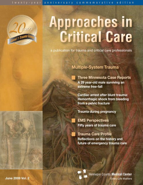
t w e n t y - y e a r a n n i v e r s a r y c o m m e m o r a t i v e e d i t i o n
Dear Readers:<br />
<strong>Medical</strong> manufacturers and hospital administrators are fond of describing<br />
new medical devices and facilities as “state of the art.” The term has always<br />
struck me. In the time it takes to ship and install a new medical device,<br />
doesn’t it usually lose “state-of-the-art” status? How long does an up-todate<br />
emergency department or intensive care unit remain “state of the art?”<br />
Between the execution of a clinical research trial and the actual<br />
implementation of its findings, will the findings become obsolete?<br />
Yet, that’s one of the primary joys of medicine—the way the field constantly<br />
evolves. It was only fifty years ago that a sharp increase in multiple-system<br />
trauma (an increase traced to motor vehicles) led to the development of<br />
what we now call emergency medical services (EMS). It was almost thirty<br />
years ago that emergency care as a certified RN specialty came into<br />
existence. And it was just twenty years ago that <strong>Hennepin</strong> <strong>County</strong> <strong>Medical</strong><br />
<strong>Center</strong> became the first in the state to become an American College of<br />
Surgeons (ACS) Level 1 trauma center.<br />
To commemorate the twenty-year anniversary of <strong>Hennepin</strong>’s trauma<br />
designation, in this issue we present several recent multiple-system trauma<br />
cases that clearly benefited from the “state of the art.” Without the recent<br />
evolutions in trauma care and resuscitation, the patients presented here<br />
would likely have died early in their clinical course. In the sections called<br />
EMS Perspectives and <strong>Trauma</strong> Care Profile, you’ll hear from long-time<br />
EMS, MD, and RN providers about where trauma care has come in the last<br />
50 years and where it is going in the next 50 years.<br />
The theme for our fall issue will be bedside ethics. If you have an<br />
interesting case study you’d like to contribute, see the author’s guidelines<br />
on the Approaches in Critical Care Web site at www.hcmc.org/approaches.<br />
We’d love to hear from you.<br />
Sincerely,<br />
Michelle H. Biros, MD, MS<br />
Approaches in Critical Care editor-in-chief<br />
Department of Emergency Medicine<br />
<strong>Hennepin</strong> <strong>County</strong> <strong>Medical</strong> <strong>Center</strong><br />
®<br />
Every Life Matters
Contents Volume 2 | Approaches in Critical Care June 2009<br />
Approaches in Critical Care<br />
Editor-in-Chief<br />
Michelle Biros, MD, MS<br />
Managing Editor<br />
Linda Zespy<br />
EMS Perspectives Editor<br />
Robert Ball, EMT-P<br />
Graphic Designer<br />
Karen Olson<br />
Marketing Director<br />
Ted Blank<br />
Patient Care Director,<br />
Critical Care and<br />
Emergency Services<br />
Kendall Hicks, RN<br />
Patient Care Director,<br />
Behavioral and<br />
Rehabilitative Services<br />
Joanne Hall, RN<br />
Printer<br />
Sexton Printing<br />
Photographers<br />
Raoul Benavides<br />
Lisa Fleming<br />
Karen Olson<br />
Clinical Reviewer<br />
Arthur Ney, MD, FACS<br />
Events Calendar Editor<br />
Susan Altmann<br />
Case Reports<br />
2 A 29 year-old male surviving an extreme free-fall<br />
Chad E. Roline, MD<br />
5 Cardiac arrest after blunt trauma: Hemorrhagic shock from bleeding<br />
from a pelvic fracture<br />
Mohamed A. Ibrahim, MD<br />
8 <strong>Trauma</strong> during pregnancy<br />
Mike Galle, MD<br />
10 <strong>Trauma</strong> Care Profile<br />
Arthur Ney, MD, FACS, Hillie Prose, RN, and Ernest Ruiz, MD reflect on<br />
the history and future of trauma care<br />
12 EMS Perspectives<br />
Fifty years of trauma care<br />
15 Photo Essay<br />
The evolution of emergency care<br />
17 Calendar of Events<br />
19 News Notes<br />
To submit an article<br />
Contact managing editor Linda Zespy at approaches@hcmed.org. The editors reserve the right to<br />
reject editorial or scientific materials for publication in Approaches in Critical Care. The views<br />
expressed in this journal do not necessarily represent those of <strong>Hennepin</strong> <strong>County</strong> <strong>Medical</strong> <strong>Center</strong>, its<br />
editors, or its staff members.<br />
Copyright<br />
Copyright 2009, <strong>Hennepin</strong> <strong>County</strong> <strong>Medical</strong> <strong>Center</strong>. Approaches in Critical Care is published twice per<br />
year by <strong>Hennepin</strong> <strong>County</strong> <strong>Medical</strong> <strong>Center</strong>, 701 Park Avenue, Minneapolis, Minnesota 55415.<br />
Subscriptions<br />
To subscribe, send an email to approaches@hcmed.org with your name and full mailing address.<br />
Approaches in Critical Care | Commemorative Issue | June 2009 | 1
Case Reports<br />
Treating <strong>Multiple</strong>-<strong>System</strong> <strong>Trauma</strong>:<br />
Three Case Reports<br />
Managing patients with multiple-system<br />
trauma is a complex undertaking. A<br />
multidisciplinary approach is essential<br />
for good patient outcomes. A skillful<br />
resuscitation requires a sound understanding<br />
of trauma and resuscitation<br />
principles and good technical skills.<br />
The clinicians involved must engage in<br />
reasoned and rapid decision-making as<br />
well as excellent communication. The<br />
stakes are high; most patients with<br />
multiple-system trauma are young and<br />
otherwise healthy and, if they survive<br />
their injury, have the potential to lead<br />
full and productive lives.<br />
The following case reports involve a<br />
diverse set of patients with multiplesystem<br />
trauma and describe the care<br />
decisions made in each instance.<br />
A 29 year-old male surviving an<br />
extreme free-fall<br />
by Chad E. Roline, MD<br />
Department of Emergency Medicine<br />
<strong>Hennepin</strong> <strong>County</strong> <strong>Medical</strong> <strong>Center</strong><br />
Abstract<br />
Although there is a general correlation<br />
between the height of a fall and the<br />
likelihood of death, a number of complex<br />
factors can make survival possible<br />
in free-fall impact events from extreme<br />
heights. This case involves a 29 yearold<br />
male who survived a 17-story fall<br />
without long-term disability.<br />
Case report<br />
Following a night of heavy alcohol consumption,<br />
a 29 year-old male returned<br />
with his friends to a downtown<br />
Minneapolis hotel. He then reportedly<br />
began running down a hallway and collided<br />
with a double-paned, floor-to-ceiling<br />
window at the end of the corridor.<br />
He crashed through the glass and fell,<br />
feet first, 17 stories. Initially, he landed<br />
on an asphalt-covered metal awning<br />
one floor above the ground. The structure<br />
collapsed to the street upon<br />
impact. He was initially unconscious at<br />
the scene and required fire department<br />
extrication from the awning.<br />
Upon arrival at the emergency department,<br />
the patient’s vital signs were:<br />
blood pressure 118/68, pulse 94, respirations<br />
21, and oxygen saturation<br />
100% on 10 liters face mask. He was<br />
awake and extremely combative, complaining<br />
of severe pain in his lower<br />
extremities. In order to facilitate his<br />
evaluation, and because of the serious<br />
mechanism of injury and worry about<br />
life-threatening injuries, he was intubated<br />
with rapid-sequence intubation.<br />
Despite successful endotracheal tube<br />
placement, the patient was noted to<br />
have intermittently poor oxygen saturation<br />
observed on pulse oximetry. His<br />
breath sounds were decreased bilaterally<br />
and a large amount of crepitus was<br />
appreciated throughout the neck and<br />
anterior chest wall. His abdomen was<br />
obese and soft. His pelvis was stable<br />
to anterior compression and he had no<br />
obvious step-off along the length of his<br />
spine. His right lower leg was noted to<br />
have an open tibia-fibula fracture.<br />
A portable chest radiograph was significant<br />
for bilateral pneumothoraces,<br />
which were managed with chest tubes.<br />
Computed tomography (CT) scans of<br />
the head, cervical spine, chest,<br />
abdomen, and pelvis demonstrated<br />
2 | Approaches in Critical Care | Commemorative Issue | June 2009
Case Reports<br />
The trauma team at <strong>Hennepin</strong> treats a patient in the stabilization room of the emergency department.<br />
bilateral pulmonary contusions and a large amount<br />
of air in the mediastinum and anterior chest wall<br />
extending into the neck. No intracranial or intraabdominal<br />
injuries were found. His blood alcohol<br />
level was measured at 0.247 g/dL.<br />
Due to the tenuous nature of the patient’s oxygen<br />
saturations, a bronchoscopy was performed<br />
by the emergency physician. This revealed<br />
blood several centimeters proximal to the carina.<br />
Because of concern that this could indicate a<br />
tracheal tear, cardiothoracic surgery was consulted.<br />
A repeat bronchoscopy was performed in<br />
the operating room with no obvious tracheal<br />
defect appreciated with blood again identified<br />
above the carina. He was cleared by cardiovascular<br />
surgery and underwent intramedullary nail<br />
fixation of his fractures. A third bronchoscopy<br />
was performed 72 hours after admission and<br />
again showed no tracheal tears or lacerations.<br />
Following this procedure, the patient was successfully<br />
extubated and his chest tubes were<br />
removed. He quickly improved and was transferred<br />
out of the surgical intensive care unit. The<br />
patient continued to do well on the floor and was<br />
discharged home on hospital day seven. The<br />
patient had an uneventful post-hospital course<br />
and recovered fully from his orthopedic injury.<br />
Discussion<br />
Falls are a common cause of trauma presenting<br />
to the emergency department and second only<br />
to motor vehicle collisions in causing traumarelated<br />
deaths in the U.S.<br />
Falls from extreme heights are a small subset of<br />
these events. These falls can be due to occupation-related<br />
accidents, such as mishaps during<br />
construction, acts of violence when a person is<br />
pushed, or self-inflicted events where a patient<br />
leaps from a high vantage point as an act of<br />
attempted suicide or psychotic behavior. In<br />
1996, Risser estimated that, using 12 feet as an<br />
average height of one story of a building, the<br />
height lethal to 50% and 90% of the population<br />
for elevated falls was four and seven stories,<br />
respectively.<br />
Over the years, several investigators have<br />
attempted to identify factors that contribute to<br />
the survival of that rare individual who withstands<br />
a free-fall from heights that would be<br />
Approaches in Critical Care | Commemorative Issue | June 2009 | 3
Case Reports<br />
expected to be fatal. In 1965, Snyder performed<br />
a review of 137 cases of people surviving such<br />
extreme falls. His report included personal<br />
investigation of each site and analysis of the<br />
details surrounding each incident. Snyder concluded<br />
that most survivors were young, overall<br />
healthy males who fell in a feet-first orientation.<br />
He also concluded that alcohol intoxication may<br />
reduce trauma in some falls by creating a state<br />
of “muscular relaxation.” However, it may be that<br />
this study simply confirmed that young, intoxicated<br />
males are more likely to suffer such a fall in<br />
the first place, due to increased risk-taking<br />
behavior in this population.<br />
“Being young and healthy clearly<br />
helped his survival, while the influence<br />
of his intoxication is more a matter<br />
of debate.”<br />
Most important to consider in free-fall survival<br />
are basic physics principles, including:<br />
Impact velocity. Impact velocity is<br />
intrinsically related to the distance of the fall.<br />
As an example of the effect of distance, one<br />
study estimated that a person falling from 32<br />
stories would reach a terminal velocity of<br />
approximately 120 miles per hour.<br />
Impact energy. Impact energy involves the<br />
kinetic energy accumulated by the body in<br />
free-fall, which on impact is converted into<br />
mechanical energy and heat. The<br />
mechanical energy that is not absorbed by<br />
the impact surface is transmitted into the<br />
body resulting in injury.<br />
Impact force. Impact force is determined by<br />
the mass of the patient and the amount of<br />
deceleration on impact. The degree of deceleration<br />
is influenced by the substance (e.g.<br />
concrete, sand, etc.) encountered.<br />
Free-falls are associated with a unique pattern<br />
of injuries. In several studies of patients who fell<br />
from a significant height, the lower limbs were<br />
the most frequent anatomical location of injury<br />
cited, with the most common lower-limb injury<br />
being calcaneal fractures. The same axial loading<br />
forces causing these fractures are transmitted<br />
to the spine. Most vulnerable is the junction<br />
of the thoracic and lumbar spines due to the<br />
more mobile nature of the lumbar spine relative<br />
to the thoracic spine.<br />
The predominant lethal injuries following free-fall<br />
events are massive head injuries, liver injuries,<br />
and, less commonly, thoracic injuries.<br />
Devastating brain and head injuries are a<br />
frequent cause of immediate death in these<br />
patients. Liver injuries are most often large<br />
lacerations with the right lobe most commonly<br />
involved. Thoracic injuries can include<br />
pulmonary injuries, a hemothorax, a pneumothorax,<br />
and thoracic aorta injuries. It is theorized<br />
that thoracic injuries are more likely when the<br />
patient lands on the buttocks as the impact<br />
forces cannot be dissipated by flexion of the<br />
hips and knees, resulting in a much larger force<br />
being transmitted to the trunk.<br />
The patient above illustrates several important<br />
concepts in free-fall injuries. He was young,<br />
healthy, and intoxicated. Being young and<br />
healthy clearly helped his survival, while the<br />
influence of his intoxication is more a matter of<br />
debate. In addition, he fell feet first and landed<br />
on an object that collapsed, allowing the time of<br />
his deceleration to the concrete to be significantly<br />
prolonged. The extended duration of his<br />
deceleration decreased his impact force to a<br />
survivable level.<br />
In conclusion, free-fall trauma should be recognized<br />
as a distinct form of blunt trauma that<br />
presents with a constellation of injuries that<br />
should be anticipated, with injury severity<br />
dependent on more factors than simply the<br />
height of the fall. While in general, greater<br />
height predicts a higher mortality, this case illustrates<br />
how in rare circumstances such falls can<br />
be survived. <br />
4 | Approaches in Critical Care | Commemorative Issue | June 2009
Case Reports<br />
Cardiac arrest after blunt trauma:<br />
Hemorrhagic shock from bleeding from<br />
a pelvic fracture<br />
by Mohamed A. Ibrahim, MD<br />
Department of Surgery<br />
<strong>Hennepin</strong> <strong>County</strong> <strong>Medical</strong> <strong>Center</strong><br />
Abstract<br />
The most common cause of shock in the trauma<br />
patient is loss of circulating blood volume from<br />
hemorrhage. While pre-hospital cardiac arrest<br />
due to blunt trauma is rarely survivable, the<br />
patients with the best outcome from traumatic<br />
arrest generally have treatable injuries, receive<br />
early endotracheal intubation, and undergo<br />
prompt transport to a trauma center for definitive<br />
management. This case report presents the successful<br />
outcome of a patient with traumatic cardiac<br />
arrest from hemorrhagic shock and illustrates<br />
the importance of well-coordinated prehospital<br />
care and aggressive resuscitation.<br />
Case report<br />
A 59 year-old male road construction worker<br />
with no major medical problems was run over<br />
and dragged by a heavy gravel truck at a road<br />
construction site. At the scene, he was alert and<br />
appropriate with a Glasgow Coma Scale score<br />
of 15 with stable vital signs. He complained of<br />
severe left hip pain and less severe right leg<br />
pain. Obvious injuries noted at the scene<br />
included large contusions and abrasions to the<br />
right side of his abdomen and chest. He also<br />
had a degloving injury to his right leg. He was<br />
transported by ground ambulance to the<br />
nearest hospital.<br />
In the emergency department, initial plain radiographs<br />
demonstrated unstable comminuted<br />
pelvic ring fractures (see Figure One on the next<br />
page) and a right lower-extremity fracture. This<br />
was open with extensive soft-tissue degloving.<br />
He was hypotensive and hypothermic.<br />
Resuscitation with fluids and blood products,<br />
along with rewarming maneuvers, were ongoing.<br />
His hypotension responded to fluid resuscitation.<br />
Given the degree of injuries and initial hemodynamic<br />
instability, a decision was made very early<br />
in the course of the initial treatment to transfer<br />
Approaches in Critical Care | Commemorative Issue | June 2009 | 5
Case Reports<br />
the patient by air to a regional Level 1 trauma<br />
center for definitive management. For airway<br />
protection, stabilization, and control while in air<br />
transport, endotracheal intubation was performed.<br />
While preparing for air transport, the<br />
patient suffered rapid clinical deterioration and<br />
loss of vital signs with pulseless electrical activity<br />
(PEA) cardiac arrest.<br />
Figure Two. This coronal slice CT scan shows wide diastasis of the left<br />
sacroiliac joint.<br />
He was emergently taken to the angiography<br />
suite where a left transected superior gluteal<br />
artery was found. The left gluteal artery and<br />
internal iliac were coiled. This stabilized his<br />
blood pressure in the 120-130 systolic range.<br />
Figure One. This pelvic x-ray shows a comminuted ring fracture and large<br />
sacroiliac joint separation.<br />
Aggressive cardiopulmonary resuscitation was<br />
undertaken according to the advanced cardiac<br />
life support (ACLS) guidelines. After approximately<br />
30 minutes of aggressive ACLS, the<br />
patient regained a perfusing sinus rhythm. Along<br />
with volume resuscitation, including large<br />
amounts of blood products, the pelvic ring was<br />
stabilized using the military anti-shock trousers<br />
(MAST) garment. Once stabilized with ongoing<br />
resuscitation, he was transferred by air for definitive<br />
care. At this point, he had received eight<br />
units of packed red blood cells, four units of<br />
plasma, and five liters of crystalloids.<br />
On arrival to the Level 1 trauma center, his initial<br />
blood pressure was 70/40 with a heart rate of<br />
160. Additional resuscitation was undertaken,<br />
including administration of additional blood products<br />
and placement of a large-bore, centralaccess<br />
catheter (9 French trauma catheter).<br />
Computed tomography (CT) scans were<br />
obtained. Along with the pelvic fracture (see<br />
Figure Two on this page), the CT scan showed<br />
free fluid in the abdomen without an identified<br />
solid organ injury. This prompted an exploratory<br />
laparotomy to rule out mesenteric or hollow<br />
visceral injury. The blood was from his pelvic<br />
injury and no additional injuries were found. His<br />
right extremity injuries were treated with debridement,<br />
temporary closure, and splinting. He was<br />
transferred to the surgical intensive care unit for<br />
ongoing resuscitation and management.<br />
By hospital day two, he was alert and appropriate.<br />
He met ventilatory weaning parameters and<br />
was extubated. On hospital day four, he underwent<br />
repair of his complex pelvic fractures. His<br />
other extremity injuries were managed with multiple<br />
operative interventions as well. He was discharged<br />
to an acute inpatient rehabilitation unit<br />
on hospital day 25.<br />
Discussion<br />
The most common cause of shock in a trauma<br />
patient is the loss of circulating blood volume<br />
from ongoing hemorrhage. For that reason,<br />
6 | Approaches in Critical Care | Commemorative Issue | June 2009
Case Reports<br />
shock in a trauma patient should be presumed<br />
to be due to hemorrhage until proven otherwise.<br />
Cardiopulmonary collapse following trauma has<br />
several possible causes, including:<br />
Hypoxia from airway obstruction,<br />
pneumothorax, or tracheobronchial injury;<br />
Injury to a vital structure, such as the heart<br />
and great vessels;<br />
Severe head or spinal cord injury with<br />
secondary cardiopulmonary arrest;<br />
Severely restricted cardiac output from<br />
tension pneumothorax or pericardial<br />
tamponade; or<br />
Extreme blood loss leading to hypovolemic<br />
shock, as in this patient’s case.<br />
Hemorrhagic shock has been classified according<br />
to the magnitude of blood loss. It is worth<br />
noting that signs of shock, such as hypotension,<br />
tachycardia, and confusion, may not be evident<br />
until more than 30% of blood volume has been<br />
lost (Class III shock). Treatment should be instituted<br />
as soon as shock is suspected or identified,<br />
typically before the source of hemorrhage<br />
is located. Occasionally, especially in young,<br />
healthy patients, blood pressure initially may be<br />
maintained until a precipitous cardiovascular<br />
collapse ensues, as occurred in this case.<br />
Aggressive volume resuscitation is indicated for<br />
patients with blunt trauma and hypovolemic<br />
shock. One of the most common terminal<br />
cardiac dysrhythmias in trauma patients is<br />
pulseless electrical activity (PEA). Successful<br />
resuscitation from cardiac arrest in this<br />
situation often depends on restoration of<br />
adequate circulating blood volume.<br />
This patient’s case demonstrates the typical<br />
presentation of patients with an ongoing source<br />
of hemorrhage who initially respond well to volume<br />
resuscitation efforts but then deteriorate<br />
hemodynamically. These patients usually require<br />
operative or invasive interventions to control<br />
hemorrhage. In this case, the cause of ongoing<br />
blood loss was superior gluteal artery transsection<br />
from his pelvic fracture.<br />
The morbidity and mortality associated with<br />
pelvic fractures was appreciated centuries ago.<br />
Prior to the 1900s, the mortality rate for pelvic<br />
fractures was well over 80%. Various modalities<br />
have been developed over the years to treat<br />
pelvic hemorrhage, including the MAST garment,<br />
external fixators, packing, and angiographic<br />
embolizations. Today, with the multidisciplinary<br />
approach to the treatment of pelvic<br />
fractures—including pre-hospital personnel,<br />
emergency physicians, orthopedic specialists,<br />
trauma surgeons, and interventional radiologists—mortality<br />
is rare unless associated with<br />
large-vessel laceration.<br />
“Prior to the 1990s, the mortality<br />
rate for pelvic fractures was<br />
well over 80%.”<br />
As shown in this case, better outcomes are<br />
seen when timely interventions are instituted,<br />
even in cases involving large pelvic-vessel<br />
lacerations. Clearly, this patient’s outcome<br />
was greatly improved by the rapidity with which<br />
aggressive cardiopulmonary resuscitation and<br />
ongoing support was undertaken. Transfer for<br />
definitive therapy for his ongoing hemorrhage<br />
was pursued well in advance and coordinated<br />
with the receiving facility, where arrangements<br />
were in place and ready upon the<br />
patient’s arrival. <br />
Approaches in Critical Care | Commemorative Issue | June 2009 | 7
Case Reports<br />
⊳ To left, paramedics at the scene<br />
of the 35W bridge collapse in<br />
August of 2008.<br />
Below, <strong>Hennepin</strong> paramedics in<br />
their inaugural ride over the new<br />
35W bridge.<br />
<strong>Trauma</strong> during pregnancy<br />
by Mike Galle, MD<br />
Department of Surgery<br />
<strong>Hennepin</strong> <strong>County</strong> <strong>Medical</strong> <strong>Center</strong><br />
Abstract<br />
<strong>Trauma</strong> care is complicated in pregnant patients<br />
because of the physiologic and anatomic<br />
changes that occur during pregnancy. Vital signs<br />
may be deceiving and treatment decisions must<br />
consider the viability of the fetus as well as the<br />
traumatized pregnant patient. In the face of fetal<br />
distress, prompt obstetrical care and emergency<br />
delivery may be necessary. This case illustrates<br />
that, in the traumatized pregnant patient, the<br />
best way to ultimately care for the fetus is to stabilize<br />
the mother.<br />
Case report<br />
A 37 year-old female was discovered lying in<br />
mud at the scene of the 35W bridge collapse in<br />
Minneapolis, MN in the summer of 2008. The<br />
circumstances of her accident were not known.<br />
Pre-hospital personnel reported that she was<br />
extremely agitated, disoriented, and unable to<br />
follow commands.<br />
On presentation to the emergency department,<br />
she was found to be pregnant near term with no<br />
major external signs of musculoskeletal trauma.<br />
Her vital signs were stable, except for mild sinus<br />
tachycardia and hypertension. She was maintaining<br />
her airway and breathing on her own but<br />
was clearly confused and screaming unintelligibly.<br />
Breath sounds were clear bilaterally. She had<br />
palpable distal pulses in all extremities. All<br />
extremities moved spontaneously but, due to<br />
her confused state, she was given a Glasgow<br />
Coma Scale score of 12. Pupils were equal and<br />
reactive to light. A Focused Assessment with<br />
Sonography in <strong>Trauma</strong> (FAST) exam was performed<br />
which showed no intra-abdominal or<br />
pericardial fluid collections. The fetus was<br />
visualized and an initial fetal heart rate of 150<br />
was noted.<br />
Because of her altered mental status and<br />
extreme agitation despite intravenous sedation,<br />
the decision was made to intubate the patient to<br />
facilitate subsequent evaluation and care. After<br />
two attempts, she was successfully endotracheally<br />
intubated. A second fetal ultrasound was<br />
performed, which showed a decreased fetal<br />
heart rate. An OB/GYN specialist was present<br />
throughout the case and, when the fetal heart<br />
rate declined, the OB/GYN made the decision to<br />
take the patient to the operating room for emergent<br />
cesarean section. The diagnoses at this<br />
point, based on her clinical assessment, included<br />
blunt force trauma with likely traumatic brain<br />
injury and intrauterine fetal distress.<br />
The patient underwent immediate, uneventful<br />
cesarean section at 34 weeks gestation. There<br />
was no operative evidence of intra-abdominal<br />
injury. On entering the uterus, there was clinical<br />
evidence of placental abruption but the fetus<br />
was quickly and uneventfully extracted. A<br />
healthy, 2.8-kilogram baby was delivered and<br />
8 | Approaches in Critical Care | Commemorative Issue | June 2009
Case Reports<br />
was discharged from the hospital soon thereafter<br />
without incident.<br />
Following her cesarean section, the patient<br />
underwent CT scanning of her head, which<br />
revealed a diffuse, moderate subarachnoid<br />
hemorrhage. (See Figure One on this page.)<br />
A ventriculostomy catheter was placed but the<br />
patient had no prolonged episodes of intracranial<br />
hypertension. She continued to have altered<br />
mental status and remained intubated for some<br />
time following admission. She required tracheostomy<br />
tube placement as well as gastric<br />
feeding tube placement on hospital day 12. Her<br />
mental status slowly improved and the patient<br />
was discharged to an inpatient rehabilitation<br />
facility on hospital day 22.<br />
The patient was last seen in neurosurgery clinic<br />
in January 2008 and was doing well. Her tracheostomy<br />
tube had been removed as well as<br />
her G-tube. She complained of intermittent<br />
headaches and memory lapses but was expected<br />
to make a full neurologic recovery. Her baby<br />
was doing well.<br />
Discussion<br />
Moore in 2004 found that the leading cause of<br />
maternal mortality in the pregnant patient is<br />
obstetric complications; the second most common<br />
cause of mortality is trauma, which is estimated<br />
to occur in up to 7% of all pregnancies.<br />
The care of the pregnant patient in trauma follows<br />
the same parameters as generalized trauma<br />
care. The first priority is intervention aimed<br />
at stabilization of the mother. In the pregnant<br />
patient, there is an increase in total blood volume.<br />
Tachycardia and hypotension may not be<br />
evident until 30% of the blood volume is lost.<br />
Thus, the vital signs may not be helpful in determining<br />
hemodynamic stability. The treating<br />
physician should therefore have a low threshold<br />
for administering volume in the traumatized<br />
pregnant patient, even in the absence of altered<br />
vital signs. Frequently, the venous return to the<br />
heart is compromised due to caval compression<br />
by the uterus. This can be alleviated by tilting<br />
the patient to the left or placing her in a left<br />
lateral decubitus position or direct uterine retraction<br />
to the left.<br />
The examination of the pregnant woman in trauma<br />
should include a pelvic examination to look<br />
for vaginal blood or evidence of membrane rupture.<br />
The most common fetal injury in trauma is<br />
placental abruption, which can quickly lead to<br />
fetal demise. A deceleration in fetal heart tones<br />
is commonly associated. In the face of any hard<br />
signs of fetal compromise or uterine pathology,<br />
an immediate caesarean section may be the<br />
only way to salvage the pregnancy.<br />
This patient was presumed to have a head<br />
injury based on her clinical evaluation that was<br />
not immediately life-threatening. The fetus<br />
demonstrated decreased heart tones, however,<br />
which is an ominous sign. The best course of<br />
action in this situation was immediate delivery of<br />
the fetus. <br />
Agalar F, et al. Factors affecting mortality in urban vertical<br />
free falls: Evaluation of 180 cases. International Surgery.<br />
1999: 271-74.<br />
American Heart Association. Part 10.7: Cardiac arrest<br />
associated with trauma. Circulation. Nov 2005: 112.<br />
Beery P, Ellison C. Surgery in the pregnant patient. 17th<br />
ed. Sabiston Textbook of Surgery. Saunders. 2004.<br />
Gupta SM, et al. Blunt force lesions related to the height<br />
of a fall. American Journal of Forensic Medicine &<br />
Pathology. 1982: 35-43.<br />
Moore, et al. <strong>Trauma</strong>. 5th Ed. McGraw Hill. 2004.<br />
⊳ Figure One. This<br />
CT scan shows subarachnoid<br />
blood and a<br />
ventriculostomy catheter.<br />
Suggested Readings/Bibliographies for Case Reports<br />
Risser D, et al. Risk of dying after a free fall from height.<br />
Forensic Sci Int. 1996;78: 187.<br />
Snyder RG. Human survivability of extreme impacts in<br />
free-fall. FAA Office of Aviation Medicine Report No. AM<br />
63-15. Aug. 1963.<br />
Tan S, Porter K. Free fall trauma. <strong>Trauma</strong>. 2006: 157-167.<br />
Warner KG, Demling RH. The pathophysiology of free-fall<br />
injury. Annals of Emergency Medicine. 1986: 1088-93.<br />
Approaches in Critical Care | Commemorative Issue | June 2009 | 9
<strong>Trauma</strong> Care Profile<br />
Q and A withQ and A with<br />
Arthur Ney, MD, FACS, Hillie Prose, RN,<br />
and Ernest Ruiz, MD<br />
Arthur Ney, MD, FACS<br />
Hillie Prose, RN<br />
Ernest Ruiz, MD<br />
During their careers at <strong>Hennepin</strong><br />
<strong>County</strong> <strong>Medical</strong> <strong>Center</strong>, the three clinicians<br />
interviewed for this issue’s<br />
<strong>Trauma</strong> Care Profile witnessed the rise<br />
of emergency medicine as a specialty<br />
and actively nurtured its growth.<br />
Arthur Ney, MD, FACS (trauma<br />
surgeon and chief of trauma), Hillie<br />
Prose, RN (retired director of emergency<br />
nursing), and Ernest Ruiz, MD<br />
(retired chief of emergency medicine)<br />
shared their thoughts about how trauma<br />
and emergency care has evolved<br />
and where the specialties might go in<br />
the twenty-first century.<br />
What was the emergency<br />
department like when you started?<br />
Hillie Prose: In 1950, we were the<br />
emergency hospital for the city of<br />
Minneapolis but emergency work—the<br />
critical care—was done on the stations.<br />
The ambulance didn’t even stop in the<br />
ED. The ED was mostly an admitting<br />
area with minor injuries cared for there.<br />
Nurses ran the place and doctors,<br />
interns, and surgery residents rotated<br />
in. We didn’t even have an IV set up<br />
until 1968. Then in 1971, Dr. Ruiz was<br />
assigned to be chief of emergency<br />
medicine. There were things we needed<br />
in the ED but we didn’t have the<br />
budget, so in his garage, he built a lot<br />
of inventions to improve patient care.<br />
Ernest Ruiz: In those days, there were<br />
very few devices being developed that<br />
were particularly useful in the emergency<br />
department, so if you came up<br />
with an idea, you had to convince a<br />
corporation to take a look at it or build<br />
it yourself. As a result, I ended up<br />
trying to make a lot of things myself.<br />
Examples? I used a small inner tube to<br />
make a cushion to put under a<br />
patient’s head. It was connected to a<br />
valve that was connected to compressed<br />
air and operated with a foot<br />
pedal. So when you went to intubate a<br />
patient, you could adjust the head level<br />
until you had the best view possible.<br />
Now, there are devices that do the<br />
same thing. For a few years, we used<br />
a device I devised for auto-transfusion<br />
of blood in trauma. I used tubing that<br />
was used at the time in cardiac surgery<br />
and blood banking. It was a little complicated<br />
but finally commercial varieties<br />
of autotransfusers came out that were<br />
much easier to use.<br />
In the last 50 years what was<br />
the most important advance in<br />
trauma care?<br />
Arthur Ney: Probably the biggest<br />
change is the improved accuracy of<br />
diagnostic testing. This has resulted in<br />
a dramatic shift in the management of<br />
injured patients, with nonoperative care<br />
now being the standard for many<br />
injuries. Exploratory surgery to identify<br />
and control hemorrhage was commonplace<br />
50 years ago. Many complex<br />
injuries are now managed with minimally<br />
invasive techniques or non-operatively<br />
with physiologic resuscitation.<br />
When we first started using CT scans<br />
in the 1970s, a single slice would take<br />
about five minutes. With modern scanners,<br />
we can frequently evaluate the<br />
head, chest, and abdomen in not much<br />
longer than that. This has resulted in<br />
significant improvement in safety in<br />
10 | Approaches in Critical Care | Commemorative Issue | June 2009
<strong>Trauma</strong> Care Profile<br />
obtaining these tests and allowed the evolution<br />
of nonoperative management for many different<br />
types of injuries.<br />
Ernest Ruiz: There are many things on the list<br />
that are right up there, like the availability of<br />
blood and management of blood administration.<br />
That was a big advancement. The development<br />
of thoracic/cardiac surgery was a big advance.<br />
And we can’t forget the good pre-hospital care<br />
that came along in the early 1960s, when there<br />
was recognition of how to immobilize a patient,<br />
which led to a decrease in the number of<br />
patients with neck and spinal injuries. Another<br />
very important advance was the advent of CT<br />
and MRI scanning. In the end, the advent of<br />
emergency medicine itself, as a specialty, has<br />
been a tremendous thing. It used to be that EDs<br />
were staffed with doctors and nurses who had<br />
not been trained in trauma or emergency critical<br />
care. Now such training is available. So the<br />
advent of emergency medicine tremendously<br />
affected the quality of care.<br />
Hillie Prose: (In the 1970s), the emergency<br />
medicine program started, and <strong>Hennepin</strong>’s program<br />
was the second in the nation. We also<br />
became the first in the nation for training in<br />
emergency room nursing. In those years, there<br />
was tremendous change in how we took care of<br />
critical patients.<br />
“It may be possible to begin the<br />
imaging process in the pre-hospital<br />
phase, using ultrasound and other<br />
heretofore-unknown methods of<br />
imaging so that proper treatment can<br />
begin then and there.”<br />
What will trauma care look like in 50 years?<br />
Arthur Ney: One of the dramatic changes of the<br />
last 50 years has been in critical care and nutrition<br />
management and I think it’s an area poised<br />
for future innovation. There will likely be significant<br />
improvements in restoring the altered physiology<br />
that occurs from prolonged shock. Right<br />
now, the interventions we use often work shortterm<br />
but occasionally lead to multi-system organ<br />
failure or a systemic inflammatory response<br />
down the line. I suspect we will have genetic<br />
typing that will give us information to be able to<br />
manipulate cytokines and the inflammatory<br />
response. That could prevent many of the late<br />
deaths following trauma.<br />
Ernest Ruiz: I think the further refinement of<br />
imagery is going to be a big thing in the future. It<br />
may be possible to begin the imaging process in<br />
the pre-hospital phase, using ultrasound and<br />
other heretofore-unknown methods of imaging<br />
so that proper treatment can begin then and<br />
there. Minimally invasive surgery is going to<br />
expand and that will make the management of<br />
certain kinds of cases safer and faster for the<br />
patient. Neurosurgery has made great strides<br />
over the years with advanced imagery and the<br />
ability to operate through vascular channels<br />
rather than by opening the cranium. Perhaps<br />
there will be new ways of protecting the brain<br />
from further bleeding while that kind of treatment<br />
is getting underway. Then hopefully someday<br />
someone will figure out how to let the nerves<br />
actually reconnect, both in the brain and with<br />
spinal cord injuries.<br />
Arthur Ney: Looking back reinforces how important<br />
it is to be willing to learn new things and<br />
accept change as an important part of being a<br />
good practitioner at all levels. The key is to<br />
work these changes into standard practice and<br />
develop a system of education and mentoring<br />
that minimizes the learning curve and makes<br />
new knowledge available to everyone. <br />
Arthur Ney, MD, FACS, has been a trauma surgeon at<br />
<strong>Hennepin</strong> since 1985 and added the role of chief of trauma<br />
in 1987.<br />
Hillie Prose, RN, arrived in the emergency department in<br />
1948 and became department director in the 1960s. As the<br />
specialty evolved and physicians became more integrally<br />
involved, she became director of emergency nursing and<br />
filled that role until she retired in 1977.<br />
Ernest Ruiz, MD, became the chief of emergency medicine<br />
in 1971 and held that title until he retired in 1985.<br />
Approaches in Critical Care | Commemorative Issue | June 2009 | 11
EMS Perspectives<br />
_______________<br />
The emergency<br />
room of <strong>Hennepin</strong><br />
<strong>County</strong> <strong>Medical</strong><br />
<strong>Center</strong> in the 1960s.<br />
____________________<br />
EMS Perspectives: Fifty Years of <strong>Trauma</strong> Care<br />
by Robert Ball, EMT-P<br />
<strong>Hennepin</strong> Emergency <strong>Medical</strong> Services<br />
<strong>Hennepin</strong> <strong>County</strong> <strong>Medical</strong> <strong>Center</strong><br />
In the day-to-day challenges of<br />
emergency medical services (EMS)<br />
care, it can be easy to forget how<br />
much trauma care has changed even<br />
within the 23 years I have worked as a<br />
paramedic. In fact, it was just 20 years<br />
ago that <strong>Hennepin</strong> <strong>County</strong> <strong>Medical</strong><br />
<strong>Center</strong> became the first hospital in<br />
Minnesota to achieve the American<br />
College of Surgeons (ACS) Level 1<br />
trauma center verification.<br />
The evolution in trauma care that led to<br />
the trauma designation system, and<br />
the evolving role of EMS professionals<br />
in trauma care, is a fascinating story of<br />
persistent innovation, questioning, and<br />
willingness to try new ideas.<br />
EMS in the 1960s<br />
The original reason for the development<br />
of modern EMS in the 1960s was<br />
the skyrocketing increase in death and<br />
disability due to motor vehicle trauma.<br />
In a famous 1966 National Academy of<br />
Sciences paper entitled, “Accidental<br />
Death and Disability: The Neglected<br />
Disease of Modern Society" (many<br />
simply call it the “EMS White Paper”),<br />
authors pointed out that a soldier shot<br />
in the jungles of Vietnam had a better<br />
chance of survival than a motorist in<br />
the U.S. The original emergency medical<br />
technician (EMT) curriculum was<br />
steeped heavily in trauma; the first<br />
EMT textbook was published by the<br />
American Academy of Orthopedic<br />
Surgeons.<br />
At the same time, researchers were<br />
trying to find ways to reduce death<br />
from sudden cardiac arrest and heart<br />
attacks. In 1966, J. Frank Pantridge,<br />
MD, of Belfast, Northern Ireland<br />
equipped an ambulance with a portable<br />
electrocardiograph and medication,<br />
and began responding to cardiac calls.<br />
He was able to demonstrate that an<br />
ambulance staffed with a physician and<br />
nurse could improve care for patients<br />
with acute cardiac events.<br />
12 | Approaches in Critical Care | Commemorative Issue | June 2009
EMS Perspectives<br />
In the U.S., physician-staffed ambulances were<br />
attempted by some hospitals, most notably St.<br />
Vincent’s Hospital in New York, but the prohibitive<br />
cost of such operations caused researchers<br />
to examine whether non-physicians could perform<br />
many of the same functions. Eugene<br />
Nagel, MD, established the first experimental<br />
paramedic program with the Miami Fire<br />
Department in 1967. Because commercial cardiac<br />
equipment was not yet available, Nagel<br />
helped push the development of telemetry<br />
equipment to share patient information with a<br />
physician at a hospital.<br />
New decisions: Care vs. quick transports<br />
The late 1960s and early 1970s were a period<br />
where the EMTs who staffed most ambulance<br />
services often deepened their education in order<br />
to become paramedics. This increase in training,<br />
and a continuing increase in available tools and<br />
technology, led to loftier goals in terms of providing<br />
advanced life support for trauma patients as<br />
well as cardiac or medical patients.<br />
During this time period, it was not uncommon for<br />
paramedics arriving at the scene of a serious<br />
trauma to secure the patient to a backboard,<br />
intubate, use cardiac monitoring, place multiple,<br />
large-bore intravenous (IV) lines, and apply and<br />
inflate anti-shock trousers—all before the patient<br />
was transported to the hospital.<br />
Unfortunately, this did not improve survival rates<br />
in trauma victims and often had the opposite<br />
result. Because of this, many tiered EMS<br />
systems—systems using both basic life support<br />
(BLS) and advanced life support (ALS)<br />
ambulances—changed their dispatch priority.<br />
<strong>Trauma</strong> cases, no matter how serious, were<br />
reverted to basic life support calls. Unlike paramedics<br />
of the era, EMTs would not only quickly<br />
identify the serious trauma patient but also<br />
recognize that there was little they could do for<br />
the patient aside from transport to the closest<br />
appropriate hospital.<br />
While the use of such terms as “scoop and run”<br />
was antithetical to the concept of paramedicine,<br />
paramedics realized the need for a better understanding<br />
of the priorities in trauma and a better<br />
process of weighing the potential benefit of field<br />
management of a patient vs. the potential risks<br />
of delaying transport. Patient assessment priorities<br />
changed. Instead of merely examining the<br />
ABCs of airway, breathing and circulation, paramedics<br />
were learning to perform a fast initial<br />
exam that included level of consciousness and<br />
obvious hemorrhage. For the severely injured,<br />
only problems in those areas were managed<br />
immediately. With rare exceptions, treatments<br />
such as IV access and cardiac monitoring were<br />
no longer to be performed at the scene. To<br />
avoid unnecessary delays at the scene, even<br />
full “head-to-toe” surveys and vital signs often<br />
were performed in transit to the hospital.<br />
Where to transport?<br />
One of the remaining questions was often,<br />
“Which hospital?” By the early 1980s, designated<br />
trauma centers and trauma systems were<br />
taking hold, due in large part to the persistence<br />
of R. Adams Cowley, MD—often described as<br />
the father of trauma medicine—who convinced<br />
legislators and hospitals that patients should not<br />
always be brought to the nearest hospital but<br />
may be best served by the hospital most<br />
equipped to treat severe trauma.<br />
In Minnesota, the question of where to bring<br />
patients was easier to answer in the rural part of<br />
the state. Because of the large distances<br />
between hospitals in rural areas, the best hospital<br />
for serious trauma usually was the closest, at<br />
least until the patient could be stabilized for<br />
transfer to a larger, more trauma-oriented facility.<br />
In the greater Minneapolis area, the question<br />
was more difficult. There were many large hospitals,<br />
all of whom needed patients. In the early<br />
to mid-1980s, most hospitals in the west metro<br />
accepted critical trauma cases. Realistically, this<br />
meant many trauma patients were stabilized and<br />
transferred to another facility for tertiary care.<br />
In 1989, <strong>Hennepin</strong> <strong>County</strong> <strong>Medical</strong> <strong>Center</strong><br />
became the first ACS-verified Level 1 trauma<br />
center in Minnesota. At the time, EMS teams,<br />
who had greatly improved scene times (the<br />
EMS system required a scene time of less than<br />
10 minutes, except on extrication calls), still<br />
secured nearly every patient to a backboard.<br />
Fluids were poured in by the liter and D50<br />
sometimes was ordered for head-injured<br />
Approaches in Critical Care | Commemorative Issue | June 2009 | 13
EMS Perspectives<br />
⊳ Intern Per Wickstrom provides<br />
care in a <strong>Hennepin</strong> ambulance in the<br />
late 1960s.<br />
⊳⊳ <strong>Hennepin</strong>’s fleet of ambulances<br />
in 1964.<br />
patients, who also were being hyperventilated<br />
at rates more closely associated with a resting<br />
pulse rate than a respiratory rate.<br />
The intervening decades have seen significant<br />
changes in the pre-hospital management of<br />
trauma. Recently, EMS professionals have taken<br />
a more proactive approach to research. Rather<br />
than theorizing and using common sense, EMS<br />
agencies, medical directors, and paramedics are<br />
taking a much harder look at patient outcomes<br />
before changing practices.<br />
Today, the head-injured patient may receive<br />
some hyperventilation as part of their treatment,<br />
but often treatment is based more on end-tidal<br />
CO2 levels than mere guesswork. Intravenous<br />
access remains a staple of the trauma patient<br />
but the amount of fluid infused has changed<br />
greatly. Only the severely burned patient and<br />
the trauma patient with critical hypotension<br />
receive any significant fluid bolus. Otherwise,<br />
the access is present to administer analgesics<br />
and to be available for blood products on arrival<br />
at the hospital.<br />
On the horizon<br />
One trauma care subject that has recently been<br />
reexamined has been the use of analgesics. For<br />
decades, paramedics were warned that providing<br />
patients with medications for pain relief was<br />
to be avoided in the field, except in the case of<br />
cardiac chest pain. The concern was that pain<br />
medications could mask physical signs and<br />
symptoms or have side effects not easily treated<br />
in the field.<br />
Today, EMS professionals, along with other<br />
providers, are recognizing that reduction of<br />
suffering must improve. In some parts of the<br />
country, the synthetic narcotic fentanyl is finding<br />
a niche in EMS care. Fentanyl’s short half-life<br />
makes it easier to titrate and allows for an easier<br />
switch to different analgesics, if desired, once<br />
the patient reaches the hospital. The medication<br />
also is safe for use in hypotensive patients, thus<br />
allowing pain management even in the serious<br />
trauma patient.<br />
Even today, there are other times we in EMS do<br />
too much. Today, the severely injured and those<br />
with suspected spinal injuries continue to require<br />
immobilization. However, increasingly EMS professionals<br />
have recognized that even a treatment<br />
as simple as a backboard is not without<br />
risk and, along with the discomfort and anxiety<br />
that can occur with immobilization, may not be<br />
the best choice for some patients. We also now<br />
recognize that many traumatic cardiac arrests<br />
cannot be resuscitated and that extended resuscitation<br />
attempts can burden the family and<br />
hospitals and even sometimes create unnecessary<br />
risks for the EMS agencies transporting<br />
such patients.<br />
In the future, the art and science of EMS trauma<br />
care will continue to be an amalgamation of<br />
technology, medicine, and education. Exciting<br />
new developments are just over the horizon.<br />
Technological advances, combined with improved<br />
education, may mean being able to implement<br />
some version of imaging in the pre-hospital setting.<br />
The development of artificial blood products<br />
for trauma victims continues to face challenges,<br />
but we are on the frontier of such discoveries.<br />
The increase in use of telemedicine will mean<br />
outlying hospitals will have more resources to<br />
determine which patients need to be moved to<br />
a trauma center and which are best served by<br />
remaining in the community.<br />
Looking ahead, one thing is clear: the advances<br />
we see in the next fifty years will be every bit as<br />
monumental as those we have experienced in<br />
the last five decades. <br />
14 | Approaches in Critical Care | Commemorative Issue | June 2009
Photo Essay<br />
Twenty-Year “<strong>Trauma</strong> 1” Anniversary Commemorative Edition<br />
Photo Essay:<br />
The Evolution of Emergency Care<br />
These photos were provided by the<br />
<strong>Hennepin</strong> History Museum, which<br />
also houses equipment, supplies, and<br />
uniforms that track the progression<br />
of American medicine, beginning with<br />
<strong>Hennepin</strong>’s opening in 1887. For hours<br />
and location of the museum, visit<br />
www.hcmed.org or call 612-873-2512.<br />
<br />
In 1911, the first electric ambulance was<br />
purchased for <strong>Hennepin</strong>. The idea of<br />
motorized ambulances had received unexpected<br />
publicity a few years earlier when<br />
President William McKinley became one of<br />
the first to ride in an electric ambulance<br />
after an assassination attempt in 1901. The<br />
state-of-the-art transportation did not help<br />
save him; McKinley died eight days after<br />
being shot.<br />
<br />
In 1894, <strong>Hennepin</strong> <strong>County</strong><br />
<strong>Medical</strong> <strong>Center</strong> (then known as<br />
Minneapolis City Hospital) rented<br />
its first ambulance—a covered<br />
wagon, a team of horses, and a driver—for<br />
$1.50 per run. At the time, the hospital had<br />
been open for seven years. Daily cost for<br />
each patient was 89 cents. Like most<br />
Americans, staff members worked a sevenday<br />
week. Day shift workers worked 89<br />
hours per week while night shift workers<br />
worked 77 hours per week.<br />
<br />
Beginning in 1916, this single room served<br />
as <strong>Hennepin</strong>’s emergency department<br />
(ED). Nurses, at <strong>Hennepin</strong> and at most<br />
hospitals across the nation, were the<br />
primary staff members for the emergency<br />
room. As the workweek was shortening<br />
across the U.S., it also decreased at<br />
<strong>Hennepin</strong>, where health professionals now<br />
worked just 56 hours per week.<br />
Approaches in Critical Care | Commemorative Issue | June 2009 | 15
Photo Essay<br />
<br />
In 1963, the first trauma<br />
training course in Minnesota<br />
was organized at <strong>Hennepin</strong>.<br />
Throughout the 1960s, the<br />
concept of emergency<br />
medicine was evolving<br />
rapidly with the realization<br />
that the lessons learned on<br />
the battlefields of Korea and<br />
Vietnam could be applied on<br />
the home front.<br />
<br />
In 1967,<br />
Minnesota’s first<br />
suicide prevention<br />
hotline, housed in<br />
<strong>Hennepin</strong>’s ED, was<br />
launched. Attitudes<br />
toward suicide as a<br />
health issue were<br />
still developing; in<br />
the early 1960s,<br />
attempted suicide<br />
was a felony in<br />
North Dakota,<br />
South Dakota,<br />
and several<br />
other states.<br />
<br />
In 1938, the same room<br />
still functioned as the ED,<br />
but surgery interns joined<br />
nurses in staffing it. The<br />
following year, the standard<br />
workweek for Americans<br />
dropped again to 48 hours.<br />
<br />
In 1969, director of<br />
emergency nursing<br />
Hillie Prose, RN<br />
created a plan to<br />
help stabilize critical<br />
patients. Initially, the<br />
plan revolved around sending nurses to inpatient floors to<br />
stay with patients until they were fully stabilized. In time,<br />
the strategy involved a special stabilization room in the<br />
emergency department to help stabilize critical patients<br />
on arrival. Later, the accreditation program for Level 1<br />
trauma designation made a stabilization room a required<br />
element of providing Level 1 care to trauma patients.<br />
<br />
In 1986, care for the most<br />
urgent patients was improved<br />
when a helipad was built on<br />
top of a parking facility at<br />
<strong>Hennepin</strong>. Also in 1986,<br />
<strong>Hennepin</strong> opened an urgent<br />
care center next door to the<br />
ED. The convenience and<br />
lower cost led to thousands of<br />
such centers opening around<br />
the U.S. in the late 1970s,<br />
1980s, and 1990s.<br />
<br />
In 1989, <strong>Hennepin</strong> became the first in Minnesota<br />
to achieve Level 1 trauma verification.<br />
<br />
In the early 1990s, as part of a<br />
national trend toward eventual<br />
elimination of storage rooms like<br />
these (medical records<br />
department, 1951), the <strong>Hennepin</strong><br />
ED became a beta test site for<br />
one of the first electronic medical<br />
record systems in the country.<br />
<br />
In 2009, <strong>Hennepin</strong> celebrated<br />
twenty years as a Level 1 trauma center.<br />
16 | Approaches in Critical Care | Commemorative Issue | June 2009
Calendar of Events<br />
To register for a course,<br />
visit www.hcmc.org and<br />
click on “Professional<br />
Education and Training.”<br />
For questions or additional<br />
information, contact Susan<br />
Altmann in <strong>Medical</strong><br />
Education at <strong>Hennepin</strong><br />
<strong>County</strong> <strong>Medical</strong> <strong>Center</strong> at<br />
(612) 873-5681 or<br />
susan.altmann@hcmed.org<br />
unless another contact person<br />
is provided. Classes are<br />
at <strong>Hennepin</strong> <strong>County</strong> <strong>Medical</strong><br />
<strong>Center</strong> unless otherwise indicated.<br />
Many courses fill<br />
quickly; please register early<br />
to avoid being wait-listed.<br />
June<br />
June 5_________________________________<br />
CPR for MDs, for HCMC staff<br />
June 10________________________________<br />
CPR/BLS, for HCMC staff<br />
June 16________________________________<br />
Healthcare Provider CPR, for G1 residents<br />
June 17-22_____________________________<br />
Advanced Cardiac Life Support, for G1 residents<br />
June 17________________________________<br />
CPR for MDs, for HCMC staff<br />
June 23-24_____________________________<br />
Pediatric Advanced Life Support - Provider<br />
June 24________________________________<br />
Pediatric Advanced Life Support - Renewal<br />
July<br />
July 7__________________________________<br />
Advanced Cardiac Life Support - Instructor<br />
Renewal<br />
July 6-7________________________________<br />
Advanced Cardiac Life Support - Instructor<br />
July 8 and 10____________________________<br />
Advanced Cardiac Life Support - Provider, for<br />
HCMC staff<br />
July 9__________________________________<br />
CPR/BLS, for HCMC staff<br />
July 10_________________________________<br />
CPR for MDs, for HCMC staff<br />
July 14-15______________________________<br />
Advanced <strong>Trauma</strong> Life Support<br />
July 21-22______________________________<br />
Advanced Pediatric Life Support<br />
Approaches in Critical Care | Commemorative Issue | June 2009 | 17
Calendar of Events<br />
August<br />
August 5_______________________________<br />
Healthcare Provider CPR<br />
August 5 and 7__________________________<br />
Advanced Cardiac Life Support, for HCMC staff<br />
August 11-12____________________________<br />
Advanced Cardiac Life Support<br />
August 12______________________________<br />
Advanced Cardiac Life Support - Renewal<br />
September 22-23_________________________<br />
Pediatric Advanced Life Support - Provider<br />
September 23___________________________<br />
Pediatric Advanced Life Support - Renewal<br />
September 30 and October 2_______________<br />
Advanced Cardiac Life Support, for HCMC staff<br />
September 28 - October 2__________________<br />
First Responder<br />
August 13______________________________<br />
CPR/BLS, for HCMC staff<br />
August 7_______________________________<br />
CPR for MDs, for HCMC staff<br />
August 19______________________________<br />
CPR for MDs, for HCMC staff<br />
August 25______________________________<br />
Advanced Cardiac Life Support, renewal for<br />
HCMC staff in a.m.<br />
August 25______________________________<br />
Advanced Cardiac Life Support, renewal for<br />
HCMC staff in p.m.<br />
September<br />
September 3-4___________________________<br />
First Responder Refresher<br />
September 9____________________________<br />
Healthcare Provider CPR<br />
<strong>Hennepin</strong> Connect magnet v2 3/31/08 10:16 AM Page 5<br />
September 15___________________________<br />
CPR/BLS, for HCMC staff<br />
September 15-17_________________________<br />
Emergency <strong>Medical</strong> Technician Refresher<br />
course at Edina Training <strong>Center</strong><br />
September 11___________________________<br />
CPR for MDs, for HCMC staff<br />
Rapid access to <strong>Hennepin</strong> physicians<br />
for referrals and consults<br />
Services available 24/7<br />
1-800-424-4262<br />
612-873-4262<br />
18 | Approaches in Critical Care | Commemorative Issue | June 2009
News Notes<br />
News Notes<br />
Earth tones and<br />
natural sunlight<br />
provide a healing<br />
environment for<br />
patients and staff<br />
members.<br />
Patient rooms<br />
have a view of<br />
part of downtown<br />
Minneapolis and<br />
light-filtering<br />
window blinds.<br />
New intensive care units open<br />
Two new units, a 24-bed medical intensive<br />
care unit (MICU) and a 24-bed<br />
surgical intensive care unit (SICU),<br />
have opened at <strong>Hennepin</strong> <strong>County</strong><br />
<strong>Medical</strong> <strong>Center</strong>. The units’ openings<br />
are the first phase of a plan to concentrate<br />
inpatient medical and surgical<br />
intensive care to one critical care floor<br />
at <strong>Hennepin</strong>.<br />
“These contemporary units are<br />
designed to respond to the needs of<br />
our patients and create a comfortable,<br />
safe, healing environment in an ideal<br />
work area for our exceptional medical<br />
and surgical caregivers,” said Lynn<br />
Abrahamsen, chief executive officer at<br />
<strong>Hennepin</strong>. “We are pleased to be able<br />
to enhance the patient experience to<br />
match the excellent clinical outcomes<br />
expected at HCMC.”<br />
The MICU and SICU units were<br />
designed with input from patients,<br />
family members, and staff. The units<br />
include all private patient rooms with<br />
flat-screen televisions, thermostats,<br />
and light-filtering window blinds.<br />
Nursing work stations, placed between<br />
every two rooms, support direct views<br />
of the patient.<br />
_______________________________<br />
New neurological studies<br />
begin in Minnesota emergency<br />
departments<br />
Two pharmaceutical treatments, one<br />
for continuous seizures and the other<br />
for strokes, will be studied in Minnesota<br />
emergency departments as part of the<br />
Neurologic Emergencies Treatment<br />
Trial (NETT) Network, a nationwide<br />
effort to improve the outcomes of neurological<br />
emergency patients through<br />
interventional clinical research.<br />
Approaches in Critical Care | Commemorative Issue | June 2009 | 19
News Notes<br />
New neurological studies begin in<br />
Minnesota emergency departments cont.<br />
The first trial, called RAMPART (Rapid<br />
Anticonvulsant Medication Prior to Arrival Trial)<br />
is a double-blind, randomized clinical trial to<br />
evaluate the efficacy of midazolam (Versed)<br />
given intramuscularly vs. lorazepam (Ativan)<br />
given intravenously.<br />
The second trial, called ALIAS (Albumin in<br />
Acute Ischemic Stroke) provides ischemic<br />
stroke patients with a high dose of Albumin or a<br />
placebo of intravenous fluid in addition to the<br />
regular course of medications given to ischemic<br />
stroke patients. The study seeks to determine<br />
whether the Albumin is associated with a fuller<br />
recovery. Investigators will continue to track<br />
patients for 12 months after their emergency<br />
department visit.<br />
_______________________________________<br />
<strong>Hennepin</strong> recognized for outstanding<br />
patient satisfaction<br />
<strong>Hennepin</strong> <strong>County</strong> <strong>Medical</strong> <strong>Center</strong> has been<br />
recognized by HealthPartners for outstanding<br />
achievement in patient satisfaction.<br />
Each year, HealthPartners conducts more than<br />
2,000 interviews with HealthPartners members<br />
who have been hospitalized in Minnesota during<br />
the previous year. This year’s results show that<br />
more than 75% of participating <strong>Hennepin</strong><br />
patients rated themselves as “very satisfied”<br />
with the care they received at <strong>Hennepin</strong>.<br />
Just four Minnesota hospitals—<strong>Hennepin</strong><br />
<strong>County</strong> <strong>Medical</strong> <strong>Center</strong>, Woodwinds Hospital,<br />
Lakeview Hospital, and Ridgeview <strong>Medical</strong><br />
<strong>Center</strong>—achieved a rate of over 75%.<br />
_______________________________________<br />
Pediatric stroke protocol available<br />
Pediatric stroke is a rare but often deadly affliction.<br />
According to a recent article in the journal<br />
Stroke, an estimated 2,904 children were<br />
admitted to U.S. hospitals with ischemic stroke<br />
between 2000 and 2003. A new pediatric stroke<br />
protocol, now in use at <strong>Hennepin</strong> <strong>County</strong><br />
<strong>Medical</strong> <strong>Center</strong>, is available to other interested<br />
medical facilities. The protocal provides uniform,<br />
standardized acute stroke care guidelines for<br />
pediatric patients.<br />
The protocol addresses care for children 4-18<br />
years of age with a new onset neurological<br />
deficit (without evidence of intracerebral hemorrhage).<br />
As part of the protocol, a diagnostic<br />
angiography must demonstrate an arterial<br />
occlusion, and intra-arterial thrombolytics must<br />
be initiated within eight hours of symptom onset.<br />
For more details, see the protocol at<br />
www.hcmc.org/approaches.<br />
_______________________________________<br />
New Web site provides medical<br />
education<br />
A new medical education Web site offers<br />
instructional videos, online courses, and an<br />
extensive database of diagnostic images to help<br />
users learn more about the practice of emergency<br />
medicine.<br />
The Web site, maintained by the Department of<br />
Emergency Medicine at <strong>Hennepin</strong> <strong>County</strong><br />
<strong>Medical</strong> <strong>Center</strong>, recently was recognized by the<br />
American Thoracic Society for its high-quality<br />
educational images.<br />
To view the site, visit www.hqmeded.com.<br />
“This year’s results show that more<br />
than 75% of participating <strong>Hennepin</strong><br />
patients rated themselves as ‘very<br />
satisfied’ with the care they received<br />
at <strong>Hennepin</strong>.”<br />
20 | Approaches in Critical Care | Commemorative Issue | June 2009
For more information<br />
To download additional resources for<br />
critical care clinicians, please visit the<br />
Approaches in Critical Care Web site<br />
at www.hcmc.org/approaches.<br />
There, you’ll find:<br />
<br />
<br />
<br />
<br />
<br />
An electronic version of<br />
Approaches in Critical Care that<br />
you can email to colleagues<br />
A pediatric stroke protocol<br />
An adult traumatic brain injury<br />
ED protocol<br />
A pediatric traumatic brain injury<br />
ED protocol<br />
Free stroke care materials,<br />
including a stroke care protocol, a<br />
Cincinnatti Prehospital Stroke<br />
Care badge card for EMS<br />
professionals, patient education<br />
fact sheets, and more<br />
®<br />
Every Life Matters
The medical illustration on this issue’s cover was<br />
produced in France in the mid-1700s as a collaboration<br />
between noted printmaker Jacques Gautier d’Agoty<br />
and the surgeon Jacques-Francois-Marie Duverney.<br />
The printmaker created separate copper plates, each<br />
engraved with a different part of the image and coated<br />
with a different color of ink. The ink impressions were<br />
then overlaid onto paper.<br />
701 Park Avenue, PR P1<br />
Minneapolis, Minnesota 55415<br />
PRESORTED<br />
STANDARD<br />
U.S. POSTAGE<br />
PAID<br />
MINNEAPOLIS, MN<br />
PERMIT NO. 3273<br />
CHANGE SERVICE REQUESTED