HCMC_P_049062 - Hennepin County Medical Center
HCMC_P_049062 - Hennepin County Medical Center
HCMC_P_049062 - Hennepin County Medical Center

You also want an ePaper? Increase the reach of your titles
YUMPU automatically turns print PDFs into web optimized ePapers that Google loves.
Dear Readers:<br />
This issue of Approaches in Critical Care is focused on the art and science of<br />
resuscitation. Perhaps no area of medicine has received more scientific, public, or<br />
media attention. For at least the last 2 decades, the drama of severe illness and<br />
injury and the tension-provoking attempts to intervene with catastrophe have fueled<br />
countless TV and film dramas, news specials, documentaries and stories in print.<br />
Concurrently, the science and art of resuscitation has also been observed by a<br />
wide audience of medical and non-medical people. While the viewing and reading<br />
audiences who follow a medical story appreciate the fabulous outcomes often<br />
achieved, they may have come to expect nothing less. It is very unlikely that the<br />
nature of resuscitation, and the miracle it often represents, is fully appreciated<br />
or understood.<br />
While those involved in a successful resuscitation may ultimately be viewed as<br />
heroes, only those who are actually present and fully understand what is<br />
happening, realize that this outcome is very hard won. Rarely are the nitty-gritty<br />
details, procedural misadventures or difficulties, or the complexity of bedside<br />
decision-making in resuscitation described. In this issue we present four case<br />
reports of severely ill or injured persons and the blow-by-blow descriptions of the<br />
steps, procedures, technologies and management decisions involved.<br />
Regardless of eventual patient outcome, a well disciplined, thoughtful and cutting<br />
edge resuscitation effort is impressive. I felt my anxiety rising as I read these cases,<br />
and tried to will a happy and quick resolution into being. I marveled at the amazing<br />
technologies that enhanced the likelihood of favorable outcome for these patients.<br />
The evolution of medicine is thrilling to witness, as we all have or will do, during the<br />
course of our careers. Bringing scientific discoveries to the bedside equips us to<br />
achieve even more from our efforts to improve patient care. While we cannot<br />
become complacent in our quest to find ever more successful methods and tools<br />
for resuscitation, it is also important to remain appreciative of the tremendous<br />
human effort and perseverance it takes to save a life.<br />
Sincerely,<br />
Michelle H. Biros, MD, MS<br />
Approaches in Critical Care Editor-in-Chief<br />
Department of Emergency Medicine<br />
<strong>Hennepin</strong> <strong>County</strong> <strong>Medical</strong> <strong>Center</strong><br />
®<br />
Every Life Matters
Contents Volume 9 | Approaches in Critical Care | January 2013<br />
Approaches in Critical Care<br />
Editor-in-Chief<br />
Michelle Biros, MD, MS<br />
Managing Editor<br />
Mary Bensman<br />
Graphic Designer<br />
Karen Olson<br />
Public Relations Director<br />
Tom Hayes<br />
Printer<br />
Sexton Printing<br />
Photographers and<br />
Image Sources<br />
Raoul Benavides<br />
Karen Olson<br />
<strong>HCMC</strong> History Museum<br />
<strong>HCMC</strong> Department of<br />
Emergency Medicine<br />
Images from the History<br />
of Medicine (IHM)<br />
Canadian <strong>Medical</strong><br />
Association Journal<br />
Clinical Reviewers<br />
Joseph Clinton, MD<br />
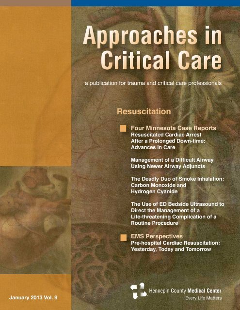
2 Case Reports<br />
Resuscitated Cardiac Arrest after a Prolonged Down-time: Advances in Care<br />
Steve Smith, MD and Brian Mahoney, MD<br />
6 Management of a Difficult Airway Using Newer Airway Adjuncts<br />
Emily Ragaini, MD<br />
8 A Memorable Case of Hypothermia<br />
Ernie Ruiz, MD<br />
9 The Deadly Duo of Smoke Inhalation: Carbon Monoxide and Hydrogen Cyanide<br />
Ben Orozco, MD<br />
12 The Use of ED Bedside Ultrasound to Direct the Management of a Life-threatening<br />
Complication of a Routine Procedure<br />
Brian Driver, MD<br />
13 EMS Perspectives<br />
Pre-hospital Cardiac Resuscitation: Yesterday, Today and Tomorrow<br />
Robert Ball, BA, EMT-P<br />
16 Calendar of Events<br />
18 News Notes<br />
Events Calendar Editor<br />
Susan Altmann<br />
To submit an article<br />
Contact the managing editor at approaches@hcmed.org. The editors reserve the right to reject the editorial<br />
or scientific materials for publication in Approaches in Critical Care. The views expressed in this journal do<br />
not necessarily represent those of <strong>Hennepin</strong> <strong>County</strong> <strong>Medical</strong> <strong>Center</strong>, or its staff members.<br />
Copyright<br />
Copyright 2013, <strong>Hennepin</strong> <strong>County</strong> <strong>Medical</strong> <strong>Center</strong>. Approaches in Critical Care is published twice per year by<br />
<strong>Hennepin</strong> <strong>County</strong> <strong>Medical</strong> <strong>Center</strong>, 701 Park Avenue, Minneapolis, Minnesota 55415.<br />
Subscriptions<br />
To subscribe, send an email to approaches@hcmed.org with your name and full mailing address.<br />
Approaches in Critical Care | January 2013 | 1
Case Reports<br />
Resuscitation: Four Case Reports<br />
“...the medical<br />
community<br />
did not widely<br />
recognize and<br />
promote<br />
artificial<br />
respiration<br />
combined<br />
with chest<br />
compressions<br />
as a key part of<br />
resuscitation<br />
following<br />
cardiac arrest<br />
until the middle<br />
of the 20th<br />
century.”<br />
The effectiveness of cardiopulmonary<br />
resuscitation, (CPR) is often seen in<br />
movies and television as a highly effective<br />
way to save the life of someone who is not<br />
breathing. In fact, a 1996 study published<br />
in the New England Journal of Medicine<br />
showed that the CPR success rate in TV<br />
shows was 75% for immediate circulation,<br />
and 67% survival to discharge. 1 This gives<br />
the general public an unrealistic expectation<br />
of a successful outcome when, on average,<br />
the actual survival rates for a patient who<br />
gets CPR after a cardiac arrest is 5-10<br />
percent. Where CPR is followed by<br />
defibrillation, within three to five minutes of<br />
VF cardiac arrest, survival rates rise to<br />
about 30 percent. 2<br />
The case reports in this issue are more in<br />
line with the reality of how lives are saved<br />
in our emergency departments. In addition,<br />
the history and improving technology of<br />
pre-hospital resuscitation is also detailed in<br />
the Emergency <strong>Medical</strong> Perspectives<br />
feature that follows the case studies.<br />
Amazing stuff when you consider that the<br />
medical community did not widely recognize<br />
and promote artificial respiration combined<br />
with chest compressions as a key part of<br />
resuscitation following cardiac arrest until<br />
the middle of the 20th century.<br />
The “ABC’s of Resuscitation” was written<br />
by Peter Safar in 1957 and CPR was first<br />
promoted as a technique for the public to<br />
learn in the 1970s. Ernie Ruiz, MD, a<br />
founding member of Emergency Services<br />
at <strong>HCMC</strong>, considers this one of the most<br />
significant advances in resuscitation during<br />
his tenure as a resuscitation surgeon and<br />
emergency physician.<br />
“These techniques were applied to all<br />
forms of resuscitation in the 1960s. In my<br />
opinion, we should also look to advancements<br />
in fiber optic instrumentation that enabled<br />
quicker and safer airway management and<br />
advances in pre-hospital care and emergency<br />
care coordination in urban and rural areas.<br />
In addition, there have been advances in<br />
medical imaging, CT, MRI and transvascular<br />
techniques that are used to<br />
discover and repair conditions such as<br />
coronary occlusions and cerebral aneurysms.<br />
Not to mention, the digital age in general.”<br />
The cases that follow are examples.<br />
References<br />
1. (Diem, S.j.; Diem, Susan J MD; Lantos, John D<br />
MD; Tulsky, James A MD (1996-06-13).<br />
“Cardiopulmonary Resuscitation on Television-<br />
Miracles and Misinformation”)<br />
2. Cardiopulmonary Resuscitation Statistics<br />
(http://www.americanheart.org)<br />
Resuscitated Cardiac Arrest after<br />
a Prolonged Down-time: Advances<br />
in Care<br />
Steve Smith, MD and Brian Mahoney, MD<br />
Department of Emergency Medicine<br />
<strong>Hennepin</strong> <strong>County</strong> <strong>Medical</strong> <strong>Center</strong> <br />
Abstract<br />
Cardiac arrest remains a major cause of<br />
mortality in the US. However, new<br />
advances in out-of-hospital, emergency<br />
department, and intensive care have<br />
provided hope for improved outcomes.<br />
Here we describe a case of a patient who<br />
benefited from new technologies that were<br />
applied to all aspects of his acute care.<br />
Case Report <br />
An athletic male in his 40s was biking on a<br />
trail when bystanders witnessed him go<br />
down but, reportedly, without significant<br />
trauma. They found him unresponsive and<br />
without a pulse. They started chest<br />
compressions and called 911. First<br />
responders and paramedics were dispatched<br />
at T = 1 minute. The subsequent events are<br />
as follows:<br />
T = 5 minutes: Minneapolis Fire<br />
Department (MPD, first responders)<br />
arrived. They continued CPR, placed a<br />
King airway, with the ResQPod ® (Inspiratory<br />
Threshold Device [ITD]) applied between<br />
King and Valve-Mask. Respirations were<br />
delivered at 10 per minute, as guided by<br />
the flashing light on the ITD. An automatic<br />
2 | Approaches in Critical Care | January 2013
Case Reports<br />
external defibrillator (AED) was applied, which<br />
advised “shock.” They delivered one shock and<br />
continuous chest compressions were resumed.<br />
T = 9 minutes: EMS (paramedics) arrived and found<br />
the patient in full arrest, the King airway working and<br />
MPD performing manual chest compressions.<br />
T = 10 minutes: The LUCAS device was placed and<br />
automated chest compressions were begun.<br />
T = 13 minutes: The patient’s rhythm was checked<br />
and found to be ventricular fibrillation. Biphasic<br />
defibrillation at 150 Joules was administered and<br />
LUCAS compressions were continued.<br />
T = 14 minutes: IV access was obtained while chest<br />
compressions continued.<br />
T = 15 minutes: Epinephrine 1 mg IV was given.<br />
T = 16 minutes: Defibrillation at 180 Joules was<br />
performed and CPR resumed.<br />
T = 20 minutes: Defibrillation was repeated at 180<br />
Joules and CPR resumed.<br />
T = 24 minutes: Defibrillation at 200 Joules was<br />
performed and CPR resumed.<br />
T = 25 minutes: A second dose of Epinephrine 1 mg<br />
IV was given.<br />
T = 27 minutes: Defibrillation at 200 Joules was<br />
performed with conversion of VF to sinus tachycardia.<br />
Thus, the patient had at least 27 minutes without a<br />
perfusing rhythm. He was then transported.<br />
T = 31 minutes: He arrived at the emergency<br />
department at <strong>Hennepin</strong> <strong>County</strong> <strong>Medical</strong> <strong>Center</strong>.<br />
Figure One. The ED ECG showing an ST- segment elevation<br />
Myocardial infarction<br />
The emergency department ECG (Figure One) was<br />
recorded at T = 35 minutes and showed massive ST<br />
elevation in the anterior leads. The catheterization lab<br />
was activated at T = 36 minutes (5 minutes after<br />
patient arrival in the emergency department). The<br />
King airway was removed and the patient was<br />
orotracheally intubated. As the patient was no longer<br />
in arrest, the ITD was removed. At this point, his<br />
pulse was 120 bpm and BP 130/80. On further<br />
examination, his pupils were noted to be equal and<br />
reactive to light, but the patient was completely<br />
comatose with no response to pain. A chest X-ray<br />
showed mild pulmonary edema. An emergency<br />
department bedside ultrasound showed the expected<br />
anterior wall motion abnormality and moderately<br />
decreased left ventricular function, consistent with<br />
acute anterior STEMI. The potassium was 4.5 mEq/L<br />
and the total CO2 was 12 mEq/L; thus, there was<br />
only mild acidosis.<br />
While waiting for the cath team, therapeutic<br />
hypothermia was begun. A Thermistor Foley was<br />
placed for temperature monitoring and feedback to<br />
the temperature control device, which in this case<br />
was to be the Arctic Sun external cooling system.<br />
The patient was given 10 mg vecuronium to prevent<br />
shivering. A propofol 40 mg bolus and drip were<br />
started in case sedation was necessary, after which<br />
the patient became mildly hypotensive to 88 systolic;<br />
propofol was discontinued and lorazepam 4 mg was<br />
given instead. To help prevent recurrent ventricular<br />
fibrillation, 2 g of magnesium was given, as well as a<br />
lidocaine load (100 mg IV bolus, then another 50 mg<br />
5 minutes later, then an additional 50 mg 5 minutes<br />
after the second dose, for a total of 200 mg) and a<br />
lidocaine drip at 3 mg/minute was begun. Amiodarone<br />
was intentionally avoided due to its beta blocking<br />
properties; patients with large anterior MI are at high<br />
risk of cardiogenic shock, especially when there is<br />
tachycardia. The patient received an IV load of<br />
heparin (5000 U), a rectal ASA, and 600 mg of<br />
clopidogrel (Plavix ® ) through an orogastric tube.<br />
Eptifibatide and Heparin boluses and drips were<br />
administered. The Arctic Sun cooling pads were<br />
applied and cooling was initiated, with a goal<br />
temperature of 33.5 degrees Celsius.<br />
At T = 69 minutes, the patient left the emergency<br />
department for the cath lab.<br />
At T = 73 minutes, the patient arrived in the cath lab.<br />
At T = 83 minutes, angiography of the left coronary<br />
system identified the culprit lesion in the mid left<br />
anterior descending coronary artery (LAD).<br />
At T = 87 minutes, the wire crossed the occlusion in<br />
the mid-LAD and flow was restored, for a door-toballoon<br />
time of 56 minutes.<br />
Thrombus in the LAD was suctioned out. Ostial<br />
stenosis of the first diagonal artery was angioplastied<br />
Approaches in Critical Care | January 2013 | 3
Case Reports<br />
Figure Two. The cardiac monitor strip showing a wide complex<br />
tachycardia, likely AIVR<br />
Figure Three. The Inspiratory<br />
Threshold Device<br />
Figure Four. The ResQPump ®<br />
and stents were placed at each lesion. Following<br />
PCI, an intra-aortic balloon pump was placed due to<br />
cardiogenic shock with elevated left ventricular enddiastolic<br />
pressure and resulting pulmonary edema.<br />
(This has since been shown to be ineffective in<br />
cardiogenic shock in STEMI.) 1<br />
The patient developed an intermittent wide complex<br />
rhythm which at first was interpreted as ventricular<br />
tachycardia (VT) (Figure Two). He was started briefly<br />
on amiodarone and it was subsequently stopped<br />
when the rhythm was reassessed. The diagnosis of<br />
VT was not certain because of a relatively slow heart<br />
rate. Instead, an accelerated idioventricular rhythm<br />
(AIVR) was considered. AIVR is differentiated from<br />
ventricular tachycardia by the heart rate: AIVR is < 120<br />
bpm, while ventricular tachycardia is almost always ><br />
120 bpm. AIVR is a common reperfusion dysrhythmia<br />
and is good evidence of reperfusion of STEMI (in<br />
other words, a good sign). It does not require any<br />
treatment unless associated with hypotension.<br />
By 24 hours, the balloon pump could be removed.<br />
Serial troponin I peaked at 13.7 ng/ml, which is far<br />
lower than expected for a STEMI of this size and<br />
suggests that total occlusion time was brief. Initial<br />
transthoracic echocardiogram on day 2 showed an<br />
ejection fraction (EF) of 15-20% (normal, 65-70%);<br />
with septal and apical wall motion abnormalities<br />
(WMA). Thus, as is usual, the myocardium was<br />
“stunned,” though not all infarcted. The EF greatly<br />
improved by 2 days later, when a repeated<br />
echocardiogram revealed an EF 35% and<br />
corresponding improvement in WMA.<br />
At 48 hours, the patient had completed the<br />
hypothermia protocol without complication and was<br />
extubated, at which time he was following simple<br />
commands but had anterograde amnesia. By 72<br />
hours, his neurological status had greatly improved;<br />
anterograde amnesia was resolving but there was no<br />
memory of events. He recovered fully and was<br />
discharged on prasugrel, aspirin, lisinopril, carvedilol,<br />
atorvastatin, and eplerenone. After cardiac<br />
rehabilitation, his ejection fraction returned to normal<br />
and he was again able to bike 50 miles at a time. He<br />
has had no further symptoms.<br />
Discussion<br />
More information on Compression Decompression<br />
CPR, the Inspiratory Threshold Device, and the<br />
LUCAS device is available at http://www.naph.org/<br />
Homepage-Sections/Explore/Innovations/Heart-<br />
Health/<strong>Hennepin</strong>-<strong>County</strong>-<strong>Medical</strong>-<strong>Center</strong>.aspx.<br />
In January 2011, investigators, including several from<br />
<strong>HCMC</strong>, published the results of a randomized trial<br />
comparing compression-decompression CPR to<br />
standard CPR in out-of-hospital cardiac arrest, and<br />
found that 74 (9%) of 840 patients survived to 1 year<br />
in the intervention group compared with 48 (6%) of<br />
813 controls (p=0·03), with equivalent cognitive skills,<br />
disability ratings, and emotional-psychological status<br />
in both groups. 2 In the compression-decompression<br />
group, first responders used both the ITD (ResQPod ® ),<br />
(Figure Three) and a specially designed compressiondecompression<br />
device called the ResQPump ®<br />
(Figure Four). The ResQPump ® has a suction cup<br />
that attaches to the chest, and when pulled up<br />
forcefully, may create 30 pounds of negative<br />
pressure in the chest, augmenting the pumping<br />
action of the chest. The ITD is placed on the<br />
endotracheal tube and has a valve that stays closed<br />
for a fraction of a second, preventing that negative<br />
pressure from drawing air down the endotracheal<br />
tube, ensuring that there will be negative pressure to<br />
draw blood into the chest and into the heart, for<br />
further pumping out to the body with each<br />
compression. The two together, then, increased<br />
survival with good neurologic outcome (Modified<br />
Rankin Score of 3 or less) by 50%.<br />
The LUCAS device (Figure Five) combines<br />
mechanical compression that is uniform and does not<br />
fatigue with suction for decompression, but with less<br />
suction. It provides 2 inches of chest compression at<br />
4 | Approaches in Critical Care | January 2013
Case Reports<br />
Figure Five.<br />
The LUCAS device<br />
several pounds of negative force. Newer software<br />
automatically adjusts the plunger to ensure good<br />
contact with the sternum. It is unlike human CPR,<br />
which degrades due to fatigue or distraction, and is<br />
often done at a rate too slow or too fast for optimal<br />
blood flow, does not allow for full chest wall recoil as<br />
the provider tends to lean on the chest, and must be<br />
stopped during administration of shocks. It has been<br />
shown to improve blood flow in experimental models<br />
and anecdotal reports in humans. We have found in<br />
some cases that it creates exceptional blood<br />
pressure in ED cardiac arrest patients, as measured<br />
by arterial line; and it is more likely than manual<br />
chest compression to keep the brain perfused (and<br />
therefore alive) in prolonged arrest. The LUCAS<br />
device also allows safe transport of the patient with<br />
ongoing CPR, thus making it possible to go to the<br />
cath lab with ongoing CPR.<br />
Therapeutic Hypothermia<br />
In 2002, 2 randomized studies in the New England<br />
Journal of Medicine compared therapeutic cooling for<br />
24 hours to standard care. 3, 4 These trials, and<br />
evidence from dog studies, form the basis of this now<br />
standard therapy for comatose survivors of cardiac<br />
arrest. In these studies, patients who were<br />
resuscitated from pulseless ventricular fibrillation<br />
(VF) or tachycardia (VT) were randomized within 3<br />
hours, and those who underwent hypothermia were<br />
sedated, chemically paralyzed, and externally cooled<br />
to a target temperature of 32 to 34 degrees Celsius,<br />
where they were kept for 24 hours before rewarming.<br />
Only 8% of cardiac arrest patients met eligibility<br />
criteria. The primary endpoint was good neurologic<br />
outcome at 6 months, which, when combining the 2<br />
studies, was achieved in the hypothermia group in<br />
54% vs. 37%. Mortality was 43% vs. 58%. Adverse<br />
events were not different between the groups.<br />
Based on this, the International Liaison Committee<br />
on Resuscitation advised use of therapeutic<br />
hypothermia, to 32-34 degrees for 12 to 24 hours, for<br />
unconscious adult patients with return of spontaneous<br />
circulation after out-of-hospital ventricular fibrillation<br />
arrest. 5 Therapeutic hypothermia requires intubation<br />
and paralysis and very intensive monitoring for<br />
electrolyte shifts and dysrhythmias.<br />
Conclusion<br />
Since 2003, we at <strong>Hennepin</strong> <strong>County</strong> <strong>Medical</strong> <strong>Center</strong><br />
have cooled comatose survivors of cardiac arrest.<br />
We have decided to broaden the indications for the<br />
therapy beyond those with VF or VT to those patients<br />
who have been resuscitated from asystole, pulseless<br />
electrical activity (PEA) and respiratory etiologies of<br />
cardiac arrest, as long as they have return of<br />
spontaneous circulation within 60 minutes of arrest.<br />
From January 2008 through December 2011,<br />
<strong>Hennepin</strong> had 129 patients resuscitated after cardiac<br />
arrests who were eligible for and underwent<br />
therapeutic hypothermia: 86 had VF or VT, and 43<br />
had PEA or asystole. Of the 129, 61 (47%) survived<br />
with good neurologic outcome; 6 of these had PEA or<br />
asystole as the initial rhythm. In total, 57 (44%) died,<br />
35 of whom had asystole or PEA. There were 11<br />
survivors, but with poor neurologic outcomes; 2 of<br />
these had asystole or PEA. The 56 of 86 (65%) with<br />
a shockable rhythm survived with good neurologic<br />
outcome. Most of these were before the use of the<br />
LUCAS device.<br />
This case and our survival statistics illustrate the<br />
rapid advancements occurring in the management of<br />
patients with cardiac arrest. Combining new therapies,<br />
such as those described in this case report, has the<br />
potential to improve survival even more. <br />
References<br />
1. Thiele H, Zeymer U, Neumann FJ, et al. Intraaortic balloon<br />
support for myocardial infarction with cardiogenic shock. N Engl J<br />
Med 2012; 367(14):1287-96.<br />
2. Aufderheide TP, Frascone RJ, Wayne MA, et al. Standard<br />
cardiopulmonary resuscitation versus active compressiondecompression<br />
cardiopulmonary resuscitation with augmentation<br />
of negative intrathoracic pressure for out-of-hospital cardiac arrest:<br />
a randomised trial. Lancet 2011;377(9762):301-11.<br />
3. Bernard SA, Gray TW, Buist MD, et al. Treatment of comatose<br />
survivors of out-of-hospital cardiac arrest with induced<br />
hypothermia. N Engl J Med 2002; 346(8):557-63.<br />
4. The_Hypothermia_after_Cardiac_Arrest_Study_Group. Mild<br />
therapeutic hypothermia to improve the neurologic outcome after<br />
cardiac arrest. N Engl J Med 2002; 346(8):549-56.<br />
5. Nolan JP, Morley PT, Hoek TL, Hickey RW. Therapeutic<br />
hypothermia after cardiac arrest. An advisory statement by the<br />
Advancement Life support Task Force of the International Liaison<br />
committee on Resuscitation. Resuscitation 2003;57(3):231-5.<br />
Approaches in Critical Care | January 2013 | 5
Case Reports<br />
Management of a Difficult Airway Using<br />
Newer Airway Adjuncts<br />
Emily Ragaini, MD<br />
Departments of Emergency Medicine<br />
<strong>Hennepin</strong> <strong>County</strong> <strong>Medical</strong> <strong>Center</strong><br />
Abstract<br />
Angioedema is a well documented side effect of<br />
angiotensin converter enzyme (ACE) inhibitors.<br />
Cases of severe angioedema can present challenges<br />
in acute airway management. We describe the use of<br />
several airway adjuncts in the management of a case<br />
of severe angioedema in a morbidly obese patient.<br />
Case Report<br />
A 37-year-old morbidly obese female with a history of<br />
hypertension was at home when she began to notice<br />
swelling of her tongue. Initially, the swelling was<br />
minimal, but over the next 2 hours progressed until<br />
her tongue filled her mouth and it was difficult for her<br />
to speak. A family member called 911 and paramedics<br />
were dispatched. They gave her 50 mg IV Benadryl<br />
en route to the hospital. She did not realize she was<br />
on a combination antihypertensive pill containing<br />
amlodipine and benazepril.<br />
At T = 0 minutes, the patient arrived in the<br />
emergency department at <strong>Hennepin</strong> <strong>County</strong> <strong>Medical</strong><br />
<strong>Center</strong>. On initial examination, the patient was<br />
observed to be breathing through her nose. Her<br />
tongue was swollen and firm and filled the<br />
oropharynx. The submental area was also swollen<br />
and protruding over her lower teeth. She was mildly<br />
tachypnic and sitting upright on cart. There was<br />
firmness of her submandibular area. She had no<br />
stridor or wheezing. Her emergency department<br />
management proceeded as follows:<br />
T = 4 minutes: She was given IV solumedrol, IV<br />
Zantac, IM epinephrine without improvement. Given<br />
the rapidly progressing tongue swelling, we<br />
recommended intubation and the patient was in<br />
agreement with this.<br />
T = 27 minutes: An initial attempt at fiberoptic<br />
guided nasotracheal intubation was made after IV<br />
ketamine was given for sedation. Despite multiple<br />
doses of sedation, the patient remained very agitated<br />
and unable to be intubated.<br />
T = 49 minutes: She was given IV etomidate and<br />
blind nasotracheal intubation was attempted. However,<br />
she remained too agitated and nearly threw herself<br />
off the cart during the final attempt. She was<br />
repositioned on the cart while being bagged. It was<br />
noted on the cardiac monitor that she had a run of<br />
bigeminy, then a short run of ventricular tachycardia.<br />
T = 56 minutes: A crichothyrotomy tray was opened<br />
at the bedside. Rapid sequence intubation was<br />
attempted after the patient was given IV etomidate<br />
and succinylcholine. This attempt used the C-MAC<br />
videolaryngoscope with a Macintosh size 4 blade.<br />
However, because of the massive tongue swelling,<br />
we were not able to pass a blade deep enough to<br />
visualize the vocal cords. Her oxygen saturation<br />
dropped into the 80%s on pulse oximetry (POX).<br />
An intubating laryngeal mask airway (ILMA) was<br />
placed and bag-value ventilation restored her to a<br />
POX of 100%.<br />
“Angioedema is a well documented side<br />
effect of angiotensin converter enzyme<br />
(ACE) inhibitors. Cases of severe<br />
angioedema can present challenges in<br />
acute airway management.”.”<br />
T = 61 minutes: An attempt to pass an endotracheal<br />
tube (ETT) was made through the ILMA but was not<br />
successful. The ETT was therefore removed. At that<br />
time, her POX was noted to be 83%.<br />
T = 64 minutes: The patient was noted to be<br />
bradycardic with heart rate in the 40s. She was given<br />
0.5 mg IV atropine with a good response in heart rate.<br />
T = 69 minutes: An ETT was passed through ILMA.<br />
She was then easily bagged. However, her POX<br />
dropped into the 70%s, with a nadir of 68%. Fiberoptic<br />
scope confirmed that the ETT was in the trachea, but<br />
appeared to be sitting low. The ETT pulled back.<br />
T = 78 minutes: A CXR showed the ETT to be<br />
positioned about 1 cm above the carina. It was pulled<br />
back 2 cm. Given her low POX, a bedside ultrasound<br />
was performed to rule out pneumothorax; this<br />
showed sliding lung signs present bilaterally.<br />
Etomidate was given for sedation and a propofol<br />
infusion started.<br />
T = 97 minutes: Her SpO2 improved to only 89%<br />
despite adequate bagging and 100% FiO2. The<br />
decision was made to remove the ILMA from around<br />
ETT. During the attempt to remove ILMA using the<br />
stabilizer rod, the ETT pilot balloon tubing became<br />
caught and snapped, causing the ETT cuff to deflate,<br />
and the ETT became dislodged.<br />
T = 99 minutes: At this point, the patient’s neck was<br />
re-prepped for possible crichothyrotomy and the<br />
ILMA was replaced.<br />
T = 107 minutes: An ETT was passed through ILMA<br />
and the ILMA was removed with ETT in place.<br />
T = 116 minutes: A CXR confirmed good placement<br />
of the ETT.<br />
6 | Approaches in Critical Care | January 2013
Case Reports<br />
Figure One. The C-MAC videolaryngoscope. The camera is fixed<br />
in the handle, with the lens within the blade. Images are projected<br />
onto a video screen that allows others besides the intubator to<br />
view the anatomy during the intubation procedure. Its usefulness<br />
is demonstrated in a case of difficult boogie placement, seen at<br />
http://www.hqmeded.com/video/40879557<br />
invaginated scolex is clearly seen inside of the cystic cavity.<br />
T = 124 minutes: She began to cough against the<br />
vent and was, therefore, paralyzed with vecuronium.<br />
An ABG revealed a mild acidosis with a pH of 7.19.<br />
Her EKG showed sinus tachycardia with lateral<br />
t wave inversions, concerning for demand ischemia.<br />
T = 164 minutes: The patient was taken to CT<br />
scanner for a CT of the neck with IV contrast to rule<br />
out Ludwig’s angina. Her CT showed a massively<br />
swollen tongue filling the airway and protruding from<br />
the mouth, with significant narrowing of the airway.<br />
There was only mild swelling of the sublingual and<br />
submandibular areas, suggesting that angioedema<br />
was more likely the cause of swelling than a soft<br />
tissue abscess.<br />
T = 184 minutes: She was transferred to the MICU<br />
with a secure airway.<br />
The patient’s benazepril was discontinued. Serial<br />
troponins were followed, given her abnormal ECG<br />
post -intubation and the witnessed episodes of<br />
bigeminy and ventricular tachycardia during<br />
intubation attempts. Her troponin peaked at 0.193<br />
ng/mL. Transthoracic echocardiogram showed a<br />
normal left ventricular ejection fraction, but she was<br />
noted to have an area of hypokinesis in the mid<br />
portion of the intraventricular septum, concerning for<br />
atypical stress cardiomyopathy.<br />
On hospital day 2, the patient had significant<br />
improvement in her tongue swelling. Given her<br />
extremely difficult intubation in the emergency<br />
department, she was extubated over an exchange<br />
catheter with anesthesia at the bedside. She<br />
tolerated extubation well. She was transferred to the<br />
medicine floor and discharged the next day. She was<br />
instructed to discard her amlodipine- benazepril<br />
combination pills at home, and given a new<br />
prescription for amlodipine alone. She was advised to<br />
avoid ACE inhibitors and an allergy alert was placed<br />
in her chart.<br />
Discussion<br />
Two important airway adjuncts were used in the<br />
management of this difficult airway. The C-MAC<br />
videolaryngoscope (Figure One) uses a modified<br />
Macintosh laryngoscope blade with an integrated<br />
video camera, which is directed toward the blade tip.<br />
More details of its functions and an example of its<br />
usefulness are illustrated in a video that shows<br />
difficulty passing a boogie. See http://www.hqmeded.<br />
com/video/40879557. The video screen image allows<br />
the attending physician to visualize the airway as the<br />
resident is intubating (see video). It also allows for<br />
recording of the intubation to facilitate image review<br />
and teaching opportunities. Studies have demonstrated<br />
a greater proportion of successful intubations and a<br />
greater percentage of Cormack-Lehane grade I or II<br />
views when compared with direct laryngoscopy.<br />
Figure Two. The intubating laryngeal mask airway (ILMA). The<br />
procedure for its placement can be viewed at http://www.hqmeded.<br />
com/video/13164204.<br />
The ILMA (Figure Two) is a supraglottic airway device<br />
that is useful for difficult airway management. The<br />
Approaches in Critical Care | January 2013 | 7
Case Reports<br />
laryngeal mask airway (LMA) was developed in the<br />
1980s and initially used in the operating room as an<br />
alternative to bag-valve-mask (BVM) ventilation. It<br />
has become a popular airway adjunct in the<br />
emergency setting for the management of difficult<br />
airways. The intubating LMA (ILMA) was developed<br />
in the late 1990s and allows an endotracheal tube to<br />
be passed through the LMA.<br />
Placement of the ILMA is a simple, single operator<br />
procedure: (http://www.hqmeded.com/video/<br />
13164204). The ILMA cuff is checked for leaks. The<br />
cuff is then deflated against a flat surface. A water<br />
soluble lubricant should be applied to the posterior<br />
surface of the mask. The ILMA is inserted, while<br />
holding the handle, into the oropharynx against the<br />
hard palate, with the mask opening toward the<br />
tongue. It is advanced until resistance is met, and the<br />
cuff then inflated. The handle can be held like a<br />
skillet, and lifted toward the ceiling, to insure an<br />
adequate seal of the mask. The BVM is directly<br />
attached to the ILMA. Either an ETT or the Fastrach<br />
Silicone Tube (FTST, a tube specially designed for<br />
intubation through the ILMA) can be used. At<br />
<strong>Hennepin</strong>, a standard ETT is often used. Placement<br />
of this is facilitated by inserting the tube with the<br />
curvature opposite of how it is held during standard<br />
intubation. A stabilizer rod is used to maintain the<br />
ETT in position, while the ILMA is deflated and<br />
removed from around the ETT. McGill forceps can<br />
also be used to stabilize the ETT in the oropharynx<br />
as the ILMA is removed.<br />
As illustrated in this case report, the ILMA is an<br />
easily placed airway adjunct that can be used in the<br />
management of difficult airway in the emergency<br />
department. An important learning point from this<br />
case is ensuring that the stabilizer rod is removed<br />
temporarily to allow passage of the pilot balloon for<br />
the ETT. On the first attempt at removal of the ILMA<br />
around the ETT, this was not done, and ultimately<br />
resulted in dislodgement of the ETT. On the subsequent<br />
attempt, this was performed and the ILMA was<br />
removed from around the ETT without difficulty.<br />
Overall, the use of the ILMA was instrumental in<br />
avoiding a surgical airway in this patient. <br />
A Memorable Case of Hypothermia<br />
by Ernie Ruiz, MD<br />
Ernie Ruiz, MD<br />
Founding member of<br />
Emergency Services<br />
<strong>Hennepin</strong> <strong>County</strong> <strong>Medical</strong> <strong>Center</strong><br />
A young woman was found by police laying on the<br />
sidewalk of a Minneapolis city street on a very cold<br />
morning in midwinter in 1975-1976. She was cold<br />
and appeared dead. The city's morgue vehicle was<br />
summoned. While awaiting the van, the woman took<br />
a breath. The ambulance was summoned and the<br />
patient was delivered to the <strong>Hennepin</strong> Emergency<br />
Department. She was without a pulse as she was<br />
transferred to the resuscitation cart. An ECG monitor<br />
showed ventricular fibrillation. While the patient was<br />
being prepared for resuscitation, I called Dr. John<br />
Haglin, Head of Cardiovascular Surgery (CVP),<br />
because during my surgery training he and I<br />
discussed the possibility of using a heart lung<br />
machine to warm very cold patients. Dr. Haglin<br />
agreed that this patient was a good candidate. He<br />
called Dr. Per Wickstrom, who was his Chief<br />
Resident on CVP, and the patient was moved to the<br />
operating room. On the heart lung machine, the<br />
patient warmed to normal temperature in just a few<br />
minutes. She was able to go home in a few days.<br />
She was normal again.<br />
This was the first time that this method of re-warming<br />
was used and reported. It quickly became the world<br />
standard for severe hypothermia resuscitation. This<br />
was a world's first for <strong>Hennepin</strong>. <br />
8 | Approaches in Critical Care | January 2013
Case Reports<br />
The Deadly Duo of Smoke Inhalation: Carbon<br />
Monoxide and Hydrogen Cyanide<br />
Ben Orozco, MD and Jon Cole, MD<br />
Departments of Emergency Medicine<br />
<strong>Hennepin</strong> <strong>County</strong> <strong>Medical</strong> <strong>Center</strong><br />
Abstract<br />
Persons found down in house fires are subject to<br />
varied mechanisms of injury, making them among the<br />
most critically ill of patients. Patients are subject to<br />
thermal injury at dermal, mucosal, and respiratory<br />
surfaces. The gases produced by combustion pose<br />
thermal and particulate damage to airways, but also<br />
may expose the patient to a hypoxic atmosphere with<br />
carbon monoxide (CO) and hydrogen cyanide gases<br />
(HCN). This case report and review of acute carbon<br />
monoxide and hydrogen cyanide gas poisoning from<br />
structure fires should help clinicians recognize these<br />
similar, but distinctly treated toxicities.<br />
Case Report<br />
A 27-year-old female rescued from an active house<br />
fire was initially unconscious and hypoxic with a<br />
Sp02 in the 40s. Paramedics intubated her on scene<br />
and end tidal C02 was 76mmHg. She was given 5mg<br />
of midazolam for sedation and transported.<br />
Upon arrival to the emergency department, vitals<br />
were SpO2 of 90% on 100% FiO2, pulse 139, and<br />
blood pressure of 180/63 mmHg. The position of the<br />
endotracheal tube was confirmed with auscultation<br />
and an end tidal CO2 waveform. The primary survey<br />
was remarkable for diffuse rhonchi. IV hydration with<br />
Ringer's Lactate solution was begun through large<br />
bore IV access. The burn team was present shortly<br />
after patient arrival. The secondary survey revealed<br />
sluggish 2mm pupils, minimal response to pain,<br />
carbonaceous sputum, and a 9% body surface area<br />
partial thickness burn to the abdomen. Chest X-ray<br />
showed pulmonary infiltrates. The patient was<br />
sedated with propofol and paralyzed. Co-oximetry<br />
gave a markedly elevated carboxyhemoglobin<br />
(COHb) level and labs returned with a measured<br />
COHb of 41%. Arterial pH was 6.8 with a marked<br />
metabolic acidosis. Ventilator settings were<br />
advanced, and hydroxocobalamin (Cyanokit ® ) was<br />
started for presumed cyanide poisoning with 12.5g<br />
of sodium thiosulfate to follow. Of note, the serum<br />
lactate later resulted at 18mmol/L.<br />
Arterial and central lines were placed and<br />
myringotomies performed. The patient was<br />
transferred to the hyperbaric oxygen (HBO) chamber<br />
where she was treated with a 90 minute 2.4<br />
atmosphere dive by a hyperbaracist. At admission to<br />
the burn unit, the patient was able to follow commands<br />
with each extremity and open her eyes to voice. Her<br />
SpO2 was 90% on 60% FIO2 and her HR 98 bpm,<br />
and BP 179/103. Her serum lactate had fallen to<br />
3.8mmol/L and pH had improved to 7.3 with serum<br />
bicarbonate of 24. Over the next two days, her<br />
pulmonary status worsened. She had bronchoscopy<br />
with lavage and advanced ventilator management<br />
with pulmonology consultation. She had a tracheostomy<br />
placed on hospital day three. Her hospital course<br />
was complicated by pneumonia, and underlying<br />
asthma. Eventually antibiotics, vasopressors, and the<br />
ventilator were weaned and withdrawn. She received<br />
daily wound care, physical and occupational therapy,<br />
and was discharged neurologically intact without<br />
a tracheostomy.<br />
Discussion<br />
Carbon Monoxide Poisoning<br />
Carbon monoxide (CO) is produced in fires as result<br />
of incomplete combustion and is colorless and<br />
odorless. CO levels within structure fires routinely<br />
exceed the immediately-dangerous-to-life-and-health<br />
(IDLH) standard of 1200 parts per million, as set by<br />
the US National Institute for Occupational Safety and<br />
Health (NIOSH). Once inhaled, CO binds hemoglobin<br />
with 250 times the potency of oxygen, creating<br />
carboxyhemoglobin (COHb), which shifts the<br />
hemoglobin desaturation curve to the left, impairing<br />
both oxygen content and delivery. CO also binds<br />
skeletal and cardiac myoglobin. Other significant<br />
mechanisms include protein oxidation, lipid<br />
peroxidation, neutrophil adhesion, vasodilation, and<br />
inhibition of cytochrome oxidase. CO persists with a<br />
half-life of approximately 250-300 minutes while<br />
breathing room air.<br />
Based on these mechanisms, patients may<br />
experience profound tissue hypoxia despite normal<br />
ambient oxygen. Symptoms depend on the level of<br />
exposure and functional status of the patient, but<br />
symptoms such as nausea, vomiting, and headache<br />
typify exposures with COHb levels of 15-20%. Severe<br />
toxicity may produce syncope, seizures, myocardial<br />
ischemia, cerebral infarction, and death. Such<br />
patients may present with chest pain, arrhythmia,<br />
positive biomarkers, shortness of breath, confusion,<br />
focal neurologic signs, or be comatose. COHb levels<br />
over 40% are generally associated with severe<br />
toxicity. Neurologic symptoms may be permanent.<br />
All victims of structure fires are at risk of CO<br />
poisoning. Routine pulse oximetry may be falsely<br />
Approaches in Critical Care | January 2013 | 9
Case Reports<br />
elevated and the skin may look pink. Screening<br />
should begin with co-oximetry, when available.<br />
Measured COHb should be performed in all cases<br />
where there is significant exposure or clinical symptoms.<br />
Cardiac monitoring and a 12 lead electrocardiogram<br />
(ECG) are mandatory in patients with chest pain,<br />
shortness of breath, comorbidities, or significant<br />
COHb levels. In addition to resuscitation and airway<br />
management, the mainstay of treatment for CO<br />
poisoning is oxygen. Also, 100% oxygen should be<br />
delivered via a tight fitting, non-rebreather mask or<br />
endotracheal tube as soon as possible, reducing the<br />
half-life of COHb from 300 minutes on room air to<br />
approximately 60 minutes. Myocardial depression<br />
and vasodilation may cause hypotension, needing<br />
treatment with fluids, vasopressors, and/or inotropes.<br />
should be placed on clinical findings suggesting<br />
severe toxicity rather than simple CO levels. The<br />
following indications for HBO are generally used by<br />
the Minnesota Poison Control <strong>Center</strong> and the <strong>Center</strong><br />
for Hyperbaric Medicine at <strong>Hennepin</strong> <strong>County</strong> <strong>Medical</strong><br />
<strong>Center</strong> which is open for emergencies 24/7.<br />
Indications for Hyperbaric Oxygen:<br />
1. History of loss of consciousness<br />
2. Serious toxicity including lethargy, confusion,<br />
seizures, focal neurologic deficit, ischemic chest<br />
pain, new dysrhythmia, ECG changes, hypotension<br />
3. COHb levels > 25% plus cardiovascular disease,<br />
cerebrovascular disease, age > 60years, < 2<br />
years, hemoglobin < 10, or exposure > 24hours<br />
4. COHb > 40%<br />
5. Pregnancy with COHb > 20% or signs of fetal<br />
distress<br />
6. Symptoms refractory to normobaric oxygen<br />
Figure One. The patient was transferred to the hyperbaric oxygen<br />
(HBO) chamber where she was treated with a 90 minute 2.4<br />
atmosphere dive by a hyperbaracist.<br />
Hyperbaric oxygen (HBO) therapy remains the<br />
optimum treatment for the elimination of CO from<br />
the human body and should be performed whenever<br />
available in cases of significant poisoning. The halflife<br />
of CO is reduced to a mere 20 minutes at 2.5<br />
atmospheres of 100% oxygen. The best published<br />
data suggests that HBO reduces cognitive sequelae<br />
of CO poisoning from 46% in the untreated group to<br />
24% in the group treated with HBO. Though practice<br />
will vary by institution, immediate consultation with a<br />
HBO facility is indicated when significant carbon<br />
monoxide poisoning occurs (Figure One). Importance<br />
Hydrogen Cyanide Gas Poisoning<br />
Hydrogen cyanide gas (HCN) is produced during the<br />
combustion of a variety of natural and synthetic<br />
fibers. HCN toxicity is often overlooked in fires as<br />
patients’ symptoms may be attributed to CO. HCN<br />
was detected in 59% of decedents of structure fires,<br />
and 50% of survivors in a recent Polish study. Once<br />
inhaled, HCN distributes to tissues affecting metabolic<br />
enzymes; most importantly, it binds cytochrome<br />
oxidase and halts cellular respiration. Effects vary,<br />
depending on concentration, duration of exposure,<br />
and patient factors; nonetheless, HCN is considered<br />
among the fastest poisons and may rapidly produce<br />
syncope, seizures, and cardiovascular collapse. While<br />
confusion, lethargy, hypertension, and tachycardia<br />
may occur, coma, bradycardia, and hypotension<br />
consistently precede death. It is eliminated with a<br />
variable half-life of 1 hour to > 2 days.<br />
Diagnosis relies on a timely clinical assessment<br />
rather than analytics. Like CO toxicity, HCN toxicity<br />
may result in normal pulse oximetry, and pink skin<br />
despite severe toxicity; moreover, venous oxygen<br />
saturations may be elevated. COHb levels poorly<br />
predict HCN toxicity, but serum lactate > 8mmol/L in<br />
fire victims predicts cyanide levels > 1.0μg/ml with<br />
94% sensitivity and 70% specificity. Hydroxocobalamin<br />
has emerged as the antidote of choice. It has been<br />
safely administered pre-hospital in France since the<br />
10 | Approaches in Critical Care | January 2013
Case Reports<br />
Figure Two: Photographs showing bright red discoloration of the patient's skin (A) and urine (B) after treatment with hydroxocobalamin for<br />
cyanide poisoning. Cescon D W , Juurlink D N. CMAJ 2009; 180:251-251. Used with permission.<br />
invaginated scolex is clearly seen inside of the cystic cavity.<br />
1980s and was FDA approved in the US in 2006.<br />
Hydroxocobalamin is given 5g IV and may be<br />
repeated. It rapidly binds and detoxifies circulating<br />
cyanide anions to form cyanocobalamin (vitamin<br />
B12). Adverse reactions are limited to transient<br />
reddening of the skin and bodily fluids (Figure Two),<br />
minor allergy, transient hypertension, and temporary<br />
interference with colorimetric laboratory tests. Since<br />
routine serum HCN levels are lacking, administration<br />
is empiric.<br />
Suspect and treat HCN toxicity in patients with:<br />
1. Suspected smoke inhalation (carbonaceous<br />
sputum, enclosed fire, etc.)<br />
2. Altered mental status<br />
3. Cardiovascular instability (especially systolic<br />
blood pressure < 90mmHg in adults)<br />
4. Initial serum lactate > 8.0mmol/L<br />
Given the rapid mechanism of action of both HCN<br />
and the antidote, there should be no delay in<br />
antidotal treatment of unstable patients without<br />
laboratory values. After administration of<br />
hydroxocobalamin, clinicians may consider treatment<br />
with sodium thiosulfate (STS) through a separate<br />
line. STS in conjunction with endogenous rhodanase<br />
detoxifies HCN to thiocyanate. STS is a constituent<br />
of the older cyanide antidote kit, which also included<br />
amyl, butyl, and sodium nitrite. The nitrites induce<br />
methemoglobinemia and should be avoided in<br />
patients suffering from concomitant CO toxicity.<br />
should now readily identify that her profound<br />
alteration of mental status, inhalational injury, and<br />
high lactate as nearly diagnostic of cyanide toxicity.<br />
Both conditions warrant immediate treatment. Our<br />
patient was fortunate to have received timely<br />
therapies, including emergency department<br />
resuscitation, airway management, hydroxocobalamin,<br />
sodium thiosulfate, and hyperbaric oxygen. Her<br />
ultimate full recovery would not have been possible<br />
without the continued thorough inpatient treatment<br />
from a multidisciplinary care team in the burn unit. <br />
References<br />
Baud, F. J., S. W. Borron, B. Megarbane, H. Trout, F. Lapostolle,<br />
E. Vicaut, M. Debray, and C. Bismuth. Value of Lactic Acidosis in<br />
the Assessment of the Severity of Acute Cyanide Poisoning. Crit<br />
Care Med 30, No. 9. Sep 2002: 2044-50.<br />
Cone, D. C., D. MacMillan, V. Parwani, and C. Van Gelder. Threats<br />
to Life in Residential Structure Fires. Prehosp Emerg Care 12, No.<br />
3. Jul-Sep 2008: 297-301.<br />
Grabowska, T., R. Skowronek, J. Nowicka, and H. Sybirska.<br />
Prevalence of Hydrogen Cyanide and Carboxyhaemoglobin in<br />
Victims of Smoke Inhalation During Enclosed-Space Fires: A<br />
Combined Toxicological Risk. Clin Toxicol (Phila) 50, No. 8. Sep<br />
2012: 759-63.<br />
Nelson, Lewis, and Lewis R. Goldfrank. Goldfrank's Toxicologic<br />
Emergencies. 9th ed. New York: McGraw-Hill <strong>Medical</strong>, 2011.<br />
O'Brien, D. J., D. W. Walsh, C. M. Terriff, and A. H. Hall. Empiric<br />
Management of Cyanide Toxicity Associated with Smoke Inhalation.<br />
[In Eng]. Prehosp Disaster Med 26, No. 5. Oct 2011: 374-82.<br />
Weaver, L. K., R. O. Hopkins, K. J. Chan, S. Churchill, C. G. Elliott,<br />
T. P. Clemmer, J. F. Orme, Jr., F. O. Thomas, and A. H. Morris.<br />
Hyperbaric Oxygen for Acute Carbon Monoxide Poisoning. N Engl<br />
J Med 347, No. 14. Oct 3 2002: 1057-67.<br />
Conclusion<br />
In our patient, the COHb of 41% is consistent with<br />
severe carbon monoxide toxicity; however, clinicians<br />
Approaches in Critical Care | January 2013 | 11
Case Reports<br />
5<br />
Figure One: ED ultrasound of Morison's pouch, showing free<br />
intraperitoneal fluid<br />
5<br />
Figure Two: ED ultrasound showing free fluid surrounding the bladder<br />
Use of ED Bedside Ultrasound to Direct the<br />
Management of a Life-threatening<br />
Complication of a Routine Procedure<br />
Brian Driver, MD<br />
Departments of Emergency Medicine and Internal Medicine<br />
<strong>Hennepin</strong> <strong>County</strong> <strong>Medical</strong> <strong>Center</strong><br />
Abstract<br />
Ultrasound use in the emergency department has<br />
been shown to be time and cost efficient, and in<br />
many cases, life saving. This case describes its use<br />
to direct the management of an unusual complication<br />
of routine colonoscopy.<br />
Case Report<br />
A 65-year-old woman presented to the emergency<br />
department with altered mental status eleven hours<br />
after a routine screening colonoscopy. She was at<br />
home the evening after the procedure and telephoned<br />
her neighbor for help. When he arrived, he found her<br />
collapsed on the kitchen floor and immediately called<br />
911. Emergency medical services emergently<br />
transported her to a stabilization room.<br />
On arrival, she appeared obtunded, pale, and<br />
critically ill. A bedside ultrasound was immediately<br />
performed revealing a large amount of free fluid in<br />
her peritoneal cavity, instantly diagnostic of an intraabdominal<br />
catastrophe (Figures One and Two).<br />
Blood was called for, fluids where hung, and general<br />
surgery was immediately paged to determine the<br />
need for emergency operative intervention.<br />
The differential diagnosis included, most prominently,<br />
hemoperitoneum from an unknown source of bleeding<br />
and viscus perforation with bowel contents spilling into<br />
the abdomen. An upright chest radiograph failed to<br />
show any pneumoperitoneum, making viscus perforation<br />
less likely. The patient’s mental status, blood pressure,<br />
and heart rate all improved with rapid fluid resuscitation.<br />
As she was now hemodynamically stable, a CT of her<br />
abdomen/pelvis was obtained, demonstrating severe<br />
splenic injury with active extravasation of contrast<br />
(Figure Three). She was taken emergently to the<br />
operating room where she was found to have severe<br />
avulsion of her splenic capsule. A splenectomy was<br />
performed, and she was discharged on post-operative<br />
day 7 with an otherwise uncomplicated hospital course.<br />
Discussion<br />
There are more than 14 million colonoscopies per<br />
year in the United States, with a relatively low overall<br />
complication rate of about 5 per 1,000 procedures.<br />
Splenic injury from a colonoscopy is exceedingly<br />
rare, with only approximately 95 case reports in the<br />
English literature. This case is the first documented<br />
report in which bedside ultrasound was utilized to<br />
facilitate rapid diagnosis and treatment.<br />
This particular injury occurs when excessive force<br />
is applied downward at the splenic flexure during<br />
colonoscopy, exerting traction on the splenocolic<br />
ligament and ultimately the splenic capsule, which<br />
then pulls free from the spleen (Figure Four). This<br />
leaves the splenic parenchyma open to the abdomen,<br />
with resultant, and often severe, bleeding. Usually, 75%<br />
of patients with this injury will present within 24 hours<br />
of colonoscopy. However, this diagnosis of colonoscopy.<br />
5<br />
Figure Three: Abdominal Ct scan showing free fluid and active contrast<br />
extravasation at the injury<br />
12 | Approaches in Critical Care | January 2013
EMS Perspectives<br />
5<br />
Figure Four: Excessive<br />
downward force at the splenic<br />
flexure stretches the splenic<br />
ligament and capsule, which<br />
then pulls off from the spleen<br />
cystic cavity.<br />
However, this diagnosis can present as<br />
late as 10 days post-procedure. Almost all<br />
patients present with abdominal pain, but<br />
only variably with back pain, shoulder pain,<br />
and shock.<br />
Treatment options include splenectomy, as<br />
in our case, but also splenic artery embolization<br />
in select institutions and, if the injury is<br />
small, careful observation in an intensive<br />
care unit.<br />
Conclusion<br />
Bedside ultrasound is crucial to making a<br />
rapid diagnosis of hemoperitoneum in<br />
critically ill patients. It can detect as little as<br />
400 mL and 150 mL of fluid in Morison’s<br />
pouch and the pelvis, respectively.<br />
Ultrasound should be utilized in every<br />
critically ill individual who presents with<br />
unexplained hemodynamic instability to rule<br />
out intra-abdominal hemorrhage. In this<br />
case, an intra-abdominal catastrophe was<br />
detected within seconds of arrival, expediting<br />
the patient’s definitive treatment. <br />
References<br />
Ghevariya, V., Kevorkian, N., Asarian, A., Anand, S., &<br />
Krishnaiah, M. (2011). Splenic Injury from Colonoscopy.<br />
Southern <strong>Medical</strong> Journal, 104(7), 515–520.<br />
Shankar, S., & Rowe, S. (2011). Splenic injury after<br />
colonoscopy: case report and review of literature. The<br />
Ochsner journal, 11(3), 276–281.<br />
Kuenssberg Jehle, Von, D., Stiller, G., & Wagner, D.<br />
(2003). Sensitivity in detecting free intraperitoneal fluid<br />
with the pelvic views of the FAST exam. American<br />
Journal of Emergency Medicine, 21(6), 476–478.<br />
Figure 4 is taken from the Ghevariya article<br />
Pre-hospital Cardiac<br />
Resuscitation: Yesterday,<br />
Today and Tomorrow<br />
by Robert Ball, BA, EMT-P<br />
<strong>Hennepin</strong> <strong>County</strong> <strong>Medical</strong> <strong>Center</strong><br />
More than 300,000 people are treated for<br />
Sudden Cardiac Arrest (SCA) by emergency<br />
medical services (EMS) each year 1 . Cardiac<br />
arrest remains one of the areas where prehospital<br />
care can have the largest impact<br />
on patient outcome. It is also an area that<br />
has changed dramatically from the early<br />
days of EMS to today, and will likely<br />
continue to change into the future.<br />
The 1950s and 1960s<br />
The basics of cardiac arrest management<br />
were developed in the late 1950s when<br />
Guy Knickerbocker, William Kouwenhoven,<br />
PhD, and James Jude, MD, realized that<br />
pushing on the chest improved circulation<br />
and external cardiac massage was<br />
possible. This technique remained largely<br />
unchanged until the 1960s, when it was<br />
combined with Dr. Peter Safar’s research<br />
on artificial respiration leading to the birth<br />
of cardio-pulmonary resuscitation (CPR) 2 .<br />
From the 1960s well into the 1990s, CPR<br />
changed little, but EMS changed a great<br />
deal, as progressive communities began<br />
using ambulances that were (or could be)<br />
staffed with a physician for cardiac arrest<br />
calls. In Minneapolis, at <strong>Hennepin</strong> <strong>County</strong><br />
General Hospital (as it was then known), a<br />
donated “infant ambulance” was refitted to<br />
include “mobile coronary unit”.<br />
In a cardiac emergency, this ambulance<br />
was dispatched to the emergency department<br />
to pick up a physician (normally a resident)<br />
and a nurse to provide advanced care (EMS<br />
personnel of this era were lucky to have<br />
EMT training). A “portable” monitor/<br />
defibrillator weighing more 50 pounds also<br />
had to be retrieved with the physician. In<br />
these early days, <strong>Hennepin</strong> <strong>County</strong> General<br />
created a Basic Life Support training program<br />
for area ambulance drivers but providing<br />
more than basic CPR still required help<br />
from the hospital.<br />
Approaches in Critical Care | January 2013 | 13
EMS Perspectives<br />
Eugene Nagel, MD demonstrated that non-physicians<br />
could be trained to provide advanced cardiac care in<br />
the field under a combination of standing orders and<br />
consultations with physicians by radio. Dr. Nagel and<br />
Jim Hirschman, MD developed the original telemetry<br />
device to send ECG data to a radio receiver in a<br />
hospital, allowing physicians to hear what the<br />
paramedic on scene had to say about a patient’s<br />
condition while reading the ECG waveform in real<br />
time and directing advanced care.<br />
Meanwhile, the Seattle Fire Department created the<br />
breakthrough Medic-1 program, which combined<br />
community CPR education with advanced life support<br />
by paramedics. Seattle’s early survival rates<br />
of up to 50% were much better than the low singledigit<br />
survival in much of the nation. This program<br />
showed that pre-hospital cardiac resuscitation<br />
required a coordinated approach combining the<br />
efforts of the bystander, the professional rescuer,<br />
and the hospital.<br />
By the later 1970s, a patient living in a major city<br />
who experienced cardiac arrest might receive CPR<br />
from a bystander or from a rescuer, such as a police<br />
officer or firefighter. By then, paramedics could<br />
initiate care at the scene, control the airway, establish<br />
IV access and attach ECG electrodes while calling a<br />
physician for orders. Patients often received<br />
epinephrine, sodium bicarbonate and Isuprel (a strong<br />
beta agonist). Patients in ventricular fibrillation were<br />
often given lidocaine and defibrillated under the radio<br />
direction of a physician, often with the aid of ECG<br />
telemetry. Once initial care was established, the<br />
patient was transported with CPR in progress.<br />
Unfortunately, most patients who required CPR<br />
during transport still did not survive to admission.<br />
The 1980s and 1990s<br />
The ‘80s and ‘90s brought more gradual change, and<br />
the scope of the paramedic’s practice increased in<br />
most communities, allowing for more seamless<br />
resuscitation efforts under standing orders without<br />
continuous calls to a physician for orders. Telemetry<br />
for ECG rhythm analysis fell by the wayside as<br />
paramedic education increased. However, community<br />
involvement in CPR continued in fits and starts.<br />
Attempts to mitigate the lack of bystander action in<br />
cardiac arrest included the <strong>Medical</strong> Priority Dispatch<br />
System (MPDS), developed by Dr. Jeff Clawson.<br />
MPDS used standard questions to allow the emergency<br />
dispatcher to send the correct emergency resources<br />
to the scene while providing bystander care<br />
instructions to the caller, to assist in childbirth, help<br />
someone who is choking, or administer CPR 5 .<br />
Early defibrillation remained a key factor in resuscitation.<br />
The advent of the Automatic External Defibrillator in<br />
the ‘80s was expected to greatly increase survival<br />
because this device did not require a paramedic to<br />
determine a shockable rhythm. CPR took a back seat<br />
in many training programs, with the idea of “buying a<br />
little time” while waiting for a defibrillator.<br />
By the 90s, many EMS systems had established first<br />
responder programs using local police or nontransporting<br />
fire services to bring an AED to the<br />
patient’s side. These first responders would arrive<br />
and attach the AED and deliver defibrillation.<br />
Advanced life support was performed by paramedics<br />
on scene. Unlike the ‘60s and ‘70s, transportation of<br />
patients in cardiac arrest decreased dramatically.<br />
Patients who had a return of spontaneous circulation<br />
received continued advanced life support and<br />
transport. Those patients who did not have a return<br />
of pulse were often pronounced dead at the scene.<br />
National survival rates still hovered around 20%, so<br />
researchers began to examine why surviving sudden<br />
cardiac arrest was so elusive. Drugs came and went,<br />
often based on anecdote or animal studies. Human<br />
resuscitation research was fraught with ethical<br />
obstacles, such as the lack of informed consent from<br />
a patient in cardiac arrest. These efforts to study<br />
cardiac arrest finally led to the Utstein Template,<br />
which provided specific definitions to help researchers<br />
and providers better understand one another’s<br />
successes and roadblocks 6 . It allowed EMS systems<br />
to report and reflect on their success. Nevertheless,<br />
even in high-performing EMS systems, successful<br />
resuscitation remained less than 30% for patients in<br />
ventricular fibrillation.<br />
Today’s Rapid Access<br />
Today’s more rapid access to basic life support<br />
improves survival in cardiac arrest 7 and CPR for the<br />
lay rescuer has become even easier. Disco has<br />
returned, at least for resuscitation. “Fast and hard”<br />
compressions on the center of the chest, to the beat<br />
of the Bee Gee’s “Stayin’ Alive” and “Call 9-1-1”<br />
simplify what a layperson needs to know to initiate<br />
resuscitation. Pulse checks and rescue breathing<br />
have become a thing of the past for lay rescuers.<br />
Researchers found that Safar was right—exhaled<br />
breaths provide sufficient oxygen to preserve life –<br />
and it is more important to provide circulation<br />
because sufficient oxygen is already in the sudden<br />
cardiac arrest patient. The old approach to “ABC”;<br />
Airway, Breathing and Circulation has been replaced<br />
by “CAB”, where circulation comes first, then airway<br />
and breathing.<br />
14 | Approaches in Critical Care | January 2013
EMS Perspectives<br />
Advanced life support has changed to provide more<br />
direct support to basic care. Interventions can be<br />
performed, but should not be to the detriment of<br />
compressions. Endotracheal intubation, a mainstay in<br />
pre-hospital ALS management of cardiac arrest, has<br />
become a secondary intervention to supraglottic<br />
airways, such as the King airway, in order to reduce<br />
the need to interrupt compressions. Many of the<br />
popular drugs of the late 20th Century have fallen by<br />
the wayside. Aside from epinephrine, few drugs are<br />
routinely given during a cardiac arrest, and when<br />
they are used, it is often based on other findings or<br />
suspected causes. Sodium bicarbonate, once a<br />
standard drug in cardiac arrest, is only given if there<br />
is reason to believe the patient has acidosis, as<br />
opposed to an assumption of acidosis. In many<br />
ambulances, lidocaine is used so infrequently that it<br />
often expires without ever being used.<br />
Best Practices<br />
Now, the Cardiac Arrest Registry to Enhance Survival<br />
(CARES), sponsored by the <strong>Center</strong>s for Disease<br />
Control, Emory University and the American Heart<br />
Association, allows physicians, epidemiologists and<br />
EMS systems to identify best practices. By reporting<br />
standardized information, key stakeholders can see a<br />
direct comparison of their local survival statistics with<br />
those of the rest of the nation. These changes have<br />
resulted in increased survival rates nationwide. While<br />
a survival rate for the patient in out-of-hospital<br />
ventricular fibrillation of 30% was considered a goal<br />
for many parts of the country in the past it is now the<br />
average performance level. Today, high-performing<br />
systems, such as <strong>Hennepin</strong> EMS, achieve survival<br />
rates of 50-55% 7 .<br />
Currently, the victim of sudden cardiac arrest is more<br />
likely to receive rapid access to the 9-1-1 system. In<br />
many cases, bystander CPR is accomplished without<br />
the need for instruction by an emergency medical<br />
dispatcher. Public access defibrillators may be<br />
employed, or first responder AEDs will be used.<br />
Automated CPR devices are often deployed early to<br />
ensure continuous quality compressions. In a growing<br />
number of cases, the patient may have a return of<br />
spontaneous circulation before the paramedics even<br />
arrive, or it occurs shortly after the beginning of ALS<br />
interventions. Post-arrest patients are often cooled to<br />
controlled hypothermia to preserve neurologic function<br />
and reduce the insult of the arrest on the central<br />
nervous system. This cooling often begins in the field<br />
with the placement of ice packs. In systems such as<br />
<strong>Hennepin</strong>’s, the victim of a sudden v-fib arrest is as<br />
likely as not to walk out of the hospital alive.<br />
Looking Ahead<br />
How will the cardiac arrest patient of the future fare?<br />
There are increased opportunities for the pre-hospital<br />
resuscitation, through improved education, improved<br />
communication and improved technology. In<br />
Minnesota, CPR training is mandated for all high<br />
school students graduating on or after 2014. For<br />
those businesses that choose to register their AEDs<br />
through a HeartSafe Community program 8 , EMS<br />
dispatchers can not only provide CPR instruction if<br />
needed, but direct laypeople to the nearest AED.<br />
In the future a text message may be sent to any<br />
registered CPR provider near a scene so that they<br />
can respond and provide trained bystander CPR.<br />
Automated CPR devices may prove to be as easy to<br />
use as an AED and also find their way into the public<br />
realm. As we study the neuroprotective properties of<br />
cooling, we may see specific devices make their way<br />
into the field, such as cooling helmets to allow for<br />
quick cooling of the brain. Descendents of Left<br />
Ventricular Assist Devices (LVADs), currently in use<br />
for those patients in heart failure, could entirely<br />
eliminate cardiac compressions at the paramedic<br />
level, relying instead on a mechanical pump to better<br />
circulate blood.<br />
Conclusion<br />
For centuries, physicians have strived to stop early<br />
death. While death is the ultimate certainty for us all,<br />
sudden death from cardiac arrest may someday<br />
become a relic of the past as we find better and faster<br />
ways to restore circulation and protect the brain.<br />
References<br />
1. American Heart Association. American Heart Association<br />
develops program to increase cardiac arrest survival [Online] April<br />
2012. Available at http://newsroom.heart.org/pr/aha/american-heartassociation-develops-232125.aspx.<br />
Accessed November 29, 2012.<br />
2. Cooper JA, Cooper JD, and Cooper JM. Contemporary Reviews<br />
in Cardiovascular Medicine. Circulation, December 2006. Vol. 114:<br />
2839-2849.<br />
3.C, Staresinic. Send Freedom House! PittMed. February, 2004.<br />
4. National EMS Museum Foundation. 1967: City of Miami Fire<br />
Department Paramedic Program [Online] August 2, 2011. Available<br />
at http://www.emsmuseum.org/virtual-museum/history/articles/<br />
399754-1967-City-of-Miami-Fire-Department-Paramedic-Program.<br />
Accessed November 30, 2012.<br />
5. G, Cady. The <strong>Medical</strong> Priority Dispatch System–A System and<br />
Product Overview. National Academies of Emergency Dispatch<br />
[Online] 1999. Available at http://www.emergencydispatch.org/<br />
articles/ArticleMPDS(Cady).html. Accessed November 30, 2012.<br />
6. Cummins RO, Chamberlain DA, Abramson NS, Allen N, Baskett<br />
PJ, Becker L, Bossaert L, Delooz HH, Dick WF, and Eisenberg<br />
MS. Recommended Guidelines for Uniform Reporting of Data<br />
From Out-of-Hospital Cardiac Arrest: The Utstein Style.<br />
Circulation, 2, 1991, Vol. 84: 960-975.<br />
7. Steill IG, Wells GA, Field B, et al. Advanced Cardiac Life<br />
Support in Out-of-Hospital Cardiac Arrest, 2004. New Engl J Med.<br />
Vol. 351: 647-656.<br />
8. Cardiac Arrest Registry to Enhance Survival. MyCares Utstein<br />
Survival Report. MyCares.NET [Online] 2011. Available at<br />
https://mycares.net/index.jsp. Accessed: November 29, 2012.<br />
9. <strong>Hennepin</strong> EMS. <strong>Hennepin</strong> EMS HeartSafe Community [Online]<br />
2012. Available at http://www.hennepinems.org/projectheartsafe.<br />
Accessed November 28th, 2012.<br />
Approaches in Critical Care | January 2013 | 15
MS<br />
Calendar of Events<br />
2012 Course Dates<br />
Basic, Refresher<br />
& Advanced<br />
w w w . h c m c . o r g / e m s<br />
1-yr Paramedic Program<br />
Begins June 2013<br />
__________________________________________<br />
2-yr AAS Paramedic Degree<br />
Begins September 2013<br />
For course information, please contact Roger<br />
Younker, Course Director@612-873-4907.<br />
__________________________________________<br />
Advanced Cardiac Life Support for Providers (AHA)<br />
February 19 and 20, March 4 and 5, April 16 and 17,<br />
May 14 and 15<br />
__________________________________________<br />
Advanced Cardiac Life Support (ACLS) Provider<br />
Renewal (AHA)<br />
February 28, March 28, April 18, May 13<br />
__________________________________________<br />
Advanced Cardiac Life Support (ACLS)<br />
Heartcode Online Provider Renewal<br />
Call for details & scheduling<br />
__________________________________________<br />
Advanced Cardiac Life Support (ACLS) for<br />
Experienced Providers (AHA)<br />
February 26<br />
__________________________________________<br />
Advanced Cardiac Life Support (ACLS) Instructor<br />
(AHA)<br />
March 26 and 27<br />
__________________________________________<br />
Advanced Cardiac Life Support (ACLS) Instructor<br />
Renewal (AHA)<br />
March 27<br />
__________________________________________<br />
Advanced Pediatric Life Support (APLS)<br />
March 19 and 20<br />
__________________________________________<br />
Advanced Trauma Life Support (ATLS)<br />
January 24 and 25, March 12 and 13, May 2 and 3<br />
__________________________________________<br />
Advanced Trauma Life Support (ATLS) Instructor<br />
April 29 and 30 (tentative)<br />
__________________________________________<br />
Pediatric Advanced Life Support (PALS) for<br />
Providers (AHA)<br />
January 29 and 30, February 14 and 15, April 30 and<br />
May 1, June 24 and 25<br />
__________________________________________<br />
Pediatric Advanced Life Support (PALS) Renewal<br />
(AHA)<br />
February 7, March 7, April 11, May 7, June 27<br />
__________________________________________<br />
Pediatric Advanced Life Support (PALS)<br />
Instructor (AHA)<br />
May 23 and 24<br />
__________________________________________<br />
Pediatric Advanced Life Support (PALS)<br />
Instructor Renewal (AHA)<br />
May 24<br />
__________________________________________<br />
Trauma Nursing Core Course (TNCC)<br />
February 12 and 13, April 9 and 10<br />
__________________________________________<br />
Emergency Nurses Pediatric Course (ENPC)<br />
January 22 and 23<br />
__________________________________________<br />
Basic EKG/ACLS Preparation (EKG I)<br />
March 18<br />
__________________________________________<br />
EKG Interpretation (EKG II)–Basic 12-Lead EKG<br />
February 5, May 7<br />
__________________________________________<br />
16 | Approaches in Critical Care | January 2013
Calendar of Events<br />
EKG Interpretation (EKG III)–12-Lead Beyond<br />
the Basics<br />
May 21<br />
__________________________________________<br />
International Trauma Life Support (ITLS)<br />
Instructor<br />
January 31 and February 1<br />
__________________________________________<br />
Advanced <strong>Medical</strong> Life Support (AMLS)<br />
February 4 and February 11<br />
February 5 and February 12<br />
__________________________________________<br />
Paramedic Refresher–48 hour National Registry<br />
24 hours classroom/24 hours online<br />
February 9<br />
__________________________________________<br />
Advanced Burn Life Support (ABLS)<br />
February 22, June 11<br />
__________________________________________<br />
Healthcare Provider Cardiopulmonary<br />
Resuscitation (CPR)<br />
March 18, April 15<br />
__________________________________________<br />
Online Healthcare Provider CPR Renewal with<br />
Skills Check-off (Lab hours of MD CPR)<br />
To register, please call 612-873-5681 or email:<br />
ems.ed@hcmed.org<br />
__________________________________________<br />
Cardiopulmonary Resuscitation (CPR) Instructor<br />
April 1<br />
__________________________________________<br />
Cardiopulmonary Resuscitation (CPR) Instructor<br />
Renewal<br />
April 1<br />
__________________________________________<br />
Infant and Child Cardiopulmonary Resuscitation<br />
(CPR)<br />
March 27, May 9<br />
__________________________________________<br />
Emergency <strong>Medical</strong> Technician (EMT) Basic<br />
Online @ <strong>HCMC</strong><br />
January 7-March 20, April 1-June 10<br />
Monday & Wednesday evenings @ <strong>HCMC</strong><br />
__________________________________________<br />
Emergency <strong>Medical</strong> Technician (EMT) Refresher<br />
January 23-25, February 119-21, February 25-27,<br />
March 18-20, (South Metro Training Facility, Edina, MN)<br />
March 16, 23, and 30 (Saturdays @ <strong>HCMC</strong>)<br />
__________________________________________<br />
First Responder (FR)<br />
April 22-26<br />
__________________________________________<br />
First Responder (FR) Refresher<br />
March 21 and 22<br />
__________________________________________<br />
TEMPO TM (Tactical Emergency <strong>Medical</strong> Police<br />
Officer Program) EMT Refresher<br />
TBD–Please contact Robert Snyder@612-373-7698<br />
__________________________________________<br />
TEMPO TM First Responder (FR) Refresher<br />
TBD–Please contact Robert Snyder@612-373-7698<br />
__________________________________________<br />
Wilderness First Responder<br />
February 11-17<br />
TBD–Please contact Phil Rach@612-373-7698<br />
To register and for more information, visit<br />
www.hcmc.org/ems.htm or contact Joni<br />
Egan in the medical education department<br />
at <strong>Hennepin</strong> <strong>County</strong> <strong>Medical</strong> <strong>Center</strong> at<br />
612-873-5681 or email joni.egan@hcmed.org<br />
unless another contact person is provided.<br />
Classes are at <strong>Hennepin</strong> unless otherwise<br />
indicated. Many courses fill quickly; please<br />
register early to avoid being wait-listed.<br />
3/31/08 10:16 AM Page 5<br />
Emergency <strong>Medical</strong> Technician (EMT) Basic<br />
April 1-April 26 (South Metro Training Facility, Edina,<br />
MN)<br />
__________________________________________<br />
Emergency <strong>Medical</strong> Technician (EMT) Refresher<br />
Online @ <strong>HCMC</strong><br />
February 2, March 9<br />
16 hours online/8 hours classroom<br />
__________________________________________<br />
Rapid access to <strong>Hennepin</strong> physicians<br />
for referrals and consults<br />
Services available 24/7<br />
1-800-424-4262<br />
612-873-4262<br />
Approaches in Critical Care | January 2013 | 17
News Notes<br />
News Notes<br />
“The simulation<br />
center’s resources<br />
enable us to<br />
rehearse situations<br />
that we rarely see,<br />
and improve<br />
targeted procedures.<br />
As we acquire new<br />
technologies, we<br />
can test them in<br />
simulation before<br />
we roll them out to<br />
the institution<br />
broadly. This<br />
process enables us<br />
to achieve quality<br />
improvement in a<br />
very controlled,<br />
organized setting.<br />
Additionally, we can<br />
improve patient<br />
satisfaction scores<br />
by working with a<br />
patient in the<br />
simulation center<br />
to do patient and<br />
family counseling<br />
and discussion.<br />
It’s a powerful<br />
tool to coordinate<br />
better care.”<br />
- Danielle Hart, MD,<br />
director of the<br />
Integrated<br />
Simulation<br />
<strong>Center</strong><br />
<strong>HCMC</strong> Opens Interdisciplinary<br />
Simulation and Education <strong>Center</strong><br />
In January of 2013, <strong>HCMC</strong> opened a new<br />
simulation center designed for both student<br />
and hospital training. The $3.5 million<br />
facility offers the most cutting edge<br />
procedural technology in the metro area.<br />
The center is designed with two large<br />
rooms that can simulate an ED stabilization<br />
bay or ICU. The stabilization setting<br />
features a full range of monitoring and<br />
radiology equipment, allowing medical<br />
faculty to simulate a real trauma with the<br />
participation of a multidisciplinary team of<br />
nurses, students, residents, faculty and<br />
pharmacists. Faculty can build exact<br />
scenarios for curriculum, and learners will<br />
be able to practice procedures and<br />
simulate interventions that they may have<br />
to do in real life. The ICU environment will<br />
provide staging for a variety of situations<br />
ranging from a family meeting and<br />
discussion to a simulated code experience.<br />
Sophisticated mannequins can interact with<br />
the learner and can be programmed to<br />
create vitals and findings that simulate<br />
heart or lung exams. Students can perform<br />
surgical procedures on the mannequin in a<br />
18 | Approaches in Critical Care | January 2013<br />
realistic bedside setting. During<br />
simulations, equipment and procedural<br />
techniques will be integrated with order<br />
sets in <strong>HCMC</strong>’s electronic medical record<br />
system. Learners can work with the<br />
equipment, create a sterile field, practice a<br />
procedure on a mannequin, and enter<br />
orders in the electronic medical record.<br />
A soundproof control room allows those<br />
controlling the simulation to change vitals,<br />
medications, and scenarios in real time.<br />
Observing rooms with mirrored windows<br />
enable faculty to observe and record<br />
everything done during a simulation, so<br />
that practice sessions can be debriefed<br />
with video resources. “Debriefing is the<br />
key,” says Dr. Meghan Walsh, Chief<br />
<strong>Medical</strong> Education Officer at <strong>HCMC</strong>. “We<br />
can watch the video, and talk with students<br />
about how they felt and what they could<br />
have done differently. It’s a powerful tool<br />
for learning.”<br />
The value of the simulation center extends<br />
beyond student and resident training. “We<br />
see the center as a vehicle to bring all of<br />
our medical professionals together and<br />
build more continuity across our<br />
multidisciplinary teams,” explains Walsh.
News Notes<br />
“The simulation center’s resources enable us to<br />
rehearse situations that we rarely see, and improve<br />
targeted procedures. As we acquire new technologies,<br />
we can test them in simulation before we roll them<br />
out to the institution broadly. This process enables us<br />
to achieve quality improvement in a very controlled,<br />
organized setting. Additionally, we can improve<br />
patient satisfaction scores by working with a patient<br />
in the simulation center to do patient and family<br />
counseling and discussion. It’s a powerful tool to<br />
coordinate better care.”<br />
The <strong>Center</strong>’s director is Danielle Hart, MD. She is an<br />
Associate Program Director in <strong>HCMC</strong>’s Department<br />
of Emergency Medicine and an Assistant Professor<br />
at the University of Minnesota <strong>Medical</strong> School. Dr.<br />
Hart designed and integrated the simulation program<br />
into the Emergency Medicine Residency Program.<br />
“Simulation will revolutionize medical education at<br />
<strong>HCMC</strong>. Hands-on learning, such as this, has been<br />
proven to improve both learning and retention in<br />
healthcare providers and trainees, and allows them<br />
not only to practice medical decision-making, but<br />
also teamwork, communication, professionalism and<br />
other skills integral to delivering the best patient care<br />
and safety.”<br />
the chest and abdomen, specifically bladder, small<br />
bowel, kidney, ureter, duodenum, diaphragm, spleen,<br />
pancreas, stomach, cardiac, liver laceration and IVC<br />
injuries. ATOM is a full-day course preceded by selfstudy<br />
and self-efficacy testing that includes didactic,<br />
surgical operative laboratory and post test components.<br />
It is directed at surgical residents in light of reduced<br />
duty hours and practicing trauma surgeons.<br />
<strong>HCMC</strong> has chosen to become one of the approved<br />
sites as a demonstration of its commitment to trauma<br />
education and collaboration with outside practitioners<br />
to provide better operative outcomes.<br />
Participants will include<br />
• Trauma Fellows and Senior Surgical Residents in<br />
their fourth or fifth year of training external to<br />
<strong>HCMC</strong> as well <strong>HCMC</strong> fourth year surgical trainees;<br />
• Practicing surgeons taking call in non-trauma<br />
centers and who are expected to manage<br />
penetrating injuries;<br />
• Practicing surgeons in trauma centers who do<br />
not see a significant number of penetrating<br />
trauma cases and who want to maintain or<br />
improve their trauma operative skills; and<br />
• Military surgeons.<br />
Participating physicians will:<br />
• Find their psychomotor skills for managing<br />
trauma improved<br />
• Be better prepared to identify penetrating trauma<br />
• Be better prepared to develop a treatment plan<br />
• Be better prepared to repair penetrating injuries<br />
ATOM<br />
Advanced Trauma Surgery Course to Be Held<br />
at <strong>HCMC</strong><br />
For surgical residents, now held to an 80-hour work<br />
week, ATOM assures them operative trauma<br />
experience, as well as a response to the new<br />
regulatory requirement for simulated skills training of<br />
surgical residents. To the practicing surgeon, ATOM<br />
provides the opportunity to practice solutions to<br />
penetrating trauma. It would be <strong>HCMC</strong>’s important<br />
contribution that surgeons in both the public and<br />
private sectors be competent and confident in<br />
management of penetrating injuries.<br />
For more information or to register contact<br />
www.hcmc.org/atom<br />
<strong>Hennepin</strong> <strong>County</strong> <strong>Medical</strong> <strong>Center</strong> is one of only a few<br />
national sites offering the American College of<br />
Surgeons (ACS) Advanced Trauma Operative<br />
(ATOM) course, none other presently in Minnesota.<br />
The purpose of the course is to increase self-efficacy<br />
and surgical competence in the repair of injuries to<br />
Approaches in Critical Care | January 2013 | 19
News Notes<br />
Best of <strong>Hennepin</strong> 2013 to Challenge Your<br />
Emergency Resuscitation Skills<br />
Three learning opportunities in one weekend<br />
Friday, April 26<br />
• CALS Conference–Rural Emergency Care:<br />
Stepping up to the Challenge<br />
Saturday, April 27<br />
• Advances in Resuscitation: The Crashing Patient<br />
Sunday, April 28<br />
• Hands-On Workshops (Including Ultrasound)<br />
in the New Simulation <strong>Center</strong><br />
Best of <strong>Hennepin</strong>, 2013 will kick off with Keith Lurie,<br />
MD, an expert in Advanced Cardiac Resuscitation<br />
techniques. Participants (in person and online) will<br />
have a front row seat for the action at one of the<br />
country’s best and busiest emergency departments.<br />
Experts will present interactive case studies. YOU<br />
can make the call about what to do next in six reallife<br />
emergency scenarios in person, or online. For<br />
registration and details about this exciting opportunity<br />
visit www.hcmc.org/bestofhennepin.<br />
Upcoming Education Session for EMS<br />
EMS Update – February 20<br />
This free annual conference for EMS will be held at<br />
a new location in 2013, Minneapolis Fire Emergency<br />
Operations Training Facility in Fridley. Highlights<br />
include: Pediatric Head Trauma, Infectious Critters,<br />
Stroke Case Studies, Lessons from EMS Down<br />
Under (Australian EMS), Scene Safety-Active<br />
Shooter, Identifying Drug Use at the Scene and Fiery<br />
Scenarios & Confined Smoke Exposure. Web Cast<br />
option is available. http://ems.hcmed.org to register.<br />
7.1 Contact hours<br />
12th Annual “Trauma: Life in the<br />
ICU, April 11 – Doubletree Hotel,<br />
Minneapolis<br />
This session will feature: Facial<br />
Trauma, Subarachnoid Hemorrhage,<br />
Nutrition in the ICU, Complications<br />
of Abdominal Trauma, Workplace<br />
Moral Distress of Nurses, Hyperbaric<br />
Oxygen, Hospital Acquired Delirium, ECMO and more.<br />
Email Barbara.Gale@hcmed.org (612) 873-7176 for<br />
more information.<br />
Did you train at <strong>Hennepin</strong>?<br />
We’re looking for you.<br />
20 | Approaches in Critical Care | January 2013<br />
You are an important member of an exclusive group<br />
of physicians who share <strong>Hennepin</strong> <strong>County</strong> <strong>Medical</strong><br />
<strong>Center</strong>’s expertise and knowledge with the people of<br />
the Upper Midwest. <strong>Hennepin</strong> is committed to<br />
continue a learning and sharing relationship with our<br />
alumni and would like to stay in touch.<br />
Please submit your contact information at<br />
www.hcmc.org/alumni/updateform.htm<br />
or to R. Hoppenrath, 701 Park Ave., Mpls, MN 55415
For more information<br />
For back issues and subscription<br />
information, please visit the<br />
Approaches in Critical Care web site<br />
at www.hcmc.org/approaches.<br />
There, you’ll find:<br />
} An electronic version of<br />
Approaches in Critical Care that<br />
you can email to colleagues<br />
} Protocols, educational materials,<br />
and many other resources from<br />
past issues.<br />
®<br />
Every Life Matters
701 Park Avenue, PR LSB-3<br />
Minneapolis, Minnesota 55415<br />
PRESORTED<br />
STANDARD<br />
U.S. POSTAGE<br />
PAID<br />
TWIN CITIES, MN<br />
PERMIT NO. 3273<br />
CHANGE SERVICE REQUESTED<br />
The cover image depicts a heart and lung from<br />
an anatomical plate published in Anatomie<br />
Generale Des Visceres, thought to have been<br />
printed in Paris in 1752. The artist is Gautier<br />
d’Agoty. He produced a number of large,<br />
colorful anatomical atlases, which were noted<br />
more for their style and sometimes their<br />
shocking appearance than their usefulness<br />
to physicians.<br />
<strong>Hennepin</strong> <strong>County</strong> <strong>Medical</strong> <strong>Center</strong> is a Level I<br />
Trauma <strong>Center</strong> and public teaching hospital<br />
repeatedly recognized as one of America’s<br />
best hospitals by U.S. News & World Report.<br />
As one of the largest and oldest hospitals in<br />
Minnesota, with 469 staffed beds and more<br />
than 102,000 emergency services visits per<br />
year at our downtown Minneapolis campus,<br />
we are committed to provide the best possible<br />
care to every patient we serve today; to search<br />
for new ways to improve the care we will<br />
provide tomorrow; to educate health care<br />
providers for the future; and to ensure access<br />
to health care for all.<br />
Approaches in Critical Care | www.hcmc.org