Ischemic Stroke - Hennepin County Medical Center
Ischemic Stroke - Hennepin County Medical Center
Ischemic Stroke - Hennepin County Medical Center

Create successful ePaper yourself
Turn your PDF publications into a flip-book with our unique Google optimized e-Paper software.
Case Reports<br />
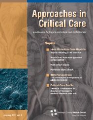
Figure One.<br />
Head CT perfusion study.<br />
The right upper quadrant<br />
shows cerebral blood volume<br />
(CBV), the left lower<br />
quadrant shows cerebral<br />
blood flow (CBF), and the<br />
right lower quadrant<br />
demonstrates mean transit<br />
time (MTT). This<br />
study shows marked<br />
asymmetry in CBF and<br />
MTT between the right<br />
and left middle cerebral<br />
artery (MCA) distributions.<br />
The reduction in<br />
the right MCA territory<br />
quantitatively meets<br />
ischemia criteria. The<br />
CBV is essentially normal<br />
in the right MCA, which<br />
suggests that this is<br />
ischemia rather than<br />
infarction.<br />
was beyond 3 hours for the intravenous<br />
thrombolytic window, beyond 6 hours for the<br />
standard intra-arterial thrombolytic window<br />
and beyond 8 hours for the current standard<br />
mechanical thrombectomy window. The initial<br />
MRI showed a relatively small area of infarction<br />
despite significant clinical symptoms.<br />
The CT perfusion was important in determining<br />
the presence of salvageable brain tissue and<br />
his candidacy for treatment despite the late<br />
time window.<br />
CT perfusion involves two contrast boluses<br />
and several timed CT cuts through the<br />
lentiform nuclei and the supraventricular white<br />
matter. The images are reconstructed to represent<br />
quantitative color maps of cerebral blood<br />
volume, cerebral blood flow and mean transit<br />
time. Comparison between the right and left<br />
hemispheres in the cerebral artery distributions<br />
can differentiate between infarction and<br />
ischemia, and delineate tissue in watershed<br />
locations that may still be viable and survive if<br />
blood flow is restored. In this case, CT perfusion<br />
was able to differentiate between the<br />
small area of infarcted brain parenchyma and<br />
the large area of ischemic tissue that was still<br />
viable and would respond to restoration of<br />
blood flow. With the advent of CT perfusion<br />
scan to differentiate between infarction and<br />
ischemia, and cerebral angiography with<br />
numerous adjunct treatment modalities such<br />
as angioplasty, intra-arterial thrombolytics and<br />
mechnical thrombectomy devices, the standard<br />
time window of 0-6 hours may be extended<br />
to a longer time window in select patients.<br />
Case Three<br />
A case of delayed intra-arterial thrombolysis<br />
in cerebrovascular accident<br />
by Lisa M. Hayden, M.D.<br />
Department of Emergency Medicine<br />
<strong>Hennepin</strong> <strong>County</strong> <strong>Medical</strong> <strong>Center</strong><br />
Abstract<br />
Though cerebrovascular accident (CVA) is<br />
considered a disease of elders, 25% of CVA<br />
occurs in patients less than 65 years of age.<br />
8 | Approaches in Critical Care | December 2008