- Page 1:
RADIATION INDUCED SIGNALING IN MAMM
- Page 4 and 5:
DECLARATION I, hereby declare that
- Page 6 and 7:
CONTENTS Page No. SYNOPSIS………
- Page 8 and 9:
2.11 Measurement of NO production
- Page 10 and 11:
PREAMBLE SYNOPSIS Ionising radiatio
- Page 12 and 13:
SYNOPSIS Aims and Objectives: To St
- Page 14 and 15:
SYNOPSIS 2.6 Immunofluorescence sta
- Page 16 and 17:
SYNOPSIS ATM gene expression, which
- Page 18 and 19:
SYNOPSIS Percentage Survival 100 80
- Page 20 and 21:
SYNOPSIS Ratio of Rad52 to Actin Ra
- Page 22 and 23:
Relative Phosphorylation 45 40 35 3
- Page 24 and 25:
SYNOPSIS Fig. 3.3A Clonogenic Cell
- Page 26 and 27:
SYNOPSIS (A) Av No. of phospho BRCA
- Page 28 and 29:
SYNOPSIS Percentage Survival 120 10
- Page 30 and 31:
SYNOPSIS Fig.3.4B. Existance of cro
- Page 32 and 33:
SYNOPSIS Fig. 3.4EDNA damage in EL-
- Page 34 and 35:
SYNOPSIS subsequent cytotoxicity ca
- Page 36 and 37:
SYNOPSIS [4] Hall, E. J. Radiobiolo
- Page 38 and 39:
CHAPTER 1 INTRODUCTION INTRODUCTION
- Page 40 and 41:
CHAPTER 1 INTRODUCTION 15.8% 9.56%
- Page 42 and 43:
CHAPTER 1 INTRODUCTION far apart an
- Page 44 and 45:
CHAPTER 1 INTRODUCTION and are more
- Page 46 and 47:
CHAPTER 1 INTRODUCTION 1.3 DNA dama
- Page 48 and 49:
CHAPTER 1 INTRODUCTION associates w
- Page 50 and 51:
CHAPTER 1 INTRODUCTION are unable t
- Page 52 and 53:
CHAPTER 1 INTRODUCTION Perhaps one
- Page 54 and 55:
CHAPTER 1 INTRODUCTION occurs at DS
- Page 56 and 57:
CHAPTER 1 INTRODUCTION related prot
- Page 58 and 59:
CHAPTER 1 INTRODUCTION damage must
- Page 60 and 61:
CHAPTER 1 INTRODUCTION At the prese
- Page 62 and 63:
CHAPTER 1 INTRODUCTION predominant
- Page 64 and 65:
CHAPTER 1 INTRODUCTION provide a me
- Page 66 and 67:
CHAPTER 1 INTRODUCTION hypersensiti
- Page 68 and 69:
CHAPTER 1 INTRODUCTION cycle-specif
- Page 70 and 71:
CHAPTER 1 INTRODUCTION 1.4 Overview
- Page 72 and 73:
CHAPTER 1 INTRODUCTION 1.4.1 Radiat
- Page 74 and 75:
CHAPTER 1 INTRODUCTION interaction
- Page 76 and 77:
CHAPTER 1 INTRODUCTION p90S6 kinase
- Page 78 and 79:
CHAPTER 1 INTRODUCTION CD4 (formerl
- Page 80 and 81:
CHAPTER 1 INTRODUCTION unrepaired d
- Page 82 and 83:
CHAPTER 1 INTRODUCTION well to the
- Page 84 and 85:
CHAPTER 1 INTRODUCTION 1.6 Radiatio
- Page 86 and 87:
CHAPTER 1 INTRODUCTION becomes pote
- Page 88 and 89:
CHAPTER 2 MATERIALS AND METHODS MAT
- Page 90 and 91:
CHAPTER 2 MATERIALS AND METHODS 2.2
- Page 92 and 93:
CHAPTER 2 MATERIALS AND METHODS res
- Page 94 and 95:
CHAPTER 2 MATERIALS AND METHODS Arr
- Page 96 and 97:
CHAPTER 2 MATERIALS AND METHODS PBS
- Page 98 and 99:
CHAPTER 2 MATERIALS AND METHODS the
- Page 100 and 101:
CHAPTER 2 MATERIALS AND METHODS 2.1
- Page 102 and 103:
CHAPTER 3 RESULTS RESULTS 93
- Page 104 and 105:
CHAPTER 3 RESULTS fractionated regi
- Page 106 and 107:
CHAPTER 3 RESULTS On comparison of
- Page 108 and 109:
CHAPTER 3 RESULTS Table 3.1.2A List
- Page 110 and 111:
CHAPTER 3 RESULTS 80 NM_002185 IL7R
- Page 112 and 113:
CHAPTER 3 RESULTS SAA2 149 AU134977
- Page 114 and 115:
CHAPTER 3 RESULTS 221 LOC643167 RNA
- Page 116 and 117:
CHAPTER 3 RESULTS 299 BF244402 FBXO
- Page 118 and 119:
CHAPTER 3 RESULTS CDK4) 57 AU152107
- Page 120 and 121:
CHAPTER 3 RESULTS 134 AL045793 METT
- Page 122 and 123:
CHAPTER 3 RESULTS 58 AF237813 ABAT
- Page 124 and 125:
CHAPTER 3 RESULTS 127 AI809749 ORMD
- Page 126 and 127:
CHAPTER 3 RESULTS 31 BE615699 UNC84
- Page 128 and 129:
CHAPTER 3 RESULTS 47 AF026303 SULT1
- Page 130 and 131:
CHAPTER 3 RESULTS 117 BF062193 CYor
- Page 132 and 133:
CHAPTER 3 RESULTS 187 AL036662 ---
- Page 134 and 135:
CHAPTER 3 RESULTS 52 AV686514 EMP2
- Page 136 and 137:
CHAPTER 3 RESULTS exposed to 10 Gy
- Page 138 and 139:
CHAPTER 3 RESULTS Fig.3.1.4 Gene ex
- Page 140 and 141:
CHAPTER 3 RESULTS Fig. 3.1.6 Radiat
- Page 142 and 143:
CHAPTER 3 RESULTS Fig. 3.1.7 Transl
- Page 144 and 145:
CHAPTER 3 RESULTS 3.1.9 Stabilizati
- Page 146 and 147:
CHAPTER 3 RESULTS 23±1.5% (Fig. 3.
- Page 148 and 149:
CHAPTER 3 RESULTS whereas only the
- Page 150 and 151:
CHAPTER 3 RESULTS Fig.3.2.2 Gene ex
- Page 152 and 153:
CHAPTER 3 RESULTS Fig.3.2.4 Gene ex
- Page 154 and 155:
CHAPTER 3 RESULTS with equal doses
- Page 156 and 157:
CHAPTER 3 RESULTS Fig.3.3.1.2 Forma
- Page 158 and 159:
CHAPTER 3 RESULTS Fig.3.3.1.3 Phosp
- Page 160 and 161:
CHAPTER 3 RESULTS (7.5±0.64) (Fig.
- Page 162 and 163:
CHAPTER 3 RESULTS Fig.3.3.1.6 Phosp
- Page 164 and 165:
CHAPTER 3 RESULTS Fig.3.3.1.7b Phos
- Page 166 and 167:
CHAPTER 3 RESULTS 3.3.2 Oxygen beam
- Page 168 and 169:
CHAPTER 3 RESULTS in all the cells.
- Page 170 and 171:
CHAPTER 3 RESULTS 3.3.2.3 Radiation
- Page 172 and 173:
CHAPTER 3 RESULTS Fig.3.3.2.3 Phosp
- Page 174 and 175:
CHAPTER 3 RESULTS Fig.3.3.2.5 Phosp
- Page 176 and 177:
CHAPTER 3 RESULTS Fig.3.3.2.7 Apopt
- Page 178 and 179:
CHAPTER 3 RESULTS PMA stimulated U9
- Page 180 and 181:
CHAPTER 3 RESULTS Fig.3.4.2. Exista
- Page 182 and 183:
CHAPTER 3 RESULTS up-regulation of
- Page 184 and 185:
CHAPTER 3 RESULTS Fig.3.4.3.2 NO pr
- Page 186 and 187:
CHAPTER 3 RESULTS 3.4.3.4 Apoptotic
- Page 188 and 189:
CHAPTER 3 RESULTS 3.4.4 Bystander e
- Page 190 and 191:
CHAPTER 3 RESULTS Fig.3.4.4b DNA da
- Page 192 and 193:
CHAPTER 3 RESULTS Fig.3.4.5a Gene e
- Page 194 and 195:
CHAPTER 4 DISCUSSION DISCUSSION 185
- Page 196 and 197:
CHAPTER 4 DISCUSSION directly expos
- Page 198 and 199:
CHAPTER 4 DISCUSSION dose is delive
- Page 200 and 201:
CHAPTER 4 DISCUSSION A clear cause
- Page 202 and 203:
CHAPTER 4 DISCUSSION 4.2 Proton bea
- Page 204 and 205:
CHAPTER 4 DISCUSSION Although the e
- Page 206 and 207:
CHAPTER 4 DISCUSSION Similar number
- Page 208 and 209:
CHAPTER 4 DISCUSSION the mechanism
- Page 210 and 211:
CHAPTER 4 DISCUSSION Thus unlike th
- Page 212 and 213:
CHAPTER 4 DISCUSSION different from
- Page 214 and 215:
CHAPTER 4 DISCUSSION upregulates ma
- Page 216 and 217:
CHAPTER 4 DISCUSSION Numerous studi
- Page 218 and 219:
CHAPTER 4 DISCUSSION inhibition of
- Page 220 and 221:
CHAPTER 4 DISCUSSION The survival o
- Page 222 and 223:
CHAPTER 5 REFERENCES [1] Khan, F. T
- Page 224 and 225:
CHAPTER 5 REFERENCES [19] Woo, R. A
- Page 226 and 227:
CHAPTER 5 REFERENCES [35] Nghiem, P
- Page 228 and 229:
CHAPTER 5 REFERENCES [52] McKay, B.
- Page 230 and 231:
CHAPTER 5 REFERENCES [69] Cary, R.
- Page 232 and 233:
CHAPTER 5 REFERENCES [84] Uziel, T.
- Page 234 and 235:
CHAPTER 5 REFERENCES [98] Liu, Y.;
- Page 236 and 237:
CHAPTER 5 REFERENCES [111] Fei, P.;
- Page 238 and 239:
CHAPTER 5 REFERENCES [127] Frank, K
- Page 240 and 241:
CHAPTER 5 REFERENCES [141] Pierce,
- Page 242 and 243: CHAPTER 5 REFERENCES [156] Roots, R
- Page 244 and 245: CHAPTER 5 REFERENCES [171] Xia, Z.;
- Page 246 and 247: CHAPTER 5 REFERENCES (MAP) kinase c
- Page 248 and 249: CHAPTER 5 REFERENCES phosphorylatio
- Page 250 and 251: CHAPTER 5 REFERENCES [211] Anderson
- Page 252 and 253: CHAPTER 5 REFERENCES [229] Konca, K
- Page 254 and 255: CHAPTER 5 REFERENCES [245] Kastan,
- Page 256 and 257: CHAPTER 5 REFERENCES [261] Miralbel
- Page 258 and 259: CHAPTER 5 REFERENCES [278] Weizman,
- Page 260 and 261: CHAPTER 5 REFERENCES [294] Zhou, H.
- Page 262 and 263: PUBLICATIONS AND PRESENTATIONS Publ
- Page 264 and 265: Mutation Research 729 (2012) 61-72
- Page 266 and 267: S. Ghosh, M. Krishna / Mutation Res
- Page 268 and 269: S. Ghosh, M. Krishna / Mutation Res
- Page 270 and 271: S. Ghosh, M. Krishna / Mutation Res
- Page 272 and 273: S. Ghosh, M. Krishna / Mutation Res
- Page 274 and 275: S. Ghosh, M. Krishna / Mutation Res
- Page 276 and 277: Mutation Research 716 (2011) 10-19
- Page 278 and 279: 12 S. Ghosh et al. / Mutation Resea
- Page 280 and 281: 14 S. Ghosh et al. / Mutation Resea
- Page 282 and 283: 16 S. Ghosh et al. / Mutation Resea
- Page 284 and 285: 18 S. Ghosh et al. / Mutation Resea
- Page 286 and 287: Mutation Research 723 (2011) 190-19
- Page 288 and 289: 192 S. Ghosh et al. / Mutation Rese
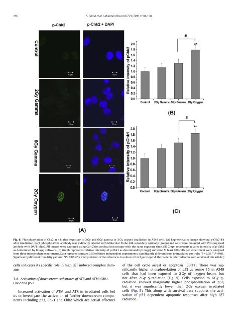
- Page 290 and 291: 194 S. Ghosh et al. / Mutation Rese
- Page 294 and 295: 198 S. Ghosh et al. / Mutation Rese
- Page 296 and 297: Cancer Invest Downloaded from infor
- Page 298 and 299: Cancer Invest Downloaded from infor
- Page 300 and 301: Cancer Invest Downloaded from infor
- Page 302 and 303: Cancer Invest Downloaded from infor
- Page 304 and 305: 1568 I. J. Radiation Oncology d Bio
- Page 306 and 307: 1570 I. J. Radiation Oncology d Bio
- Page 308 and 309: 1572 I. J. Radiation Oncology d Bio
- Page 310: 1574 I. J. Radiation Oncology d Bio