

LIFE01200604005 Shri Somnath Ghosh - Homi Bhabha National ...
LIFE01200604005 Shri Somnath Ghosh - Homi Bhabha National ...
LIFE01200604005 Shri Somnath Ghosh - Homi Bhabha National ...

You also want an ePaper? Increase the reach of your titles
YUMPU automatically turns print PDFs into web optimized ePapers that Google loves.
64 S. <strong>Ghosh</strong>, M. Krishna / Mutation Research 729 (2012) 61–72<br />
(A)<br />
rray)<br />
Fold Change (Microar<br />
(B)<br />
Fold Change (RT-PC CR)<br />
3.6<br />
3.4<br />
Control<br />
3.2<br />
5X2Gy<br />
3.0<br />
2.8<br />
2.6<br />
2.4<br />
2.2<br />
2.0<br />
1.8<br />
1.6<br />
1.4<br />
1.2<br />
1.0<br />
0.8<br />
0.6<br />
0.4<br />
0.2<br />
0.0<br />
CDK1A(p21) GADD45A TLR3 FDXR<br />
Genes<br />
3.6<br />
3.4<br />
3.2<br />
Control<br />
3.0<br />
5X2Gy<br />
2.8<br />
2.6<br />
2.4<br />
2.2<br />
2.0<br />
1.8<br />
1.6<br />
1.4<br />
1.2<br />
1.0<br />
0.8<br />
0.6<br />
0.4<br />
0.2<br />
0.0<br />
CDK1A(p21) GADD45A TLR3 FDXR<br />
Genes<br />
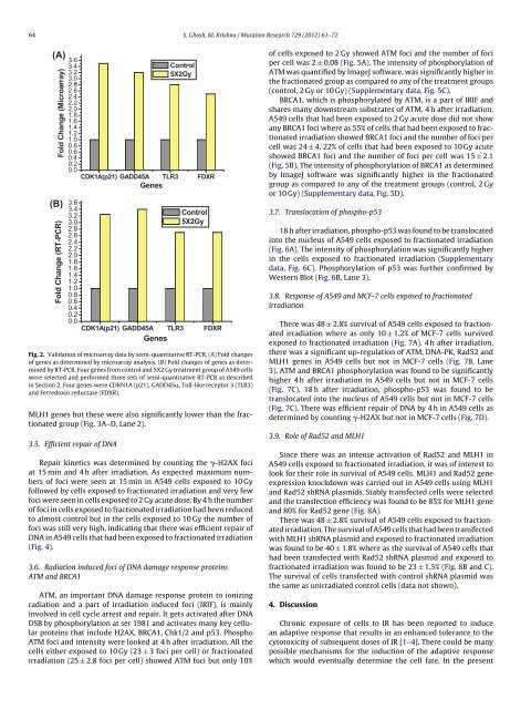
Fig. 2. Validation of microarray data by semi-quantitative RT-PCR. (A) Fold changes<br />
of genes as determined by microarray analysis. (B) Fold changes of genes as determined<br />
by RT-PCR. Four genes from control and 5X2 Gy treatment group of A549 cells<br />
were selected and performed three sets of semi-quantitative RT-PCR as described<br />
in Section 2. Four genes were CDKN1A (p21), GADD45, Toll-like receptor 3 (TLR3)<br />
and Ferredoxin reductase (FDXR).<br />
MLH1 genes but these were also significantly lower than the fractionated<br />
group (Fig. 3A–D, Lane 2).<br />
3.5. Efficient repair of DNA<br />
Repair kinetics was determined by counting the -H2AX foci<br />
at 15 min and 4 h after irradiation. As expected maximum numbers<br />
of foci were seen at 15 min in A549 cells exposed to 10 Gy<br />
followed by cells exposed to fractionated irradiation and very few<br />
foci were seen in cells exposed to 2 Gy acute dose. By 4 h the number<br />
of foci in cells exposed to fractionated irradiation had been reduced<br />
to almost control but in the cells exposed to 10 Gy the number of<br />
foci was still very high, indicating that there was efficient repair of<br />
DNA in A549 cells that had been exposed to fractionated irradiation<br />
(Fig. 4).<br />
3.6. Radiation induced foci of DNA damage response proteins<br />
ATM and BRCA1<br />
ATM, an important DNA damage response protein to ionizing<br />
radiation and a part of irradiation induced foci (IRIF), is mainly<br />
involved in cell cycle arrest and repair. It gets activated after DNA<br />
DSB by phosphorylation at ser 1981 and activates many key cellular<br />
proteins that include H2AX, BRCA1, Chk1/2 and p53. Phospho<br />
ATM foci and intensity were looked at 4 h after irradiation. All the<br />
cells either exposed to 10 Gy (23 ± 3 foci per cell) or fractionated<br />
irradiation (25 ± 2.8 foci per cell) showed ATM foci but only 10%<br />
of cells exposed to 2 Gy showed ATM foci and the number of foci<br />
per cell was 2 ± 0.08 (Fig. 5A). The intensity of phosphorylation of<br />
ATM was quantified by ImageJ software, was significantly higher in<br />
the fractionated group as compared to any of the treatment groups<br />
(control, 2 Gy or 10 Gy) (Supplementary data, Fig. 5C).<br />
BRCA1, which is phosphorylated by ATM, is a part of IRIF and<br />
shares many downstream substrates of ATM. 4 h after irradiation,<br />
A549 cells that had been exposed to 2 Gy acute dose did not show<br />
any BRCA1 foci where as 55% of cells that had been exposed to fractionated<br />
irradiation showed BRCA1 foci and the number of foci per<br />
cell was 24 ± 4. 22% of cells that had been exposed to 10 Gy acute<br />
showed BRCA1 foci and the number of foci per cell was 15 ± 2.1<br />
(Fig. 5B). The intensity of phosphorylation of BRCA1 as determined<br />
by ImageJ software was significantly higher in the fractionated<br />
group as compared to any of the treatment groups (control, 2 Gy<br />
or 10 Gy) (Supplementary data, Fig. 5D).<br />
3.7. Translocation of phospho-p53<br />
18 h after irradiation, phospho-p53 was found to be translocated<br />
into the nucleus of A549 cells exposed to fractionated irradiation<br />
(Fig. 6A). The intensity of phosphorylation was significantly higher<br />
in the cells exposed to fractionated irradiation (Supplementary<br />
data, Fig. 6C). Phosphorylation of p53 was further confirmed by<br />
Western Blot (Fig. 6B, Lane 3).<br />
3.8. Response of A549 and MCF-7 cells exposed to fractionated<br />
irradiation<br />
There was 48 ± 2.8% survival of A549 cells exposed to fractionated<br />
irradiation where as only 10 ± 1.2% of MCF-7 cells survived<br />
exposed to fractionated irradiation (Fig. 7A). 4 h after irradiation,<br />
there was a significant up-regulation of ATM, DNA-PK, Rad52 and<br />
MLH1 genes in A549 cells but not in MCF-7 cells (Fig. 7B, Lane<br />
3). ATM and BRCA1 phosphorylation was found to be significantly<br />
higher 4 h after irradiation in A549 cells but not in MCF-7 cells<br />
(Fig. 7C). 18 h after irradiation, phospho-p53 was found to be<br />
translocated into the nucleus of A549 cells but not in MCF-7 cells<br />
(Fig. 7C). There was efficient repair of DNA by 4 h in A549 cells as<br />
determined by counting -H2AX but not in MCF-7 cells (Fig. 7D).<br />
3.9. Role of Rad52 and MLH1<br />
Since there was an intense activation of Rad52 and MLH1 in<br />
A549 cells exposed to fractionated irradiation, it was of interest to<br />
look for their role in survival of A549 cells. MLH1 and Rad52 gene<br />
expression knockdown was carried out in A549 cells using MLH1<br />
and Rad52 shRNA plasmids. Stably transfected cells were selected<br />
and the transfection efficiency was found to be 85% for MLH1 gene<br />
and 80% for Rad52 gene (Fig. 8A).<br />
There was 48 ± 2.8% survival of A549 cells exposed to fractionated<br />
irradiation. The survival of A549 cells that had been transfected<br />
with MLH1 shRNA plasmid and exposed to fractionated irradiation<br />
was found to be 40 ± 1.8% where as the survival of A549 cells that<br />
had been transfected with Rad52 shRNA plasmid and exposed to<br />
fractionated irradiation was found to be 23 ± 1.5% (Fig. 8B and C).<br />
The survival of cells transfected with control shRNA plasmid was<br />
the same as unirradiated control cells (data not shown).<br />
4. Discussion<br />
Chronic exposure of cells to IR has been reported to induce<br />
an adaptive response that results in an enhanced tolerance to the<br />
cytotoxicity of subsequent doses of IR [1–4]. There could be many<br />
possible mechanisms for the induction of the adaptive response<br />
which would eventually determine the cell fate. In the present


