Low molecular weight chitosan nanoparticulate system at low N:P ...
Low molecular weight chitosan nanoparticulate system at low N:P ...
Low molecular weight chitosan nanoparticulate system at low N:P ...

Create successful ePaper yourself
Turn your PDF publications into a flip-book with our unique Google optimized e-Paper software.
Intern<strong>at</strong>ional Journal of Nanomedicine<br />
Dovepress<br />
open access to scientific and medical research<br />
Open Access Full Text Article<br />
Original Research<br />
<strong>Low</strong> <strong>molecular</strong> <strong>weight</strong> <strong>chitosan</strong> <strong>nanoparticul<strong>at</strong>e</strong><br />
<strong>system</strong> <strong>at</strong> <strong>low</strong> N:P r<strong>at</strong>io for nontoxic<br />
polynucleotide delivery<br />
Mohamad Alameh<br />
Diogo DeJesus<br />
Myriam Jean<br />
Vincent Darras<br />
Marc Thibault<br />
Marc Lavertu<br />
Michael D Buschmann<br />
Abderrazzak Merzouki<br />
Institute of Biomedical Engineering,<br />
Department of Chemical Engineering,<br />
École Polytechnique, Montréal,<br />
Canada<br />
Correspondence: Abderrazzak Merzouki<br />
Institute of Biomedical Engineering,<br />
Department of Chemical Engineering,<br />
École Polytechnique, PO Box 6079,<br />
St<strong>at</strong>ion Centre-ville, Montréal<br />
(Québec), Canada H3C 3A7<br />
Tel +1 514 340 5121 ext 4799<br />
Fax +1 514 340 5227<br />
Email abderrazzak.merzouki@polymtl.ca<br />
Abstract: Chitosan, a n<strong>at</strong>ural polymer, is a promising <strong>system</strong> for the therapeutic delivery of both<br />
plasmid DNA and synthetic small interfering RNA. Reports <strong>at</strong>tempting to identify the optimal<br />
parameters of <strong>chitosan</strong> for synthetic small interfering RNA delivery were inconclusive with<br />
high <strong>molecular</strong> <strong>weight</strong> <strong>at</strong> high amine-to-phosph<strong>at</strong>e (N:P) r<strong>at</strong>ios apparently required for efficient<br />
transfection. Here we show, for the first time, th<strong>at</strong> <strong>low</strong> <strong>molecular</strong> <strong>weight</strong> <strong>chitosan</strong> (LMW-CS)<br />
formul<strong>at</strong>ions <strong>at</strong> <strong>low</strong> N:P r<strong>at</strong>ios are suitable for the in vitro delivery of small interfering RNA.<br />
LMW-CS nanoparticles <strong>at</strong> <strong>low</strong> N:P r<strong>at</strong>ios were positively charged (ζ-potential ∼20 mV) with<br />
an average size be<strong>low</strong> 100 nm as demonstr<strong>at</strong>ed by dynamic light sc<strong>at</strong>tering and environmental<br />
scanning electron microscopy, respectively. Nanoparticles were spherical, a shape promoting<br />
decreased cytotoxicity and enhanced cellular uptake. Nanoparticle stability was effective for <strong>at</strong><br />
least 20 hours <strong>at</strong> N:P r<strong>at</strong>ios above two in a slightly acidic pH of 6.5. At a higher basic pH of 8,<br />
these nanoparticles were unravelled due to <strong>chitosan</strong> neutraliz<strong>at</strong>ion, exposing their polynucleotide<br />
cargo. Cellular uptake ranged from 50% to 95% in six different cell lines as measured by<br />
cytometry. Increasing <strong>chitosan</strong> <strong>molecular</strong> <strong>weight</strong> improved nanoparticle stability as well as the<br />
ability of nanoparticles to protect the oligonucleotide cargo from nucleases <strong>at</strong> supraphysiological<br />
concentr<strong>at</strong>ions. The highest knockdown efficiency was obtained with the specific formul<strong>at</strong>ion<br />
92-10-5 th<strong>at</strong> combines sufficient nuclease protection with effective intracellular release. This<br />
<strong>system</strong> <strong>at</strong>tained .70% knockdown of the messenger RNA, similar to commercially available<br />
lipoplexes, without apparent cytotoxicity. Contrary to previous reports, our d<strong>at</strong>a demonstr<strong>at</strong>e<br />
th<strong>at</strong> LMW-CS <strong>at</strong> <strong>low</strong> N:P r<strong>at</strong>ios are efficient and nontoxic polynucleotide delivery <strong>system</strong>s<br />
capable of transfecting a plethora of cell lines.<br />
Keywords: siRNA, nonviral delivery <strong>system</strong>, <strong>chitosan</strong>, gene silencing, RecQL1, ApoB<br />
Introduction<br />
RNA interference (RNAi), an evolutionary endogenous gene regul<strong>at</strong>ion mechanism<br />
based on double-stranded RNA (short hairpin RNA, microRNA, Piwi-interacting<br />
RNA, and small interfering RNA [siRNA]), has provided a potential new class of<br />
therapeutics. 1 Since its discovery in Caenorhabditis elegans, 2 RNAi has been proven<br />
effective in mammalian cells 1,3–11 and has reached clinical trials. 1,12–14 However, direct<br />
delivery of RNAi-inducing entities such as synthetic siRNA or short hairpin RNA<br />
continues to be problem<strong>at</strong>ic owing to their rapid extracellular/intracellular degrad<strong>at</strong>ion<br />
by nucleases (ie, RNAse and DNAse), limited blood stability, poor cellular uptake,<br />
and nonspecific targeting. 15–17 As a consequence, the transl<strong>at</strong>ion of RNAi into a clinical<br />
therapeutic reality is still pending resolution of these issues.<br />
submit your manuscript | www.dovepress.com<br />
Dovepress<br />
http://dx.doi.org/10.2147/IJN.S26571<br />
Intern<strong>at</strong>ional Journal of Nanomedicine 2012:7 1399–1414<br />
1399<br />
© 2012 Alameh et al, publisher and licensee Dove Medical Press Ltd. This is an Open Access article<br />
which permits unrestricted noncommercial use, provided the original work is properly cited.
Alameh et al<br />
Chemical modific<strong>at</strong>ion of synthetic siRNAs has provided<br />
resistance to nuclease degrad<strong>at</strong>ion and improved blood<br />
stability. 18–22 For example, selective addition of a phosphorothio<strong>at</strong>e<br />
linkage or substitution with 2′-O-methyl on the C2<br />
position of specific riboses increases nuclease resistance of<br />
siRNAs without compromising activity. 14,19,20 Nevertheless,<br />
some chemical modific<strong>at</strong>ions can increase cytotoxicity,<br />
off-target effects and reduce messenger RNA (mRNA)<br />
hybridiz<strong>at</strong>ion. 23–27 Despite progress achieved through chemical<br />
modific<strong>at</strong>ion to increase siRNA half-life, transfection efficiency,<br />
cellular targeting, and uptake remain as obstacles to<br />
effective delivery. Therefore, packaging <strong>system</strong>s which can<br />
both protect and transport chemically unmodified/ modified<br />
siRNA to target cells are required.<br />
Liposomes/Lipoplexes have been extensively used as<br />
nonviral vehicles for plasmid and RNAi entities and pose<br />
toxicity concerns. For example, the repe<strong>at</strong>ed administr<strong>at</strong>ion<br />
of lipid-based delivery vehicles caused phospholipidosis. 28<br />
Intravenous injection of stable nucleic acid-lipid particles<br />
has successfully targeted the liver to silence the apolipoprotein<br />
B (ApoB) gene in mice and nonhuman prim<strong>at</strong>es. 10<br />
However, a significant 20-fold transient elev<strong>at</strong>ion in serum<br />
transaminases (aspart<strong>at</strong>e transaminase, alanine transaminase)<br />
indic<strong>at</strong>ive of hep<strong>at</strong>ocellular necrosis was identified <strong>at</strong> the<br />
effective dose. Liposomal formul<strong>at</strong>ions of nucleic acids are<br />
known inducers of inflamm<strong>at</strong>ory cytokines including tumor<br />
necrosis factor-alpha, interferon-gamma, and interleukin-6<br />
which may be rel<strong>at</strong>ed to liver damage. 29 Polyethylene glycol<br />
(PEG) modific<strong>at</strong>ion of liposomes (PEGyl<strong>at</strong>ion), for the<br />
purpose of reducing their toxicity, was also demonstr<strong>at</strong>ed to<br />
elicit acute hypersensitivity after repe<strong>at</strong>ed dosing. 30–32 Similarly,<br />
the highly studied c<strong>at</strong>ionic family of polymers such as<br />
polyethylenimine demonstr<strong>at</strong>ed high gene transfer efficiency<br />
but was also associ<strong>at</strong>ed with significant toxicity issues 1,33<br />
limiting their broad use in clinical trials. Polyethylenimine<br />
cytotoxicity was characterized as a two-phase process where<br />
the polyc<strong>at</strong>ion-cell interaction induces loss of cell membrane<br />
integrity and the induction of programmed cell de<strong>at</strong>h. Insights<br />
into polyethylenimine toxicity highlight the importance of<br />
polyc<strong>at</strong>ion/organelle interactions – ie, mitochondria and<br />
lysosomes – on the induction of toxicity. 34,35 In general, c<strong>at</strong>ionic<br />
polymers display less toxicity associ<strong>at</strong>ed with cytokine<br />
induction – immune activ<strong>at</strong>ion – compared to their c<strong>at</strong>ionic<br />
lipid counterparts. 36<br />
Chitosan, a family of c<strong>at</strong>ionic polymers of β-1-4 N-acetylglucosamine<br />
and D-glucosamine residues, has been extensively<br />
studied for the delivery of plasmid DNA (pDNA)<br />
and siRNA both in vitro and in vivo. 3,8,17,37–43 Chitosan<br />
Dovepress<br />
properties include mucoadhesivity, 44 biocomp<strong>at</strong>ibility,<br />
biodegradability, 45 nontoxicity, and <strong>low</strong> cost of production.<br />
Primary amine residues confer a polyc<strong>at</strong>ionic n<strong>at</strong>ure to<br />
<strong>chitosan</strong> <strong>at</strong> pH values be<strong>low</strong> its pKa (∼6.5) thus enabling it<br />
to condense polyanionic compounds such as nucleic acids.<br />
Electrost<strong>at</strong>ic interaction between <strong>chitosan</strong> and nucleic acids<br />
leads to the spontaneous form<strong>at</strong>ion of nanoparticles of<br />
different sizes and shapes. 46 The ability of <strong>chitosan</strong>-based<br />
nanoparticles to transfect cells efficiently depends on several<br />
parameters such as: (1) the degree of deacetyl<strong>at</strong>ion (DDA),<br />
which represents the fraction of ionizable monomers; (2) the<br />
average <strong>molecular</strong> <strong>weight</strong> (M n<br />
), proportional to chain length,<br />
and (3) the amine-to-phosph<strong>at</strong>e (N:P) charge r<strong>at</strong>io represented<br />
by the amine-(<strong>chitosan</strong>)-to-phosph<strong>at</strong>e (DNA or RNA)<br />
r<strong>at</strong>io used to form nanoparticles.<br />
We have previously demonstr<strong>at</strong>ed th<strong>at</strong> maximiz<strong>at</strong>ion of<br />
in vitro transfection efficiency for the delivery of pDNA<br />
depends on a fine balance between these tunable parameters<br />
of <strong>chitosan</strong> 38–40 and found maximum transgene expression for<br />
DDA:M n<br />
values th<strong>at</strong> run along a diagonal from high DDA/<br />
<strong>low</strong> M n<br />
to <strong>low</strong> DDA/high M n<br />
. 38 We have also demonstr<strong>at</strong>ed<br />
th<strong>at</strong> specific <strong>chitosan</strong> formul<strong>at</strong>ions [DDA, M n<br />
, and N:P r<strong>at</strong>io]<br />
efficiently express transgene in vivo. 37,41<br />
We also demonstr<strong>at</strong>ed th<strong>at</strong> specific formul<strong>at</strong>ions are able<br />
to trigger an anti-transgene immune response; 37 therefore,<br />
nanoparticles can be designed based on the fine-tuning of<br />
<strong>chitosan</strong> parameters for applic<strong>at</strong>ion-specific purposes such<br />
as genetic vaccin<strong>at</strong>ion or gene therapy.<br />
The structural differences between pDNA and siRNA<br />
are believed to affect the complex<strong>at</strong>ion/stability of nanoparticles<br />
and optimal parameters required for effective<br />
delivery. Chitosan has also been used for siRNA delivery<br />
both in vitro and in vivo. 1,8,10,17,43 However, and despite<br />
<strong>at</strong>tempts to identify optimal physicochemical parameters for<br />
siRNA delivery, 43 inconclusive results have been observed<br />
in the liter<strong>at</strong>ure due to experimental discrepancies. 8,17 For<br />
example, it was reported th<strong>at</strong> intermedi<strong>at</strong>e DDA (80%)<br />
and high Mw (64–170 kDa) <strong>chitosan</strong> were more efficient<br />
than <strong>low</strong> <strong>molecular</strong> <strong>weight</strong> <strong>chitosan</strong> (LMW-CS) (10 kDa)<br />
in delivering siRNA. 17,43 However, high <strong>molecular</strong> <strong>weight</strong><br />
<strong>chitosan</strong>s are found to be cytotoxic, 47–49 thus potentially<br />
limiting their use in future clinical trials. Additionally, most<br />
of the reports evalu<strong>at</strong>ing the physicochemical parameters of<br />
<strong>chitosan</strong>/siRNA nanoparticles were performed <strong>at</strong> high N:P<br />
r<strong>at</strong>ios (N:P .25). 8,17,43 Such formul<strong>at</strong>ions bring significant<br />
practical problems including limited dosing due to aggreg<strong>at</strong>ion<br />
and the nonspecific effects of large quantities of soluble<br />
<strong>chitosan</strong>. 50 Here, we investig<strong>at</strong>e, for the first time, the ability<br />
1400<br />
submit your manuscript | www.dovepress.com<br />
Dovepress<br />
Intern<strong>at</strong>ional Journal of Nanomedicine 2012:7
Dovepress<br />
of specific LMW-CS formul<strong>at</strong>ions (92-10-5, 80-80-10,<br />
80-40-5, and 80-10-10) [DDA, M n<br />
, and N:P r<strong>at</strong>io] <strong>at</strong> <strong>low</strong><br />
N:P r<strong>at</strong>ios to in vitro deliver siRNA targeting: (1) the RecQL1<br />
DNA helicase mRNA in the colon adenocarcinoma RecQL1<br />
overexpressing cell line (LS174T) and (2) ApoB mRNA in<br />
the hep<strong>at</strong>ocarcinoma-derived cell line (HepG2). The choice<br />
of these two targets resides in their relevance to cancer and<br />
<strong>at</strong>herosclerosis, respectively. 6,7,9,51,52 We also explored the<br />
ability of these formul<strong>at</strong>ions to transfect multiple cell lines<br />
such as A549, AsPC1, HEK293, and Raw264.7 without<br />
apparent toxicity. In this study, we hypothesized th<strong>at</strong>, contrary<br />
to previous liter<strong>at</strong>ure, 8,17,42,43 <strong>low</strong> Mw <strong>chitosan</strong>s (LMW-CS)<br />
complexed <strong>at</strong> <strong>low</strong> N:P r<strong>at</strong>ios represent suitable formul<strong>at</strong>ions<br />
for siRNA delivery and gene knockdown; similar to our<br />
observ<strong>at</strong>ions with pDNA. 37–41 Additionally, we hypothesized<br />
th<strong>at</strong> <strong>low</strong> N:P r<strong>at</strong>ios assure sufficient protection and efficient<br />
delivery of the siRNA cargo. Moreover, we explore the<br />
physicochemical properties of these specific formul<strong>at</strong>ions<br />
with the prospect of optimizing nanoparticle transfection<br />
and silencing efficiencies. Our results demonstr<strong>at</strong>e, for the<br />
first time, th<strong>at</strong> LMW-CSs <strong>at</strong> <strong>low</strong> N:P r<strong>at</strong>ios are effective and<br />
nontoxic delivery <strong>system</strong>s for polynucleotide and siRNA<br />
delivery for in vitro gene silencing.<br />
M<strong>at</strong>erials and methods<br />
Synthesis of siRNAs and dsODNs<br />
siRNAs targeting the RecQL1 DNA helicase and ApoB<br />
mRNAs were synthesized using a novel RNA synthesis chemistry,<br />
the 5′-silyl-2′-orthoester protecting groups (2′-ACE) 54<br />
combined with a standard phosphoramitide solid-phase technology<br />
by Dharmacon (Thermo Scientific, Dharmacon RNAi<br />
Technologies, Lafayette, CO). RecQL1 mRNA-specific<br />
siRNA (siRNA-RecQL1) contains the sense sequence of<br />
5′-GUUCAGACCACUUCAGCUUdTdT-3′ and antisense<br />
5′-AAGCUGAAGUGGUCUGAACdTdT-3′ whereas ApoB<br />
mRNA-specific siRNA (siRNA-ApoB) contains the sense<br />
sequence of 5′-GUCAUCACACUGAAUACCAAU-3′ and<br />
antisense 5′-AUUGGUAUUCAGUGUGAUGACAC-3′.<br />
Mock siRNA was also used as a neg<strong>at</strong>ive control. Mock siRNA<br />
is a nontargeting siRNA (Dharmacon, D-001710-01-05)<br />
designed to have minimal targeting of known genes in human,<br />
mouse, and r<strong>at</strong> cells.<br />
Double-stranded oligodeoxynucleotides (dsODNs,<br />
21 bp) encoding the same sequences and mimicking<br />
siRNA physicochemical properties were used for<br />
nanoparticle characteriz<strong>at</strong>ion. The double-stranded<br />
oligodeoxynucleotide (dsODN) sequences were synthesized<br />
using the phosphoramidite chemistry (Integr<strong>at</strong>ed DNA<br />
Chitosan for polynucleotide delivery<br />
Technologies Inc, Coralville, IO) and used for size and<br />
zeta potential determin<strong>at</strong>ion, nanoparticle stability, and<br />
nuclease protection assays. For confocal microscopy and f<strong>low</strong><br />
cytometry analysis, 6-carboxyfluorescein (6FAM) 5′-labeled<br />
dsODNs were used (Integr<strong>at</strong>ed DNA Technologies Inc).<br />
The r<strong>at</strong>ionale for using dsODN for <strong>chitosan</strong> nanoparticle<br />
physicochemical characteriz<strong>at</strong>ion is their siRNA-mimicking<br />
properties. These mimicking properties are due to similarities<br />
<strong>at</strong> the structural level (double-stranded structure, length,<br />
and nucleotide overhangs) between siRNA and dsODNs.<br />
Additionally, charge densities are similar between siRNA<br />
and dsODNs due to identical phosph<strong>at</strong>e residue numbers on<br />
their backbone. The main differences between siRNA and<br />
dsODNs lie in the substitution of uracil to thymine (U → T)<br />
in the dsODN sequences, and in the deoxyribosil<strong>at</strong>ion of the<br />
dsODN sugar backbone.<br />
Prepar<strong>at</strong>ion and characteriz<strong>at</strong>ion<br />
of depolymerized <strong>chitosan</strong><br />
Clinical-grade <strong>chitosan</strong> <strong>at</strong> different DDAs was obtained from<br />
BioSynthec Inc, (Laval, QC, Canada) and depolymerized<br />
using nitrous acid to achieve specific number-average<br />
<strong>molecular</strong> <strong>weight</strong> targets (M n<br />
) of 80, 40, and 10 kDa. Chitosan<br />
number- and <strong>weight</strong>-average <strong>molecular</strong> <strong>weight</strong>s (M n<br />
and M w<br />
)<br />
were determined by gel perme<strong>at</strong>ion chrom<strong>at</strong>ography using<br />
a Shimadzu LC-20AD isocr<strong>at</strong>ic pump, autosampler<br />
SIL-20AC HT, oven CTO-20AC coupled with a Dawn<br />
HELEOS II multiangle laser light sc<strong>at</strong>tering detector<br />
(Wy<strong>at</strong>t Technology Co, Santa Barbara, CA), a Viscostar II<br />
(Wy<strong>at</strong>t Technology Co), an Optilab rEX interferometric<br />
refractometer (Wy<strong>at</strong>t Technology Co), and two Shodex<br />
OHpak (SB-806M HQ and SB-805 HQ; Showa Denko<br />
America, Inc, New York, NY) columns eluted with a pH 4.5<br />
acetic acid (0.15 M)/sodium acet<strong>at</strong>e (0.1 M)/sodium azide<br />
(4 mM) buffer. 54,55 The injection volume was 100 µL, the<br />
f<strong>low</strong> r<strong>at</strong>e 0.8 mL min −1 and the temper<strong>at</strong>ure 25°C. The dn/dc<br />
value was previously calcul<strong>at</strong>ed for <strong>chitosan</strong> with a DDA<br />
of 92% (for a laser’s wavelength of 658 nm) and is equal<br />
to 0.208 and 0.201 for <strong>chitosan</strong> with 80% DDA. The degree<br />
of deacetyl<strong>at</strong>ion was determined by 1 H NMR according to<br />
our previous reports. 38,56<br />
Prepar<strong>at</strong>ion of <strong>chitosan</strong> nanoparticles<br />
Chitosans with specific M n<br />
and DDA (Table 1) were dissolved<br />
overnight on a rotary mixer <strong>at</strong> 0.5% (w/v) in hydrochloric<br />
acid using a glucosamine:HCl r<strong>at</strong>io of 1:1 <strong>at</strong> a final concentr<strong>at</strong>ion<br />
of 5 mg/mL. Sterile filtered solutions were then<br />
diluted with deionized w<strong>at</strong>er to obtain the desired r<strong>at</strong>io<br />
Intern<strong>at</strong>ional Journal of Nanomedicine 2012:7<br />
submit your manuscript | www.dovepress.com<br />
Dovepress<br />
1401
Alameh et al<br />
Table 1 Physicochemical characteristics of bulk <strong>chitosan</strong>s<br />
Chitosan DDA (%) a M n<br />
(kDa) b M w<br />
(kDa) PDI c N:P r<strong>at</strong>io d<br />
92-10 92 10 11.8 1.5 5<br />
80-10 80 10 14.5 1.3 10<br />
80-40 80 40 53.0 1.3 5<br />
80-80 80 80 110.9 1.6 5<br />
Notes: a As determined by 1H NMR; b as determined by gel perme<strong>at</strong>ion<br />
chrom<strong>at</strong>ography (GPC); c M w<br />
/M n<br />
; d nanopartice N:P r<strong>at</strong>io follwing complex<strong>at</strong>ion with<br />
either dsODN or siRNA used in this study.<br />
Abbrevi<strong>at</strong>ions: DDA, degree of deacetyl<strong>at</strong>ion; M n<br />
, number average <strong>molecular</strong><br />
<strong>weight</strong>; M w<br />
, specific <strong>molecular</strong> <strong>weight</strong>; PDI, polydispersity index; N:P, amine to<br />
phosph<strong>at</strong>e; dsODN, double-stranded oligodeoxynucleotides.<br />
(N:P) of amine (<strong>chitosan</strong> deacetyl<strong>at</strong>ed groups) to phosph<strong>at</strong>e<br />
(dsODNs/siRNA nucleic acids). Nanoparticles (92-10-5,<br />
80-10-10, 80-40-5, and 80-80-5) were then prepared by<br />
rapid mixing (pipetting) of 100 µL of diluted <strong>chitosan</strong> solution<br />
to 100 µL of dsODNs or siRNA <strong>at</strong> a concentr<strong>at</strong>ion of<br />
0.05 µg/µL or 100 nM.<br />
Nanoparticle size and ζ-potential analysis<br />
The size of <strong>chitosan</strong>/dsODN-RecQL1 and <strong>chitosan</strong>/dsODN-<br />
ApoB nanoparticles – intensity average diameter – was<br />
determined by dynamic light sc<strong>at</strong>tering <strong>at</strong> an angle of 173°<br />
<strong>at</strong> room temper<strong>at</strong>ure using the Malvern Zetasizer Nano ZS<br />
(Malvern, Worcestershire, UK). Fol<strong>low</strong>ing nanoparticle form<strong>at</strong>ion,<br />
samples were diluted in 10 mM NaCl <strong>at</strong> a r<strong>at</strong>io of 1:10<br />
and measured in triplic<strong>at</strong>e. The ζ-potential was measured in<br />
triplic<strong>at</strong>e using laser Doppler velocimetry <strong>at</strong> 25°C on the<br />
same instrument with the viscosity and dielectric constant<br />
of pure w<strong>at</strong>er used for calcul<strong>at</strong>ions.<br />
Environmental scanning electron<br />
microscopy (ESEM)<br />
Chitosan/dsODN-RecQL1 and <strong>chitosan</strong>/dsODN-ApoB nanoparticles<br />
were sprayed on silicon wafer substr<strong>at</strong>e then sputterco<strong>at</strong>ed<br />
with gold (Agar Manual Sputter Co<strong>at</strong>er; Marivac<br />
Inc, Montreal, QC, Canada) and imaged using a Quanta 200<br />
FEG Environmental Scanning Electron Microscope (FEI<br />
Inc, Hillsboro, OR). Observ<strong>at</strong>ions were performed <strong>at</strong> 20 kV<br />
using the high-vacuum mode. The average particle diameter<br />
(± standard devi<strong>at</strong>ion) was determined using the XT Docu<br />
image analysis software (FEI Inc).<br />
Nanoparticle stability assessment<br />
by polyacrylamide gel electrophoresis<br />
The stability of <strong>chitosan</strong>/dsODN nanoparticles <strong>at</strong> different<br />
pHs (6.5 and 8) and for different incub<strong>at</strong>ion times<br />
(0.5, 4, and 24 hours) was assessed using polyacrylamide<br />
gel electrophoresis. Upon form<strong>at</strong>ion, nanoparticles were<br />
Dovepress<br />
mixed <strong>at</strong> a r<strong>at</strong>io of 1:1 with 2-(N-morpholino)ethanesulfonic<br />
acid buffer (MES 1X) (20 mM MES, 8 mM sodium acet<strong>at</strong>e,<br />
pH 6.5) or Tris-acet<strong>at</strong>e (TAE)-EDTA buffer (TAE 1X) (2 M<br />
Tris-acet<strong>at</strong>e, 50 mM EDTA, pH 8). The samples were then<br />
migr<strong>at</strong>ed on a 13% polyacrylamide gel (BioRad Labor<strong>at</strong>ories,<br />
Mississauga, ON, Canada) for 2 hours <strong>at</strong> 100 mV in<br />
either MES or TAE buffer. Gels were stained with 0.5 µg/mL<br />
ethidium bromide solution (BioRad Labor<strong>at</strong>ories) to visualize<br />
dsODNs. Gel document<strong>at</strong>ion and image analysis were<br />
performed using a Bio-Vision 3000 (Vilbert Lourm<strong>at</strong>,<br />
Marne-la-Vallée, France) and the Vision-Capt software,<br />
respectively.<br />
Nuclease protection assay<br />
The level of protection against nuclease <strong>at</strong>tack offered by<br />
<strong>chitosan</strong> formul<strong>at</strong>ions (92-10-5, 80-80-10, 80-40-5, and<br />
80-80-5) was assessed electrophoretically on a 5% agarose<br />
gel. Chitosan/dsODN-RecQL1 and <strong>chitosan</strong>/dsODN-ApoB<br />
nanoparticles <strong>at</strong> different DDA, M w<br />
, and N:P r<strong>at</strong>ios were<br />
incub<strong>at</strong>ed with 0.5, 1, 2, 5, or 10 units of DNAse I (Sigma-<br />
Aldrich, Oakville, ON, Canada) per µg of dsODNs in 20 µL<br />
of MES-MgCl 2<br />
buffer (20 mM MES, 1 mM MgCl 2<br />
, pH 6.5)<br />
for 30 minutes <strong>at</strong> 37°C. The reaction was stopped by adding<br />
2 µL of EDTA (50 mM) (Sigma-Aldrich). To ensure proper<br />
migr<strong>at</strong>ion of the nondigested dsODNs, samples were tre<strong>at</strong>ed<br />
with Streptomyces griseus type III <strong>chitosan</strong>ase (Sigma-Aldrich)<br />
<strong>at</strong> 10 mU/µL for 1.5 hours <strong>at</strong> 37°C and stopped by placing<br />
the samples <strong>at</strong> −20°C for 15 minutes as previously described. 3<br />
Samples were migr<strong>at</strong>ed <strong>at</strong> 90 V during 1 hour then stained with<br />
0.5 µg/mL ethidium bromide solution before visualiz<strong>at</strong>ion.<br />
Captured images were analyzed using Vision-Capt software<br />
(v 15.06; Vilber Lourm<strong>at</strong>, Paris, France). Rel<strong>at</strong>ive amounts<br />
of dsODN-RecQL1 or dsODN-ApoB (%) were determined<br />
by comparison of the integr<strong>at</strong>ed signal intensity of nucleasetre<strong>at</strong>ed<br />
samples versus nontre<strong>at</strong>ed samples.<br />
In vitro cell transfection<br />
Cell culture<br />
All cell lines were purchased from American Type Cell<br />
Culture (Manassas, VA). The HepG2 cell line was cultured in<br />
minimal essential medium (MEM). The HEK293, Raw294.7,<br />
and LS174T cell lines were cultured in high-glucose<br />
Dulbecco’s modified eagle’s media (DMEM-HG). The A549<br />
and AsPC1 cell lines were cultured in F12-K and Roswell<br />
Park Memorial Institute medium media, respectively. All<br />
cell culture media contained 1.85 g/L of sodium bicarbon<strong>at</strong>e<br />
(NaHCO 3<br />
) and were supplemented with 10% fetal bovine<br />
serum (Cedarlane Labor<strong>at</strong>ories, Burlington, ON, Canada).<br />
1402<br />
submit your manuscript | www.dovepress.com<br />
Dovepress<br />
Intern<strong>at</strong>ional Journal of Nanomedicine 2012:7
Dovepress<br />
All cell lines were cultured <strong>at</strong> 37°C in a 5% CO 2<br />
incub<strong>at</strong>or.<br />
For transfection, cells were pl<strong>at</strong>ed in 96-well or 24-well<br />
culture pl<strong>at</strong>es (Corning, <strong>Low</strong>ell, MA) to obtain a ∼50%<br />
confluence the day of transfection using 100 µL/well or<br />
500 µL/well, respectively, of complete culture medium.<br />
Cell transfection<br />
For in vitro transfection, DMEM-HG was prepared with<br />
0.976 g/L of MES and 0.84 g/L of NaHCO 3<br />
<strong>at</strong> a pH of 6.5.<br />
Transfection media containing 10% fetal bovine serum was<br />
equilibr<strong>at</strong>ed overnight <strong>at</strong> 37°C in a 5% CO 2<br />
incub<strong>at</strong>or and<br />
the pH was adjusted to 6.5 using sterile HCl (1N) prior to<br />
transfection. For siRNA transfection performed in a 96-well<br />
pl<strong>at</strong>e, <strong>chitosan</strong>/siRNA nanoparticles were prepared as<br />
described above, 30 minutes before use. A 100 µL siRNA<br />
solution <strong>at</strong> a concentr<strong>at</strong>ion of 0.05 µg/µL was used for siRNA<br />
complex<strong>at</strong>ion with <strong>chitosan</strong> <strong>at</strong> a 1:1 r<strong>at</strong>io (v/v). Fol<strong>low</strong>ing<br />
complex<strong>at</strong>ion, nanoparticles were incub<strong>at</strong>ed in a ghost<br />
pl<strong>at</strong>e containing the transfection media (DMEM-HG + fetal<br />
bovine serum) <strong>at</strong> a final concentr<strong>at</strong>ion of 1.35 ng/µL; equivalent<br />
to 10 pmol per well of siRNA. For dsODN transfection performed<br />
in a 24-well pl<strong>at</strong>e, nanoparticles were complexed<br />
as described above and incub<strong>at</strong>ed <strong>at</strong> a final concentr<strong>at</strong>ion<br />
of 8.07 ng/µL, equivalent to 60 pmol per well of dsODNs.<br />
Pl<strong>at</strong>es containing nanoparticles were equilibr<strong>at</strong>ed for 10 minutes<br />
<strong>at</strong> 37°C, 5% CO 2<br />
. Medium over cells was aspir<strong>at</strong>ed and<br />
replenished with either 500 µL (24-well pl<strong>at</strong>e) or 100 µL per<br />
well (96-well pl<strong>at</strong>e) of the transfection medium containing<br />
dsODN- or siRNA-based nanoparticles. Cells were incub<strong>at</strong>ed<br />
with <strong>chitosan</strong>/siRNA or <strong>chitosan</strong>/dsODN nanoparticles<br />
until analysis 24 hours post transfection. The commercially<br />
available liposome, DharmaFECT (Dharmacon RNAi<br />
Technologies), was used as a positive control and both<br />
untre<strong>at</strong>ed cells and uncomplexed siRNA/dsODN-tre<strong>at</strong>ed<br />
cells were used as neg<strong>at</strong>ive controls.<br />
Transfection with DharmaFECT<br />
DharmaFECT was used as a positive control for transfection<br />
efficiency in all tested cell lines. DharmaFECT/dsODN<br />
(f<strong>low</strong> cytometry and confocal microscopy) or DharmaFECT/<br />
siRNA (qPCR and viability assay) lipoplexes (1:2 [w/v] r<strong>at</strong>io)<br />
were prepared fol<strong>low</strong>ing the manufacturer’s protocol.<br />
In vitro cell viability assay<br />
Nanoparticle toxicity was evalu<strong>at</strong>ed using the alamarBlue ®<br />
prolifer<strong>at</strong>ion assay (Invitrogen, Carlsbad, CA). The principle<br />
of the assay is based on the n<strong>at</strong>ural reducing power of<br />
viable cells to convert resazurin, a blue and nonfluorescent<br />
Chitosan for polynucleotide delivery<br />
compound, into resofurin; a red and fluorescent molecule.<br />
Viable cells continuously convert resazurin to resofurin,<br />
thereby providing a quantit<strong>at</strong>ive measure of viability.<br />
Transfection was performed as described above using<br />
<strong>chitosan</strong>-siRNA nanoparticles. Five thousand cells/well<br />
were seeded 24 hours before transfection. To allevi<strong>at</strong>e the<br />
experimental bias from the effect of RecQL1 gene silencing<br />
on cell viability, nontargeting siRNA (siRNA mock)<br />
was used instead. Twenty-four hours post transfection with<br />
<strong>chitosan</strong>-based nanoparticles, 20 µL of alamarBlue reagent,<br />
pre-warmed <strong>at</strong> 37°C was added to each well and incub<strong>at</strong>ed<br />
for another 4 hours. At the end of the incub<strong>at</strong>ion 100 µL of<br />
media containing reduced alamarBlue dye was transferred<br />
to a black Corning 96-well pl<strong>at</strong>e and read on an infinite 200<br />
fluorescence pl<strong>at</strong>e reader (Tecan Systems, San Jose, CA)<br />
with excit<strong>at</strong>ion 560 nm, emission 590 nm and a cut-off of<br />
570 nm. Cells without the addition of alamarBlue were used<br />
as blank and dimethyl sulfoxide was used as a positive control<br />
of toxicity. The viability of nontransfected control cells<br />
was arbitrarily defined as 100%. The rel<strong>at</strong>ive cell viability<br />
was calcul<strong>at</strong>ed using the fol<strong>low</strong>ing formula: (fluorescence<br />
intensity sample<br />
/fluorescence intensity control<br />
) × 100.<br />
Uptake analysis by f<strong>low</strong> cytometry<br />
and confocal microscopy<br />
Fluorescence-activ<strong>at</strong>ed cell sorting (FACS) analysis<br />
The cellular uptake of dsODNs was determined by transfecting<br />
AsPC1, A549, LS174T, HepG2, HEK293, and Raw264.7<br />
cell lines with nanoparticles formed with (6FAM) 5′labeled<br />
dsODNs. Twenty-four hours post-transfection, cells were<br />
<strong>chitosan</strong>ase tre<strong>at</strong>ed for 60 minutes to elimin<strong>at</strong>e any cell<br />
surface-associ<strong>at</strong>ed nanoparticles left from the transfection as<br />
described previously. 3 Afterward, cells were washed twice<br />
with phosph<strong>at</strong>e-buffered saline, trypsinized, and resuspended<br />
in phosph<strong>at</strong>e-buffered saline. The analysis of cell uptake was<br />
made using a BD Canto f<strong>low</strong> cytometer (Becton Dickinson,<br />
San Jose, CA). For each sample, 20,000 events were counted<br />
and to exclude cell debris, dead cells, and aggreg<strong>at</strong>ed cells,<br />
a collection g<strong>at</strong>e was established using a dot plot of the<br />
forward light sc<strong>at</strong>ter against the side sc<strong>at</strong>ter. Nontransfected<br />
cells were used as neg<strong>at</strong>ive controls to discrimin<strong>at</strong>e (6FAM)<br />
positive cells from auto-fluorescence.<br />
Confocal microscopy<br />
For nanoparticle internaliz<strong>at</strong>ion analysis, the LS174T,<br />
HepG2, HEK293, and Raw264.7 cell lines were seeded on<br />
35 mm glass-bottom culture dishes (M<strong>at</strong>Tek, A shland, MA) <strong>at</strong><br />
40,000 cells/dish using 500 µL of complete culture medium.<br />
Intern<strong>at</strong>ional Journal of Nanomedicine 2012:7<br />
submit your manuscript | www.dovepress.com<br />
Dovepress<br />
1403
Alameh et al<br />
Nanoparticles were formed with fluorescent rhodamine B<br />
isothiocyan<strong>at</strong>e-labeled <strong>chitosan</strong> and dsODNs labeled with<br />
6FAM on their 5′ extremities (Integr<strong>at</strong>ed DNA Technologies).<br />
Prior to imaging, cell membranes were stained with 5 µg/mL<br />
of Cell Mask Deep Red (Invitrogen, Burlington, ON,<br />
Canada). Images were taken in multitrack mode using a<br />
Zeiss LSM 510 META confocal Axioplan 200 microscope<br />
(Carl Zeiss AG, Feldbach, Switzerland). Chitosan and<br />
dsODNs were visualized as red and green pseudocolors,<br />
respectively. The sp<strong>at</strong>ial overlap of these two colors produced<br />
yel<strong>low</strong> which permitted a qualit<strong>at</strong>ive assessment of<br />
colocaliz<strong>at</strong>ion.<br />
Quantit<strong>at</strong>ive PCR (qPCR) analysis<br />
of RecQL1 and ApoB mRNA knockdown<br />
RNA extraction and assessment methods<br />
(yield, purity, and integrity)<br />
RNA extraction was performed using the RNA XS ®<br />
extraction kit from Macherey-Nagel (Biolynx, Montréal,<br />
QC, Canada) according to the manufacturer’s protocol<br />
fol<strong>low</strong>ing <strong>chitosan</strong>ase tre<strong>at</strong>ment, as described previously. 3<br />
Total RNA was quantified and RNA integrity was measured<br />
using the Agilent BioAnalyzer 2100 (Agilent Technologies,<br />
Mississauga, ON, Canada) fol<strong>low</strong>ing the manufacturer’s<br />
protocol. RNA integrity was evalu<strong>at</strong>ed by the r<strong>at</strong>io of 28S/18S<br />
ribosomal RNA 57 and the RNA integrity number (RIN). The<br />
Agilent 2100 BioAnalyzer uses autom<strong>at</strong>ed microfluidics,<br />
capillary electrophoresis, and fluorescence to evalu<strong>at</strong>e RNA<br />
integrity. The RIN is a rel<strong>at</strong>ive measure of RNA quality<br />
th<strong>at</strong> is based largely on electrophoretic trace analysis. The<br />
BioAnalyzer 2100 autom<strong>at</strong>ically computes RIN, where an<br />
ideal nondegraded RNA sample has RIN = 10.<br />
Reverse transcription<br />
Total RNA was reverse transcribed in a final volume of<br />
20 µL using the First Strand cDNA Transcriptor Kit (Roche<br />
Diagnostics, Laval, QC, Canada) with oligodT primers as<br />
described by the manufacturer’s protocol. Samples were<br />
stored <strong>at</strong> −20°C.<br />
Gene expression assays<br />
The RecQL1 and ApoB mRNA expression level was determined<br />
using assays designed with the Universal Probe<br />
Library (UPL) from Roche (Roche Applied Science, Laval,<br />
QC, Canada). Endogenous control (hypoxanthine guanine<br />
phosphoribosyl transferase) and glyceraldehyde 3-phosph<strong>at</strong>e<br />
dehydrogenase expression levels were determined<br />
using pre-valid<strong>at</strong>ed TaqMan ® Gene Expression Assays<br />
Dovepress<br />
(Applied Bio<strong>system</strong>s, Carlsbad, CA). RecQL1 and ApoB<br />
mRNA (target detection) reactions for 384-well pl<strong>at</strong>e form<strong>at</strong>s<br />
were performed using 1.5 µL of cDNA samples (25–50 ng),<br />
5 µL of the Fast Universal qPCR MasterMix (Applied Bio<strong>system</strong>s)<br />
2 µM of each primer, and 1 µM of a Universal Probe<br />
Library probe (RecQL1 [probe #29]/ApoB [probe #55]) in a<br />
total volume of 10 µL. For endogenous control assessment,<br />
reactions were performed using identical volumes of cDNA<br />
and, Fast Universal qPCR Master Mix, 0.5 µL of the TaqMan<br />
Gene Expression Assay (20×) and 2.5 µL of w<strong>at</strong>er in a total<br />
volume of 10 µL.<br />
Detection and analysis<br />
The ABI PRISM ® 7900HT Sequence Detection System<br />
(Applied Bio<strong>system</strong>s) was used to detect the amplific<strong>at</strong>ion<br />
level and was programmed with an initial step of<br />
3 minutes <strong>at</strong> 95°C, fol<strong>low</strong>ed by 45 cycles of 5 seconds <strong>at</strong><br />
95°C and 30 seconds <strong>at</strong> 60°C. All reactions were run in<br />
triplic<strong>at</strong>e and the average values of Cts (threshold cycle)<br />
were used for quantific<strong>at</strong>ion. Glyceraldehyde 3-phosph<strong>at</strong>e<br />
dehydrogenase and hypoxanthine guanine phosphoribosyl<br />
transferase were used as endogenous controls. The rel<strong>at</strong>ive<br />
quantific<strong>at</strong>ion of target genes was determined using the<br />
ρρCT method. Briefly, the Ct values of target genes were<br />
normalized to an endogenous control gene (endogenous<br />
control) (∆CT = Ct target<br />
– Ct endoC<br />
) and compared with a<br />
calibr<strong>at</strong>or: ∆∆CT = ∆Ct target<br />
– ∆Ct calibr<strong>at</strong>or<br />
. Rel<strong>at</strong>ive expression<br />
(RQ) was calcul<strong>at</strong>ed using the Sequence Detection System<br />
2.2.2 software using the RQ = 2 −∆∆CT formula.<br />
St<strong>at</strong>istical analysis<br />
The st<strong>at</strong>istical analysis was performed using St<strong>at</strong>istica<br />
9.0 Software (STATSOFT; St<strong>at</strong>istica, Tulsa, OK). D<strong>at</strong>a<br />
are expressed as mean ± standard devi<strong>at</strong>ion. St<strong>at</strong>istical<br />
significance was determined with one-way analysis of variance,<br />
fol<strong>low</strong>ed by Tukey’s post hoc test. The results were<br />
considered significant and highly significant (P , 0.05 and<br />
P , 0.01, respectively).<br />
Results<br />
Size and ζ-potential of <strong>chitosan</strong><br />
nanoparticles<br />
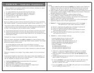
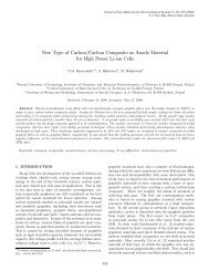
All formul<strong>at</strong>ions of <strong>chitosan</strong>/dsODN nanoparticles were in<br />
the range of 41–109 nm as measured by environmental scanning<br />
electron microscopy (ESEM) and dynamic light sc<strong>at</strong>tering<br />
(Figure 1 and Table 2). Chitosan/dsODN nanoparticles<br />
showed higher size values with increasing M n<br />
. No st<strong>at</strong>istically<br />
significant differences were observed when comparing DDAs<br />
1404<br />
submit your manuscript | www.dovepress.com<br />
Dovepress<br />
Intern<strong>at</strong>ional Journal of Nanomedicine 2012:7
Dovepress<br />
Chitosan for polynucleotide delivery<br />
Figure 1 Environmental scanning electron microscopy images of spherical <strong>chitosan</strong>/dsODN nanoparticles. (A) 92-10-5 <strong>chitosan</strong>/dsODN-RecQL1 nanoparticles; (B) 80-40-5<br />
<strong>chitosan</strong>/dsODN-RecQL1 nanoparticles; (C) 80-10-10 <strong>chitosan</strong>/dsODN-RecQL1 nanoparticles; (D) 92-10-5 <strong>chitosan</strong>/dsODN-ApoB nanoparticles; (E) 80-80-5 <strong>chitosan</strong>/<br />
dsODN-ApoB nanoparticles, and (F) 80-10-10 <strong>chitosan</strong>/dsODN-ApoB nanoparticles.<br />
Abbrevi<strong>at</strong>ions: ApoB, apolipoprotein B; dsODN, double-stranded oligodeoxynucleotide.<br />
for these specific formul<strong>at</strong>ions. The excess <strong>chitosan</strong> in all<br />
formul<strong>at</strong>ions resulted in positively charged nanoparticles as<br />
shown by ζ-potential measurements (Table 2).<br />
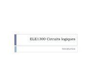
Chitosan/dsODN nanoparticle stability<br />
Chitosan-based nanoparticles were incub<strong>at</strong>ed for 0.5, 4, and<br />
20 hours in two different buffers (pH 6.5 and 8) to assess the<br />
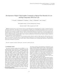
effect of time and pH on nanoparticle stability (Figure 2).<br />
Nanoparticles were stable up to 20 hours <strong>at</strong> an N:P r<strong>at</strong>io<br />
above 2 in slightly acidic buffers (pH 6.5). At 4 hours fol<strong>low</strong>ing<br />
nanoparticle form<strong>at</strong>ion, and under slightly acidic<br />
Table 2 Size and zeta potential values obtained by dynamic light<br />
sc<strong>at</strong>tering for <strong>chitosan</strong>/dsODN-RecQL1 and <strong>chitosan</strong>/dsODN-<br />
ApoB nanoparticles<br />
ODN Chitosan Size DLS<br />
(nm)<br />
Size ESEM<br />
(nm)<br />
RecQL1 92-10-5 63 ± 8 54 ± 6 23 ± 1<br />
RecQL1 80-40-5 86 ± 9 97 ± 12 18 ± 1<br />
RecQL1 80-10-10 91 ± 7 73 ± 9 18 ± 2<br />
ApoB 92-10-5 45 ± 4 66 ± 5 21 ± 2<br />
ApoB 80-80-5 100 ± 8 75 ± 13 16 ± 1<br />
ApoB 80-10-10 64 ± 6 67 ± 7 19 ± 2<br />
Zeta potential<br />
(mV)<br />
Notes: Values are mean ± SD; n = 3.<br />
Abbrevi<strong>at</strong>ions: ApoB, apolipoprotein B; dsODN, double-stranded oligodeoxynucleotides;<br />
DLS, dynamic light sc<strong>at</strong>tering; ESEM, environmental scanning electron<br />
microscopy; SD, standard devi<strong>at</strong>ion.<br />
conditions, no detectable dsODNs were observed <strong>at</strong> N:P<br />
r<strong>at</strong>ios of 2 or higher (Figure 2A and C). On the contrary,<br />
dsODN release was observed for the same N:P r<strong>at</strong>ios <strong>at</strong> a pH<br />
of 8 (Figure 2B and D). Longer exposure time – 20 hours – <strong>at</strong><br />
a pH of 6.5 resulted in increased dsODN-ApoB release <strong>at</strong> an<br />
N:P r<strong>at</strong>io of 2. This p<strong>at</strong>tern was not observed for the dsODN-<br />
RecQL1 sequence. This may be due to sequence/structural<br />
differences between the two dsODNs. Furthermore, our<br />
results <strong>at</strong> a pH of 8 show a rapid partial-to-complete dsODN<br />
release after 0.5 hour <strong>at</strong> an N:P r<strong>at</strong>io of 2 (Figure 2B and D).<br />
At N:P r<strong>at</strong>io 10 and for the same pH of 8, <strong>chitosan</strong> showed<br />
a partial release of dsODNs indic<strong>at</strong>ing the effect of excess<br />
<strong>chitosan</strong> on preserving stability. Overall, our specific <strong>chitosan</strong><br />
formul<strong>at</strong>ions assured nanoparticle stability for a minimum<br />
period of 20 hours <strong>at</strong> an N:P r<strong>at</strong>io above 2 in slightly acidic<br />
near-neutral pH environments.<br />
Nanoparticle protection assay<br />
For effective gene expression and/or inhibition, nucleic acids<br />
entrapped in the delivery vehicle must be protected from degrad<strong>at</strong>ion<br />
by enzymes such as serum nucleases. 58 The ability<br />
of <strong>chitosan</strong>-based nanoparticles to protect siRNA mimicking<br />
dsODN sequences was assessed using a DNAse I protection<br />
assay against different <strong>chitosan</strong> formul<strong>at</strong>ions complexed<br />
with dsODN-RecQL1 or dsODN-ApoB. Upon incub<strong>at</strong>io n<br />
Intern<strong>at</strong>ional Journal of Nanomedicine 2012:7<br />
submit your manuscript | www.dovepress.com<br />
Dovepress<br />
1405
Alameh et al<br />
Dovepress<br />
N:P<br />
A<br />
0<br />
20 h 4 h<br />
0.5 h 20 h 4 h<br />
0.5 h<br />
0.5 2 10 0 0.5 2 10 0 0.5 2 10<br />
C N:P 0 0.5 2 10 0 0.5 2 10 0 0.5 2 10<br />
B<br />
D<br />
Figure 2 Chitosan nanoparticle temporal stability. Stability was assessed <strong>at</strong> 0.5, 4, and 24 hours after complex form<strong>at</strong>ion using polyacrylamide gel electrophoresis <strong>at</strong> a pH<br />
of 6.5 (MES 1X) and pH8 (TAE 1X). Chitosan 92-10 <strong>at</strong> different N:P r<strong>at</strong>ios (0.5, 2, and 10) was complexed with: (A) dsODN-RecQL1 <strong>at</strong> a pH of 6.5; (B) dsODN-RecQL1<br />
<strong>at</strong> a pH of 8; (C) dsODN-ApoB <strong>at</strong> a pH of 6.5, and (D) dsODN-ApoB <strong>at</strong> a pH of 8. Unstable nanoparticles release dsODNs which become visible fol<strong>low</strong>ing EtBr staining on<br />
polyacrylamide gel fol<strong>low</strong>ing ethidium bromide staining of the polyacrylamide gel.<br />
Abbrevi<strong>at</strong>ions: ApoB, apolipoprotein B; dsODN, double-stranded oligodeoxynucleotide; N:P, amine to phosph<strong>at</strong>e.<br />
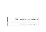
with DNAse I, naked dsODN-RecQL1 and dsODN-ApoB<br />
(controls) were completely degraded (Figure 3A–D, lane 3).<br />
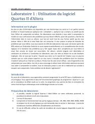
In contrast, DNAse I protection assay showed th<strong>at</strong> all <strong>chitosan</strong>s<br />
tested protected dsODNs from degrad<strong>at</strong>ion <strong>at</strong> DNAse I<br />
concentr<strong>at</strong>ions ,2 units DNAse I per µg dsODN (Figure 3).<br />
Chitosan formul<strong>at</strong>ions demonstr<strong>at</strong>ed an average of ∼80%<br />
protection of dsODNs <strong>at</strong> DNAse I concentr<strong>at</strong>ions of 0.5 U/µg<br />
(Figure 3). The ability of LMW-CS (92-10, 80-40, 80-80,<br />
and 80-10) to protect dsODNs from nuclease degrad<strong>at</strong>ion<br />
decreased with increased concentr<strong>at</strong>ions of DNAse I. Our<br />
results show th<strong>at</strong> protection decreased from ∼50% <strong>at</strong> a DNAse<br />
I concentr<strong>at</strong>ion of 1 U/µg to less than ∼20% <strong>at</strong> 2 U/µg (92-10<br />
and 80-10). Moreover, our results suggest th<strong>at</strong> higher M n<br />
<strong>chitosan</strong><br />
(80-40 and 80-80) offers a slightly better protection of<br />
dsODNs as compared to <strong>low</strong>er M n<br />
<strong>chitosan</strong> (92-10 and 80-10)<br />
<strong>at</strong> high DNAse I concentr<strong>at</strong>ions (2 U/µg) (Figure 3A–D). The<br />
enhanced cargo protection observed with higher <strong>molecular</strong><br />
<strong>weight</strong> <strong>chitosan</strong>s is consistent with previous studies where<br />
higher binding affinities between high Mw <strong>chitosan</strong>s and<br />
nucleic acids was demonstr<strong>at</strong>ed. 59 Altogether, our results<br />
show th<strong>at</strong> DNAse I protection is considerable when using<br />
intermedi<strong>at</strong>e to <strong>low</strong> DDA/M n<br />
and preserves approxim<strong>at</strong>ely<br />
60% of nucleic acid when using 1 unit of DNAse I per µg<br />
of dsODNs.<br />
In vitro cell uptake analysis by f<strong>low</strong><br />
cytometry and confocal microscopy<br />
Nanoparticle internaliz<strong>at</strong>ion into cells can be another r<strong>at</strong>elimiting<br />
step for effective drug delivery <strong>system</strong>s. In general,<br />
efficient nanoparticle internaliz<strong>at</strong>ion depends on several<br />
factors, such as the cell type, the physicochemical surface<br />
properties of the nanoparticles, and the bio–nano interface. 60<br />
The internaliz<strong>at</strong>ion of RecQL1- and ApoB-bearing nanoparticles<br />
was assessed in two different sets of relevant cell lines<br />
using f<strong>low</strong> cytometry (FACS). For the assessment of (6FAM)<br />
dsODN-RecQL uptake, transfection and FACS analysis were<br />
performed on AsPC1, A549, and LS174T cancer cell lines<br />
whereas (6FAM) dsODN-ApoB uptake was performed on<br />
HEK293, HepG2, and Raw269.7 cell lines. Our FACS results<br />
show th<strong>at</strong> cell uptake using <strong>chitosan</strong>/(6FAM) dsODN-ApoB<br />
nanoparticles achieved levels comparable to the commercially<br />
used lipoplex (DharmaFECT) (Figures 4 and 5), demonstr<strong>at</strong>ing<br />
the internaliz<strong>at</strong>ion efficiency of LMW-CS formul<strong>at</strong>ions<br />
in different cell lines. Moreover, our results indic<strong>at</strong>e th<strong>at</strong><br />
different <strong>chitosan</strong> formul<strong>at</strong>ions show st<strong>at</strong>istically significant<br />
differences in their cell uptake efficiency, with LMW-CSs<br />
92-10-5 and 80-10-10 more easily internalized compared<br />
to the higher <strong>molecular</strong> <strong>weight</strong> 80-80-5 and 80-40-5, in a<br />
cell-line-dependent manner. Interestingly, the A549 and<br />
HEK293 cell lines demonstr<strong>at</strong>ed no st<strong>at</strong>istical differences<br />
in uptake efficiency between the different <strong>chitosan</strong> formul<strong>at</strong>ions<br />
(Figures 4 and 5A). However, the A549 and HEK293<br />
cell lines showed st<strong>at</strong>istically significant increases in uptake<br />
when compared to the LS174T and Raw264.7 cell lines, again<br />
highlighting some important cell-type dependencies.<br />
In general, LMW-CS (92-10-5 and 80-10-10) showed<br />
higher uptake efficiency, ranging from approxim<strong>at</strong>ely 65% to<br />
95% depending on the transfected cell line (Figures 4 and 5A).<br />
These results are in accordance with confocal microscopy<br />
d<strong>at</strong>a, where images represent<strong>at</strong>ive of the whole popul<strong>at</strong>ion<br />
show th<strong>at</strong> the vast majority of cells for each of the four cell<br />
types imaged show nanoparticle internaliz<strong>at</strong>ion (Figure 6).<br />
The lack of colocaliz<strong>at</strong>ion <strong>at</strong> 24 hours between dsODNs and<br />
<strong>chitosan</strong> indic<strong>at</strong>es th<strong>at</strong> complete release of the dsODN cargo<br />
1406<br />
submit your manuscript | www.dovepress.com<br />
Dovepress<br />
Intern<strong>at</strong>ional Journal of Nanomedicine 2012:7
Dovepress<br />
Chitosan for polynucleotide delivery<br />
A<br />
B<br />
DNase units:<br />
20 bp<br />
Rel<strong>at</strong>ive quantity of<br />
dsODN-RecQL1 (%)<br />
100<br />
80<br />
60<br />
40<br />
20<br />
0<br />
dsODN-RecQL1<br />
C1 C2 C3<br />
0<br />
C1<br />
92-10-5/dsODN-RecQL1<br />
92-10-5/dsODN-RecQL1<br />
80-40-5 dsODN-RecQL1<br />
80-40-5/dsODN-RecQL1<br />
80-10-10/dsODN-RecQL1<br />
0 0.5 0 0.5 1 2 5 0 0.5 1 2 5 0 0.5 1 2 5<br />
C2<br />
C3<br />
80-10-10/dsODN-RecQL1<br />
C<br />
D<br />
Rel<strong>at</strong>ive quantity<br />
of dsODN-ApoB (%)<br />
DNase units:<br />
20 bp<br />
100<br />
80<br />
60<br />
40<br />
20<br />
0<br />
dsODN-ApoB<br />
C1<br />
0<br />
C2<br />
C3<br />
92-10-5/dsODN-ApoB<br />
92-10-5/dsODN-ApoB<br />
80-80-5/dsODN-ApoB<br />
80-80-5/dsODN-ApoB<br />
80-10-10/dsODN-ApoB<br />
0 0.5 0 0.5 1 2 5 0 0.5 1 2 5 0 0.5 1 2 5<br />
C1 C2 C3<br />
80-10-10/dsODN-ApoB<br />
Figure 3 Nuclease protection assays of <strong>chitosan</strong>/dsODN nanocomplexes. (A) Chitosan (92-10-5, 80-40-5 or 80-10-10) complexed with dsODN-RecQL1. (B) dsODN-RecQL1<br />
remaining after the DNAse I digestion was assessed using the signal intensity of the tre<strong>at</strong>ed samples with the control (ie, 0 U DNAse I = 100% intensity). This comparison was<br />
made between the samples of the same <strong>chitosan</strong> formul<strong>at</strong>ion. (C) Chitosans (92-10-5, 80-80-5 or 80-10-10) complexed with dsODN-ApoB. (D) dsODN-ApoB remaining<br />
after the DNAse I digestion was similarly assessed as in (B).<br />
Abbrevi<strong>at</strong>ions: ApoB, apolipoprotein B; dsODN, double-stranded oligodeoxynucleotide.<br />
100<br />
<br />
<br />
<br />
<br />
<br />
DharmaFECT<br />
92-10-5<br />
80-40-5<br />
80-10-10<br />
80<br />
<br />
Uptake (% of cells)<br />
60<br />
40<br />
20<br />
0<br />
AsPC1<br />
A549<br />
LS174T<br />
Figure 4 Cellular uptake of dsODN-RecQL1 nanoparticles 24 hours post transfection in AspC1, A549, and LS174T cancer cell lines. Chitosan formul<strong>at</strong>ions 92-10-5,<br />
80-40-5, and 80-10-10 were complexed to (6FAM) 5′ labeled dsODN-RecQL1 and transfected <strong>at</strong> 60 pmol/well 24 hours prior to fluorescence-activ<strong>at</strong>ed cell sorting analysis.<br />
DharmaFECT was used as the positive uptake control.<br />
Notes: Values are mean ± SD; n = 3; **P . 0.01.<br />
Abbrevi<strong>at</strong>ions: dsODN, double-stranded oligodeoxynucleotide; SD, standard devi<strong>at</strong>ion.<br />
Intern<strong>at</strong>ional Journal of Nanomedicine 2012:7<br />
submit your manuscript | www.dovepress.com<br />
Dovepress<br />
1407
Alameh et al<br />
Dovepress<br />
Uptake (% of cells)<br />
100<br />
80<br />
60<br />
40<br />
A<br />
DharmaFECT<br />
92-10-5<br />
80-80-5<br />
80-10-5<br />
<br />
<br />
<br />
Uptake of HepG2 cells<br />
100<br />
80<br />
60<br />
40<br />
20<br />
0<br />
B<br />
<br />
F44<br />
*<br />
P69<br />
DharmaFECT<br />
92-10-5<br />
80-80-5<br />
80-10-10<br />
<br />
?<br />
P74<br />
20<br />
0<br />
HepG2<br />
HEK293<br />
Raw264.7<br />
Figure 5 Cellular uptake of dsODN-ApoB nanoparticles 24 hours post transfection in HEK293, Raw269.7, and HepG2 cell lines. Chitosan formul<strong>at</strong>ions 92-10-5, 80-80-5,<br />
and 80-10-10 were complexed to (6FAM) 5′ labeled dsODN-ApoB and transfected <strong>at</strong> 60 pmol/well 24 hours prior to fluorescence-activ<strong>at</strong>ed cell sorting analysis. (A) Uptake<br />
efficiency of dsODN-ApoB in percentage (%). (B) Uptake efficiency of dsODN-ApoB in HepG2 cells <strong>at</strong> different passage numbers. DharmaFECT was used as the positive<br />
uptake control.<br />
Notes: Values are mean ± SD; n = 3; *P . 0.05; **P . 0.01.<br />
Abbrevi<strong>at</strong>ions: ApoB, apolipoprotein B; dsODN, double-stranded oligodeoxynucleotide; SD, standard devi<strong>at</strong>ion.<br />
Figure 6 Confocal imaging of <strong>chitosan</strong>/dsODN nanocomplex uptake 24 hours post transfection. Chitosan 92-10 (DDA, M n<br />
) was labeled with rhodamine (red) and dsODNs<br />
were 5′ labeled with (6FAM) (green). Chitosan 92-10 was complexed to dsODNs <strong>at</strong> an N:P r<strong>at</strong>io of 5. Cell membranes were stained prior to imaging with CellMask (blue)<br />
to differenti<strong>at</strong>e between internalized and membrane-bound nanoparticles. Images shown represent each separ<strong>at</strong>e channel, with dsODNs in green, <strong>chitosan</strong> in red, membrane<br />
in blue, differential interference contrast image in grey, and the merged images shown on the bottom left quadrant. (A) LS174T cells transfected with <strong>chitosan</strong>/dsODN-<br />
RecQL1 nanoparticles. (B) HepG2 cells transfected with <strong>chitosan</strong>/dsODN-ApoB nanoparticles. (C) HEK293 cells transfected with <strong>chitosan</strong>/dsODN-ApoB nanoparticles.<br />
(D) Raw 294.7 cells transfected with <strong>chitosan</strong>/dsODN-ApoB nanoparticles.<br />
Abbrevi<strong>at</strong>ions: ApoB, apolipoprotein B; dsODN, double-stranded oligodeoxynucleotide; N:P, amine to phosph<strong>at</strong>e.<br />
1408<br />
submit your manuscript | www.dovepress.com<br />
Dovepress<br />
Intern<strong>at</strong>ional Journal of Nanomedicine 2012:7
Dovepress<br />
was achieved 24 hours post transfection. Furthermore, the<br />
diffuse staining p<strong>at</strong>tern of dsODNs seen in most transfected<br />
cells suggests th<strong>at</strong> complexes have escaped endocytic vesicles<br />
(Figure 6), consistent with previous live cell imaging work<br />
using <strong>chitosan</strong>–plasmid DNA nanoparticles. 40<br />
Specific gene silencing and cell<br />
cytotoxicity evalu<strong>at</strong>ion of <strong>chitosan</strong><br />
nanoparticles in different cell lines<br />
Gene silencing occurs when complementarity is achieved<br />
between the siRNA seed region and target mRNA. 1<br />
Chitosan-specif ic formul<strong>at</strong>ions (92-10-5, 80-40-5,<br />
80-10-10, and 80-80-5) were assessed for mRNA knockdown<br />
in two different cell lines relevant to cancer and<br />
<strong>at</strong>herosclerosis, targeted by RecQL1 and ApoB siRNA,<br />
respectively. qPCR analysis revealed inhibition of RecQL1<br />
and ApoB since their coding mRNAs were downregul<strong>at</strong>ed<br />
more than twofold (Figure 7). More specifically, in LS174T<br />
cells, <strong>chitosan</strong> 92-10-5 showed a high level of silencing<br />
(∼80%) of RecQL1, similar to the current commercial gold<br />
standard liposomal formul<strong>at</strong>ion (∼80%), used here as a<br />
p ositive control. Formul<strong>at</strong>ions 80-40-5 and 80-10-10 also<br />
induced significant silencing but to a <strong>low</strong>er degree than<br />
92-10-5 and also with an increase of nonspecific mock<br />
Chitosan for polynucleotide delivery<br />
silencing, especially for formul<strong>at</strong>ion 80-10-10, for reasons<br />
th<strong>at</strong> remain to be elucid<strong>at</strong>ed. For the HepG2 cell line, only the<br />
best performing 92-10-5 was tested and induced significant<br />
silencing (∼55% versus ∼80% for positive control) of ApoB<br />
but slightly <strong>low</strong>er than RecQL1 for LS174T. Importantly, our<br />
results showed th<strong>at</strong> silencing efficiency with <strong>chitosan</strong> reached<br />
similar levels to the positive control, with a markedly reduced<br />
cytotoxicity from the delivery <strong>system</strong> as assessed using the<br />
alamarBlue assay (Figure 8).<br />
Discussion<br />
In this study, we evalu<strong>at</strong>ed the efficiency of specific <strong>low</strong><br />
<strong>molecular</strong> <strong>weight</strong> <strong>chitosan</strong> (LMW-CS) formul<strong>at</strong>ions <strong>at</strong><br />
<strong>low</strong> N:P r<strong>at</strong>ios for the in vitro delivery of siRNA targeting<br />
either RecQL1 or ApoB genes. RecQL1 is a DNA helicase<br />
playing a major role in homologous recombin<strong>at</strong>ion, maintenance<br />
of genomic stability, and DNA repair <strong>at</strong> damaged<br />
replic<strong>at</strong>ion forks. 52,61 Overexpression of RecQL1 has been<br />
implic<strong>at</strong>ed in cancer by preventing cell apoptosis. 6,7,62 As<br />
for ApoB, it is a major gene involved in <strong>at</strong>herosclerosis<br />
through its essential role in the form<strong>at</strong>ion of very <strong>low</strong> density<br />
lipoprotein which will therefore gener<strong>at</strong>e <strong>low</strong> density<br />
lipoproteins fol<strong>low</strong>ing triacylglycerol hydrolyz<strong>at</strong>ion in the<br />
circul<strong>at</strong>ion. 51,63,64<br />
100<br />
siRNA RecQL1<br />
siRNA ApoB<br />
siRNA mock<br />
80<br />
Silencing (%)<br />
60<br />
40<br />
<br />
<br />
<br />
20<br />
<br />
<br />
<br />
0<br />
DharmaFECT<br />
92-10-5 80-40-5 80-10-10 DharmaFECT 92-10-5<br />
LS174T<br />
HepG2<br />
Figure 7 Real-time polymerase chain reaction analysis of the inhibition of RecQL1 and ApoB gene expression in specific cell lines. LS174T cells were transfected with <strong>chitosan</strong><br />
(92-10-5, 80-40-5, and 80-10-10)/siRNA-RecQL1 nanoparticles, whereas HepG2 cells were transfected with <strong>chitosan</strong> (92-10-5)/siRNA-ApoB nanoparticles. The inhibition<br />
percentage was obtained by comparing the transfected and nontransfected cells, using the ∆∆CT method.<br />
Notes: Values are mean ± SD; n = 3; **P . 0.01.<br />
Abbrevi<strong>at</strong>ions: ApoB, apolipoprotein B; siRNA, small interfering RNA; SD, standard devi<strong>at</strong>ion.<br />
Intern<strong>at</strong>ional Journal of Nanomedicine 2012:7<br />
submit your manuscript | www.dovepress.com<br />
Dovepress<br />
1409
Alameh et al<br />
Dovepress<br />
140<br />
120<br />
siRNA Mock<br />
siRNA ApoB<br />
Viability (% of control)<br />
100<br />
80<br />
60<br />
40<br />
20<br />
0<br />
DharmaFECT<br />
92-10-5<br />
80-40-5<br />
80-10-10<br />
DMSO<br />
DharmaFECT<br />
92-10-5<br />
80-80-5<br />
80-10-10<br />
DMSO<br />
LS174T<br />
HepG2<br />
Figure 8 Cell viability assessment using the alamarBlue ® assay 24 hours post transfection with different <strong>chitosan</strong>/siRNA formul<strong>at</strong>ions. To allevi<strong>at</strong>e the apoptotic effect of<br />
RecQL1 gene silencing for a proper assessment of <strong>chitosan</strong>-siRNA toxicity, mock siRNA was used for transfection in the LS174T cell line. The HepG2 cell line was transfected<br />
with ApoB siRNA. DharmaFECT was used for comparison purposes whereas dimethyl sulfoxide was used as a positive control of toxicity.<br />
Notes: Values are presented as mean ± SD; n = 3.<br />
Abbrevi<strong>at</strong>ions: ApoB, apolipoprotein B; siRNA, small interfering RNA.<br />
Nanoparticle size is one parameter affecting uptake and<br />
intracellular trafficking, both considered as potential r<strong>at</strong>elimiting<br />
steps for effective gene therapy. 41,42 For instance,<br />
nano-sized particles have been shown to be internalized more<br />
efficiently than micro-sized particles. 65–67 In this study, LMW-<br />
CS-based nanoparticles ranged in size from 41–110 nm,<br />
a size range promoting uptake, prolonged blood circul<strong>at</strong>ion,<br />
higher tissue penetr<strong>at</strong>ion, and a rel<strong>at</strong>ively free passage from<br />
the mononuclear phagocyte <strong>system</strong>. 48,68–70 Therefore, our<br />
results show th<strong>at</strong> these specific LMW-CS nanoparticles <strong>at</strong><br />
<strong>low</strong> N:P r<strong>at</strong>ios meet performance criteria (Table 3) and are<br />
potentially relevant for in vivo administr<strong>at</strong>ion. The different<br />
<strong>chitosan</strong> parameters – DDA, M n<br />
, and N:P r<strong>at</strong>ios – used<br />
in this study did not significantly affect nanoparticle size,<br />
with higher <strong>molecular</strong> <strong>weight</strong> <strong>chitosan</strong> promoting a slightly<br />
increased size (Table 1). Our results are in contrast to previously<br />
published reports where the authors found increased<br />
nanoparticle size for <strong>low</strong>er <strong>molecular</strong> <strong>weight</strong> <strong>chitosan</strong>. 43<br />
This discrepancy may be due to differences in experimental<br />
conditions and to the high N:P r<strong>at</strong>io used in Liu et al and<br />
Howard et al reports 8,43 versus <strong>low</strong> N:P r<strong>at</strong>ios reported in our<br />
study. ESEM analysis revealed th<strong>at</strong> these small nanoparticles<br />
were of spherical shape consistent with previous findings for<br />
pDNA, 37 siRNA, 8 and dsODNs. 71 The effect of nanoparticle<br />
shape on internaliz<strong>at</strong>ion efficiency showed spherical particles<br />
of similar size being internalized 500% more efficiently<br />
than rod-shaped particles. 72–74 This is mainly explained by<br />
increased membrane-wrapping time required for elong<strong>at</strong>ed<br />
particles and gre<strong>at</strong>er thermodynamic forces required for<br />
their engulfment. 60,74 It was previously demonstr<strong>at</strong>ed th<strong>at</strong><br />
the morphology of <strong>chitosan</strong>-pDNA nanoparticles is strongly<br />
dependent upon their charge r<strong>at</strong>ios, and the vari<strong>at</strong>ion of the<br />
l<strong>at</strong>ter resulted in nanoparticles with different topological conform<strong>at</strong>ions<br />
including spherical, 75 toroidal, 76,77 and globular<br />
morphologies. 76,78 Chitosan-based nanoparticle shape may<br />
also seem to be affected by the type of nucleic acid – pDNA<br />
or siRNA/dsODN – used for complex<strong>at</strong>ion and the process<br />
of nanoparticle form<strong>at</strong>ion; ie, ionic gel<strong>at</strong>ion. The fact th<strong>at</strong><br />
these LMW-CS nanoparticles demonstr<strong>at</strong>ed a reproducible<br />
p<strong>at</strong>tern of spherical particles <strong>at</strong> <strong>low</strong> N:P r<strong>at</strong>ios may be indic<strong>at</strong>ive<br />
of higher internaliz<strong>at</strong>ion efficiency than nanoparticles of<br />
different topological conform<strong>at</strong>ions.<br />
1410<br />
submit your manuscript | www.dovepress.com<br />
Dovepress<br />
Intern<strong>at</strong>ional Journal of Nanomedicine 2012:7
Dovepress<br />
Chitosan for polynucleotide delivery<br />
Table 3 Safety and performance criteria for the development of<br />
effective nonviral gene delivery <strong>system</strong>s<br />
C<strong>at</strong>egories<br />
Physical and<br />
chemical properties<br />
Activity in cell<br />
based assays*<br />
Performance and<br />
safety in animal<br />
models<br />
Performance criteria**<br />
• Nanoparticle form<strong>at</strong>ion/assembly <strong>at</strong> a nucleic<br />
acid scale of 10 mg, from the perspective of<br />
size, shape, aggreg<strong>at</strong>ion, and charge, as well as<br />
from an efficacy standpoint (such as toxicity<br />
and transfection efficacy), should be<br />
similar/reproducible each time<br />
nanoparticles are formed <strong>at</strong> this and <strong>low</strong>er<br />
concentr<strong>at</strong>ions<br />
• Assemblies less than 300 nm, PDI (,0.3) and<br />
no less than 80% incorpor<strong>at</strong>ion efficiency<br />
• No aggreg<strong>at</strong>ion in 50% mouse/human serum<br />
• Chemical stability of the assembly for .30 days<br />
• Preferably amenable to freeze-drying<br />
without any loss of its performance criteria<br />
• .50% reduc<strong>at</strong>ion in target mRNA by target<br />
specific siRNA <strong>at</strong> concentr<strong>at</strong>ions ,100 nm<br />
in 10% serum containing media<br />
• ,10% reduction in target mRNA by control<br />
siRNA <strong>at</strong> concentr<strong>at</strong>ions ,100 nm in 10%<br />
serum containing media<br />
• .5-fold window between target gene silencing<br />
IC50 and IC50 for reduction in viability<br />
• Activity in <strong>at</strong> least relevant 3 cell lines to the<br />
delivery <strong>system</strong> under evalu<strong>at</strong>ion<br />
• .50% reduction in target mRNA levels by<br />
target siRNA and ,10% reduction in target<br />
mRNA levels in target tissue <strong>at</strong> 1 mg/kg dose<br />
by control siRNA by 24–48 hr<br />
• Demonstr<strong>at</strong>ion of RNAi-medi<strong>at</strong>ed target<br />
mRNA cleavage by 5’-RACE<br />
• ,10-fold cytokine induction (TNFα, IFNβ,<br />
IL6) and ,10-fold increase in ALT and AST<br />
<strong>at</strong> 3 mg/kg siRNA dose<br />
• No effect on body <strong>weight</strong> and normal blood<br />
clinical chemistry and hem<strong>at</strong>ology d<strong>at</strong>a<br />
• Lack of immunogenicity<br />
• Lack of auto-antibodies and anti-DNA<br />
antibodies<br />
• Lack of <strong>system</strong>ic toxicity<br />
• No effect on body <strong>weight</strong> and normal blood<br />
clinical chemistry and hem<strong>at</strong>ology d<strong>at</strong>a<br />
Notes: *If the delivery pl<strong>at</strong>form incorpor<strong>at</strong>es a targeting moiety evidence should be<br />
provided for targeting by (a) using cell lines with differential expression of targeted<br />
receptor and (b) using assemblies with “active” and “inactive or mutant” targeting<br />
moieties; **performance criteria described here are for both siRNA and plasmid DNA<br />
delivery <strong>system</strong>s. The criteria in this table has been adapted from inform<strong>at</strong>ion in US<br />
FDA 84,85 and US Pharmacopeia. 86<br />
Abbrevi<strong>at</strong>ions: PDI, polydispersity index; mRNA, messenger RNA; siRNA, small<br />
interfering RNA; IC50, half maximal inhibitory concentr<strong>at</strong>ion; RACE, rapid amplific<strong>at</strong>ion<br />
of cDNA ends; ALT, alanine transaminase; AST, aspart<strong>at</strong>e transaminase.<br />
Nanoparticle stability and nucleic acid protection are<br />
important parameters for efficient nucleic acid delivery. Our<br />
results of nuclease protection indic<strong>at</strong>e th<strong>at</strong> all LMW-CS formul<strong>at</strong>ions<br />
tested were able to protect dsODNs <strong>at</strong> supraphysiological<br />
concentr<strong>at</strong>ions of nucleases. Nuclease protection<br />
is of gre<strong>at</strong> importance for nucleic acid delivery <strong>system</strong>s<br />
through maintenance of cargo bioavailability and improved<br />
pharmacokinetic profile, thereby increasing the therapeutic<br />
potential of these nanoparticles. Increasing <strong>chitosan</strong> <strong>molecular</strong><br />
<strong>weight</strong> resulted in an enhanced cargo protection (Figure 3)<br />
in agreement with previous findings. 17,38,42,43,46 Nevertheless,<br />
enhancing the ability of nanoparticles to protect their siRNA<br />
from degrad<strong>at</strong>ion may render their intracellular disassembly<br />
more difficult, as demonstr<strong>at</strong>ed with high <strong>molecular</strong> <strong>weight</strong><br />
<strong>chitosan</strong>-pDNA nanoparticles. 40 Further characteriz<strong>at</strong>ion of<br />
nanoparticle stability by gel retard<strong>at</strong>ion assays show th<strong>at</strong><br />
LMW-CS used <strong>at</strong> <strong>low</strong> N:P r<strong>at</strong>ios can effectively complex and<br />
compact dsODNs into stable particles. We found LMW-CS<br />
nanoparticles <strong>at</strong> N:P r<strong>at</strong>ios above 2 to be stable in slightly<br />
acidic buffers for <strong>at</strong> least 20 hours. These interesting findings<br />
are in contrast with most previous studies using <strong>chitosan</strong>siRNA<br />
nanoparticles, where high M n<br />
and high N:P r<strong>at</strong>ios are<br />
usually required to achieve particle stability. 8,17,42,43,79 This<br />
discrepancy can be explained by the <strong>low</strong>er pH (pH 6.5) of the<br />
electrophoresis buffer in our study compared to the commonly<br />
used TAE buffer <strong>at</strong> a pH of 8 for <strong>chitosan</strong>-based nanoparticle<br />
characteriz<strong>at</strong>ion, 8,17,42,43,80 a difference th<strong>at</strong> was clearly<br />
highlighted by our gel retard<strong>at</strong>ion assay performed <strong>at</strong> both<br />
pHs (Figure 2). The use of a <strong>low</strong>er pH in the electrophoresis<br />
buffer results in higher degrees of <strong>chitosan</strong> ioniz<strong>at</strong>ion which<br />
transl<strong>at</strong>es to stronger electrost<strong>at</strong>ic <strong>at</strong>traction to the polyanionic<br />
nucleic acid and hence more stable nanoparticles. This simple<br />
modific<strong>at</strong>ion of the pH permits <strong>low</strong>er N:P r<strong>at</strong>ios than those<br />
observed previously 8,17,42,43,80 to achieve nanoparticle stability.<br />
A direct consequence of this modific<strong>at</strong>ion transl<strong>at</strong>es into<br />
reduced dosing, aggreg<strong>at</strong>ion, and other undesirable nonspecific<br />
effects of large quantities of soluble <strong>chitosan</strong> for in vivo<br />
delivery where nanoparticles are to be injected <strong>at</strong> physiological<br />
pH values close to the <strong>chitosan</strong> pK a<br />
of 6.5.<br />
In general, efficient nanoparticle internaliz<strong>at</strong>ion depends<br />
on factors such as cell type, physicochemical surface properties<br />
of the nanoparticles, and the bio–nano interface. 60 In this<br />
report, we demonstr<strong>at</strong>ed th<strong>at</strong> LMW-CS nanoparticles were<br />
efficiently internalized in multiple cell lines. The uptake<br />
efficiency as measured by f<strong>low</strong> cytometry ranged from 50%<br />
(Raw269.7) to 95% (A549 and HEK293), depending on the<br />
cell line. St<strong>at</strong>istical analysis of uptake efficiency intercell lines<br />
showed meaningful differences when comparing the A549<br />
and HEK293 (high uptake) to the LS174T and Raw269.7<br />
(medium uptake), indic<strong>at</strong>ing a cell-line dependency of<br />
<strong>chitosan</strong> uptake. The cell-line dependency of <strong>chitosan</strong> nanoparticles<br />
uptake was previously suggested to be associ<strong>at</strong>ed<br />
with different endocytic p<strong>at</strong>hways. 81,82 F<strong>low</strong> cytometry d<strong>at</strong>a<br />
Intern<strong>at</strong>ional Journal of Nanomedicine 2012:7<br />
submit your manuscript | www.dovepress.com<br />
Dovepress<br />
1411
Alameh et al<br />
showed LMW-CS nanoparticles to be efficiently internalized<br />
to levels similar or higher than commercially available<br />
liposomal <strong>system</strong>s such as DharmaFECT.<br />
Finally, the transfection efficiency of LMW-CS nanoparticles<br />
as measured by gene-silencing efficacy was evalu<strong>at</strong>ed in<br />
two different cell lines: RecQL1 in LS174T cells and ApoB in<br />
HepG2 cells. The ability of these <strong>chitosan</strong> formul<strong>at</strong>ions to efficiently<br />
silence gene expression reached more than a twofold<br />
specific mRNA knockdown; with <strong>chitosan</strong> 92-10-5 being the<br />
most efficient and specific in the LS174T cell line. Other <strong>low</strong><br />
<strong>molecular</strong> <strong>weight</strong> formul<strong>at</strong>ions also achieved good levels of<br />
gene silencing in the LS174T cell line. Interestingly, <strong>chitosan</strong><br />
80-10-10 achieved a high level of silencing with a concomitant<br />
increase in silencing when delivering mock siRNA. This<br />
intriguing observ<strong>at</strong>ion is currently under investig<strong>at</strong>ion in our<br />
labor<strong>at</strong>ory. The <strong>chitosan</strong> formul<strong>at</strong>ion 92-10-5 complexed to<br />
ApoB siRNA showed <strong>low</strong>er target mRNA knockdown in<br />
HepG2 when compared to the LS174T cell lines targeted<br />
with the RecQL1 siRNA. The silencing efficiency correl<strong>at</strong>ed<br />
well with uptake efficiency as observed by f<strong>low</strong> cytometry<br />
where <strong>chitosan</strong> 92-10-5 showed both high uptake and high<br />
silencing efficiencies. Despite structural differences between<br />
pDNA and siRNA, 46 the <strong>chitosan</strong> formul<strong>at</strong>ion 92-10-5 has<br />
shown the highest transfection efficiencies for both siRNA<br />
and pDNA to d<strong>at</strong>e. 37–41 Taken together, our results show th<strong>at</strong><br />
LWM-CS nanoparticles <strong>at</strong> <strong>low</strong> N:P r<strong>at</strong>ios can achieve efficient<br />
uptake and gene silencing in vitro, serving as a proof<br />
of concept for their use as efficient siRNA delivery vectors in<br />
cancer and <strong>at</strong>herosclerotic animal models. Although in vitro<br />
and in vivo performance criteria differ, no consensus on such<br />
performances has been established. For in vivo performance,<br />
safety remains the major issue, with guidance available from<br />
the US Food and Drug Administr<strong>at</strong>ion for the development of<br />
gene and cell therapy products. 83 Therefore, the development<br />
of nonviral drug delivery <strong>system</strong>s for in vivo use should take<br />
into account physicochemical criteria, cell-based criteria,<br />
and, most importantly, in vivo performance and safety criteria<br />
(Table 3). The <strong>low</strong> <strong>molecular</strong> <strong>weight</strong>, <strong>low</strong> N:P <strong>system</strong><br />
presented here meets many of these criteria and has already<br />
been demonstr<strong>at</strong>ed as efficient in vivo for plasmid DNA<br />
delivery. 37,41 Thus a complete characteriz<strong>at</strong>ion of the safety<br />
and in vivo performance of our LMW-CS <strong>system</strong> delivering<br />
RecQL1 and ApoB targeting siRNA is currently under investig<strong>at</strong>ion<br />
in animal models of cancer and <strong>at</strong>herosclerosis.<br />
Acknowledgments<br />
This work was supported by the N<strong>at</strong>ional Science and<br />
Engineering Research Council (NSERC) and by the Groupe<br />
Dovepress<br />
de Recherche en Sciences et Technologies Biomédicales of<br />
the Fonds de la Recherche en Santé Quebec. We are gr<strong>at</strong>eful<br />
to Dr Monica Nelea for the ESEM analyses.<br />
Disclosure<br />
The authors declare no conflicts of interest in this work.<br />
References<br />
1. de Fougerolles A, Vornlocher HP, Maraganore J, Lieberman J. Interfering<br />
with disease: a progress report on siRNA-based therapeutics. N<strong>at</strong> Rev<br />
Drug Discov. 2007;6(6):443–453.<br />
2. Fire A, Xu S, Montgomery MK, Kostas SA, Driver SE, Mello CC.<br />
Potent and specific genetic interference by double-stranded RNA in<br />
Caenorhabditis elegans. N<strong>at</strong>ure. 1998;391(6669):806–811.<br />
3. Alameh M, Jean M, DeJesus D, Buschmann MD, Merzouki A.<br />
Chitosanase-based method for RNA isol<strong>at</strong>ion from cells transfected<br />
with <strong>chitosan</strong>/siRNA nanocomplexes for real-time RT-PCR in gene<br />
silencing. Int J Nanomedicine. 2010;5:473.<br />
4. Bantounas I, Phylactou LA, Uney JB. RNA interference and the use of<br />
small interfering RNA to study gene function in mammalian <strong>system</strong>s.<br />
J Mol Endocrinol. 2004;33(3):545–557.<br />
5. Castanotto D, Rossi JJ. The promises and pitfalls of RNA-interferencebased<br />
therapeutics. N<strong>at</strong>ure. 2009;457(7228):426–433.<br />
6. Futami K, Kumagai E, Makino H, et al. Induction of mitotic cell de<strong>at</strong>h<br />
in cancer cells by small interference RNA suppressing the expression<br />
of RecQL1 helicase. Cancer Sci. 2008;99(1):71–80.<br />
7. Futami K, Kumagai E, Makino H, et al. Anticancer activity of RecQL1<br />
helicase siRNA in mouse xenograft models. Cancer Sci. 2008;<br />
99(6):1227–1236.<br />
8. Howard KA, Rahbek UL, Liu X, et al. RNA interference in vitro and<br />
in vivo using a novel <strong>chitosan</strong>/siRNA nanoparticle <strong>system</strong>. Mol Ther.<br />
2006;14(4):476–484.<br />
9. Soutschek J, Akinc A, Bramlage B, et al. Therapeutic silencing of an<br />
endogenous gene by <strong>system</strong>ic administr<strong>at</strong>ion of modified siRNAs.<br />
N<strong>at</strong>ure. 2004;432(7014):173–178.<br />
10. Zimmermann TS, Lee AC, Akinc A, et al. RNAi-medi<strong>at</strong>ed gene silencing<br />
in non-human prim<strong>at</strong>es. N<strong>at</strong>ure. 2006;441(7089):111–114.<br />
11. Elbashir SM, Harborth J, Lendeckel W, Yalcin A, Weber K, Tuschl T.<br />
Duplexes of 21-nucleotide RNAs medi<strong>at</strong>e RNA interference in cultured<br />
mammalian cells. N<strong>at</strong>ure. 2001;411(6836):494–498.<br />
12. Whelan J. First clinical d<strong>at</strong>a on RNAi. Drug Discov Today. 2005;10(15):<br />
1014–1015.<br />
13. Corey DR. RNA learns from antisense. N<strong>at</strong> Chem Biol. 2007;3(1):<br />
8–11.<br />
14. Corey DR. Chemical modific<strong>at</strong>ion: the key to clinical applic<strong>at</strong>ion of<br />
RNA interference? J Clin Invest. 2007;117(12):3615–3622.<br />
15. Stein CA. Phosphorothio<strong>at</strong>e antisense oligodeoxynucleotides: questions<br />
of specificity. Trends Biotechnol. 1996;14(5):147–149.<br />
16. Urban-Klein B, Werth S, Abuharbeid S, Czubayko F, Aigner A.<br />
RNAi-medi<strong>at</strong>ed gene-targeting throught <strong>system</strong>ic applic<strong>at</strong>ion of<br />
polyethylenimine (PEI)-complexed siRNA in vivo. Gene Ther. 2004;<br />
12(5):461–466.<br />
17. K<strong>at</strong>as H, Alpar HO. Development and characteris<strong>at</strong>ion of <strong>chitosan</strong><br />
nanoparticles for siRNA delivery. J Control Release. 2006;115(2):<br />
216–225.<br />
18. Elmen J, Thonberg H, Ljungberg K, et al. Locked nucleic acid (LNA)<br />
medi<strong>at</strong>ed improvements in siRNA stability and functionality. Nucleic<br />
Acids Res. 2005;33(1):439–447.<br />
19. Judge AD, Bola G, Lee AC, MacLachlan I. Design of noninflamm<strong>at</strong>ory<br />
synthetic siRNA medi<strong>at</strong>ing potent gene silencing in vivo. Mol Ther.<br />
2006;13(3):494–505.<br />
20. Whitehead KA, Langer R, Anderson DG. Knocking down b arriers:<br />
advances in siRNA delivery. N<strong>at</strong> Rev Drug Discov. 2009;8(2):<br />
129–138.<br />
1412<br />
submit your manuscript | www.dovepress.com<br />
Dovepress<br />
Intern<strong>at</strong>ional Journal of Nanomedicine 2012:7
Dovepress<br />
Chitosan for polynucleotide delivery<br />
21. Bramsen JB, Laursen MB, Nielsen AF, et al. A large-scale che mical<br />
modific<strong>at</strong>ion screen identifies design rules to gener<strong>at</strong>e siRNAs with<br />
high activity, high stability and <strong>low</strong> toxicity. Nucleic Acids Res.<br />
2009;37(9):2867–2881.<br />
22. Layzer JM, McCaffrey AP, Tanner AK, Huang Z, Kay MA,<br />
Sullenger BA. In vivo activity of nuclease-resistant siRNAs. RNA.<br />
2004;10(5):766–771.<br />
23. Weyermann J, Lochmann D, Georgens C, Zimmer A. Albuminprotamine-oligonucleotide-nanoparticles<br />
as a new antisense delivery<br />
<strong>system</strong>. Part 2: cellular uptake and effect. Eur J Pharm Biopharm. 2005;<br />
59(3):431–438.<br />
24. Amarzguioui M, Holen T, Babaie E, Prydz H. Tolerance for mut<strong>at</strong>ions<br />
and chemical modific<strong>at</strong>ions in a siRNA. Nucleic Acids Res. 2003;<br />
31(2):589–595.<br />
25. Parrish S, Fleenor J, Xu S, Mello C, Fire A. Functional an<strong>at</strong>omy of a<br />
dsRNA trigger: differential requirement for the two trigger strands in<br />
RNA interference. Mol Cell. 2000;6(5):1077–1087.<br />
26. Braasch DA, Jensen S, Liu Y, et al. RNA interference in<br />
m ammalian cells by chemically-modified RNA. Biochemistry. 2003;<br />
42(26):7967–7975.<br />
27. Harborth J, Elbashir SM, Vandenburgh K, et al. Sequence, chemical,<br />
and structural vari<strong>at</strong>ion of small interfering RNAs and short hairpin<br />
RNAs and the effect on mammalian gene silencing. Antisense Nucleic<br />
Acid Drug Dev. 2003;13(2):83–105.<br />
28. Reasor MJ, Kacew S. Drug-induced phospholipidosis: are there functional<br />
consequences? Exp Biol Med (Maywood). 2001;226(9):825–830.<br />
29. Tousignant JD, G<strong>at</strong>es AL, Ingram LA, et al. Comprehensive analysis<br />
of the acute toxicities induced by <strong>system</strong>ic administr<strong>at</strong>ion of c<strong>at</strong>ionic<br />
lipid:plasmid DNA complexes in mice. Hum Gene Ther. 2000;<br />
11(18):2493–2513.<br />
30. Ishida T, Ichihara M, Wang X, et al. Injection of PEGyl<strong>at</strong>ed liposomes<br />
in r<strong>at</strong>s elicits PEG-specific IgM, which is responsible for rapid elimin<strong>at</strong>ion<br />
of a second dose of PEGyl<strong>at</strong>ed liposomes. J Control Release.<br />
2006;112(1):15–25.<br />
31. Judge A, McClintock K, Phelps JR, MacLachlan I. Hypersensitivity and<br />
loss of disease site targeting caused by antibody responses to PEGyl<strong>at</strong>ed<br />
liposomes. Mol Ther. 2006;13(2):328–337. Epub November 7, 2005.<br />
32. Semple SC, Harasym TO, C<strong>low</strong> KA, Ansell SM, Klimuk SK, Hope MJ.<br />
Immunogenicity and rapid blood clearance of liposomes containing<br />
polyethylene glycol-lipid conjug<strong>at</strong>es and nucleic acid. J Pharmacol<br />
Exp Ther. 2005;312(3):1020–1026. Epub November 3, 2004.<br />
33. Boeckle S, von Gersdorff K, van der Piepen S, Culmsee C,<br />
Wagner E, Ogris M. Purific<strong>at</strong>ion of polyethylenimine polyplexes<br />
highlights the role of free polyc<strong>at</strong>ions in gene transfer. J Gene Med.<br />
2004;6(10):1102–1111.<br />
34. Moghimi SM, Symonds P, Murray JC, Hunter AC, Debska G, Szewczyk A.<br />
A two-stage poly(ethylenimine)-medi<strong>at</strong>ed cytotoxicity: implic<strong>at</strong>ions for<br />
gene transfer/therapy. Mol Ther. 2005;11(6):990–995.<br />
35. Hunter AC, Moghimi SM. C<strong>at</strong>ionic carriers of genetic m<strong>at</strong>erial<br />
and cell de<strong>at</strong>h: a mitochondrial tale. Biochim Biophys Acta. 2010;<br />
1797(6–7):1203–1209.<br />
36. Al-Dosari MS, Gao X. Nonviral gene delivery: principle, limit<strong>at</strong>ions, and<br />
recent progress. AAPS J. 2009;(4):671–681. Epub October 16, 2009.<br />
37. Jean M, Smaoui F, Lavertu M, et al. Chitosan-plasmid nanoparticle<br />
formul<strong>at</strong>ions for IM and SC delivery of recombinant FGF-2 and<br />
PDGF-BB or gener<strong>at</strong>ion of antibodies. Gene Ther. 2009;16(9):<br />
1097–1110.<br />
38. Lavertu M, Méthot S, Tran-Khanh N, Buschmann MD. High efficiency<br />
gene transfer using <strong>chitosan</strong>/DNA nanoparticles with specific combin<strong>at</strong>ions<br />
of <strong>molecular</strong> <strong>weight</strong> and degree of deacetyl<strong>at</strong>ion. Biom<strong>at</strong>erials.<br />
2006;27(27):4815–4824.<br />
39. Nimesh S, Thibault MM, Lavertu M, Buschmann MD. Enhanced gene<br />
delivery medi<strong>at</strong>ed by <strong>low</strong> <strong>molecular</strong> <strong>weight</strong> <strong>chitosan</strong>/DNA complexes:<br />
effect of pH and serum. Mol Biotechnol. 2010;46(2):182–196.<br />
40. Thibault M, Nimesh S, Lavertu M, Buschmann MD. Intracellular trafficking<br />
and decondens<strong>at</strong>ion kinetics of <strong>chitosan</strong>-pDNA polyplexes. Mol<br />
Ther. 2010;18(10):1787–1795.<br />
41. Jean M, Alameh M, Buschmann MD, Merzouki A. Effective and safe<br />
gene-based delivery of GLP-1 using <strong>chitosan</strong>/plasmid-DNA therapeutic<br />
nanocomplexes in an animal model of Type 2 Diabetes. Gene Ther.<br />
2011;18(8):807–816.<br />
42. Howard KA, Kjems J. Polyc<strong>at</strong>ion-based nanoparticle delivery for<br />
improved RNA interference therapeutics. Expert Opin Biol Ther.<br />
2007;7(12):1811–1822.<br />
43. Liu X, Howard KA, Dong M, et al. The influence of polymeric properties<br />
on <strong>chitosan</strong>/siRNA nanoparticle formul<strong>at</strong>ion and gene silencing.<br />
Biom<strong>at</strong>erials. 2007;28(6):1280–1288.<br />
44. de Campos AM, Diebold Y, Carvalho EL, Sanchez A, Alonso MJ.<br />
Chitosan nanoparticles as new ocular drug delivery <strong>system</strong>s: in vitro<br />
stability, in vivo f<strong>at</strong>e, and cellular toxicity. Pharm Res. 2004;21(5):<br />
803–810.<br />
45. Onishi H, Machida Y. Biodegrad<strong>at</strong>ion and distribution of w<strong>at</strong>er-soluble<br />
<strong>chitosan</strong> in mice. Biom<strong>at</strong>erials. 1999;20(2):175–182.<br />
46. Mao S, Sun W, Kissel T. Chitosan-based formul<strong>at</strong>ions for delivery of<br />
DNA and siRNA. Adv Drug Deliv Rev. 2010;62(1):12–27.<br />
47. Richardson SC, Kolbe HV, Duncan R. Potential of <strong>low</strong> <strong>molecular</strong><br />
mass <strong>chitosan</strong> as a DNA delivery <strong>system</strong>: biocomp<strong>at</strong>ibility, body<br />
distribution and ability to complex and protect DNA. Int J Pharm.<br />
1999;178(2):231–243.<br />
48. Huang M, Khor E, Lim LY. Uptake and cytotoxicity of <strong>chitosan</strong> molecules<br />
and nanoparticles: effects of <strong>molecular</strong> <strong>weight</strong> and degree of<br />
deacetyl<strong>at</strong>ion. Pharm Res. 2004;21(2):344–353.<br />
49. Wiegand C, Winter D, Hipler UC. Molecular-<strong>weight</strong>-dependent toxic<br />
effects of <strong>chitosan</strong>s on the human ker<strong>at</strong>inocyte cell line HaCaT. Skin<br />
Pharmacol Physiol. 2010;23(3):164–170.<br />
50. Ma PL, Buschmann MD, Winnik FM. One-step analysis of DNA/<br />
<strong>chitosan</strong> complexes by field-f<strong>low</strong> fraction<strong>at</strong>ion reveals particle size and<br />
free <strong>chitosan</strong> content. Biomacromolecules. 2010;11(3):549–554.<br />
51. Olofsson SO, Boren J. Apolipoprotein B: a clinically important apolipoprotein<br />
which assembles <strong>at</strong>herogenic lipoproteins and promotes the<br />
development of <strong>at</strong>herosclerosis. J Intern Med 2005;258(5):395–410.<br />
52. Sharma S, Doherty KM, Brosh RM Jr. Mechanisms of RecQ helicases<br />
in p<strong>at</strong>hways of DNA metabolism and maintenance of genomic stability.<br />
Biochem J. 2006;398(3):319–337.<br />
53. Scaringe SA, Wincott FE, Caruthers MH. Novel RNA synthesis<br />
method using 5′-silyl-2′-orthoester protecting groups. J Am Chem Soc.<br />
1998;120:11820–11821.<br />
54. Nguyen S, Hisiger S, Jolicoeur M, Winnik FM, Buschmann MD.<br />
Fraction<strong>at</strong>ion and characteriz<strong>at</strong>ion of <strong>chitosan</strong> by analytical SEC<br />
and 1 H-NMR after semi-prepar<strong>at</strong>ive SEC. Carbohydr Polym.<br />
2009;75:636–646.<br />
55. Nguyen S, Winnik FM, Buschmann MD. Improved reproducibility in<br />
the determin<strong>at</strong>ion of the <strong>molecular</strong> <strong>weight</strong> of <strong>chitosan</strong> by analytical size<br />
exclusion chrom<strong>at</strong>ography. Carbohydr Polym. 2009;75:528–533.<br />
56. Lavertu M, Xia Z, Serreqi AN, et al. A valid<strong>at</strong>ed 1 H-NMR method for<br />
the determin<strong>at</strong>ion of the degree of deacetyl<strong>at</strong>ion of <strong>chitosan</strong>. J Pharm<br />
Biomed Anal. 2003;32(6):1149–1158.<br />
57. Skrypina NA, Timofeeva AV, Khaspekov GL, Savochkina LP,<br />
Beabealashvilli R. Total RNA suitable for <strong>molecular</strong> biology analysis.<br />
J Biotechnol. 2003;105(1–2):1–9.<br />
58. Quong D, Neufeld RJ. DNA protection from extracapsular nucleases,<br />
within <strong>chitosan</strong>- or poly-L-lysine-co<strong>at</strong>ed algin<strong>at</strong>e beads. Biotechnol<br />
Bioeng. 1998;60(1):124–134.<br />
59. Ma PL, Lavertu M, Winnik FM, Buschmann MD. New insights into<br />
<strong>chitosan</strong>-DNA interactions using isothermal titr<strong>at</strong>ion microcalorimetry.<br />
Biomacromolecules. 2009;10(6):1490–1499.<br />
60. Nel AE, Madler L, Velegol D, et al. Understanding biophysicochemical<br />
interactions <strong>at</strong> the nano-bio interface. N<strong>at</strong> M<strong>at</strong>er. 2009;8(7):543–557.<br />
61. Wu L, Hickson ID. DNA helicases required for homologous recombin<strong>at</strong>ion<br />
and repair of damaged replic<strong>at</strong>ion forks. Annu Rev Genet.<br />
2006;40:279–306.<br />
62. Kawabe T, Tsuyama N, Kitao S, et al. Differential regul<strong>at</strong>ion of human<br />
RecQ family helicases in cell transform<strong>at</strong>ion and cell cycle. Oncogene.<br />
2000;19(41):4764–4772.<br />
Intern<strong>at</strong>ional Journal of Nanomedicine 2012:7<br />
submit your manuscript | www.dovepress.com<br />
Dovepress<br />
1413
Alameh et al<br />
Dovepress<br />
63. Schaefer JR, Scharnagl H, Baumstark MW, et al. Homozygous familial<br />
defective apolipoprotein B-100. Enhanced removal of apolipoprotein<br />
E-containing VLDLs and decreased production of LDLs. Arterioscler<br />
Thromb Vasc Biol. 1997;17(2):348–353.<br />
64. Itabe H. Oxid<strong>at</strong>ive modific<strong>at</strong>ion of LDL: its p<strong>at</strong>hological role in<br />
<strong>at</strong> herosclerosis. Clin Rev Allergy Immunol. 2009;37(1):4–11.<br />
65. Gref R, Domb A, Quellec P, et al. The controlled intravenous delivery<br />
of drugs using PEG-co<strong>at</strong>ed sterically stabilized nanospheres. Adv Drug<br />
Deliv Rev. 1995;16:215–233.<br />
66. Bivas-Benita M, Romeijn S, Junginger HE, Borchard G. PLGA-PEI<br />
nanoparticles for gene delivery to pulmonary epithelium. Eur J Pharm<br />
Biopharm. 2004;58(1):1–6.<br />
67. Panyam J, Labhasetwar V. Biodegradable nanoparticles for drug<br />
and gene delivery to cells and tissue. Adv Drug Deliv Rev. 2003;<br />
55(3):329–347.<br />
68. Guy J, Drabek D, Antoniou M. Delivery of DNA into mammalian cells<br />
by receptor-medi<strong>at</strong>ed endocytosis and gene therapy. Mol Biotechnol.<br />
1995;3(3):237–248.<br />
69. Desai MP, Labhasetwar V, Amidon GL, Levy RJ. Gastrointestinal uptake<br />
of biodegradable microparticles: effect of particle size. Pharm Res.<br />
1996;13(12):1838–1845.<br />
70. Seymour LW. Passive tumor targeting of soluble macromolecules<br />
and drug conjug<strong>at</strong>es. Crit Rev Ther Drug Carrier Syst. 1992;<br />
9(2):135–187.<br />
71. Jean M, Alameh M, De Jesus D, et al. Chitosan-based therapeutic<br />
nanocomplexes for combin<strong>at</strong>ion gene therapy and gene silencing of<br />
in vitro cell lines relevant to type 2 diabetes. Eur J Pharm Sci. 2011;<br />
45(1–2):138–149. Epub November 9, 2011.<br />
72. Chithrani BD, Chan WC. Elucid<strong>at</strong>ing the mechanism of cellular uptake<br />
and removal of protein-co<strong>at</strong>ed gold nanoparticles of different sizes and<br />
shapes. Nano Lett. 2007;7(6):1542–1550.<br />
73. Chithrani BD, Ghazani AA, Chan WC. Determining the size and shape<br />
dependence of gold nanoparticle uptake into mammalian cells. Nano<br />
Lett. 2006;6(4):662–668.<br />
74. Verma A, Stellacci F. Effect of surface properties on nanoparticle-cell<br />
interactions. Small. 2010;6(1):12–21.<br />
75. Liu W, Sun S, Cao Z, et al. An investig<strong>at</strong>ion on the physicochemical<br />
properties of <strong>chitosan</strong>/DNA polyelectrolyte complexes. Biom<strong>at</strong>erials.<br />
2005;26(15):2705–2711.<br />
76. Erbacher P, Zou S, Bettinger T, Steffan AM, Remy JS. Chitosan-based<br />
vector/DNA complexes for gene delivery: biophysical characteristics<br />
and transfection ability. Pharm Res. 1998;15(9):1332–1339.<br />
77. Danielsen S, Strand S, de Lange Davies C, Stokke BT. Glycosaminoglycan<br />
destabiliz<strong>at</strong>ion of DNA-<strong>chitosan</strong> polyplexes for gene delivery<br />
depends on <strong>chitosan</strong> chain length and GAG properties. Biochim Biophys<br />
Acta. 2005;1721(1–3):44–54. Epub November 2, 2004.<br />
78. Köping-Höggård M, Mel’nikova YS, Vårum KM, Lindman B, Ar tursson<br />
P. Rel<strong>at</strong>ionship between the physical shape and the efficiency of oligomeric<br />
<strong>chitosan</strong> as a gene delivery <strong>system</strong> in vitro and in vivo. J Gene<br />
Med. 2003;5(2):130–141.<br />
79. Gao S, Dagnaes-Hansen F, Nielsen EJ, et al. The effect of chemical<br />
modific<strong>at</strong>ion and nanoparticle formul<strong>at</strong>ion on stability and biodistribution<br />
of siRNA in mice. Mol Ther. 2009;17(7):1225–1233.<br />
80. Ji AM, Su D, Che O, et al. Functional gene silencing medi<strong>at</strong>ed by c hitosan/<br />
siRNA nanocomplexes. Nanotechnology. 2009;20(40):405103.<br />
81. Bishop NE. An Upd<strong>at</strong>e on Non-cl<strong>at</strong>hrin-co<strong>at</strong>ed Endocytosis. Rev Med<br />
Virol. 1997;7(4):199–209.<br />
82. Huang M, Ma Z, Khor E, Lim LY. Uptake of FITC-<strong>chitosan</strong> nanoparticles<br />
by A549 cells. Pharm Res. 2002;19(10):1488–1494.<br />
83. Center for Biologics Evalu<strong>at</strong>ion and Research, US Food and Drug<br />
Administr<strong>at</strong>ion. Guidance for Industry: Consider<strong>at</strong>ions for Plasmid<br />
DNA Vaccines for Infectious Disease Indic<strong>at</strong>ions. Rockville, MD;<br />
2007. Available from: http://www.fda.gov/BiologicsBloodVaccines/<br />
GuidanceComplianceRegul<strong>at</strong>oryInform<strong>at</strong>ion/Guidances/Vaccines/<br />
ucm074770.htm. Accessed January 1, 2012.<br />
84. Center for Biologics Evalu<strong>at</strong>ion and Research. Guidance for Industry:<br />
Consider<strong>at</strong>ions for Plasmid DNA Vaccines for Infectious Disease Indic<strong>at</strong>ions.<br />
Rockville, MD: US FDA; 2007. Available from: http://iccvam.<br />
niehs.nih.gov/SuppDocs/FedDocs/FDA/FDA_plasdnavac.pdf. Accessed<br />
February 23, 2012.<br />
85. Center for Biologics Evalu<strong>at</strong>ion and Research. Guidance for FDA<br />
Reviewers and Sponsors: Content and Review of Chemistry, Manufacturing,<br />
and Control (CMC) Inform<strong>at</strong>ion for Human Gene Therapy<br />
Investig<strong>at</strong>ional New Drug Applic<strong>at</strong>ions (INDs). Rockville, MD: US<br />
FDA; 2008. Available from: http://www.fda.gov/downloads/BiologicsBloodVaccines/GuidanceComplianceRegul<strong>at</strong>oryInform<strong>at</strong>ion/Guidances/CellularandGeneTherapy/ucm078694.pdf.<br />
Accessed February 23,<br />
2012.<br />
86. US Pharmacopeial Convention. Gene therapy products. In: Second<br />
Supplement to USP 34-NF 29. Rockville, MD: United St<strong>at</strong>es Pharmacopeial;<br />
2011:5135–5158.<br />
Intern<strong>at</strong>ional Journal of Nanomedicine<br />
Publish your work in this journal<br />
The Intern<strong>at</strong>ional Journal of Nanomedicine is an intern<strong>at</strong>ional, peerreviewed<br />
journal focusing on the applic<strong>at</strong>ion of nanotechnology<br />
in diagnostics, therapeutics, and drug delivery <strong>system</strong>s throughout<br />
the biomedical field. This journal is indexed on PubMed Central,<br />
MedLine, CAS, SciSearch®, Current Contents®/Clinical Medicine,<br />
Submit your manuscript here: http://www.dovepress.com/intern<strong>at</strong>ional-journal-of-nanomedicine-journal<br />
Dovepress<br />
Journal Cit<strong>at</strong>ion Reports/Science Edition, EMBase, Scopus and the<br />
Elsevier Bibliographic d<strong>at</strong>abases. The manuscript management <strong>system</strong><br />
is completely online and includes a very quick and fair peer-review<br />
<strong>system</strong>, which is all easy to use. Visit http://www.dovepress.com/<br />
testimonials.php to read real quotes from published authors.<br />
1414<br />
submit your manuscript | www.dovepress.com<br />
Dovepress<br />
Intern<strong>at</strong>ional Journal of Nanomedicine 2012:7