The primate cranial base: ontogeny, function and - Harvard University
The primate cranial base: ontogeny, function and - Harvard University The primate cranial base: ontogeny, function and - Harvard University
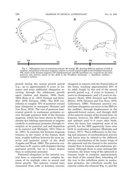
124 YEARBOOK OF PHYSICAL ANTHROPOLOGY [Vol. 43, 2000 Fig. 3. Midsagittal view of nonhuman primate (A) human (B), showing different patterns of drift of occipital, and position of foramen magnum, relative to overall cranial length in the Frankfurt horizontal. FM, center of the foramen magnum; OP (opisthocranium) and PR (prosthion) are, respectively the most posterior and anterior points on the skull in the Frankfurt horizontal. , depository surfaces; resorptive surfaces. growth during the neural growth period (e.g., up to approximately 6 years in humans) and some additional elongation occurring through the adolescent growth spurt (Ashton and Spence, 1958; Scott, 1958; Riolo et al., 1974; Sirianni and Swindler, 1979; Sirianni, 1985). The SOS contributes to roughly 70% of posterior cranial base elongation in macaques (Sirianni and Van Ness, 1978). The rest of posterior basicranial growth in nonhuman primates occurs through posterior drift of the foramen magnum, which has been shown by fluorochrome dye labeling experiments to migrate caudally in nonhuman primates through resorption at its posterior end and deposition at its anterior end (Michejda, 1971; Giles et al., 1981). In contrast, the foramen magnum remains in the center of the human skull base, roughly halfway between the most anterior and posterior points of the skull (Lugoba and Wood, 1990). The posterior cranial base in H. sapiens still elongates during postnatal growth, but to a lesser degree than in nonhuman primates. Postnatal elongation in the anterior cranial base is somewhat more complex because of its multiple roles in neural and facial growth. The anterior cranial base (measured from sella to foramen caecum) elongates in concert with the frontal lobes of the brain, reaching approximately 95% of its adult length by the end of the neural growth period (e.g., 6 years in humans, 3 years in chimpanzees, and 1.2 years in macaques) (Scott, 1958; Sirianni and Newell- Morris, 1978; Sirianni and Van Ness, 1978; Lieberman, 1998). Postnatal anterior cranial base elongation can occur in the SES (in the midline), through displacement in the sphenoid-frontal suture, and through drift of the anterior margin of the frontal bone. In humans, however, the SES remains active and unfused until 6–8 years after birth, when the brain has completed most of its growth, but the SES apparently fuses near birth in nonhuman primates (Michejda and Lamey, 1971). These differences in the timing and sequence of synchondroseal activity and fusion may be related to the different relative contributions of the lesser wings of the sphenoid and the frontal to the anterior cranial floor in humans and nonhuman primates. Although there is some intraspecific variation, the lesser wing of the sphenoid in humans tends to comprise approximately one third of the cranial floor, extending all the way to the cribriform plate; in nonhuman primates, the cribriform usually lies entirely within the ethmoid (Fig. 4), and the
D.E. Lieberman et al.] PRIMATE CRANIAL BASE 125 Fig. 4. Superior view of cranial base in Homo sapiens (left) and Pan troglodytes (right) (after Aiello and Dean, 1991). FC, foramen, caecum; PS, planum sphenoideum point; SP, sphenoidale; S, sella. Note similar orientation of the inferior petrosal posterior surface (PPip) in the two species (data from Spoor, 1997). In addition, the lesser wing of the sphenoid and the cribriform plate comprise a much greater percentage of the midline anterior cranial base in humans than in chimpanzees. lesser wing of the sphenoid makes up less than one tenth of the cranial floor (Van der Linden and Enlow, 1971; Aiello and Dean, 1990; McCarthy, 2001). Differences in the sequence of synchondroseal fusion may also be related to differences in the timing and nature of cranial base angulation in human vs. nonhuman primates (Jeffery, 1999; see below). While the anterior cranial base grows solely during the neural growth phase (it reaches adult size at the same time as the brain), the more inferior portions of the anterior cranial base continue to grow as part of the face after the neural growth phase, forming the ethmomaxillary complex (Enlow, 1990). This complex grows downward and forward mostly through drift and displacement. In addition, the sphenoid sinus drifts anteriorly. Since the ethmoid (with the exception of the cribriform plate) primarily grows as part of the ethmomaxillary complex, its postnatal growth is most properly treated in a review of facial growth. Medio-lateral growth. How the cranial base widens is important because of its various interactions with neurocranial and facial shape (Lieberman et al., 2000; see below). The increases in width of the anterior and posterior cranial fossae occur primarily from drift (in which the external and internal surfaces of the squamae are depository and resorptive, respectively), and from intramembranous bone growth in sutures with some component of lateral orientation, such as the fronto-ethmoid and occipitomastoid sutures (Sperber, 1989). Lateral growth in the middle cranial fossa is slightly more complicated. The sphenoid body does not widen much (Kodama, 1976a,b; Sasaki and Kodama, 1976). Instead, most increases in middle cranial fossa width presumably occur in the spheno-temporal suture and through lateral drift of the squamous portions of the sphenoid. Increases in cerebellum and brain-stem size have been implicated in changes in the orientation of the petrous pyramids (Fig. 4), which are more coronally oriented externally (but not internally) in humans than in nonhuman primates (Dean, 1988). Spoor (1997) found that petrous pyramid orientation in a broad interspecific sample of pri-
- Page 1 and 2: YEARBOOK OF PHYSICAL ANTHROPOLOGY 4
- Page 3 and 4: D.E. Lieberman et al.] PRIMATE CRAN
- Page 5 and 6: D.E. Lieberman et al.] PRIMATE CRAN
- Page 7: D.E. Lieberman et al.] PRIMATE CRAN
- Page 11 and 12: D.E. Lieberman et al.] PRIMATE CRAN
- Page 13 and 14: D.E. Lieberman et al.] PRIMATE CRAN
- Page 15 and 16: D.E. Lieberman et al.] PRIMATE CRAN
- Page 17 and 18: D.E. Lieberman et al.] PRIMATE CRAN
- Page 19 and 20: D.E. Lieberman et al.] PRIMATE CRAN
- Page 21 and 22: D.E. Lieberman et al.] PRIMATE CRAN
- Page 23 and 24: D.E. Lieberman et al.] PRIMATE CRAN
- Page 25 and 26: D.E. Lieberman et al.] PRIMATE CRAN
- Page 27 and 28: D.E. Lieberman et al.] PRIMATE CRAN
- Page 29 and 30: D.E. Lieberman et al.] PRIMATE CRAN
- Page 31 and 32: Species or specimen IRE1 1 IRE5 2 (
- Page 33 and 34: D.E. Lieberman et al.] PRIMATE CRAN
- Page 35 and 36: D.E. Lieberman et al.] PRIMATE CRAN
- Page 37 and 38: D.E. Lieberman et al.] PRIMATE CRAN
- Page 39 and 40: D.E. Lieberman et al.] PRIMATE CRAN
- Page 41 and 42: D.E. Lieberman et al.] PRIMATE CRAN
- Page 43 and 44: D.E. Lieberman et al.] PRIMATE CRAN
- Page 45 and 46: D.E. Lieberman et al.] PRIMATE CRAN
- Page 47 and 48: D.E. Lieberman et al.] PRIMATE CRAN
- Page 49 and 50: D.E. Lieberman et al.] PRIMATE CRAN
- Page 51 and 52: D.E. Lieberman et al.] PRIMATE CRAN
- Page 53: D.E. Lieberman et al.] PRIMATE CRAN
124 YEARBOOK OF PHYSICAL ANTHROPOLOGY [Vol. 43, 2000<br />
Fig. 3. Midsagittal view of nonhuman <strong>primate</strong> (A) human (B), showing different patterns of drift of<br />
occipital, <strong>and</strong> position of foramen magnum, relative to overall <strong>cranial</strong> length in the Frankfurt horizontal.<br />
FM, center of the foramen magnum; OP (opisthocranium) <strong>and</strong> PR (prosthion) are, respectively the most<br />
posterior <strong>and</strong> anterior points on the skull in the Frankfurt horizontal. , depository surfaces; <br />
resorptive surfaces.<br />
growth during the neural growth period<br />
(e.g., up to approximately 6 years in humans)<br />
<strong>and</strong> some additional elongation occurring<br />
through the adolescent growth<br />
spurt (Ashton <strong>and</strong> Spence, 1958; Scott,<br />
1958; Riolo et al., 1974; Sirianni <strong>and</strong> Swindler,<br />
1979; Sirianni, 1985). <strong>The</strong> SOS contributes<br />
to roughly 70% of posterior <strong>cranial</strong><br />
<strong>base</strong> elongation in macaques (Sirianni <strong>and</strong><br />
Van Ness, 1978). <strong>The</strong> rest of posterior basi<strong>cranial</strong><br />
growth in nonhuman <strong>primate</strong>s occurs<br />
through posterior drift of the foramen<br />
magnum, which has been shown by fluorochrome<br />
dye labeling experiments to migrate<br />
caudally in nonhuman <strong>primate</strong>s through resorption<br />
at its posterior end <strong>and</strong> deposition<br />
at its anterior end (Michejda, 1971; Giles et<br />
al., 1981). In contrast, the foramen magnum<br />
remains in the center of the human skull<br />
<strong>base</strong>, roughly halfway between the most anterior<br />
<strong>and</strong> posterior points of the skull<br />
(Lugoba <strong>and</strong> Wood, 1990). <strong>The</strong> posterior <strong>cranial</strong><br />
<strong>base</strong> in H. sapiens still elongates during<br />
postnatal growth, but to a lesser degree<br />
than in nonhuman <strong>primate</strong>s.<br />
Postnatal elongation in the anterior <strong>cranial</strong><br />
<strong>base</strong> is somewhat more complex because<br />
of its multiple roles in neural <strong>and</strong><br />
facial growth. <strong>The</strong> anterior <strong>cranial</strong> <strong>base</strong><br />
(measured from sella to foramen caecum)<br />
elongates in concert with the frontal lobes of<br />
the brain, reaching approximately 95% of<br />
its adult length by the end of the neural<br />
growth period (e.g., 6 years in humans, 3<br />
years in chimpanzees, <strong>and</strong> 1.2 years in macaques)<br />
(Scott, 1958; Sirianni <strong>and</strong> Newell-<br />
Morris, 1978; Sirianni <strong>and</strong> Van Ness, 1978;<br />
Lieberman, 1998). Postnatal anterior <strong>cranial</strong><br />
<strong>base</strong> elongation can occur in the SES (in<br />
the midline), through displacement in the<br />
sphenoid-frontal suture, <strong>and</strong> through drift<br />
of the anterior margin of the frontal bone. In<br />
humans, however, the SES remains active<br />
<strong>and</strong> unfused until 6–8 years after birth,<br />
when the brain has completed most of its<br />
growth, but the SES apparently fuses near<br />
birth in nonhuman <strong>primate</strong>s (Michejda <strong>and</strong><br />
Lamey, 1971). <strong>The</strong>se differences in the timing<br />
<strong>and</strong> sequence of synchondroseal activity<br />
<strong>and</strong> fusion may be related to the different<br />
relative contributions of the lesser wings of<br />
the sphenoid <strong>and</strong> the frontal to the anterior<br />
<strong>cranial</strong> floor in humans <strong>and</strong> nonhuman <strong>primate</strong>s.<br />
Although there is some intraspecific<br />
variation, the lesser wing of the sphenoid in<br />
humans tends to comprise approximately<br />
one third of the <strong>cranial</strong> floor, extending all<br />
the way to the cribriform plate; in nonhuman<br />
<strong>primate</strong>s, the cribriform usually lies<br />
entirely within the ethmoid (Fig. 4), <strong>and</strong> the


