The primate cranial base: ontogeny, function and - Harvard University
The primate cranial base: ontogeny, function and - Harvard University The primate cranial base: ontogeny, function and - Harvard University
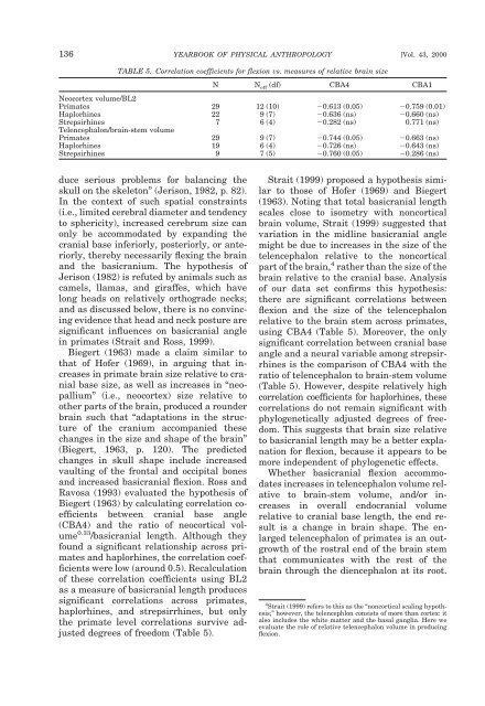
136 YEARBOOK OF PHYSICAL ANTHROPOLOGY [Vol. 43, 2000 TABLE 5. Correlation coefficients for flexion vs. measures of relative brain size N N eff (df) CBA4 CBA1 Neocortex volume/BL2 Primates 29 12 (10) 0.613 (0.05) 0.759 (0.01) Haplorhines 22 9 (7) 0.636 (ns) 0.660 (ns) Strepsirhines 7 6 (4) 0.282 (ns) 0.771 (ns) Telencephalon/brain-stem volume Primates 29 9 (7) 0.744 (0.05) 0.663 (ns) Haplorhines 19 6 (4) 0.726 (ns) 0.643 (ns) Strepsirhines 9 7 (5) 0.760 (0.05) 0.286 (ns) duce serious problems for balancing the skull on the skeleton” (Jerison, 1982, p. 82). In the context of such spatial constraints (i.e., limited cerebral diameter and tendency to sphericity), increased cerebrum size can only be accommodated by expanding the cranial base inferiorly, posteriorly, or anteriorly, thereby necessarily flexing the brain and the basicranium. The hypothesis of Jerison (1982) is refuted by animals such as camels, llamas, and giraffes, which have long heads on relatively orthograde necks; and as discussed below, there is no convincing evidence that head and neck posture are significant influences on basicranial angle in primates (Strait and Ross, 1999). Biegert (1963) made a claim similar to that of Hofer (1969), in arguing that increases in primate brain size relative to cranial base size, as well as increases in “neopallium” (i.e., neocortex) size relative to other parts of the brain, produced a rounder brain such that “adaptations in the structure of the cranium accompanied these changes in the size and shape of the brain” (Biegert, 1963, p. 120). The predicted changes in skull shape include increased vaulting of the frontal and occipital bones and increased basicranial flexion. Ross and Ravosa (1993) evaluated the hypothesis of Biegert (1963) by calculating correlation coefficients between cranial base angle (CBA4) and the ratio of neocortical volume 0.33 /basicranial length. Although they found a significant relationship across primates and haplorhines, the correlation coefficients were low (around 0.5). Recalculation of these correlation coefficients using BL2 as a measure of basicranial length produces significant correlations across primates, haplorhines, and strepsirrhines, but only the primate level correlations survive adjusted degrees of freedom (Table 5). Strait (1999) proposed a hypothesis similar to those of Hofer (1969) and Biegert (1963). Noting that total basicranial length scales close to isometry with noncortical brain volume, Strait (1999) suggested that variation in the midline basicranial angle might be due to increases in the size of the telencephalon relative to the noncortical part of the brain, 4 rather than the size of the brain relative to the cranial base. Analysis of our data set confirms this hypothesis: there are significant correlations between flexion and the size of the telencephalon relative to the brain stem across primates, using CBA4 (Table 5). Moreover, the only significant correlation between cranial base angle and a neural variable among strepsirrhines is the comparison of CBA4 with the ratio of telencephalon to brain-stem volume (Table 5). However, despite relatively high correlation coefficients for haplorhines, these correlations do not remain significant with phylogenetically adjusted degrees of freedom. This suggests that brain size relative to basicranial length may be a better explanation for flexion, because it appears to be more independent of phylogenetic effects. Whether basicranial flexion accommodates increases in telencephalon volume relative to brain-stem volume, and/or increases in overall endocranial volume relative to cranial base length, the end result is a change in brain shape. The enlarged telencephalon of primates is an outgrowth of the rostral end of the brain stem that communicates with the rest of the brain through the diencephalon at its root. 4 Strait (1999) refers to this as the “noncortical scaling hypothesis;” however, the telencephlon consists of more than cortex: it also includes the white matter and the basal ganglia. Here we evaluate the role of relative telencephalon volume in producing flexion.
D.E. Lieberman et al.] PRIMATE CRANIAL BASE 137 Increasing the size of the outer cortex of the telencephalon (the neocortex) while still connecting to the rest of the brain through the diencephalon might be expected to generate a more spheroid shape, regardless of any functional constraints on skull or brain shape. In other words, the telencephalon may be spheroidal because of the geometry of its connections and the way it develops, rather than for any functional or adaptive reason. Alternately, a spheroidal cerebrum may minimize “wiring length” in the brain, a potentially important principle of design in neural architecture (Allman and Kaas, 1974; Barlow, 1986; Mitchison, 1991; Cherniak, 1995; Van Essen, 1997). Accordingly, a spheroid telencephalon may optimize neocortical wiring lengths as well as minimize the distance from all points in the cerebrum to the diencephalon, a structure through which all connections to the rest of the brain must pass (Ross and Henneberg, 1995). Another possible advantage of a flexed basicranium derives from the in vitro experiments of Demes (1985), showing that the angulation of the cranial base in combination with a spherical neurocranium helps distribute applied stresses efficiently over a large area and decreases stresses in the anterior cranial base during loading of the temporomandibular joint. This interesting model, however, requires further testing. Whether the spheroid shape of the telencephalon is a functional adaptation or a structural consequence of geometry and developmental processes remains to be determined. Nevertheless, the presence of the cerebellum, and ultimately of the brain stem, prevents caudal expansion of the telencephalon, making rostral expansion of the telencephalon the easiest route. This would cause the especially large human brain to develop a kink of the kind measured by Hofer, which in turn may cause flexion of the basicranium. If this hypothesis is correct, then some proportion of the variation in basicranial angle among primates is caused by intrinsic changes in brain shape, and not the relationship between the size of the brain and the base on which it sits. One caution (noted above) is that ontogenetic data suggest that the interspecific variation in cranial base angle and shape presented above is partially a consequence of variables other than relative encephalization or intrinsic brain shape. Ontogenetic data are useful because they allow one to examine temporal relationships among predicted causal factors. The human ontogenetic data provide mixed support for the hypothesis that cranial base angulation reflects relative encephalization. Jeffery (1999) found no significant relationship between CBA1 and IRE1 during the second fetal trimester in humans, when brain growth is especially rapid; but Lieberman and McCarthy (1999) found that the human cranial base flexes rapidly during the first 2 postnatal years, when most brain growth occurs. Why relative brain size in humans correlates with cranial base angle after birth but not before remains to be explained. In addition, and in contrast to humans, the cranial base in all nonhuman primates so far analyzed extends rather than flexes during the period of postnatal brain growth, and continues to extend throughout the period of facial growth, after brain growth has ceased. In Pan, for example, approximately 88% of cranial base extension (CBA1) occurs after the brain has reached 95% adult size (Lieberman and McCarthy, 1999). Similar results characterize other genera (e.g., Macaca; Sirianni and Swindler, 1985; Schneiderman, 1992). Ontogenetic data do not disprove the hypothesis that variation in cranial base angle is related to brain size, but instead highlight the likelihood that the processes which generate variation in cranial base angle are polyphasic and multifactorial. Notably, the ontogenetic data suggest that the tight structural relationship between the face and the anterior cranial base (discussed below) is also an important influence on cranial base angle. This suggests that a large proportion of the interspecific variation in CBA, IRE, and other aspects of neural size and shape reported above is explained by interactions between the brain and the cranial base prior to the end of the neural growth phase. Thereafter, other factors (especially those related to the face) influence the shape of the cranial base. One obvious way to test this hypothesis is to compare the
- Page 1 and 2: YEARBOOK OF PHYSICAL ANTHROPOLOGY 4
- Page 3 and 4: D.E. Lieberman et al.] PRIMATE CRAN
- Page 5 and 6: D.E. Lieberman et al.] PRIMATE CRAN
- Page 7 and 8: D.E. Lieberman et al.] PRIMATE CRAN
- Page 9 and 10: D.E. Lieberman et al.] PRIMATE CRAN
- Page 11 and 12: D.E. Lieberman et al.] PRIMATE CRAN
- Page 13 and 14: D.E. Lieberman et al.] PRIMATE CRAN
- Page 15 and 16: D.E. Lieberman et al.] PRIMATE CRAN
- Page 17 and 18: D.E. Lieberman et al.] PRIMATE CRAN
- Page 19: D.E. Lieberman et al.] PRIMATE CRAN
- Page 23 and 24: D.E. Lieberman et al.] PRIMATE CRAN
- Page 25 and 26: D.E. Lieberman et al.] PRIMATE CRAN
- Page 27 and 28: D.E. Lieberman et al.] PRIMATE CRAN
- Page 29 and 30: D.E. Lieberman et al.] PRIMATE CRAN
- Page 31 and 32: Species or specimen IRE1 1 IRE5 2 (
- Page 33 and 34: D.E. Lieberman et al.] PRIMATE CRAN
- Page 35 and 36: D.E. Lieberman et al.] PRIMATE CRAN
- Page 37 and 38: D.E. Lieberman et al.] PRIMATE CRAN
- Page 39 and 40: D.E. Lieberman et al.] PRIMATE CRAN
- Page 41 and 42: D.E. Lieberman et al.] PRIMATE CRAN
- Page 43 and 44: D.E. Lieberman et al.] PRIMATE CRAN
- Page 45 and 46: D.E. Lieberman et al.] PRIMATE CRAN
- Page 47 and 48: D.E. Lieberman et al.] PRIMATE CRAN
- Page 49 and 50: D.E. Lieberman et al.] PRIMATE CRAN
- Page 51 and 52: D.E. Lieberman et al.] PRIMATE CRAN
- Page 53: D.E. Lieberman et al.] PRIMATE CRAN
136 YEARBOOK OF PHYSICAL ANTHROPOLOGY [Vol. 43, 2000<br />
TABLE 5. Correlation coefficients for flexion vs. measures of relative brain size<br />
N N eff (df) CBA4 CBA1<br />
Neocortex volume/BL2<br />
Primates 29 12 (10) 0.613 (0.05) 0.759 (0.01)<br />
Haplorhines 22 9 (7) 0.636 (ns) 0.660 (ns)<br />
Strepsirhines 7 6 (4) 0.282 (ns) 0.771 (ns)<br />
Telencephalon/brain-stem volume<br />
Primates 29 9 (7) 0.744 (0.05) 0.663 (ns)<br />
Haplorhines 19 6 (4) 0.726 (ns) 0.643 (ns)<br />
Strepsirhines 9 7 (5) 0.760 (0.05) 0.286 (ns)<br />
duce serious problems for balancing the<br />
skull on the skeleton” (Jerison, 1982, p. 82).<br />
In the context of such spatial constraints<br />
(i.e., limited cerebral diameter <strong>and</strong> tendency<br />
to sphericity), increased cerebrum size can<br />
only be accommodated by exp<strong>and</strong>ing the<br />
<strong>cranial</strong> <strong>base</strong> inferiorly, posteriorly, or anteriorly,<br />
thereby necessarily flexing the brain<br />
<strong>and</strong> the basicranium. <strong>The</strong> hypothesis of<br />
Jerison (1982) is refuted by animals such as<br />
camels, llamas, <strong>and</strong> giraffes, which have<br />
long heads on relatively orthograde necks;<br />
<strong>and</strong> as discussed below, there is no convincing<br />
evidence that head <strong>and</strong> neck posture are<br />
significant influences on basi<strong>cranial</strong> angle<br />
in <strong>primate</strong>s (Strait <strong>and</strong> Ross, 1999).<br />
Biegert (1963) made a claim similar to<br />
that of Hofer (1969), in arguing that increases<br />
in <strong>primate</strong> brain size relative to <strong>cranial</strong><br />
<strong>base</strong> size, as well as increases in “neopallium”<br />
(i.e., neocortex) size relative to<br />
other parts of the brain, produced a rounder<br />
brain such that “adaptations in the structure<br />
of the cranium accompanied these<br />
changes in the size <strong>and</strong> shape of the brain”<br />
(Biegert, 1963, p. 120). <strong>The</strong> predicted<br />
changes in skull shape include increased<br />
vaulting of the frontal <strong>and</strong> occipital bones<br />
<strong>and</strong> increased basi<strong>cranial</strong> flexion. Ross <strong>and</strong><br />
Ravosa (1993) evaluated the hypothesis of<br />
Biegert (1963) by calculating correlation coefficients<br />
between <strong>cranial</strong> <strong>base</strong> angle<br />
(CBA4) <strong>and</strong> the ratio of neocortical volume<br />
0.33 /basi<strong>cranial</strong> length. Although they<br />
found a significant relationship across <strong>primate</strong>s<br />
<strong>and</strong> haplorhines, the correlation coefficients<br />
were low (around 0.5). Recalculation<br />
of these correlation coefficients using BL2<br />
as a measure of basi<strong>cranial</strong> length produces<br />
significant correlations across <strong>primate</strong>s,<br />
haplorhines, <strong>and</strong> strepsirrhines, but only<br />
the <strong>primate</strong> level correlations survive adjusted<br />
degrees of freedom (Table 5).<br />
Strait (1999) proposed a hypothesis similar<br />
to those of Hofer (1969) <strong>and</strong> Biegert<br />
(1963). Noting that total basi<strong>cranial</strong> length<br />
scales close to isometry with noncortical<br />
brain volume, Strait (1999) suggested that<br />
variation in the midline basi<strong>cranial</strong> angle<br />
might be due to increases in the size of the<br />
telencephalon relative to the noncortical<br />
part of the brain, 4 rather than the size of the<br />
brain relative to the <strong>cranial</strong> <strong>base</strong>. Analysis<br />
of our data set confirms this hypothesis:<br />
there are significant correlations between<br />
flexion <strong>and</strong> the size of the telencephalon<br />
relative to the brain stem across <strong>primate</strong>s,<br />
using CBA4 (Table 5). Moreover, the only<br />
significant correlation between <strong>cranial</strong> <strong>base</strong><br />
angle <strong>and</strong> a neural variable among strepsirrhines<br />
is the comparison of CBA4 with the<br />
ratio of telencephalon to brain-stem volume<br />
(Table 5). However, despite relatively high<br />
correlation coefficients for haplorhines, these<br />
correlations do not remain significant with<br />
phylogenetically adjusted degrees of freedom.<br />
This suggests that brain size relative<br />
to basi<strong>cranial</strong> length may be a better explanation<br />
for flexion, because it appears to be<br />
more independent of phylogenetic effects.<br />
Whether basi<strong>cranial</strong> flexion accommodates<br />
increases in telencephalon volume relative<br />
to brain-stem volume, <strong>and</strong>/or increases<br />
in overall endo<strong>cranial</strong> volume<br />
relative to <strong>cranial</strong> <strong>base</strong> length, the end result<br />
is a change in brain shape. <strong>The</strong> enlarged<br />
telencephalon of <strong>primate</strong>s is an outgrowth<br />
of the rostral end of the brain stem<br />
that communicates with the rest of the<br />
brain through the diencephalon at its root.<br />
4 Strait (1999) refers to this as the “noncortical scaling hypothesis;”<br />
however, the telencephlon consists of more than cortex: it<br />
also includes the white matter <strong>and</strong> the basal ganglia. Here we<br />
evaluate the role of relative telencephalon volume in producing<br />
flexion.


