The primate cranial base: ontogeny, function and - Harvard University
The primate cranial base: ontogeny, function and - Harvard University The primate cranial base: ontogeny, function and - Harvard University
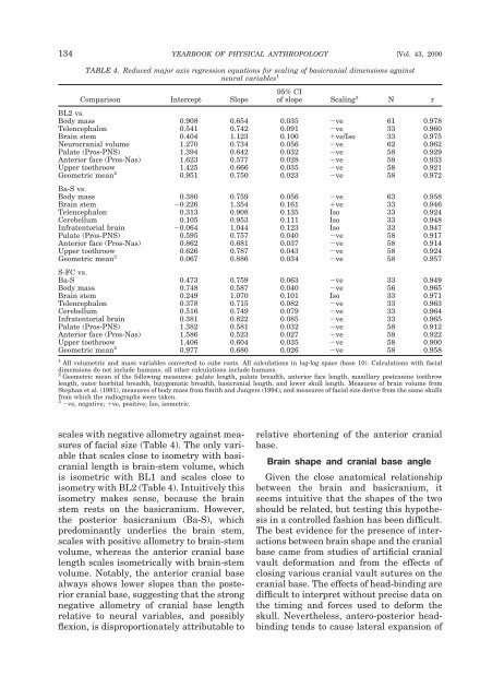
134 YEARBOOK OF PHYSICAL ANTHROPOLOGY [Vol. 43, 2000 TABLE 4. Reduced major axis regression equations for scaling of basicranial dimensions against neural variables 1 Comparison Intercept Slope 95% CI of slope Scaling 3 N r BL2 vs. Body mass 0.908 0.654 0.035 ve 61 0.978 Telencephalon 0.541 0.742 0.091 ve 33 0.960 Brain stem 0.404 1.123 0.100 ve/Iso 33 0.975 Neurocranial volume 1.270 0.734 0.056 ve 62 0.962 Palate (Pros-PNS) 1.394 0.642 0.032 ve 58 0.929 Anterior face (Pros-Nas) 1.623 0.577 0.028 ve 58 0.933 Upper toothroow 1.425 0.666 0.035 ve 58 0.921 Geometric mean 2 0.951 0.750 0.023 ve 58 0.972 Ba-S vs. Body mass 0.380 0.759 0.056 ve 63 0.958 Brain stem 0.226 1.354 0.161 ve 33 0.946 Telencephalon 0.313 0.908 0.135 Iso 33 0.924 Cerebellum 0.105 0.953 0.111 Iso 33 0.948 Infratentorial brain 0.064 1.044 0.123 Iso 33 0.947 Palate (Pros-PNS) 0.595 0.757 0.040 ve 58 0.917 Anterior face (Pros-Nas) 0.862 0.681 0.037 ve 58 0.914 Upper toothroow 0.626 0.787 0.043 ve 58 0.924 Geometric mean 2 0.067 0.886 0.034 ve 58 0.957 S-FC vs. Ba-S 0.473 0.759 0.063 ve 33 0.949 Body mass 0.748 0.587 0.040 ve 56 0.965 Brain stem 0.249 1.070 0.101 Iso 33 0.971 Telencephalon 0.378 0.715 0.082 ve 33 0.963 Cerebellum 0.516 0.749 0.079 ve 33 0.964 Infratentorial brain 0.381 0.822 0.085 ve 33 0.965 Palate (Pros-PNS) 1.382 0.581 0.032 ve 58 0.912 Anterior face (Pros-Nas) 1.586 0.523 0.027 ve 58 0.922 Upper toothroow 1.406 0.604 0.035 ve 58 0.900 Geometric mean 2 0.977 0.680 0.026 ve 58 0.958 1 All volumetric and mass variables converted to cube roots. All calculations in log-log space (base 10). Calculations with facial dimensions do not include humans; all other calculations include humans. 2 Geometric mean of the following measures: palate length, palate breadth, anterior face length, maxillary postcanine toothrow length, outer biorbital breadth, bizygomatic breadth, basicranial length, and lower skull length. Measures of brain volume from Stephan et al. (1981), measures of body mass from Smith and Jungers (1994), and measures of facial size derive from the same skulls from which the radiographs were taken. 3 ve, negative; ve, positive; Iso, isometric. scales with negative allometry against measures of facial size (Table 4). The only variable that scales close to isometry with basicranial length is brain-stem volume, which is isometric with BL1 and scales close to isometry with BL2 (Table 4). Intuitively this isometry makes sense, because the brain stem rests on the basicranium. However, the posterior basicranium (Ba-S), which predominantly underlies the brain stem, scales with positive allometry to brain-stem volume, whereas the anterior cranial base length scales isometrically with brain-stem volume. Notably, the anterior cranial base always shows lower slopes than the posterior cranial base, suggesting that the strong negative allometry of cranial base length relative to neural variables, and possibly flexion, is disproportionately attributable to relative shortening of the anterior cranial base. Brain shape and cranial base angle Given the close anatomical relationship between the brain and basicranium, it seems intuitive that the shapes of the two should be related, but testing this hypothesis in a controlled fashion has been difficult. The best evidence for the presence of interactions between brain shape and the cranial base came from studies of artificial cranial vault deformation and from the effects of closing various cranial vault sutures on the cranial base. The effects of head-binding are difficult to interpret without precise data on the timing and forces used to deform the skull. Nevertheless, antero-posterior headbinding tends to cause lateral expansion of
D.E. Lieberman et al.] PRIMATE CRANIAL BASE 135 Fig. 9. Bivariate plot of the measure by Hofer (1969) of basicranial flexion and CBA4 against his measure of brain flexion. Hofer’s brain angle is the anterior angle between Forel’s axis, from the most antero-inferior point on the frontal lobe to the most postero-inferior point on the occipital lobe, and Meynert’s axis, from the ventral edge of the junction between the pons and medulla to the caudal recess of the interpeduncular fossa. The data of Hofer (1969) measure anterior angles between lines, rather than the inferior angles favored by recent workers (e.g., Ross and Ravosa, 1993). The plotted data therefore represent the complement of Hofer’s angles. the cranial base along with a slight increase in CBA; conversely, annular head-binding tends to cause medio-lateral narrowing and antero-posterior elongation of the cranial base, also with a slight increase in CBA (Antón, 1989; Cheverud et al., 1992; Kohn et al., 1993). Natural or experimentally induced premature closure of sutures (synostoses) in the cranial vault have similarly predictable effects. For example, bilateral coronal synostoses cause antero-posterior shortening of the cranial base (Babler, 1989; David et al., 1989), and unilateral coronal synostoses (plagiocephaly) cause marked asymmetry in the cranial vault, cranial base, and face. Interspecific analyses of the relationship between brain shape and cranial base shape in primates are rare. Hofer (1965, 1969) measured the orientation of the cerebral hemispheres relative to the brain stem in primates using two axes: Forel’s axis, from the most antero-inferior point on the frontal lobe to the most postero-inferior point on the occipital lobe, measuring the orientation of the inferior surface of the cerebral hemispheres; and Meynert’s axis, from the ventral edge of the junction between the pons and medulla to the caudal recess of the interpeduncular fossa, quantifying the orientation of the brain stem. Hofer (1965) also measured the angle of the midline cranial base using a modified version of the angle of Landzert (1866), similar to the CBA4 used by Ross and Ravosa (1993). Figure 9, a plot of the measure by Hofer of basicranial angle against his measure of brain angle, illustrates that these variables are highly correlated and scale isometrically with each other (i.e., have a slope of 1.0). As the cerebrum flexes on the brain stem, the planum sphenoideum flexes relative to the clivus. The explanation of Hofer (1969) for this phenomenon is that the telencephalon becomes more spherical as it enlarges, to minimize surface area relative to volume. An alternative hypothesis is that increasing the antero-posterior diameter of the head “would be disastrous, making larger animals unusually long-headed, and would pro-
- Page 1 and 2: YEARBOOK OF PHYSICAL ANTHROPOLOGY 4
- Page 3 and 4: D.E. Lieberman et al.] PRIMATE CRAN
- Page 5 and 6: D.E. Lieberman et al.] PRIMATE CRAN
- Page 7 and 8: D.E. Lieberman et al.] PRIMATE CRAN
- Page 9 and 10: D.E. Lieberman et al.] PRIMATE CRAN
- Page 11 and 12: D.E. Lieberman et al.] PRIMATE CRAN
- Page 13 and 14: D.E. Lieberman et al.] PRIMATE CRAN
- Page 15 and 16: D.E. Lieberman et al.] PRIMATE CRAN
- Page 17: D.E. Lieberman et al.] PRIMATE CRAN
- Page 21 and 22: D.E. Lieberman et al.] PRIMATE CRAN
- Page 23 and 24: D.E. Lieberman et al.] PRIMATE CRAN
- Page 25 and 26: D.E. Lieberman et al.] PRIMATE CRAN
- Page 27 and 28: D.E. Lieberman et al.] PRIMATE CRAN
- Page 29 and 30: D.E. Lieberman et al.] PRIMATE CRAN
- Page 31 and 32: Species or specimen IRE1 1 IRE5 2 (
- Page 33 and 34: D.E. Lieberman et al.] PRIMATE CRAN
- Page 35 and 36: D.E. Lieberman et al.] PRIMATE CRAN
- Page 37 and 38: D.E. Lieberman et al.] PRIMATE CRAN
- Page 39 and 40: D.E. Lieberman et al.] PRIMATE CRAN
- Page 41 and 42: D.E. Lieberman et al.] PRIMATE CRAN
- Page 43 and 44: D.E. Lieberman et al.] PRIMATE CRAN
- Page 45 and 46: D.E. Lieberman et al.] PRIMATE CRAN
- Page 47 and 48: D.E. Lieberman et al.] PRIMATE CRAN
- Page 49 and 50: D.E. Lieberman et al.] PRIMATE CRAN
- Page 51 and 52: D.E. Lieberman et al.] PRIMATE CRAN
- Page 53: D.E. Lieberman et al.] PRIMATE CRAN
134 YEARBOOK OF PHYSICAL ANTHROPOLOGY [Vol. 43, 2000<br />
TABLE 4. Reduced major axis regression equations for scaling of basi<strong>cranial</strong> dimensions against<br />
neural variables 1<br />
Comparison Intercept Slope<br />
95% CI<br />
of slope Scaling 3 N r<br />
BL2 vs.<br />
Body mass 0.908 0.654 0.035 ve 61 0.978<br />
Telencephalon 0.541 0.742 0.091 ve 33 0.960<br />
Brain stem 0.404 1.123 0.100 ve/Iso 33 0.975<br />
Neuro<strong>cranial</strong> volume 1.270 0.734 0.056 ve 62 0.962<br />
Palate (Pros-PNS) 1.394 0.642 0.032 ve 58 0.929<br />
Anterior face (Pros-Nas) 1.623 0.577 0.028 ve 58 0.933<br />
Upper toothroow 1.425 0.666 0.035 ve 58 0.921<br />
Geometric mean 2 0.951 0.750 0.023 ve 58 0.972<br />
Ba-S vs.<br />
Body mass 0.380 0.759 0.056 ve 63 0.958<br />
Brain stem 0.226 1.354 0.161 ve 33 0.946<br />
Telencephalon 0.313 0.908 0.135 Iso 33 0.924<br />
Cerebellum 0.105 0.953 0.111 Iso 33 0.948<br />
Infratentorial brain 0.064 1.044 0.123 Iso 33 0.947<br />
Palate (Pros-PNS) 0.595 0.757 0.040 ve 58 0.917<br />
Anterior face (Pros-Nas) 0.862 0.681 0.037 ve 58 0.914<br />
Upper toothroow 0.626 0.787 0.043 ve 58 0.924<br />
Geometric mean 2 0.067 0.886 0.034 ve 58 0.957<br />
S-FC vs.<br />
Ba-S 0.473 0.759 0.063 ve 33 0.949<br />
Body mass 0.748 0.587 0.040 ve 56 0.965<br />
Brain stem 0.249 1.070 0.101 Iso 33 0.971<br />
Telencephalon 0.378 0.715 0.082 ve 33 0.963<br />
Cerebellum 0.516 0.749 0.079 ve 33 0.964<br />
Infratentorial brain 0.381 0.822 0.085 ve 33 0.965<br />
Palate (Pros-PNS) 1.382 0.581 0.032 ve 58 0.912<br />
Anterior face (Pros-Nas) 1.586 0.523 0.027 ve 58 0.922<br />
Upper toothroow 1.406 0.604 0.035 ve 58 0.900<br />
Geometric mean 2 0.977 0.680 0.026 ve 58 0.958<br />
1 All volumetric <strong>and</strong> mass variables converted to cube roots. All calculations in log-log space (<strong>base</strong> 10). Calculations with facial<br />
dimensions do not include humans; all other calculations include humans.<br />
2 Geometric mean of the following measures: palate length, palate breadth, anterior face length, maxillary postcanine toothrow<br />
length, outer biorbital breadth, bizygomatic breadth, basi<strong>cranial</strong> length, <strong>and</strong> lower skull length. Measures of brain volume from<br />
Stephan et al. (1981), measures of body mass from Smith <strong>and</strong> Jungers (1994), <strong>and</strong> measures of facial size derive from the same skulls<br />
from which the radiographs were taken.<br />
3 ve, negative; ve, positive; Iso, isometric.<br />
scales with negative allometry against measures<br />
of facial size (Table 4). <strong>The</strong> only variable<br />
that scales close to isometry with basi<strong>cranial</strong><br />
length is brain-stem volume, which<br />
is isometric with BL1 <strong>and</strong> scales close to<br />
isometry with BL2 (Table 4). Intuitively this<br />
isometry makes sense, because the brain<br />
stem rests on the basicranium. However,<br />
the posterior basicranium (Ba-S), which<br />
predominantly underlies the brain stem,<br />
scales with positive allometry to brain-stem<br />
volume, whereas the anterior <strong>cranial</strong> <strong>base</strong><br />
length scales isometrically with brain-stem<br />
volume. Notably, the anterior <strong>cranial</strong> <strong>base</strong><br />
always shows lower slopes than the posterior<br />
<strong>cranial</strong> <strong>base</strong>, suggesting that the strong<br />
negative allometry of <strong>cranial</strong> <strong>base</strong> length<br />
relative to neural variables, <strong>and</strong> possibly<br />
flexion, is disproportionately attributable to<br />
relative shortening of the anterior <strong>cranial</strong><br />
<strong>base</strong>.<br />
Brain shape <strong>and</strong> <strong>cranial</strong> <strong>base</strong> angle<br />
Given the close anatomical relationship<br />
between the brain <strong>and</strong> basicranium, it<br />
seems intuitive that the shapes of the two<br />
should be related, but testing this hypothesis<br />
in a controlled fashion has been difficult.<br />
<strong>The</strong> best evidence for the presence of interactions<br />
between brain shape <strong>and</strong> the <strong>cranial</strong><br />
<strong>base</strong> came from studies of artificial <strong>cranial</strong><br />
vault deformation <strong>and</strong> from the effects of<br />
closing various <strong>cranial</strong> vault sutures on the<br />
<strong>cranial</strong> <strong>base</strong>. <strong>The</strong> effects of head-binding are<br />
difficult to interpret without precise data on<br />
the timing <strong>and</strong> forces used to deform the<br />
skull. Nevertheless, antero-posterior headbinding<br />
tends to cause lateral expansion of


