Femtosecond Photoemission Investigation of Electron Dynamics at ...
Femtosecond Photoemission Investigation of Electron Dynamics at ...
Femtosecond Photoemission Investigation of Electron Dynamics at ...

Create successful ePaper yourself
Turn your PDF publications into a flip-book with our unique Google optimized e-Paper software.
Università C<strong>at</strong>tolica del Sacro Cuore<br />
Dipartimento di M<strong>at</strong>em<strong>at</strong>ica e Fisica<br />
<strong>Femtosecond</strong> <strong>Photoemission</strong> <strong>Investig<strong>at</strong>ion</strong><br />
<strong>of</strong> <strong>Electron</strong> <strong>Dynamics</strong> <strong>at</strong> Surfaces<br />
Ph.D. Thesis<br />
Emanuele Pedersoli<br />
Brescia, 2006
Scuola di Dottor<strong>at</strong>o di Ricerca in<br />
Fisica, Astr<strong>of</strong>isica e Fisica Applic<strong>at</strong>a<br />
XIX ciclo<br />
Università degli Studi di Milano<br />
Facoltà di Scienze M<strong>at</strong>em<strong>at</strong>iche, Fisiche e N<strong>at</strong>urali<br />
Dipartimento di Fisica, Milano (Italy)<br />
Università C<strong>at</strong>tolica del Sacro Cuore<br />
Facoltà di Scienze M<strong>at</strong>em<strong>at</strong>iche, Fisiche e N<strong>at</strong>urali<br />
Dipartimento di M<strong>at</strong>em<strong>at</strong>ica e Fisica, Brescia (Italy)<br />
(Sede Consorzi<strong>at</strong>a)<br />
Pr<strong>of</strong>. Gianpaolo Bellini (Director)<br />
Pr<strong>of</strong>. Fulvio Parmigiani (Supervisor)
Contents<br />
Introduction 1<br />
Overview . . . . . . . . . . . . . . . . . . . . . . . . . . . . . . . . . . 1<br />
Outline . . . . . . . . . . . . . . . . . . . . . . . . . . . . . . . . . . . 3<br />
I Photoc<strong>at</strong>hodes 5<br />
1 Quantum efficiency <strong>of</strong> copper photoc<strong>at</strong>hodes 7<br />
1.1 Introduction . . . . . . . . . . . . . . . . . . . . . . . . . . . . . . 7<br />
1.2 Vectorial photoelectric effect . . . . . . . . . . . . . . . . . . . . . 8<br />
1.3 Quantum efficiency measurements . . . . . . . . . . . . . . . . . 11<br />
1.3.1 Experimental setup and sample characteriz<strong>at</strong>ion . . . . . 11<br />
1.3.2 Experimental results . . . . . . . . . . . . . . . . . . . . . 16<br />
1.4 Discussion . . . . . . . . . . . . . . . . . . . . . . . . . . . . . . . 16<br />
1.5 Conclusion . . . . . . . . . . . . . . . . . . . . . . . . . . . . . . 19<br />
Acknowledgments . . . . . . . . . . . . . . . . . . . . . . . . . . . . . . 19<br />
2 Monte Carlo transverse emittance study on Cs 2 Te 21<br />
2.1 Introduction . . . . . . . . . . . . . . . . . . . . . . . . . . . . . . 21<br />
2.2 Theoretical model to describe Cs 2 Te . . . . . . . . . . . . . . . . 22<br />
2.3 Photoemitted electron angular distribution . . . . . . . . . . . . 25<br />
I
II<br />
CONTENTS<br />
2.3.1 Trajectory randomiz<strong>at</strong>ion upon sc<strong>at</strong>tering . . . . . . . . . 27<br />
2.3.2 Quasi elastic sc<strong>at</strong>tering . . . . . . . . . . . . . . . . . . . 29<br />
2.4 Transverse emittance . . . . . . . . . . . . . . . . . . . . . . . . . 34<br />
2.4.1 Cs 2 Te transverse emittance calcul<strong>at</strong>ions . . . . . . . . . . 35<br />
2.4.2 Effects <strong>of</strong> surface aging and contamin<strong>at</strong>ion . . . . . . . . . 36<br />
2.5 Conclusions . . . . . . . . . . . . . . . . . . . . . . . . . . . . . . 38<br />
Acknowledgments . . . . . . . . . . . . . . . . . . . . . . . . . . . . . . 38<br />
II Surface and Image Potential St<strong>at</strong>es on Noble Metals 39<br />
3 Phase shift model 41<br />
3.1 Introduction . . . . . . . . . . . . . . . . . . . . . . . . . . . . . . 42<br />
3.2 Solution for the phase shift model . . . . . . . . . . . . . . . . . 43<br />
3.3 Effective mass <strong>of</strong>f the image potential st<strong>at</strong>es . . . . . . . . . . . . 45<br />
3.3.1 Dependence <strong>of</strong> φ C on the position in the gap . . . . . . . 45<br />
( )<br />
3.3.2 Binding energy ε n k‖ and effective mass m ∗ . . . . . . . 47<br />
4 Experimental Setup 51<br />
4.1 <strong>Femtosecond</strong> pulsed amplified Ti:Sapphire laser system . . . . . . 51<br />
4.2 Pulse modific<strong>at</strong>ion and characteriz<strong>at</strong>ion . . . . . . . . . . . . . . 53<br />
4.3 Ultrahigh vacuum chamber . . . . . . . . . . . . . . . . . . . . . 54<br />
4.4 <strong>Photoemission</strong> Measurements . . . . . . . . . . . . . . . . . . . . 57<br />
5 Spin orbit splitting on Au(111) surface st<strong>at</strong>e 61<br />
5.1 Introduction . . . . . . . . . . . . . . . . . . . . . . . . . . . . . . 61<br />
5.2 Discussion <strong>of</strong> experimental results . . . . . . . . . . . . . . . . . . 64<br />
5.3 Conclusions . . . . . . . . . . . . . . . . . . . . . . . . . . . . . . 67<br />
6 Comparison between theory and experiment on Cu(111) and<br />
Cu(100) surface electronic st<strong>at</strong>es. 69<br />
6.1 Introduction . . . . . . . . . . . . . . . . . . . . . . . . . . . . . . 70
CONTENTS<br />
III<br />
6.2 Theoretical framework . . . . . . . . . . . . . . . . . . . . . . . . 72<br />
6.3 Experiment . . . . . . . . . . . . . . . . . . . . . . . . . . . . . . 73<br />
6.4 Results and discussion . . . . . . . . . . . . . . . . . . . . . . . . 73<br />
6.5 Conclusions . . . . . . . . . . . . . . . . . . . . . . . . . . . . . . 79<br />
Acknowledgments . . . . . . . . . . . . . . . . . . . . . . . . . . . . . . 82<br />
7 Role <strong>of</strong> <strong>at</strong>hermal electrons in non-linear photoemission from<br />
Ag(100) 83<br />
7.1 Introduction . . . . . . . . . . . . . . . . . . . . . . . . . . . . . . 84<br />
7.2 Two-photon or three-photon photoemission . . . . . . . . . . . . 85<br />
7.3 <strong>Photoemission</strong> autocorrel<strong>at</strong>ion . . . . . . . . . . . . . . . . . . . . 89<br />
7.4 Conclusions . . . . . . . . . . . . . . . . . . . . . . . . . . . . . . 94<br />
8 Angle resolved photoemission study <strong>of</strong> image potential st<strong>at</strong>es<br />
and surface st<strong>at</strong>e on Cu(111) 95<br />
8.1 Introduction . . . . . . . . . . . . . . . . . . . . . . . . . . . . . . 96<br />
8.2 Results and discussion . . . . . . . . . . . . . . . . . . . . . . . . 97<br />
8.3 Conclusions . . . . . . . . . . . . . . . . . . . . . . . . . . . . . . 103<br />
9 Image potential st<strong>at</strong>e effective mass vari<strong>at</strong>ion with hot electrons<br />
popul<strong>at</strong>ion on Cu(111) 105<br />
9.1 Introduction . . . . . . . . . . . . . . . . . . . . . . . . . . . . . . 106<br />
9.2 Experimental Setup . . . . . . . . . . . . . . . . . . . . . . . . . 106<br />
9.3 Results and discussion . . . . . . . . . . . . . . . . . . . . . . . . 107<br />
9.4 Conclusions . . . . . . . . . . . . . . . . . . . . . . . . . . . . . . 113<br />
Appendix 115<br />
A Calcul<strong>at</strong>ions about the phase shift model 115<br />
A.1 Energy <strong>of</strong> the image potential st<strong>at</strong>e . . . . . . . . . . . . . . . . . 115<br />
A.1.1 Nearly free electron model . . . . . . . . . . . . . . . . . . 115
IV<br />
CONTENTS<br />
A.1.2 Total energy <strong>of</strong> the image potential st<strong>at</strong>e . . . . . . . . . 116<br />
A.1.3 Energy associ<strong>at</strong>ed with the imaginary part q . . . . . . . 119<br />
A.1.4 Non-kinetic part <strong>of</strong> the energy . . . . . . . . . . . . . . . 119<br />
A.2 Wavefunction <strong>of</strong> the image potential st<strong>at</strong>e . . . . . . . . . . . . . 120<br />
A.2.1 Schrödinger’s equ<strong>at</strong>ion . . . . . . . . . . . . . . . . . . . . 120<br />
A.2.2 Calcul<strong>at</strong>ion <strong>of</strong> the ψ k (r) deriv<strong>at</strong>ives . . . . . . . . . . . . 121<br />
A.2.3 Solution <strong>of</strong> Schrödinger’s equ<strong>at</strong>ion . . . . . . . . . . . . . 121<br />
A.2.4 Phase φ C due to the reflection on the crystal surface . . . 123<br />
A.3 Hydrogen-like Rydberg series <strong>of</strong> st<strong>at</strong>es . . . . . . . . . . . . . . . 124<br />
List <strong>of</strong> Public<strong>at</strong>ions 127<br />
Bibliography 129
Introduction<br />
Overview<br />
Ultrafast pulsed lasers have allowed in recent years the investig<strong>at</strong>ion <strong>of</strong> physical<br />
phenomena in the femtosecond timescale through time resolved techniques.<br />
In solid st<strong>at</strong>e spectroscopy they are a powerful tool to study topics such as<br />
electron interactions, transiently popul<strong>at</strong>ed empty band st<strong>at</strong>es, ultrafast linear<br />
and non-linear photoemission phenomena; the gener<strong>at</strong>ion <strong>of</strong> short electron<br />
bunches is another important achievement <strong>of</strong> fast light sources. Future devices<br />
will provide X ray femtosecond pulses to implement time resolved diffraction<br />
and <strong>at</strong>tosecond pulses to probe faster electronic processes.<br />
<strong>Femtosecond</strong> pulses are very vers<strong>at</strong>ile in photoemission spectroscopy. The<br />
short dur<strong>at</strong>ion <strong>of</strong> the excit<strong>at</strong>ion, in photoc<strong>at</strong>hodes with fast response, can produce<br />
short electron bunches. Harmonic or sum frequency gener<strong>at</strong>ion and parametric<br />
amplific<strong>at</strong>ion in non-linear optical crystals allow tunability <strong>of</strong> the exciting<br />
photon from the infrared to the ultraviolet range. The high peak intensity <strong>of</strong><br />
short light pulses gener<strong>at</strong>es phenomena such as coherent many-photon photoemission<br />
from occupied st<strong>at</strong>es and non-linear photoemission from normally empty<br />
st<strong>at</strong>es popul<strong>at</strong>ed by relax<strong>at</strong>ion <strong>of</strong> non-equilibrium excited electrons.<br />
Impinging with ultrafast laser light on a solid surface it is possible both to<br />
control some changes in electronic dynamics, by the vari<strong>at</strong>ion <strong>of</strong> photon energy,<br />
1
2 Introduction<br />
light intensity, direction and polariz<strong>at</strong>ion, and to probe them, by photoemission<br />
spectroscopy. In this thesis two main topics were investig<strong>at</strong>ed: the first is the<br />
theoretical and experimental analysis <strong>of</strong> the femtosecond photoemission yield <strong>of</strong><br />
Cu and Cs 2 Te photoc<strong>at</strong>hodes; the second deals with photoemission from surface<br />
and image potential st<strong>at</strong>es on noble metals.<br />
New gener<strong>at</strong>ion light sources like free electron lasers are expected, in a few<br />
years, to supply femtosecond coherent X ray pulses. These devices are based on<br />
the acceler<strong>at</strong>ion <strong>of</strong> short electron bunches photoemitted by femtosecond laser<br />
pulses: the quality <strong>of</strong> the electron beam has to be studied and improved by the<br />
point <strong>of</strong> view <strong>of</strong> time dur<strong>at</strong>ion, quantum efficiency and velocity distribution.<br />
The quantum efficiency <strong>of</strong> copper photoc<strong>at</strong>hodes were investig<strong>at</strong>ed varying the<br />
angle <strong>of</strong> incidence between the light beam and the normal to the sample surface,<br />
evidencing an enhanced efficiency in p polariz<strong>at</strong>ion which is not connected to<br />
absorption increase: this phenomenon is known as vectorial photoelectric effect,<br />
here explained in terms <strong>of</strong> non-local conductivity tensor. The thermal emittance<br />
<strong>of</strong> photoelectrons from Cs 2 Te thin films was also calcul<strong>at</strong>ed by a Monte Carlo<br />
simul<strong>at</strong>ion, considering the effects <strong>of</strong> electron phonon sc<strong>at</strong>tering and discussing<br />
the dependence <strong>of</strong> thermal emittance and quantum yield on the electron affinity;<br />
the results highlight the importance <strong>of</strong> considering the m<strong>at</strong>erial contamin<strong>at</strong>ion.<br />
The investig<strong>at</strong>ion <strong>of</strong> surface Shockley st<strong>at</strong>es and image potential st<strong>at</strong>es on<br />
low indexes faces <strong>of</strong> noble metals allowed to go through several topics concerning<br />
surface electron dynamics. In particular, image potential st<strong>at</strong>es popul<strong>at</strong>ion<br />
is a two dimensional nearly free electron gas whose internal interactions are<br />
very sensitive to the laser induced popul<strong>at</strong>ion <strong>of</strong> the near projected bulk bands:<br />
different electron behaviors can be caused by laser pulses and revealed by effective<br />
mass and inverse lifetimes measurements performed through non-linear<br />
photoemission spectroscopy. Experimental d<strong>at</strong>a are compared with theoretical<br />
calcul<strong>at</strong>ions and can be qualit<strong>at</strong>ively explained by the phase shift model.<br />
Shockley surface st<strong>at</strong>es were also investig<strong>at</strong>ed, obtaining results in agreement<br />
with liter<strong>at</strong>ure; the most interesting case is Au(111) whose surface st<strong>at</strong>e shows
Outline 3<br />
a spin orbit splitting due to a spin polariz<strong>at</strong>ion <strong>of</strong> the splitted branches <strong>of</strong> its<br />
parabolic dispersion.<br />
Outline<br />
This thesis is composed <strong>of</strong> two main parts.<br />
Part I deals with the characteriz<strong>at</strong>ion <strong>of</strong> photoc<strong>at</strong>hodes th<strong>at</strong> can be used<br />
as a source <strong>of</strong> femtosecond electrons bunches: in Chap. 1 experiments on copper<br />
photoc<strong>at</strong>hodes are reported, in which we measured the photoemission total<br />
yield, finding for p polarized light a quantum efficiency enhancement known as<br />
vectorial photoelectric effect [1]; Chap. 2 reports the results <strong>of</strong> Monte Carlo<br />
calcul<strong>at</strong>ions on thermal emittance and quantum yield <strong>of</strong> Cs 2 Te irradi<strong>at</strong>ed by a<br />
light beam [2].<br />
In Part II we study the behavior <strong>of</strong> Shockley surface st<strong>at</strong>es and image potential<br />
st<strong>at</strong>es on noble metals. Chap. 3 reports the phase shift model, the theoretical<br />
description <strong>of</strong> the image potential st<strong>at</strong>es; calcul<strong>at</strong>ions are shown in App. A. In<br />
Chap. 4 we show the experimental setup used for experiments described in the<br />
following.<br />
The two following chapters describe some measurements confirming theoretical<br />
calcul<strong>at</strong>ions: measurements on the spin orbit splitting <strong>of</strong> the Shockley<br />
surface st<strong>at</strong>es Au(111) are shown in Chap. 5; Chap. 6 reports theoretical calcul<strong>at</strong>ions<br />
on Cu(111) and Cu(100) surface electronic st<strong>at</strong>es, compared with the<br />
corresponding experimental results [3].<br />
The last three chapters deal with the behavior <strong>of</strong> image potential st<strong>at</strong>es in<br />
presence <strong>of</strong> an <strong>at</strong>hermal electrons popul<strong>at</strong>ion due to femtosecond laser pulses<br />
excit<strong>at</strong>ion. Chap. 7 presents time resolved pump & probe experiments studying<br />
non-linear photoemission from Ag(100) and the influence <strong>of</strong> <strong>at</strong>hermal hot electrons<br />
on it [4]. The last two chapters investig<strong>at</strong>e the behavior <strong>of</strong> the Cu(111)<br />
image potential st<strong>at</strong>e depending on the empty projected bulk bands popul<strong>at</strong>ion<br />
by hot electrons excited by the laser pulse: Chap. 8 presents two-photon photoe-
4 Introduction<br />
mission spectroscopy, performed both in resonance with the energy difference<br />
between Shockley and image potential st<strong>at</strong>e and out <strong>of</strong> resonance [5]; two-color<br />
photoemission spectroscopy, th<strong>at</strong> allows to control the hot electrons popul<strong>at</strong>ion<br />
via the pump intensity, and two-photon photoemission are reported in Chap. 9.
Part I<br />
Photoc<strong>at</strong>hodes<br />
5
Chapter 1<br />
Quantum efficiency <strong>of</strong><br />
copper photoc<strong>at</strong>hodes<br />
Quantum efficiency measurements <strong>of</strong> single photon photoemission from a<br />
Cu(111) single crystal and a Cu polycrystal photoc<strong>at</strong>hodes, irradi<strong>at</strong>ed by 150 fs<br />
6.28 eV laser pulses, are reported over a broad range <strong>of</strong> the incidence angle θ,<br />
both in s and p polariz<strong>at</strong>ions. The maximum quantum efficiency (about 4 ×<br />
10 −4 ) for polycrystalline Cu is obtained in p polariz<strong>at</strong>ion <strong>at</strong> an angle <strong>of</strong> incidence<br />
θ = 65 ◦ , gre<strong>at</strong>er than the angle <strong>of</strong> maximum light absorption; in fact we observe<br />
a photoemission enhancement in p polariz<strong>at</strong>ion which can not be explained in<br />
terms <strong>of</strong> optical absorption: this phenomenon is known as vectorial photoelectric<br />
effect. Issues concerning surface roughness and symmetry consider<strong>at</strong>ions are<br />
addressed; an explan<strong>at</strong>ion in terms <strong>of</strong> non-local conductivity tensor is proposed.<br />
1.1 Introduction<br />
Although studied both theoretically and experimentally for more than a<br />
century, photoemission from solids is still non-completely understood, even for<br />
7
8 Chapter 1. Quantum efficiency <strong>of</strong> copper photoc<strong>at</strong>hodes<br />
well known systems such as noble metals photoc<strong>at</strong>hodes. Nevertheless, a better<br />
knowledge <strong>of</strong> this m<strong>at</strong>ter is requested for many theoretical and applic<strong>at</strong>ive<br />
aims, one <strong>of</strong> which is the advent <strong>of</strong> the fourth gener<strong>at</strong>ion free electron laser<br />
sources [6, 7, 8]. A fundamental issue regards the photoc<strong>at</strong>hode m<strong>at</strong>erial for the<br />
laser-driven photoinjector devices, to obtain short electron bunches with high<br />
charge density and low emittance. Metal photoc<strong>at</strong>hodes are good candid<strong>at</strong>es,<br />
having a high reliability, long lifetime and a fast time response between 1 and<br />
10 fs. However, two major drawbacks limit their usefulness: the small quantum<br />
efficiency and the high work function, requiring light source in the ultraviolet<br />
for efficient linear photoemission.<br />
In this chapter we analyze the vari<strong>at</strong>ion <strong>of</strong> the quantum efficiency <strong>of</strong> copper<br />
photoc<strong>at</strong>hodes as a function <strong>of</strong> light polariz<strong>at</strong>ion and incidence angle between<br />
the normal to the sample surface and the direction <strong>of</strong> the impinging light beam.<br />
Results evidence an enhancement <strong>of</strong> the quantum efficiency for p polariz<strong>at</strong>ion<br />
as compared to wh<strong>at</strong> would be expected taking into account only the electromagnetic<br />
absorption process, in accordance with the phenomenon known as<br />
vectorial photoelectric effect, which can be rel<strong>at</strong>ed to a rapid sp<strong>at</strong>ial vari<strong>at</strong>ion<br />
<strong>of</strong> the electric field through the sample surface.<br />
1.2 Vectorial photoelectric effect<br />
The quantum efficiency dependence on angle <strong>of</strong> incidence and light polariz<strong>at</strong>ion<br />
is a long standing problem [9, 10, 11, 12, 13] th<strong>at</strong> largely remains to be<br />
understood. Our d<strong>at</strong>a are well fitted by a phenomenological model [11] th<strong>at</strong><br />
keeps into account only light absorption, without any explan<strong>at</strong>ion in terms <strong>of</strong><br />
microscopic quantum physics.<br />
If we consider a light beam impinging on a surface sample, we can decompose<br />
its electric field vector E inside the sample in his components E ‖ = E p‖ + E s<br />
parallel and E ⊥ = E p⊥ perpendicular to the surface <strong>of</strong> the sample, where E p<br />
and E s are the p and s polarized field components respectively, as shown in
1.2. Vectorial photoelectric effect 9<br />
Figure 1.1: Represent<strong>at</strong>ion <strong>of</strong> angles θ and θ t and wave vectors k and k t for<br />
incident and transmitted light and field components addressed in the text; the<br />
polariz<strong>at</strong>ion angle α is also shown. A real index <strong>of</strong> refraction n is assumed for<br />
the present figure.<br />
Fig. 1.1. Calling ε ‖ = ε p‖ + ε s and ε ⊥ = ε p⊥ the electromagnetic energy inside<br />
the sample due to the light with electric vector E ‖ and E ⊥ respectively, we<br />
assume th<strong>at</strong> the number <strong>of</strong> photoemitted electrons is simply proportional to the<br />
absorbed energy, but with different efficiency due to polariz<strong>at</strong>ion, and then we<br />
write the number <strong>of</strong> photoemitted electrons n f (θ) as a function <strong>of</strong> the angle <strong>of</strong><br />
incidence as<br />
n f (θ) = aε ‖ (θ) + bε ⊥ (θ)<br />
= a[ε ‖ (θ) + (b/a)ε ⊥ (θ)].<br />
(1.1)<br />
The definition <strong>of</strong> Quantum efficiency Q(θ) can now be given as the r<strong>at</strong>io<br />
between the number <strong>of</strong> photoelectron n f (θ) and the number <strong>of</strong> photons f impinging<br />
on the sample surface, resulting<br />
Q(θ) = n f (θ)<br />
f<br />
= a[ε ‖(θ) + (b/a)ε ⊥ (θ)]<br />
, (1.2)<br />
f<br />
th<strong>at</strong> can be normalized dividing by the quantum efficiency <strong>at</strong> normal incidence,
10 Chapter 1. Quantum efficiency <strong>of</strong> copper photoc<strong>at</strong>hodes<br />
obtaining<br />
Q(θ)<br />
Q(0) = [a/f][ε ‖(θ) + (b/a)ε ⊥ (θ)]<br />
= ε ‖(θ)<br />
[a/f]ε ‖ (0) ε ‖ (0) + r ε ⊥(θ)<br />
ε ‖ (0) , (1.3)<br />
where a, b and r = b/a are parameters th<strong>at</strong> do not depend on the angle <strong>of</strong><br />
incidence θ, and ε ⊥ (0) = 0.<br />
We can now deal with two particular conditions <strong>of</strong> the polariz<strong>at</strong>ion: with p<br />
polarized light we can write<br />
while for s polariz<strong>at</strong>ion we have E ⊥ = 0 and so<br />
Q p (θ)<br />
Q p (0) = ε p‖(θ)<br />
ε p‖ (0) + r ε p⊥(θ)<br />
ε p‖ (0) , (1.4)<br />
Q s (θ)<br />
Q s (0) = ε s(θ)<br />
ε s (0) . (1.5)<br />
The three r<strong>at</strong>ios in the right parts <strong>of</strong> Eq. (1.4), (1.5) can be calcul<strong>at</strong>ed from<br />
the classical theory <strong>of</strong> electrodynamics [11, 14]: for a metal like Cu, whose complex<br />
index <strong>of</strong> refraction is n = 0.98 + 1.49i [15], all non-reflected photons are<br />
absorbed before penetr<strong>at</strong>ing more than about 10 nm into the sample; we can<br />
identify the electromagnetic energies inside the solid ε ‖ (θ) and ε ⊥ (θ) with the<br />
total absorptions 1−R p (θ) and 1−R s (θ), where R p (θ) and R s (θ) are the reflectivities<br />
calcul<strong>at</strong>ed from the Fresnel laws for p and s polariz<strong>at</strong>ion respectively.<br />
Calling θ t the complex angle between the transmitted beam and the normal<br />
to the surface (see Fig. 1.1) and defining l = (n 2 − sin 2 θ) 1/2 = n cos(θ t ), we<br />
obtain<br />
R p (θ) = |n2 cos θ − l| 2<br />
|n 2 cos θ + l| 2 , R | cos θ − l|2<br />
s(θ) =<br />
| cos θ + l| 2 (1.6)<br />
and the quantum efficiency r<strong>at</strong>ios eventually become<br />
Q p (θ)<br />
Q p (0) = 1 − R p(θ)<br />
1 − R p (0) · |E p‖| 2 + r|E p⊥ | 2<br />
|E p‖ | 2 + |E p⊥ | 2 ,<br />
Q s (θ)<br />
Q s (0) = 1 − R s(θ)<br />
1 − R s (0) . (1.7)
1.3. Quantum efficiency measurements 11<br />
We note th<strong>at</strong> quantum efficiency <strong>of</strong> s polariz<strong>at</strong>ion light is expected to be simply<br />
proportional to the light absorption governed by Fresnel laws, while in p<br />
polariz<strong>at</strong>ion a vectorial photoelectric effect is predicted with a characteristic<br />
parameter r; in this case, the maximum quantum efficiency is obtained <strong>at</strong> an<br />
incident angle θ M different from the pseudo Brewster angle θ B <strong>of</strong> maximum<br />
light absorption.<br />
At normal incidence the electric field polariz<strong>at</strong>ion is always parallel to the<br />
surface, allowing to write Q p (0) = Q s (0) = Q(0); in Fig. 1.2 we show, as<br />
an example for n = 0.98 + 1.49i, r = 10 and Q(0) = 1 × 10 −4 , a graph <strong>of</strong> the<br />
calcul<strong>at</strong>ed value <strong>of</strong> the quantum efficiency as a function <strong>of</strong> the angle <strong>of</strong> incidence<br />
θ and <strong>of</strong> the angle <strong>of</strong> polariz<strong>at</strong>ion α, defined as drawn in Fig. 1.1.<br />
1.3 Quantum efficiency measurements<br />
Quantum efficiency measurements <strong>of</strong> single photon photoemission from a<br />
Cu(111) single crystal and a Cu polycrystal photoc<strong>at</strong>hodes, irradi<strong>at</strong>ed by laser<br />
beams <strong>of</strong> 150 fs pulse dur<strong>at</strong>ion and hν = 6.28 eV photon energy, are reported<br />
over a broad range <strong>of</strong> incidence angles, both in s and p polariz<strong>at</strong>ions.<br />
1.3.1 Experimental setup and sample characteriz<strong>at</strong>ion<br />
The experimental setup is described in Fig. 1.3.<br />
Our source is an amplified Ti:Sapphire laser supplying pulses with a dur<strong>at</strong>ion<br />
less than 150 fs and an average power <strong>of</strong> 600 mW <strong>at</strong> 1 kHz repetition r<strong>at</strong>e, giving<br />
a pulse energy <strong>of</strong> 600 µJ and a peak power <strong>of</strong> about 10 10 W. The laser is tuned<br />
on a near infrared wavelength <strong>of</strong> 790 nm whose ultraviolet fourth harmonic<br />
with photon energy hν = 6.28 eV is used in our photoemission experiments.<br />
This photon is obtained by a double process <strong>of</strong> second harmonic gener<strong>at</strong>ion<br />
in a beta-barium-bor<strong>at</strong>e (βBBO) crystal: the first step is in phase m<strong>at</strong>ching<br />
and guarantees a high efficiency; for the doubling <strong>of</strong> the hν = 3.14 eV second
12 Chapter 1. Quantum efficiency <strong>of</strong> copper photoc<strong>at</strong>hodes<br />
Figure 1.2: Calcul<strong>at</strong>ed value <strong>of</strong> the quantum efficiency as a function <strong>of</strong> θ and α<br />
for n = 0.98 + 1.49i, r = 10 and Q(0) = 1 × 10 −4 .
1.3. Quantum efficiency measurements 13<br />
Figure 1.3: Experimental setup for quantum efficiency measurement on copper<br />
photoc<strong>at</strong>hodes.
14 Chapter 1. Quantum efficiency <strong>of</strong> copper photoc<strong>at</strong>hodes<br />
harmonic photons there is no phase m<strong>at</strong>ching allowed, but the small yield <strong>of</strong><br />
fourth harmonic photons requested for photoemission experiments is obtained<br />
in a thin (200 µm) BBO crystal. After the non-linear optics steps, we obtain<br />
three collinear beams <strong>of</strong> different colors: they are sp<strong>at</strong>ially dispersed by a MgF 2<br />
wedge prism, with minimal temporal and pulse front tilt distortions, and the<br />
beam with the desired photon energy hν = 6.28 eV is selected by a pinhole and<br />
impinges on the sample in the ultra high vacuum (UHV) chamber through a<br />
beam splitter and an optical flange.<br />
The ultra high vacuum chamber has a base pressure less than 2×10 −10 mbar<br />
<strong>at</strong> room temper<strong>at</strong>ure; samples are cleaned by cycles <strong>of</strong> Ar + sputtering followed<br />
by annealing <strong>at</strong> 500 ◦ C. This procedure is continued until the proper value <strong>of</strong><br />
the work function (4.65 eV for the polycrystal and 4.94 eV for the single crystal<br />
[15]) is measured in photoemission spectra acquired by a time <strong>of</strong> flight (ToF)<br />
spectrometer; in these conditions a clear low energy electron diffraction (LEED)<br />
p<strong>at</strong>tern for the Cu(111) sample is obtained. <strong>Photoemission</strong> spectra are also<br />
acquired during total yield measurements in order to measure the sample work<br />
function and monitor possible onset <strong>of</strong> sample contamin<strong>at</strong>ions and space charge<br />
effects.<br />
A more efficient third harmonic conversion, supplying a photon energy hν =<br />
4.71 eV, is the most usual to study polycrystal photoc<strong>at</strong>hodes, but for our<br />
purposes it is too close to the copper work function. We preferred the fourth<br />
harmonic photon because its higher energy allows to obtain linear photoemission<br />
also from samples with higher work function, due to the surface l<strong>at</strong>tice structure<br />
<strong>of</strong> the single crystal or to the onset <strong>of</strong> sample contamin<strong>at</strong>ion. Moreover,<br />
ultraviolet short laser pulses induce the removal <strong>of</strong> oxide contaminants and the<br />
breaking <strong>of</strong> chemical bonds on the surface, contributing to maintain the sample<br />
cleanliness; this effect improves with shorter wavelengths [16, 17]: working with<br />
a 6.28 eV photon energy should thus help to increase the duty time <strong>of</strong> machines<br />
based on Cu photoc<strong>at</strong>hodes.<br />
Several <strong>at</strong>omic force microscopy (AFM) scans <strong>of</strong> the samples surface, with
1.3. Quantum efficiency measurements 15<br />
Figure 1.4: Atomic force microscopy images <strong>of</strong> the two samples’ surfaces. Measured<br />
route mean squared roughness is 20 nm for the Cu polycrystal and 2 nm<br />
for the Cu(111) single crystal.<br />
sizes ranging from 1 × 1 µm 2 to 60 × 60 µm 2 , give values <strong>of</strong> the root mean<br />
squared roughness h rms ≃ 20 nm for the Cu polycrystal and h rms ≃ 2 nm for<br />
the Cu(111) single crystal (see Fig. 1.4).<br />
The quantum efficiency Q is the r<strong>at</strong>io between the number <strong>of</strong> photoemitted<br />
electrons n f , obtained from the photocurrent measured from the sample<br />
with a Keithley 6485 Picoammeter, and the number <strong>of</strong> incident photons f, detected<br />
measuring on a Tektronix TDS3054B digital oscilloscope the output <strong>of</strong> a<br />
Hamam<strong>at</strong>su R928 photomultiplier tube. The laser peak intensity on the target,<br />
regul<strong>at</strong>ed by a λ/2 wavepl<strong>at</strong>e coupled with a polarizer, is I ≃ 5 × 10 5 W/cm 2 :<br />
these conditions avoid space charge and guarantee th<strong>at</strong> non-linear photoemission<br />
is negligible if compared to one-photon photoemission, th<strong>at</strong> is predominant.<br />
A small part <strong>of</strong> the impinging beam is directed to the photomultiplier through<br />
a beam splitter and three mirrors in order to <strong>at</strong>tenu<strong>at</strong>e the light power not to<br />
s<strong>at</strong>ur<strong>at</strong>e the instrument. Due to experimental condition, only p polarized light<br />
intensity can be measured; s polarized light is renormalized supposing th<strong>at</strong> <strong>at</strong>
16 Chapter 1. Quantum efficiency <strong>of</strong> copper photoc<strong>at</strong>hodes<br />
normal incidence there can not be any difference between the two polariz<strong>at</strong>ions.<br />
1.3.2 Experimental results<br />
The quantum efficiency for linear photoemission in the femtosecond regime<br />
is measured as a function <strong>of</strong> the incidence angle θ in the angular range −55 ◦ ≤<br />
θ ≤ +80 ◦ , both in s and p polariz<strong>at</strong>ions on a Cu polycrystal and a Cu(111)<br />
single crystal. Results are plotted in Fig. 1.5.<br />
Experimental d<strong>at</strong>a obtained on the two samples are plotted for both p (circles)<br />
and s (triangles) polariz<strong>at</strong>ions and fitted by Eq. (1.7) (lines); dashed<br />
lines represent the behavior expected taking into account light absorption only<br />
(r = 1), for p polariz<strong>at</strong>ion.<br />
For p polarized light, an enhancement <strong>of</strong> the quantum efficiency is evident in<br />
both samples as compared to wh<strong>at</strong> would be expected taking into account only<br />
the electromagnetic absorption process. D<strong>at</strong>a are in good agreement with the<br />
model described in Sec. 1.2, showing a vectorial photoelectric effect. The fitting<br />
procedures give the best values <strong>of</strong> the fitting parameter r <strong>of</strong> Eq. (1.7): r = 13 for<br />
the polycrystalline Cu and r = 9 for the Cu(111) single crystal. The maximum<br />
value <strong>of</strong> Q does not occur <strong>at</strong> the pseudo Brewster angle <strong>of</strong> incidence θ B = 57 ◦ ,<br />
where there is maximum absorption, but is shifted by about 8 ◦ toward grazing<br />
incidence: the maximum quantum efficiency Q ≃ 4 × 10 −4 , obtained with p<br />
polariz<strong>at</strong>ion <strong>at</strong> θ = 65 ◦ , is four times the value <strong>at</strong> normal incidence.<br />
Eq. (1.7) is in agreement with d<strong>at</strong>a for s polariz<strong>at</strong>ion too, showing th<strong>at</strong> in<br />
this case the photoemission yield n f is simply proportional to the number <strong>of</strong><br />
absorbed photons f (1 − R s (θ)), according to the Fresnel laws.<br />
1.4 Discussion<br />
Experimental d<strong>at</strong>a exposed in Sec. 1.3 show a behavior in good agreement<br />
with the vectorial photoelectric effect described in Sec. 1.2.
1.4. Discussion 17<br />
Figure 1.5: Measurements <strong>of</strong> quantum efficiency dependence on the angle <strong>of</strong><br />
incidence θ for a Cu polycrystal and a Cu(111) single crystal for p (circles) and<br />
s (triangles) polarized light. Fits, based on Eq. (1.7), are reported as solid lines.<br />
The dashed lines are calcul<strong>at</strong>ed taking into account Fresnel absorption only.
18 Chapter 1. Quantum efficiency <strong>of</strong> copper photoc<strong>at</strong>hodes<br />
At the light <strong>of</strong> our d<strong>at</strong>a, it is important to investig<strong>at</strong>e the physical mechanisms<br />
th<strong>at</strong> enhances the photoelectron yield due to E ⊥ compared to the one<br />
due to E ‖ .<br />
The crystalline symmetry, important when dealing with polariz<strong>at</strong>ion dependent<br />
photoemission, play no role in the present experiment. Both in the Cu(111)<br />
single crystal, where symmetry consider<strong>at</strong>ions could apply, and in the polycrystalline<br />
Cu, where any contribution rel<strong>at</strong>ed to symmetry is canceled by the random<br />
orient<strong>at</strong>ion <strong>of</strong> the single crystals domains composing the sample, E ⊥ is<br />
about 10 times more effective than E ‖ as a cause <strong>of</strong> the photoemission process.<br />
<strong>Photoemission</strong> enhancement due to surface roughness has been recently investig<strong>at</strong>ed<br />
[18, 19, 20]. In the present case surface roughness enhancement can<br />
be ruled out: the observed vectorial photoelectric effect is comparable on both<br />
samples, despite their surface roughnesses differ by an order <strong>of</strong> magnitude, as<br />
can be seen in the <strong>at</strong>omic force microscopy scans shown in Fig. 1.4. The compar<strong>at</strong>ive<br />
study <strong>of</strong> the single crystal Cu(111) and polycrystalline Cu c<strong>at</strong>hodes<br />
allows to clarify th<strong>at</strong> our experiment is not dependent on sample morphology.<br />
Therefore, we seek for an explan<strong>at</strong>ion in terms <strong>of</strong> a more general mechanism.<br />
Solutions <strong>of</strong> the Maxwell equ<strong>at</strong>ions on an ideal jellium-vacuum interface for an<br />
impinging plane electromagnetic wave <strong>of</strong> frequency ω, evidence an electromagnetic<br />
field sp<strong>at</strong>ially varying on the length scale <strong>of</strong> about 1 Å on the jellium side<br />
[21]. The sp<strong>at</strong>ially varying electromagnetic field is due to the non-local character<br />
<strong>of</strong> the conductivity tensor. This is calcul<strong>at</strong>ed using free electron-like wave<br />
functions, so it does not depend on the symmetry <strong>of</strong> the crystal. The m<strong>at</strong>rix<br />
element entering the differential cross section for photoemission is composed <strong>of</strong><br />
two terms. The first is the usual electric dipole contribution, the second is due<br />
to the rapidly varying electric field. The second term prevails for ω < ω p , where<br />
ω p is the plasma frequency, and leads to an enhancement <strong>of</strong> the photocurrent<br />
for the electric field components perpendicular to the sample surface [22, 23].<br />
In the present experiment, ħω = 6.28 eV and ħω p ∼ 19 eV [24]. This mechanism<br />
explains an enhancement <strong>of</strong> the quantum efficiency for p polarized incident
1.5. Conclusion 19<br />
radi<strong>at</strong>ion while not affecting the results for s polarized light. Furthermore, it<br />
does not depend on surface roughness or a particular symmetry <strong>of</strong> the crystal.<br />
We therefore propose it as the main microscopic mechanism to explain our<br />
experimental evidences.<br />
1.5 Conclusion<br />
Quantum efficiency measurements on Cu photoc<strong>at</strong>hodes, irradi<strong>at</strong>ed with<br />
150 fs laser pulses <strong>at</strong> hν = 6.28 eV, are reported over a broad range <strong>of</strong> incident<br />
angles in both s and p polariz<strong>at</strong>ions. A quantum efficiency enhancement<br />
is found for light with electric field perpendicular to the sample’s surface, showing<br />
a vectorial photoelectric effect. The maximum value <strong>of</strong> quantum efficiency<br />
Q ≃ 4 × 10 −4 is four times bigger than the one measured <strong>at</strong> normal incidence<br />
and is achieved with p polarized light impinging on the sample <strong>at</strong> an incidence<br />
angle <strong>of</strong> θ = 65 ◦ , gre<strong>at</strong>er than the pseudo Brewster angle θ B .<br />
<strong>Investig<strong>at</strong>ion</strong> <strong>of</strong> both a Cu(111) single crystal and a Cu polycrystal allows<br />
us to rule out a microscopic processes based on symmetry consider<strong>at</strong>ions and<br />
surface roughness to explain our d<strong>at</strong>a. An explan<strong>at</strong>ion in terms <strong>of</strong> a rapidly<br />
varying effective field, due to the non-local character <strong>of</strong> the conductivity tensor,<br />
is suggested.<br />
Acknowledgments<br />
This work was supported by the U.S. Department <strong>of</strong> Energy, Office <strong>of</strong> Science<br />
under Contract No. DE-AC03-76SF00098.
Chapter 2<br />
Monte Carlo transverse<br />
emittance study on Cs 2 Te<br />
The thermal emittance <strong>of</strong> photoelectrons in Cs 2 Te thin films is investig<strong>at</strong>ed<br />
by a Monte Carlo simul<strong>at</strong>ion. The effects <strong>of</strong> electron-phonon sc<strong>at</strong>tering are<br />
discussed and the thermal emittance calcul<strong>at</strong>ed for a radi<strong>at</strong>ion wavelength <strong>of</strong><br />
265 nm and a spot radius <strong>of</strong> 1.5 mm, finding a value <strong>of</strong> ɛ n,rms = 0.56 mrad mm.<br />
The dependence <strong>of</strong> ɛ n,rms and the quantum yield on the electron affinity is also<br />
investig<strong>at</strong>ed. The d<strong>at</strong>a show the importance <strong>of</strong> considering aging and contamin<strong>at</strong>ion<br />
<strong>of</strong> the m<strong>at</strong>erial to assess its emittance.<br />
2.1 Introduction<br />
High quality electron beams are a fundamental tool in many applic<strong>at</strong>ion areas<br />
ranging from electron microscopy to acceler<strong>at</strong>or technology. In this context<br />
the transverse emittance ɛ n,rms <strong>of</strong> an electron beam <strong>at</strong> the surface <strong>of</strong> the photoc<strong>at</strong>hode<br />
is an important parameter: it is a measure <strong>of</strong> the beam spread in both<br />
real and momentum spaces and it constitutes the lower limit for the emittance<br />
21
22 Chapter 2. Monte Carlo transverse emittance study on Cs 2 Te<br />
th<strong>at</strong> can be gener<strong>at</strong>ed by an injector.<br />
Semiconductor photoc<strong>at</strong>hodes have <strong>at</strong>tracted <strong>at</strong>tention because <strong>of</strong> their high<br />
quantum yield [25]. In particular, Cs 2 Te is a semiconductor compound under<br />
investig<strong>at</strong>ion as a photoemitter under short laser pulse excit<strong>at</strong>ion for the<br />
new gener<strong>at</strong>ion free electron lasers (FELs) and advanced synchrotron radi<strong>at</strong>ion<br />
sources [6, 7, 8, 26]. Despite the fact th<strong>at</strong> Cs 2 Te has been studied extensively,<br />
in the past most <strong>of</strong> the efforts have been devoted to issues such as the energy<br />
distribution <strong>of</strong> the photoemitted electrons and the beam dynamics <strong>of</strong> the photoejected<br />
electron bunch under high electric field gradients, a typical topic in<br />
acceler<strong>at</strong>or physics. The issues <strong>of</strong> the photoemitted electrons’ angular distribution<br />
and thermal emittance <strong>at</strong> the c<strong>at</strong>hode have received compar<strong>at</strong>ively little<br />
<strong>at</strong>tention, nevertheless being a relevant topic both under an applic<strong>at</strong>ive and<br />
fundamental point <strong>of</strong> view.<br />
In this work we investig<strong>at</strong>e, via Monte Carlo simul<strong>at</strong>ions, the photoemitted<br />
electron angular distribution and transverse emittance <strong>at</strong> the surface <strong>of</strong> a<br />
Cs 2 Te photoc<strong>at</strong>hode under 265 nm radi<strong>at</strong>ion wavelength, the fourth harmonic <strong>of</strong><br />
Nd:YAG fundamental wavelength; the quantum yield is also calcul<strong>at</strong>ed. Attention<br />
is devoted to the microscopic mechanisms, such as electron-phonon sc<strong>at</strong>tering,<br />
affecting the photoelectrons angular distribution; the effects <strong>of</strong> aging and<br />
contamin<strong>at</strong>ion <strong>of</strong> the photoc<strong>at</strong>hode on thermal emittance and quantum yield<br />
are also discussed.<br />
2.2 Theoretical model to describe Cs 2 Te<br />
The compound Cs 2 Te is a p type semiconductor with a band gap <strong>of</strong> 3.2 eV<br />
and an electron affinity <strong>of</strong> 0.3 eV. A schem<strong>at</strong>ic band diagram is illustr<strong>at</strong>ed in<br />
Fig. 2.1. Semiconductor m<strong>at</strong>erials are known to be good photoemitters because<br />
they are good absorbers and photoexcited electrons experience little electronelectron<br />
sc<strong>at</strong>tering. A large r<strong>at</strong>io between the band gap and the electron affinity<br />
results in a high quantum efficiency for photons with energy gre<strong>at</strong>er than E g +E a
2.2. Theoretical model to describe Cs 2 Te 23<br />
Figure 2.1: Energy band diagram for Cs 2 Te. The energy gap E g = 3.2 eV separ<strong>at</strong>es<br />
the valence band from the conduction band. The vacuum level is separ<strong>at</strong>ed<br />
from the conduction band minimum by the electron affinity E a = 0.3 eV. The<br />
zero energy reference is taken <strong>at</strong> the conduction band minimum. The electron<br />
affinity E a is the potential barrier felt by a photoexcited electron approaching<br />
the surface. The electron kinetic energy E in inside the m<strong>at</strong>erial is measured<br />
with respect to the conduction band minimum. Internal and external emission<br />
polar angles for a photoemitted electron are also shown: arrows represent the<br />
momenta <strong>of</strong> the electron inside and outside the m<strong>at</strong>erial.
24 Chapter 2. Monte Carlo transverse emittance study on Cs 2 Te<br />
and less than 2E g [27].<br />
The Monte Carlo simul<strong>at</strong>ion <strong>of</strong> photoemission from Cs 2 Te follows a scheme<br />
first introduced to study electron sc<strong>at</strong>tering mechanism in metals [28] and successfully<br />
applied to simul<strong>at</strong>e the quantum yield dependence on photon energy<br />
in Cs 2 Te [29]. Calcul<strong>at</strong>ions are based on the three step model for photoemission<br />
[30], which has proved reliable to interpret photoemission d<strong>at</strong>a in many<br />
m<strong>at</strong>erials, including Cs 2 Te [31]. In this model a photoemitted electron is first<br />
photoexcited inside the m<strong>at</strong>erial (photoexcit<strong>at</strong>ion), secondly it travels toward<br />
the sample surface (transport) and as a third step it overcomes the surface<br />
barrier (emission).<br />
We consider a 30 nm Cs 2 Te polycrystalline layer deposited on a Mo substr<strong>at</strong>e<br />
and use the microscopic parameters deduced from experimental d<strong>at</strong>a taken from<br />
Ref. [29].<br />
We assume th<strong>at</strong> every absorbed photon produces a photoexcited electron.<br />
The distributions <strong>of</strong> electron kinetic energy E in , momentum p in and depth z<br />
from the surface within the m<strong>at</strong>erial are st<strong>at</strong>istically determined <strong>at</strong> the beginning<br />
<strong>of</strong> the simul<strong>at</strong>ion as in reference [31] and are recalcul<strong>at</strong>ed for every single<br />
photoexcited electron after each sc<strong>at</strong>tering event. The probability for an electron<br />
to travel without sc<strong>at</strong>tering a distance z/ cos θ in in the forward direction to<br />
the surface is proportional to exp (−z/λ p cos θ in ), where θ in is the angle <strong>of</strong> the<br />
electron trajectory inside the sample, defined in Fig. 2.1, and λ p is the electron<br />
mean free p<strong>at</strong>h, assumed to be energy-independent. With the Cs 2 Te parameters<br />
and a photon energy hν = 4.68 eV, the excited electrons are below the threshold<br />
for the onset <strong>of</strong> electron-electron sc<strong>at</strong>tering; therefore, only quasi elastic<br />
electron-phonon sc<strong>at</strong>tering is considered. Upon an electron-phonon sc<strong>at</strong>tering<br />
event, the electron energy is reduced by an amount E p = 5 meV [29] and the<br />
momentum direction is assumed to be randomized over all solid angles with the<br />
same probability; it is equivalent to consider all values <strong>of</strong> cos θ in equiprobable.<br />
When photoexcited electrons reach the surface, they are photoemitted only<br />
if the kinetic energy associ<strong>at</strong>ed to their momentum component perpendicular
2.3. Photoemitted electron angular distribution 25<br />
to the surface is gre<strong>at</strong>er than the electron affinity E a , sufficient to overcome<br />
the surface potential barrier; otherwise electrons are reflected back into the<br />
semiconductor film. The simul<strong>at</strong>ion is iter<strong>at</strong>ed until all the initial electrons have<br />
been elimin<strong>at</strong>ed after being photoemitted, crossing the Cs 2 Te/Mo interface or<br />
having a total energy E in < E a .<br />
2.3 Photoemitted electron angular distribution<br />
When electrons are photoemitted from a c<strong>at</strong>hode with photons well above<br />
threshold, a spread in the transverse and longitudinal components <strong>of</strong> the electron<br />
momentum results.<br />
The emittance <strong>of</strong> an electron bunch is a measure <strong>of</strong> its<br />
spread both in real and momentum space and depends on the spot size, the<br />
momentum distribution and the angular distribution. The thermal emittance<br />
is the emittance <strong>at</strong> the c<strong>at</strong>hode surface, investig<strong>at</strong>ed through the photoemitted<br />
electron angular distribution.<br />
As mentioned in the previous paragraph, only electrons approaching the<br />
surface with sufficient energy associ<strong>at</strong>ed to the perpendicular momentum component<br />
will cross the semiconductor-vacuum interface. Given the total electron<br />
energy <strong>at</strong> the surface E in , an electron will be photoemitted only if the internal<br />
angle θ in <strong>at</strong> the surface is lower than a critical angle θ c ≡ arccos( √ E a /E in ).<br />
The rel<strong>at</strong>ion between the internal and external polar angles is given by a<br />
solid-st<strong>at</strong>e analog <strong>of</strong> Snell’s law due to conserv<strong>at</strong>ion <strong>of</strong> transverse momentum <strong>at</strong><br />
the interface:<br />
sin(θ in )<br />
sin(θ out ) = √<br />
Ein − E a<br />
E in<br />
, (2.1)<br />
where θ in and θ out are the polar angles for the electron trajectories inside and<br />
outside the sample surface respectively, as shown in Fig. 2.1.<br />
The calcul<strong>at</strong>ed distribution <strong>of</strong> photoemitted electrons is reported in Fig. 2.2.<br />
The distribution has a maximum for θ in ≃ 34 ◦ and all photoemitted electrons<br />
are found within an internal cone defined by an angle <strong>of</strong> value max {θ in } = 63 ◦ ,
26 Chapter 2. Monte Carlo transverse emittance study on Cs 2 Te<br />
Figure 2.2: Angular probability distribution over internal angles θ in <strong>of</strong> the photoexcited<br />
electrons th<strong>at</strong> are also photoemitted. The bin angular width is chosen<br />
equal to 0.36 ◦ . The calcul<strong>at</strong>ion is performed following 10 5 electrons trajectories<br />
and assuming a value <strong>of</strong> E a = 0.3 eV for Cs 2 Te.
2.3. Photoemitted electron angular distribution 27<br />
where {θ in } denotes the set <strong>of</strong> values acquired by θ in . A physical model to<br />
explain this distribution must be considered.<br />
Assuming a random distribution over polar angles, the probability <strong>of</strong> finding<br />
an electron in the range [θ in , θ in + dθ in ] is proportional to sin θ in dθ in ; electrons<br />
with small θ in have larger probability (proportional to exp (−z/λ p cos θ in )) to<br />
reach the surface because <strong>of</strong> the shorter distance |z/ cos θ in | to travel on their<br />
trajectory. Taking into account both <strong>of</strong> these mechanisms, integr<strong>at</strong>ing over<br />
the distance z from the surface and assuming th<strong>at</strong> λ p
28 Chapter 2. Monte Carlo transverse emittance study on Cs 2 Te<br />
Figure 2.3: Angular probability distribution over internal angles θ in <strong>of</strong> the photoexcited<br />
electrons th<strong>at</strong> are also photoemitted. The assumption <strong>of</strong> a constant<br />
θ c = max {θ c } for all the electrons, regardless <strong>of</strong> their energy, is made. The plots,<br />
obtained changing the value <strong>of</strong> E a , map a function proportional to sin(2θ in ).<br />
The bin angular width is chosen equal to 0.36 ◦ .
2.3. Photoemitted electron angular distribution 29<br />
max {θ c } as expected. Moreover, changing E a amounts to changing max {θ c },<br />
with larger cut <strong>of</strong>f angles associ<strong>at</strong>ed to lower electron affinity values.<br />
The photoemitted electron angular distribution with respect to the external<br />
emission angle θ out is reported, in polar coordin<strong>at</strong>es, in Fig. 2.4 and compared<br />
with the corresponding angular distribution over internal angles. The distribution<br />
over the external angles is smeared on the range [0, 90 ◦ ] and is peaked <strong>at</strong><br />
lower polar angles as compared to wh<strong>at</strong> would be expected invoking the first<br />
two mechanisms alone.<br />
2.3.2 Quasi elastic sc<strong>at</strong>tering<br />
We now investig<strong>at</strong>e the role <strong>of</strong> electron energy loss, due to quasi elastic sc<strong>at</strong>tering,<br />
on the photoemitted electron angular probability distribution. To this<br />
purpose, we simul<strong>at</strong>e the angular probability distributions assuming both elastic<br />
(E p = 0 meV) and quasi elastic (E p = 5 meV) sc<strong>at</strong>tering. The distributions are<br />
equivalent, as can be appreci<strong>at</strong>ed in Fig. 2.5.<br />
Each quasi elastic sc<strong>at</strong>tering event reduces the electron energy by 5 meV<br />
and a photoemitted electron is found to sc<strong>at</strong>ter, on average, about 27 times:<br />
the average photoemitted electron energy loss is about 130 meV. Therefore an<br />
important issue to be explained is the negligible effect on electrons angular<br />
distribution <strong>of</strong> a mean energy loss <strong>of</strong> about 9% <strong>of</strong> max {E in }. This energy<br />
loss mainly affects the high energy part <strong>of</strong> the photoemitted electron’s internal<br />
energy distribution.<br />
To understand the mechanism let us consider an electron with E in ≃ E a .<br />
Upon sc<strong>at</strong>tering, this electron is elimin<strong>at</strong>ed from the distribution, because electrons<br />
with E in < E a cannot be photoemitted, but its energy position can be<br />
filled with an electron sc<strong>at</strong>tered from an higher energy position. This sc<strong>at</strong>tering<br />
driven electron hopping to lower energies depopul<strong>at</strong>es the high energy tail <strong>of</strong> the<br />
distribution (compare for instance the electron probability distribution for energies<br />
above 1.1 eV in the two histograms reported in Fig. 2.6, where no higher
30 Chapter 2. Monte Carlo transverse emittance study on Cs 2 Te<br />
Figure 2.4: <strong>Electron</strong>s count over internal angles θ in (green graph) and over<br />
external angles θ out (red graph) <strong>of</strong> the photoexcited electrons th<strong>at</strong> are also<br />
photoemitted. The number <strong>of</strong> photoexcited electrons assumed in the simul<strong>at</strong>ion<br />
is 10 5 . The bin angular width is chosen equal to 0.36 ◦ . The inset shows the angle<br />
orient<strong>at</strong>ion: angles amplitudes are taken clockwise starting from the neg<strong>at</strong>ive z<br />
axis.
2.3. Photoemitted electron angular distribution 31<br />
Figure 2.5: Angular probability distribution over internal angles θ in <strong>of</strong> the photoexcited<br />
electrons th<strong>at</strong> are also photoemitted. The bin angular width is chosen<br />
equal to 0.36 ◦ .
32 Chapter 2. Monte Carlo transverse emittance study on Cs 2 Te<br />
Figure 2.6: Energy probability distribution over internal kinetic energy E in<br />
<strong>of</strong> the photoemitted electrons. E in is the electron kinetic energy inside the<br />
sample, the zero energy reference being <strong>at</strong> the bottom <strong>of</strong> the conduction band<br />
and E a = 0.3 eV. The width <strong>of</strong> the energy bin is 10 meV. The light blue and<br />
red histograms show the probability distributions calcul<strong>at</strong>ed with the sc<strong>at</strong>tering<br />
event supposed elastic (E p = 0 eV) and quasi elastic (E p = 5 meV) respectively.
2.3. Photoemitted electron angular distribution 33<br />
energy electrons are present to fill the vacancies). This effect is particularly evident<br />
inspecting the function D(E in ) = ∫ E in<br />
E a<br />
[<br />
P inel (E ′ in ) − P el(E ′ in ) ]<br />
dE ′ in , th<strong>at</strong><br />
is the integral function <strong>of</strong> the difference between the two probability distributions<br />
obtained for the cases with (P inel (E ′ in )) and without (P el(E ′ in )) sc<strong>at</strong>tering<br />
(see Fig. 2.7). The probability difference function is slightly positive for ener-<br />
Figure 2.7: Graph <strong>of</strong> the integral function D(E in ) <strong>of</strong> the difference between the<br />
two probability distribution reported in Fig. 2.5, 2.6. The dashed line is the<br />
zero probability line. The width <strong>of</strong> the energy bin is 10 meV.<br />
gies below 0.5 eV, indic<strong>at</strong>ing th<strong>at</strong> the probability distributions over this range<br />
are essentially not affected by inelastic sc<strong>at</strong>tering. For energies in the range<br />
comprised between 0.5 and 1.1 eV, D(E in ) becomes neg<strong>at</strong>ive: inelastic sc<strong>at</strong>tering<br />
increases the number <strong>of</strong> electrons with kinetic energies in this interval. For<br />
energies in excess <strong>of</strong> 1.1 eV, the function D(E in ) increases up to zero value,
34 Chapter 2. Monte Carlo transverse emittance study on Cs 2 Te<br />
<strong>at</strong>tained <strong>at</strong> E in ≃ 1.4 eV, meaning th<strong>at</strong> electrons added by inelastic sc<strong>at</strong>tering<br />
to the previous energy interval are actually transferred from the 1.1-1.4 eV energy<br />
interval. This is the mechanism we addressed as inelastic sc<strong>at</strong>tering driven<br />
electron hopping.<br />
The high energy tail <strong>of</strong> the probability distribution contributes equally to<br />
all emission angles and, for this reason, is the portion th<strong>at</strong> least affects the<br />
photoemitted electron angular distribution. This can be appreci<strong>at</strong>ed noting<br />
th<strong>at</strong> θ c is monotonic with E in and recalling the role <strong>of</strong> θ c in determining the<br />
angular electron distribution. We thus conclude th<strong>at</strong> the energy loss due to<br />
the quasi elastic electron-phonon sc<strong>at</strong>tering plays a negligible role in the photoemitted<br />
electron angular distribution. It is the trajectory randomiz<strong>at</strong>ion <strong>of</strong><br />
electron-phonon sc<strong>at</strong>tering th<strong>at</strong> strongly affects the photoemitted electron angular<br />
distribution via the mechanism explained in the previous subsection.<br />
2.4 Transverse emittance<br />
The normalized transverse root mean square emittance ɛ n,rms is a measure<br />
<strong>of</strong> the electron beam phase space density th<strong>at</strong> may be deduced by measuring<br />
the root mean square beam divergence <strong>at</strong> the c<strong>at</strong>hode for a given beam spot<br />
size; it is defined as<br />
ɛ n,rms =<br />
mc√ 1 σ2 (x)σ 2 (p x ) − σ 2 (xp x ), (2.2)<br />
where σ 2 (x) ≡ N −1 ∑ N<br />
i=1 (x i − x) 2 1 , x ≡ N −1 ∑ N<br />
i=1 x i, m is the electron<br />
rest mass and c the speed <strong>of</strong> light; the subscript i indic<strong>at</strong>es quantities referred<br />
to the i-th photoemitted electron <strong>at</strong> the surface, whereas N is the number <strong>of</strong><br />
photoemitted electrons. The correl<strong>at</strong>ion term σ 2 (xp x ) vanishes <strong>at</strong> the source,<br />
since the quantities x and p x are uncorrel<strong>at</strong>ed, and x and p x are null, hence<br />
1 The actual definition should be σ 2 (x) ≡ (N − 1) −1 P N<br />
i=1 (x i − x) 2 , however, as soon as<br />
N is big enough, we can consider (N − 1) ≃ N.
2.4. Transverse emittance 35<br />
Eq. (2.2) reduces to<br />
ɛ n,rms = 1 mc p x,rms × x rms . (2.3)<br />
2.4.1 Cs 2 Te transverse emittance calcul<strong>at</strong>ions<br />
Figure 2.8: Probability distribution for the normalized transverse momentum<br />
p x <strong>at</strong> the c<strong>at</strong>hode for the photoemitted electron. A bin <strong>of</strong> 5 × 10 −5 is used for<br />
the normalized transverse momentum.<br />
The transverse emittance <strong>of</strong> an electron beam <strong>at</strong> the photoc<strong>at</strong>hode depends<br />
on the laser beam spot size r, which can be rel<strong>at</strong>ed to x rms , and on the transverse<br />
momentum distribution p x,rms <strong>of</strong> the photoemitted electrons. The simul<strong>at</strong>ed<br />
normalized transverse momentum probability distribution <strong>at</strong> the c<strong>at</strong>hode, calcul<strong>at</strong>ed<br />
on the basis <strong>of</strong> the photoemitted electron angular and energy probability<br />
distribution obtained in the previous paragraph, is reported in Fig. 2.8. The cal-
36 Chapter 2. Monte Carlo transverse emittance study on Cs 2 Te<br />
cul<strong>at</strong>ion is performed assuming a wavelength <strong>of</strong> 265 nm. From the distribution<br />
we obtain p x,rms /mc = 7.4 × 10 −4 . We assume an hard edge laser spot size <strong>of</strong><br />
radius r = 1.5 × 10 −3 . Making use <strong>of</strong> Eq. (2.3), we find ɛ n,rms =0.56 mrad mm.<br />
2.4.2 Effects <strong>of</strong> surface aging and contamin<strong>at</strong>ion<br />
Figure 2.9: Quantum yield (right axis) and normalized transverse root mean<br />
square emittance <strong>at</strong> the c<strong>at</strong>hode ɛ n,rms (left axis) as a function <strong>of</strong> electron<br />
affinity normalized against the maximum internal electron kinetic energy.<br />
The reliability <strong>of</strong> the photoc<strong>at</strong>hode characteristics with aging and contamin<strong>at</strong>ion<br />
<strong>of</strong> the surface is one <strong>of</strong> the key issues in the choice <strong>of</strong> the photoc<strong>at</strong>hode<br />
m<strong>at</strong>erial. Surface contamin<strong>at</strong>ion, occurring during oper<strong>at</strong>ion <strong>of</strong> the radio frequency<br />
gun, affects the m<strong>at</strong>erial electron affinity: the question arises as to wh<strong>at</strong><br />
effect this may have on ɛ n,rms and the quantum yield. The results <strong>of</strong> the simul<strong>at</strong>ions<br />
are reported in Fig. 2.9 against a value <strong>of</strong> the electron affinity normalized
2.4. Transverse emittance 37<br />
with respect to the maximum electron kinetic energy E a / (hν − E g ), a quantity<br />
from now on defined as normalized electron affinity.<br />
The clean sample corresponds, with the parameters in use, to a normalized<br />
electron affinity <strong>of</strong> 0.2, thus showing a quantum yield <strong>of</strong> 12.5%, a value in good<br />
agreement with experimental d<strong>at</strong>a [32]. The graphs show th<strong>at</strong> the main concern<br />
is the rel<strong>at</strong>ively rapid drop in quantum yield th<strong>at</strong> persists as the electron affinity<br />
grows in excess <strong>of</strong> 0.3 eV (normalized electron affinity <strong>of</strong> 0.2). The decrease<br />
in the emittance is due to the preferential selection <strong>of</strong> electrons with forward<br />
directed momentum.<br />
Figure 2.10: Normalized transverse root mean square emittance <strong>at</strong> the c<strong>at</strong>hode<br />
ɛ n,rms as a function <strong>of</strong> electron affinity normalized against the maximum electron<br />
kinetic energy. The empty squares are obtained on the basis <strong>of</strong> Monte Carlo<br />
simul<strong>at</strong>ion. The full triangles are obtained applying the analytical calcul<strong>at</strong>ions<br />
reported in [33] for the case with sc<strong>at</strong>tering.
38 Chapter 2. Monte Carlo transverse emittance study on Cs 2 Te<br />
The results <strong>of</strong> the Monte Carlo simul<strong>at</strong>ions are compared in Fig. 2.10 with<br />
those obtained by applying simple analytical calcul<strong>at</strong>ions [33]. The difference<br />
among the Monte Carlo simul<strong>at</strong>ion and the analytical calcul<strong>at</strong>ion is <strong>of</strong><br />
significance for low value <strong>of</strong> E a / (hν − Eg), and amounts to almost 40% for<br />
E a = 0.3 eV (normalized electron affinity <strong>of</strong> 0.2), the value corresponding to<br />
clean Cs 2 Te. This fact underlines the relevance <strong>of</strong> st<strong>at</strong>istical approaches, as<br />
Monte Carlo simul<strong>at</strong>ions, when addressing issues rel<strong>at</strong>ed to electron dynamics<br />
within the m<strong>at</strong>erial such as thermal emittance <strong>at</strong> the c<strong>at</strong>hode.<br />
2.5 Conclusions<br />
The photoemitted electron angular distribution and the transverse emittance<br />
<strong>at</strong> the c<strong>at</strong>hode surface on Cs 2 Te are investig<strong>at</strong>ed by Monte Carlo calcul<strong>at</strong>ions.<br />
An explan<strong>at</strong>ion <strong>of</strong> the photoemitted electron angular distribution is given in<br />
terms <strong>of</strong> electron redistribution over all angles smaller than the proper critical<br />
angle for each electron. <strong>Electron</strong>-phonon sc<strong>at</strong>tering affects the photoemitted<br />
electron angular distribution through the randomiz<strong>at</strong>ion <strong>of</strong> momentum direction,<br />
whereas an average energy loss <strong>of</strong> about 0.1 eV per photoemitted electron<br />
has a negligible effect. The transverse emittance, calcul<strong>at</strong>ed for an impinging<br />
radi<strong>at</strong>ion <strong>at</strong> a wavelength <strong>of</strong> 265 nm and a laser spot size <strong>of</strong> 1.5 × 10 −3 m, is<br />
about 0.56 mrad mm. The effect <strong>of</strong> aging and contamin<strong>at</strong>ion <strong>of</strong> the c<strong>at</strong>hode on<br />
ɛ n,rms and the quantum yield is also investig<strong>at</strong>ed. Our results are an improvement<br />
<strong>of</strong> those obtained with analytical calcul<strong>at</strong>ions [33] still in use for thermal<br />
emittance studies [34], and underline the importance <strong>of</strong> st<strong>at</strong>istical approaches<br />
to study photoemission processes where sc<strong>at</strong>tering is involved.<br />
Acknowledgments<br />
This work was supported by the U.S. Department <strong>of</strong> Energy, Office <strong>of</strong> Science<br />
under Contract No. DE-AC03-76SF00098.
Part II<br />
Surface and Image Potential<br />
St<strong>at</strong>es on Noble Metals<br />
39
Chapter 3<br />
Phase shift model<br />
<strong>Electron</strong>ic bulk bands calcul<strong>at</strong>ions can be performed for three dimensional<br />
infinite crystalline solids; the presence <strong>of</strong> a surface, breaking the solid symmetry<br />
in one <strong>of</strong> the three dimensions <strong>of</strong> space, allows the rising <strong>of</strong> some surface rel<strong>at</strong>ed<br />
fe<strong>at</strong>ures like Shockley surface st<strong>at</strong>es and image potential st<strong>at</strong>es.<br />
When a bulk band structure presents an inverted gap in which bands have<br />
crossed yielding s bands <strong>at</strong> the top and p bands <strong>at</strong> the bottom <strong>of</strong> the gap, the<br />
symmetry break due to the existence <strong>of</strong> a surface allows the presence in the<br />
gap <strong>of</strong> a surface st<strong>at</strong>e whose wave function has a short penetr<strong>at</strong>ion in the solid,<br />
exponentially decaying with the distance from the surface. These st<strong>at</strong>es took<br />
their name from Shockley [35] and are well known.<br />
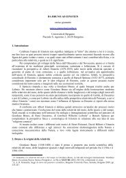
Another kind <strong>of</strong> surface st<strong>at</strong>es are the image potential st<strong>at</strong>es, described in<br />
Fig. 3.1: they represent a two dimensional free electron gas trapped in front <strong>of</strong><br />
a solid surface when electrons neither can fall into the bulk, for the presence <strong>of</strong><br />
a forbidden bulk band gap <strong>at</strong> their energy, nor can escape into the vacuum, because<br />
their presence in front <strong>of</strong> the surface repels electrons in the solid, cre<strong>at</strong>ing<br />
an image charge whose Coulomb potential represents a long range barrier th<strong>at</strong><br />
traps electron in the image potential st<strong>at</strong>e. These electrons are represented by a<br />
41
42 Chapter 3. Phase shift model<br />
standing wave function th<strong>at</strong> is multiply reflected between the crystal barrier and<br />
the Coulomb image potential barrier: the behavior <strong>of</strong> these st<strong>at</strong>es is described<br />
in the phase shift model, th<strong>at</strong> we analyze in the following <strong>of</strong> this chapter.<br />
Figure 3.1: Description <strong>of</strong> image potential st<strong>at</strong>es. The image charge inside the<br />
solid cre<strong>at</strong>es a Coulomb potential th<strong>at</strong> <strong>at</strong>tracts electrons along the z direction:<br />
they are trapped in a two dimensional free electron gas. Images are taken from<br />
Ref. [36].<br />
3.1 Introduction<br />
The phase shift model describes the wavefunctions <strong>of</strong> electrons in the image<br />
potential st<strong>at</strong>es as multiply reflected standing waves between a Coulomb<br />
boundary, due to an image charge in the solid, and a crystal boundary, due to<br />
the gap in the bulk bands. In each reflection on the crystal, the wavefunction is<br />
multiplied by a factor r C e iφ C<br />
, while a factor r B e iφ B<br />
is due to each reflection on<br />
the Coulomb image potential boundary. To obtain a st<strong>at</strong>ionary wave we impose<br />
r B = r C = 1 and φ B + φ C = 2nπ, n ∈ Z.<br />
Although the definition <strong>of</strong> a realistic potential m<strong>at</strong>ching the periodic l<strong>at</strong>tice<br />
inside the crystal and the Coulomb-like behavior outside is not trivial and several<br />
different models were proposed to handle the problem [37, 38, 39, 40, 41],<br />
all <strong>of</strong> these potentials have more or less the form described in Fig. 3.2: after
3.2. Solution for the phase shift model 43<br />
the outermost crystal layer, the bulk potential is continuously m<strong>at</strong>ched to an<br />
external Coulomb potential.<br />
Figure 3.2: Electric potential <strong>at</strong> the surface boundary between the bulk solid<br />
l<strong>at</strong>tice (z < 0) and the Coulomb potential outside the surface (z > 0).<br />
3.2 Solution for the phase shift model<br />
In this section we report some well known results <strong>of</strong> the phase shift model<br />
[42, 43]; calcul<strong>at</strong>ions are shown in App. A. We consider the vacuum energy level<br />
as the origin <strong>of</strong> the energy axis (E V = 0).<br />
In a nearly free electron model context, we can consider a band structure<br />
with a surface st<strong>at</strong>e placed in a gap <strong>of</strong> amplitude 2V g . The surface st<strong>at</strong>e’s<br />
wavefunction exponentially decays in the bulk and, as seen in Eq. (A.32), has<br />
the form<br />
ψ(r) = e qz cos(pz + δ) p, q ∈ R, (3.1)
44 Chapter 3. Phase shift model<br />
where z is the coordin<strong>at</strong>e on the axis perpendicular to the crystal surface, z < 0<br />
in the bulk, k = (p − iq)ẑ and δ is a phase we will define in Eq. (3.5); the total<br />
energy <strong>of</strong> the st<strong>at</strong>e is (Eq. (A.15))<br />
Defining<br />
E = ħ2 k‖<br />
2 √<br />
2m + ħ2 p 2<br />
2m − ħ2 q 2<br />
2m ± Vg 2 − 4 ħ2 p 2 ħ 2 q 2<br />
2m 2m . (3.2)<br />
E g = ħ2 p 2<br />
2m<br />
and<br />
ε = E − ħ2 k 2 ‖<br />
2m = ħ2 u 2<br />
2m , (3.3)<br />
we can identify an energy associ<strong>at</strong>ed with the imaginary part q <strong>of</strong> the wavevector<br />
(Eq. (A.19))<br />
ħ 2 q 2<br />
√<br />
2m = − (ε + E g) + Vg 2 + 4εE g (3.4)<br />
and a non-kinetic part <strong>of</strong> the energy (Eq. (A.21), (A.22))<br />
E − ħ2 k 2<br />
2m = V ge i2δ ,<br />
sin(2δ) = − ħ2<br />
2m 2pq/V g. (3.5)<br />
M<strong>at</strong>ching <strong>at</strong> the image plane z = z 0 the bulk image potential st<strong>at</strong>e wavefunction<br />
(3.1) for z < z 0 with the standing wave e −iuz + r C e iφ C<br />
e iuz for z > z 0<br />
with r C = 1 and imposing the function’s and first deriv<strong>at</strong>ive’s continuity, we<br />
obtain (Eq. (A.37))<br />
u is defined in Eq. (3.3).<br />
u tan(φ C /2) = p tan(pz 0 + δ) − q; (3.6)<br />
The dependence <strong>of</strong> the phase φ B added by the wavefunction reflection on<br />
the image potential on the binding energy ε can be calcul<strong>at</strong>ed analytically or<br />
numerically for several models <strong>of</strong> different complexity. The simplest is a reflection<br />
on a Coulomb potential well, whose solution can be regarded as a useful<br />
approxim<strong>at</strong>ion, yielding for the reflection phase the dependence [44]<br />
(√ )<br />
3.4 eV<br />
φ B = π<br />
− 1 ; (3.7)<br />
ε
3.3. Effective mass <strong>of</strong>f the image potential st<strong>at</strong>es 45<br />
imposing the st<strong>at</strong>ionarity condition φ B +φ C = 2nπ, n ∈ N according to Sec. A.3,<br />
we obtain a Rydberg series <strong>of</strong> hydrogen-like st<strong>at</strong>es labeled by the quantum<br />
number n, whose binding energy is (Eq. (A.42))<br />
ε n =<br />
0.85 eV<br />
(n + a) 2 , (3.8)<br />
where we introduced the quantum defect (Eq. (A.41))<br />
a = 1 (<br />
1 − φ )<br />
C<br />
. (3.9)<br />
2 π<br />
3.3 Effective mass <strong>of</strong>f the image potential st<strong>at</strong>es<br />
Image potential st<strong>at</strong>es are popul<strong>at</strong>ed by a two dimensional free electron gas:<br />
the energy part directly depending on k ‖ are then expected to show a free<br />
electron parabolic dispersion, yielding for the total energy the expression<br />
E ( k ‖<br />
)<br />
= εn<br />
(<br />
k‖<br />
)<br />
+<br />
ħ 2 k 2 ‖<br />
2m ; (3.10)<br />
in this section we want to investig<strong>at</strong>e the dependence <strong>of</strong> the binding energy<br />
ε n<br />
(<br />
k‖<br />
)<br />
on the parallel momentum k‖ .<br />
3.3.1 Dependence <strong>of</strong> φ C on the position in the gap<br />
The binding energy ε n <strong>of</strong> the image potential st<strong>at</strong>e depends on the quantum<br />
defect a which is affected by the reflection phase φ C by Eq. (3.8), (3.9); to<br />
( )<br />
predict the values for ε n k‖ we have to investig<strong>at</strong>e the behavior <strong>of</strong> φC , th<strong>at</strong><br />
depends on the position <strong>of</strong> the vacuum level with respect to the gap in the<br />
projected bulk bands, as schem<strong>at</strong>ically described in Fig. 3.3.<br />
We are interested in Shockley inverted gaps, in which the upper band is s-<br />
like and the lower band is p-like. In this case a st<strong>at</strong>e in the top <strong>of</strong> the gap has<br />
to be m<strong>at</strong>ched with even (respect to the <strong>at</strong>om positions) bulk wavefunctions<br />
and its phase is φ C = π, while in the bottom the surface st<strong>at</strong>e wavefunction
46 Chapter 3. Phase shift model<br />
Figure 3.3: Dependence <strong>of</strong> φ C on the position in the gap and its effect on<br />
the wavefunction propag<strong>at</strong>ion into the bulk. The left panel is reported from<br />
Ref. [45].
3.3. Effective mass <strong>of</strong>f the image potential st<strong>at</strong>es 47<br />
m<strong>at</strong>ches an odd function and its phase <strong>at</strong> the mirror plane is φ C = 0; the phase<br />
continuously changes from π <strong>at</strong> the top <strong>of</strong> the gap to 0 <strong>at</strong> the bottom.<br />
Obviously, st<strong>at</strong>es whose electron energy is close to any <strong>of</strong> the gap edges penetr<strong>at</strong>e<br />
in the bulk for a long depth without decaying, whereas st<strong>at</strong>es in the middle<br />
<strong>of</strong> the gap vanish faster into the solid. On the contrary, the distance between<br />
the crystal surface and the image potential st<strong>at</strong>e’s wavefunction maximum is<br />
not domin<strong>at</strong>ed by the energy distance between the st<strong>at</strong>e and the bulk bands,<br />
but continuously increases with the decreasing <strong>of</strong> φ C from the top to the bottom<br />
<strong>of</strong> the gap.<br />
This determines the behavior <strong>of</strong> the binding energy ε n : if the vacuum level<br />
is in a position th<strong>at</strong> yields the image potential st<strong>at</strong>e near the top <strong>of</strong> the gap, φ C<br />
is near to its maximum value π, the quantum defect a is small and the binding<br />
energy ε n is about 0.85/n eV; if the vacuum level is <strong>at</strong> the bottom <strong>of</strong> the gap,<br />
φ C<br />
tends to 0, a reaches its maximum value 1/2 and the binding energy ε n<br />
decreases.<br />
If we change the sample m<strong>at</strong>erial from one having an image potential st<strong>at</strong>e<br />
in the top <strong>of</strong> the gap to several others in which the st<strong>at</strong>e is positioned more and<br />
more down until the gap’s bottom, the values <strong>of</strong> φ C continuously decreases from<br />
π to 0 and the binding energy ε 1 <strong>of</strong> the n = 1 st<strong>at</strong>e continuously decreases from<br />
about 0.85 eV, close to the value obtained for Cu, to 0.85 eV/(1+1/2) 2 = 0.38 eV<br />
<strong>at</strong> the bottom <strong>of</strong> the gap.<br />
( )<br />
3.3.2 Binding energy ε n k‖ and effective mass m<br />
∗<br />
The dispersion <strong>of</strong> the band edges <strong>at</strong> the top and <strong>at</strong> the bottom <strong>of</strong> the bulk<br />
gap are different from the free electron and the image potential st<strong>at</strong>e dispersion:<br />
for this reason the energy distance between the image potential st<strong>at</strong>es and the<br />
band edges depends on k ‖ ; this dependence also influences the value <strong>of</strong> φ C and<br />
( )<br />
consequently <strong>of</strong> a and ε n k‖ , th<strong>at</strong> then depends on k‖ .<br />
The case <strong>of</strong> the n = 1 image potential st<strong>at</strong>e is described in Fig. 3.4. The
48 Chapter 3. Phase shift model<br />
Figure 3.4: Schem<strong>at</strong>ic represent<strong>at</strong>ion <strong>of</strong> the reason why the image potential<br />
st<strong>at</strong>e presents an effective mass m ∗ bigger than the free electron mass m.
3.3. Effective mass <strong>of</strong>f the image potential st<strong>at</strong>es 49<br />
upper bulk bands disperse less than a free electron: in the point A the st<strong>at</strong>e<br />
and the bands touch each other, φ C = π and the binding energy ε n<br />
(<br />
k‖ = k A<br />
)<br />
is maximum, as can be seen following the cyan free electron parabola; moving<br />
to the point k ‖ = k B , φ C decreases and the red free electron parabola passing<br />
through B has a smaller binding energy ε n<br />
(<br />
k‖ = k B<br />
)<br />
; in the center <strong>of</strong> the first<br />
Brillouin zone, φ C is minimum and the C point stands on the green free electron<br />
parabola with the smallest binding energy ε n<br />
(<br />
k‖ = 0 ) .<br />
To recover all the points found spanning all the k ‖ range, we draw a blue<br />
parabola th<strong>at</strong> is fl<strong>at</strong>ter than a free electron one: we define an effective mass<br />
m ∗ > m for which we can rewrite Eq. (3.10) with the approxim<strong>at</strong>ion<br />
E ( k ‖<br />
)<br />
= εn + ħ2 k 2 ‖<br />
2m ∗ , (3.11)<br />
where ε n = ε n<br />
(<br />
k‖ = 0 ) and the suppression <strong>of</strong> its dependence on the parallel<br />
momentum is compens<strong>at</strong>ed by the introduction <strong>of</strong> an effective mass bigger than<br />
the free electron one.
Chapter 4<br />
Experimental Setup<br />
In this chapter we describe the devices and the setup th<strong>at</strong> allowed us to<br />
perform experiments reported in the following chapters <strong>of</strong> Part II.<br />
4.1 <strong>Femtosecond</strong> pulsed amplified Ti:Sapphire<br />
laser system<br />
The pulsed beam for photoemission experiments is supplied by a femtosecond<br />
amplified Ti:Sapphire laser system.<br />
The seed <strong>of</strong> our light source is a Ti:Sapphire oscill<strong>at</strong>or: it provides 130 fs<br />
pulses with a 76 MHz repetition r<strong>at</strong>e <strong>at</strong> an average power <strong>of</strong> about 500 mW;<br />
the wavelength λ = 790 nm corresponds to a photon energy hν = 1.57 eV.<br />
This seed is amplified by a second Ti:Sapphire cavity: only one pulse over<br />
76000, stretched to avoid excessive power concentr<strong>at</strong>ion th<strong>at</strong> could damage the<br />
crystal, is trapped into the cavity, thanks to a Pockels cell th<strong>at</strong> changes the<br />
light’s polariz<strong>at</strong>ion. The pulse undergoes several passages into the pumped<br />
crystal to be amplified until it reaches the maximum allowed energy; it is then<br />
let out by a second polariz<strong>at</strong>ion rot<strong>at</strong>ion in the Pockels cell. The amplified pulse<br />
51
52 Chapter 4. Experimental Setup<br />
travels then in a compressor stage.<br />
The output <strong>of</strong> this amplified Ti:Sapphire system is a 130 fs pulsed infrared<br />
(λ = 790 nm, hν = 1.57 eV) laser with a 1 kHz repetition r<strong>at</strong>e <strong>at</strong> an average<br />
power <strong>of</strong> about 500 mW: each pulse carries 500 µJ and the pulse peak power<br />
can be estim<strong>at</strong>ed as the r<strong>at</strong>io between pulse energy and pulse dur<strong>at</strong>ion P P eak =<br />
5 × 10 −4 J/1.3 × 10 −13 s ≃ 4 × 10 9 W. The device is shown in Fig. 4.1.<br />
Figure 4.1: <strong>Femtosecond</strong> amplified Ti:Sapphire laser providing λ = 790 nm,<br />
hν = 1.57 eV infrared 130 fs pulses with a 1 MHz repetition r<strong>at</strong>e <strong>at</strong> an average<br />
power <strong>of</strong> about 500 mW: 500 µJ pulse energy and 4 × 10 9 W pulse peak power.
4.2. Pulse modific<strong>at</strong>ion and characteriz<strong>at</strong>ion 53<br />
4.2 Pulse modific<strong>at</strong>ion and characteriz<strong>at</strong>ion<br />
The laser beam provided by Ti:Sapphire oscill<strong>at</strong>or and amplifier is modified<br />
and characterized online. Devices like multilayer or metallic mirrors, lenses,<br />
λ/2 and λ/4 wavepl<strong>at</strong>es, polarizers, prisms allow to control direction, intensity,<br />
polariz<strong>at</strong>ion, spot size <strong>of</strong> the pulsed laser beam. For photoemission purposes, it’s<br />
also very important to control the light wavelength: tuning the photon energy<br />
allows us to perform different kinds <strong>of</strong> experiments, passing, for example, from<br />
linear to resonant or to indirect non-linear photoemission.<br />
Tunability is one important fe<strong>at</strong>ure <strong>of</strong> femtosecond pulsed sources th<strong>at</strong> allow<br />
to perform both non-linear optics in uniaxial crystals, to obtain harmonics <strong>of</strong><br />
the fundamental wavelength, and parametric amplific<strong>at</strong>ion, yielding continuous<br />
frequency tunability.<br />
In this work we report experiments in which three kinds <strong>of</strong> harmonic up<br />
conversion in beta-barium-bor<strong>at</strong>e (βBBO) crystals are used to obtain second<br />
(λ = 395 nm, hν = 3.14 eV), third (λ = 263 nm, hν = 4.71 eV) and fourth<br />
(λ = 197 nm, hν = 6.28 eV) harmonic <strong>of</strong> the fundamental wavelength. Second<br />
harmonic gener<strong>at</strong>ion is performed in a type I crystal; the third harmonic is<br />
obtained by a sum frequency gener<strong>at</strong>ion between this second harmonic beam and<br />
the fundamental in a type II crystal; the fourth harmonic is gener<strong>at</strong>ed frequency<br />
doubling the second harmonic beam out <strong>of</strong> phase m<strong>at</strong>ching in a 200 µm thin<br />
βBBO crystal.<br />
An almost continuous tunability from 1600 nm infrared to 250 nm ultraviolet<br />
is guaranteed by two parametric amplifiers, shown in Fig. 4.2, and their<br />
harmonics.<br />
The first is a traveling-wave optical parametric gener<strong>at</strong>or (TOPG) th<strong>at</strong> supplies<br />
130 fs pulses with an average power <strong>of</strong> about 30 mW and a wavelength<br />
tunable in the infrared between 1150 nm and 1500 nm (0.8 eV and 1.1 eV): its<br />
fourth harmonic spans the range 3.2 eV ≤ hν ≤ 4.4 eV. The second is a noncollinear<br />
optical parametric amplifier (NOPA) th<strong>at</strong> yields pulses with temporal
54 Chapter 4. Experimental Setup<br />
Figure 4.2: Parametric amplifiers. Their output and harmonics allow to perform<br />
photoemission with a photon energy tunability 0.8 eV ≤ hν ≤ 5 eV.<br />
width down to 20 fs with a tunability between the visible λ = 500 nm to the infrared<br />
λ = 750 nm ((1.6 eV and 2.5 eV) and an average power <strong>of</strong> about 10 mW,<br />
allowing to span the range 3.2 eV ≤ hν ≤ 5 eV with the second harmonic.<br />
The output wavelength is measured by an online spectrometer. Pulse dur<strong>at</strong>ion<br />
and temporal shape can be measured by an online home made fast autocorrel<strong>at</strong>or<br />
shown in Fig. 4.3.<br />
4.3 Ultrahigh vacuum chamber<br />
Experiments are carried out in a ultrahigh vacuum (UHV) chamber system<br />
shown in Fig. 4.4. A turbomolecular pump keeps a base pressure better than<br />
3 × 10 −10 mbar <strong>at</strong> room temper<strong>at</strong>ure; the chamber is made in µ-metal, ensuring<br />
th<strong>at</strong> the residual magnetic field inside is smaller than 10 mG.<br />
The laser beam enters the chamber through an optical flange to impinge on<br />
the sample held on a manipul<strong>at</strong>or with 5 degrees <strong>of</strong> freedom. Photoemitted<br />
electrons are collected by a time <strong>of</strong> flight spectrometer (ToF) th<strong>at</strong> measures<br />
their kinetic energy with a resolution <strong>of</strong> about 35 meV <strong>at</strong> E KE = 2 eV. The<br />
photoemission geometry is described in Fig. 4.5. The angle between the imping-
4.3. Ultrahigh vacuum chamber 55<br />
Figure 4.3: Home made fast autocorrel<strong>at</strong>or for online pulse characteriz<strong>at</strong>ion.
56 Chapter 4. Experimental Setup<br />
Figure 4.4: Ultra high vacuum chamber system.
4.4. <strong>Photoemission</strong> Measurements 57<br />
ing beam and the trajectory <strong>of</strong> analyzed electrons is fixed to 30 ◦ , whereas the θ<br />
angle between the normal to the sample and the position <strong>of</strong> the analyzer can be<br />
varied to perform angle resolved photoemission: the geometric dimension <strong>of</strong> the<br />
microchannel pl<strong>at</strong>e used as the analyzer detector gives an angular resolution <strong>of</strong><br />
about ∆θ = 0.8 ◦ .<br />
<strong>Photoemission</strong> measurements described in the following chapters <strong>of</strong> Part II<br />
are performed on noble metals single crystals polished with standard optical<br />
methods to a mirror finish and oriented along the (111) or (100) surface with<br />
an error <strong>of</strong> 0.2 ◦ . The sample’s surface is cleaned by cycles <strong>of</strong> Ar + sputtering<br />
and subsequent annealing <strong>at</strong> 500 ◦ C for Au and Cu, 400 ◦ C for Ag: the cleaning<br />
procedure is carried on until the proper work function for the surface under<br />
examin<strong>at</strong>ion is obtained from photoemission spectra.<br />
Once the sample is clean, a sharp low energy electron diffraction (LEED)<br />
p<strong>at</strong>tern can be seen, allowing the sample orient<strong>at</strong>ion, rot<strong>at</strong>ing the φ angle around<br />
the normal to the surface, for dispersion: along the ΓM for (111) samples, as<br />
shown in Fig. 4.5, or along the ΓX for (100) samples.<br />
4.4 <strong>Photoemission</strong> Measurements<br />
The detection angle θ between the sample normal and the analyzer axis is<br />
rel<strong>at</strong>ed to the electron momentum parallel to the surface k ‖ by<br />
√ 2mEKE<br />
k ‖ =<br />
sin θ, (4.1)<br />
ħ<br />
where E KE is the kinetic energy <strong>of</strong> the electrons photoemitted from the investig<strong>at</strong>ed<br />
st<strong>at</strong>e. The experimentally observed kinetic energy <strong>of</strong> the photoelectrons<br />
is given by<br />
E KE = hν − E i + E B + V ST , (4.2)<br />
where hν is the photon energy, E i is the electronic st<strong>at</strong>e energy with respect to<br />
the vacuum level, E B is an external potential <strong>of</strong> a few tenths <strong>of</strong> a volt applied
58 Chapter 4. Experimental Setup<br />
Figure 4.5: <strong>Photoemission</strong> geometry: varying the φ angle we can chose the direction<br />
along which to perform the dispersion measurements. The image describes<br />
the ΓM direction on the LEED p<strong>at</strong>tern <strong>of</strong> a Cu(111) sample.
4.4. <strong>Photoemission</strong> Measurements 59<br />
between the sample and the analyzer to collect the low energy electrons, and<br />
V ST = Φ S − Φ T oF is a contact potential difference due to the work function<br />
difference between the sample (Φ S ) and the time <strong>of</strong> flight spectrometer (Φ T oF =<br />
4.20 eV).<br />
<strong>Photoemission</strong> is measured with a sample to analyzer entrance slit distance<br />
<strong>of</strong> 40 mm over a total flight distance <strong>of</strong> 440 mm. To obtain reliable results, the<br />
influence <strong>of</strong> stray fields due to contact potential difference or applied electric<br />
fields between the sample and the spectrometer entrance slit must be considered.<br />
The common procedure is to compens<strong>at</strong>e the contact potential difference<br />
applying a bias voltage. However, the bias voltage compens<strong>at</strong>ion is never perfect,<br />
and the applied deceler<strong>at</strong>ing voltage distort the spectra <strong>at</strong> low electron<br />
kinetic energy and <strong>at</strong> non-normal emission angles. For this reason, in the course<br />
<strong>of</strong> these experiments, we considered a different approach. In our system the<br />
spectrometer entrance slit and the sample constitute a parallel pl<strong>at</strong>e system<br />
and the contact potential difference produces an electric field acting only on the<br />
component <strong>of</strong> the electron momentum perpendicular to the surface. In this way<br />
the component <strong>of</strong> the electron momentum parallel to the surface is conserved,<br />
while the angular distribution <strong>of</strong> photoemitted electrons is modified by changing<br />
the perpendicular electric field.<br />
We verified th<strong>at</strong>, with an uncompens<strong>at</strong>ed contact potential difference, the<br />
same k ‖ spectral distribution is obtained for different photon energies, implying<br />
th<strong>at</strong> only the normal component <strong>of</strong> the electron momentum is modified. This<br />
amounts to a vertical transl<strong>at</strong>ion in the kinetic energy versus k ‖ dispersion,<br />
conserving the curv<strong>at</strong>ure <strong>of</strong> the dispersing electronic st<strong>at</strong>es peaks and then their<br />
effective mass values; spectra deform<strong>at</strong>ion must be taken into account only<br />
for electrons photoemitted <strong>at</strong> very low kinetic energies (less than 1 eV), for<br />
which non-normal acceler<strong>at</strong>ion due to residual fields is not negligible. In the<br />
spectra shown in this work the kinetic energy <strong>of</strong> the photoelectrons include the<br />
uncompens<strong>at</strong>ed contact potential difference.
Chapter 5<br />
Spin orbit splitting on<br />
Au(111) surface st<strong>at</strong>e<br />
Linear and two-photon photoemission preliminary studies on Au(111) spin<br />
orbit splitted Shockley surface st<strong>at</strong>e are shown. The splitting is evident in linear<br />
photoemission spectra, taken in a low kinetic energy region th<strong>at</strong> prevents from<br />
a correct measurement <strong>of</strong> the effective mass in the energy versus k ‖ dispersion;<br />
in two-photon photoemission experiments the expected value <strong>of</strong> the effective<br />
mass is measured. To perform future spin polariz<strong>at</strong>ion dichroism measurements,<br />
non-linear photoemission has to be performed <strong>at</strong> low temper<strong>at</strong>ure to narrow the<br />
Shockley surface st<strong>at</strong>e linewidth and evidence the splitting.<br />
5.1 Introduction<br />
The splitting <strong>of</strong> bulk electronic levels due to spin-orbit coupling is well known.<br />
In the last decade also the spin-orbit splitting <strong>of</strong> surface Shockley st<strong>at</strong>es was<br />
discovered and investig<strong>at</strong>ed by theory and experiment [46, 47, 48, 49].<br />
61
62 Chapter 5. Spin orbit splitting on Au(111) surface st<strong>at</strong>e<br />
According to theory, the E(k ‖ ) dependence on k ‖ = ∣ ∣k ‖<br />
∣ ∣ is<br />
where E B<br />
E(k ‖ ) = E B + ħ2 k‖<br />
2<br />
2m ± γ SOk ‖ = E B ′ + ħ2 (<br />
k‖ ± ∆k ) 2<br />
, (5.1)<br />
2m<br />
is the binding energy <strong>at</strong> Γ without spin-orbit coupling, m is the<br />
electron rest mass, γ SO a parameter th<strong>at</strong> represents the coupling intensity; the<br />
last two terms <strong>of</strong> Eq. (5.1) are equal if we define ∆k = γ SO m/ħ 2 and E ′ B =<br />
E B − (ħ∆k)2<br />
2m<br />
. The splitted surface st<strong>at</strong>e is represented on the E(k ‖) versus k ‖<br />
graph by two identical parabolas horizontally shifted by an amount 2∆k; their<br />
vertexes are shifted <strong>of</strong> ±∆k from the Γ point.<br />
Spin-orbit splitting is expected on all (111) faces <strong>of</strong> noble metals, but its<br />
intensity is variable due to each solid’s electronic structure: angle resolved photoemission<br />
can resolve the two splitted fe<strong>at</strong>ures on Au(111) [46, 47, 48, 49]<br />
for which theory predicts a splitting 2∆k = 0.025 Å −1 [48], whereas for lighter<br />
noble metals the splitting is too small to be experimentally observed with nowadays<br />
available techniques (2∆k = 0.0013 Å −1 for Ag(111) [48], even smaller for<br />
Cu(111) [47]).<br />
In Fig. 5.1 two examples <strong>of</strong> spin-orbit splitting calcul<strong>at</strong>ion for the Au(111)<br />
surface st<strong>at</strong>e are reported from Ref. [48, 49]. Panel a) also reports a comparison<br />
with angle resolved photoemission d<strong>at</strong>a. The dispersion <strong>of</strong> the splitted surface<br />
st<strong>at</strong>e does not depend on the orient<strong>at</strong>ion along a high symmetry direction.<br />
While the electron popul<strong>at</strong>ion <strong>of</strong> bulk spin-orbit splitted st<strong>at</strong>es is not spin<br />
polarized, because both up and down spin are allowed on each st<strong>at</strong>e, for surface<br />
st<strong>at</strong>es each one <strong>of</strong> the splitted parabolas is almost completely spin polarized<br />
[49]: spin depending dichroism could be probed on the Au(111) surface st<strong>at</strong>e<br />
implementing angle resolved photoemission with different light polariz<strong>at</strong>ions.<br />
Since the surface st<strong>at</strong>e’s full width <strong>at</strong> half maximum (FWHM) strongly increases<br />
with temper<strong>at</strong>ure, to study the spin-orbit splitting on this st<strong>at</strong>e it is<br />
necessary to cool down the sample to some tens <strong>of</strong> K. In this chapter we show<br />
some preliminary experimental results obtained <strong>at</strong> room temper<strong>at</strong>ure with a<br />
femtosecond Ti:Sapphire laser source.
5.1. Introduction 63<br />
Figure 5.1: a) Comparison between calcul<strong>at</strong>ion (dots) and experiment (gray<br />
scale plot) on spin-orbit splitted Au(111) surface st<strong>at</strong>e, reported from Ref. [48].<br />
b) Theoretical description <strong>of</strong> the E(k ‖ ) dependence on k ‖ (Eq. (5.1)) for the<br />
spin-orbit splitted Au(111) surface st<strong>at</strong>e, reported from Ref. [49]
64 Chapter 5. Spin orbit splitting on Au(111) surface st<strong>at</strong>e<br />
5.2 Discussion <strong>of</strong> experimental results<br />
Angle resolved photoemission measurements were performed on Au(111) surface<br />
st<strong>at</strong>es. The experimental setup is described in Chap. 4. Linear photoemission<br />
was performed with a photon energy hν = 6.28 eV obtained by two frequency<br />
doubling steps <strong>of</strong> the Ti:Sapphire fundamental; two-photon photoemission<br />
spectra were also acquired impinging with photons <strong>of</strong> energy hν = 4.28 eV,<br />
fourth harmonic <strong>of</strong> the traveling-wave optical parametric gener<strong>at</strong>or output.<br />
In Fig. 5.2, we show the results <strong>of</strong> direct photoemission. In this conditions<br />
we can collect a spectrum in some tens <strong>of</strong> minutes, but the difference hν −Φ S ≃<br />
1 eV between the sample work function and the photon energy is small for<br />
our purposes: electrons leaving the sample have an energy less than 1 eV and<br />
are then acceler<strong>at</strong>ed by the chemical potential difference V ST = Φ S − Φ T oF ≃<br />
1 eV between sample and time <strong>of</strong> flight analyzer. This acceler<strong>at</strong>ion is nonnegligible<br />
and deforms spectra in the low energy region: this is the reason why<br />
the values <strong>of</strong> effective mass r<strong>at</strong>io m ∗ /m = 0.44 ± 0.04 and spin orbit splitting<br />
2∆k = 0.047 Å −1 are overestim<strong>at</strong>ed compared to the values <strong>of</strong> m ∗ /m = 0.25<br />
and 2∆k = 0.025 Å −1 found in liter<strong>at</strong>ure [47, 48, 49].<br />
To move the fe<strong>at</strong>ures we are studying to a higher kinetic energy region, we<br />
can perform non-linear photoemission spectroscopy. In this case our measurements<br />
are disturbed by the big amount <strong>of</strong> photoelectrons coming from occupied<br />
st<strong>at</strong>es lying under the surface st<strong>at</strong>e: measurement time increases and energy<br />
resolution gets worse, making it impossible to distinguish the spin-orbit splitting.<br />
Nevertheless, this configur<strong>at</strong>ion allows to measure a value for the effective<br />
mass r<strong>at</strong>io m ∗ /m = 0.24 ± 0.02 in agreement with liter<strong>at</strong>ure. This d<strong>at</strong>a are<br />
shown in Fig. 5.3.
5.2. Discussion <strong>of</strong> experimental results 65<br />
Figure 5.2: a) Angular dispersion <strong>of</strong> the linear photoemission spectra collected<br />
<strong>at</strong> hν = 6.28 eV along the ΓM direction <strong>of</strong> the Brillouin zone. b) Kinetic energy<br />
versus k ‖ momentum for the spin-orbit splitted Shockley surface st<strong>at</strong>e measured<br />
with linear photoemission on Au(111). A parabolic fit gives an effective mass <strong>of</strong><br />
m ∗ 1/m = 0.45 ± 0.04 and m ∗ 2/m = 0.42 ± 0.04 for the Shockley surface st<strong>at</strong>e; the<br />
two parabolas are horizontally shifted by 2∆k = 0.047 Å −1 . The too high values<br />
<strong>of</strong> this results are <strong>at</strong>tributed to a deform<strong>at</strong>ion <strong>of</strong> this region <strong>of</strong> the photoemission<br />
spectra, due to the significant acceler<strong>at</strong>ion <strong>of</strong> low energy electrons caused by the<br />
chemical potential difference between sample and detector.
66 Chapter 5. Spin orbit splitting on Au(111) surface st<strong>at</strong>e<br />
Figure 5.3: a) Angular dispersion <strong>of</strong> the two-photon photoemission spectra collected<br />
<strong>at</strong> hν = 4.28 eV along the ΓM direction <strong>of</strong> the Brillouin zone. b) Kinetic<br />
energy versus k ‖ momentum for the spin-orbit splitted Shockley surface st<strong>at</strong>e<br />
measured with two-photon photoemission on Au(111). A parabolic fit gives an<br />
effective mass <strong>of</strong> m ∗ /m = 0.24 ± 0.02 for the Shockley surface st<strong>at</strong>e. With this<br />
photon energy, the studied fe<strong>at</strong>ure are in a kinetic energy region th<strong>at</strong> allows to<br />
correctly measure the effective mass, but the loss <strong>of</strong> resolution prevents us from<br />
distinguishing the spin orbit splitting.
5.3. Conclusions 67<br />
5.3 Conclusions<br />
These d<strong>at</strong>a demonstr<strong>at</strong>e the possibility to observe the spin-orbit spitting<br />
on the Shockley surface st<strong>at</strong>e <strong>of</strong> Au(111) with femtosecond photoemission. To<br />
perform further experiments it is necessary to cool down to liquid helium temper<strong>at</strong>ure<br />
to have sharper peaks. Linear photoemission is a good technique to<br />
see the spin-orbit splitting and can be a good test for sample cleanliness, but for<br />
quantit<strong>at</strong>ive measurements non-linear photoemission is suggested, even though<br />
a long runtime to perform measurements must be taken into account in these<br />
conditions.
Chapter 6<br />
Comparison between theory<br />
and experiment on Cu(111)<br />
and Cu(100) surface<br />
electronic st<strong>at</strong>es.<br />
Recent advances in both the experimental resolution and in the comput<strong>at</strong>ional<br />
capabilities motiv<strong>at</strong>e new studies <strong>of</strong> surface properties. In particular,<br />
a detailed comparison between theoretical and experimental d<strong>at</strong>a is expected<br />
to provide a better insight into surface and image st<strong>at</strong>es. In this chapter we<br />
present a joint effort analyzing such fe<strong>at</strong>ures <strong>of</strong> the Cu(111) and Cu(100) surfaces.<br />
The experiments are performed by using both linear and non-linear angle<br />
resolved photoemission. From the theoretical point <strong>of</strong> view, we use the Green<br />
function embedding technique within density functional theory. We account for<br />
the image st<strong>at</strong>es by suitably modifying the effective potential in the Kohn-Sham<br />
(KS) equ<strong>at</strong>ion and the generalized boundary conditions on the Green function.<br />
69
70 Chapter 6.Theory and experiment on copper surface electronic st<strong>at</strong>es<br />
Comprehensive theoretical and experimental results on the effective masses and<br />
binding energy <strong>of</strong> the Shockley st<strong>at</strong>e and the first image st<strong>at</strong>es are reported.<br />
The theoretical and numerical calcul<strong>at</strong>ions reported in this chapter were<br />
performed by the G. P. Brivio and M. I. Trioni group <strong>of</strong> the Dipartimento di<br />
Scienza dei M<strong>at</strong>eriali dell’Università degli studi di Milano Bicocca; results are<br />
published in Ref. [3].<br />
6.1 Introduction<br />
When a solid is termin<strong>at</strong>ed by a surface, new electronic st<strong>at</strong>es are cre<strong>at</strong>ed,<br />
th<strong>at</strong> are not present in ideal infinite solids: the so called surface st<strong>at</strong>es, localized<br />
<strong>at</strong> the interface and decaying exponentially into vacuum. If their energy<br />
lies within a surface projected gap, such st<strong>at</strong>es are truly discrete and their wavefunction<br />
decays exponentially into bulk too; otherwise they display a linewidth<br />
due to hybridiz<strong>at</strong>ion with the bands, and propag<strong>at</strong>e into the solid. Such surface<br />
st<strong>at</strong>es are important for bond form<strong>at</strong>ion in surface reactivity and hence in<br />
growth kinetics [50]. A particular class <strong>of</strong> surface st<strong>at</strong>es is determined by the<br />
image potential, which shows a Coulomb tail far enough from the surface [51].<br />
Since the probability maxima <strong>of</strong> the surface st<strong>at</strong>es wavefunction are loc<strong>at</strong>ed a<br />
few Å from the topmost ion layer, those st<strong>at</strong>es show an almost free electron<br />
dispersion as a function <strong>of</strong> the surface wavevector. Their measurement may also<br />
allow the determin<strong>at</strong>ion <strong>of</strong> the detailed long range shape <strong>of</strong> the surface potential<br />
and help in working out losses in photoemission experiments [52, 53].<br />
Apart from inverse photoelectron spectroscopy, measurements <strong>of</strong> image potential<br />
st<strong>at</strong>es are recently performed with two photon photoemission (2PPE).<br />
This technique, which can also be used for detecting other types <strong>of</strong> surface st<strong>at</strong>e,<br />
is particularly suitable to probe unoccupied bound st<strong>at</strong>es <strong>at</strong> surfaces, and it can<br />
be applied to determine their energy, dispersion and lifetime. In fact the energy<br />
and momentum resolution <strong>of</strong> two photon photoemission is comparable to<br />
th<strong>at</strong> achieved by conventional angle resolved photoelectron spectroscopy. The
6.1. Introduction 71<br />
spectroscopic properties <strong>of</strong> image potential st<strong>at</strong>es are an important and timely<br />
research topic. For example, studies on the lifetime may help understanding<br />
the contribution <strong>of</strong> bulk hybridiz<strong>at</strong>ion [54, 55] and <strong>of</strong> the many-body effects<br />
[56]. Nevertheless few reliable and accur<strong>at</strong>e d<strong>at</strong>a <strong>of</strong> surface st<strong>at</strong>es, especially <strong>of</strong><br />
image ones, exist; so system<strong>at</strong>ic investig<strong>at</strong>ions, even for clean metal surfaces,<br />
are needed.<br />
From the theoretical point <strong>of</strong> view, calcul<strong>at</strong>ions <strong>of</strong> surface st<strong>at</strong>es within the<br />
density functional theory (DFT) scheme are reliable only when such st<strong>at</strong>es are<br />
occupied in the system ground st<strong>at</strong>e. This rules out the possibility <strong>of</strong> obtaining<br />
a density functional theory accur<strong>at</strong>e description <strong>of</strong> image potential st<strong>at</strong>es, also<br />
considering th<strong>at</strong> the image potential tail cannot be accounted for in any current<br />
approxim<strong>at</strong>ion <strong>of</strong> the exchange-correl<strong>at</strong>ion potential. Another difficulty stems<br />
from the fact th<strong>at</strong> the popular repe<strong>at</strong>ed slab geometry does not always allow<br />
for clearly distinguishing surface from bulk structures and may even suggest<br />
spurious ones [57, 58].<br />
In this chapter, we will pursue a joint experimental and theoretical study <strong>of</strong><br />
Shockley and image potential st<strong>at</strong>es <strong>of</strong> Cu cut along the (111) and (100) surfaces,<br />
aiming <strong>at</strong> presenting a more comprehensive set <strong>of</strong> d<strong>at</strong>a than the previous<br />
works [51, 59]. The photoemission appar<strong>at</strong>us is described in Chap. 4. Density<br />
functional calcul<strong>at</strong>ions are performed within the Green function embedding approach<br />
[60]. This method, outlined in Sec. 6.2, is particularly suited to describe<br />
spectral properties. In fact, since it takes into account a truly semi-infinite solid,<br />
it allows for distinguishing between sharp discrete surface st<strong>at</strong>es and resonant<br />
ones [61]. The image potential is added phenomenologically in the last step <strong>of</strong><br />
the solution <strong>of</strong> the Kohn-Sham equ<strong>at</strong>ion [58, 62]. All results are presented and<br />
discussed in Sec. 6.4.
72 Chapter 6.Theory and experiment on copper surface electronic st<strong>at</strong>es<br />
6.2 Theoretical framework<br />
The calcul<strong>at</strong>ions are worked out within the embedding scheme [60]. In particular,<br />
we adopt the implement<strong>at</strong>ion by Ishida [61] for the tre<strong>at</strong>ment <strong>of</strong> realistic<br />
solid surfaces. We consider a system which is infinite, periodic in the surface<br />
plane but non-periodic along the surface normal direction z. We define a finite<br />
region along z, the so called embedded region, determined by the volume in<br />
which the perturb<strong>at</strong>ion due to the surface is well screened. The problem is then<br />
solved in such a region only. For a metallic Cu substr<strong>at</strong>e, this implies considering<br />
the two topmost Cu layers on the bulk side, and a vacuum portion extending<br />
for 12 <strong>at</strong>omic units from the outermost ion layer.<br />
The calcul<strong>at</strong>ion is carried out solving a Kohn-Sham-like equ<strong>at</strong>ion in the<br />
density functional theory framework, using a Green function description and a<br />
full-potential linearized augmented plane wave (FLAPW) method. Additional<br />
terms, named embedding potentials and acting as a sort <strong>of</strong> generalized boundary<br />
conditions, appear in the Kohn-Sham hamiltonian accounting for the presence<br />
<strong>of</strong> the semi-infinite substr<strong>at</strong>e [63]. The embedding potential <strong>at</strong> the vacuum<br />
region is usually determined analytically from the Green function calcul<strong>at</strong>ed for<br />
a constant potential [64].<br />
The implement<strong>at</strong>ion used in this paper has the additional capability <strong>of</strong> tre<strong>at</strong>ing<br />
image potential st<strong>at</strong>es, which are not described correctly in density functional<br />
theory by the local or semi-local approxim<strong>at</strong>ions commonly used for the<br />
evalu<strong>at</strong>ion <strong>of</strong> the exchange and correl<strong>at</strong>ion functional. In fact such functionals<br />
determine an exponential asymptotic decay <strong>of</strong> the potential outside a surface,<br />
instead <strong>of</strong> the correct image-like behavior V (z) ∝ −1/4z. We follow the<br />
phenomenological approach developed by Nekovee and Inglesfield [62] and just<br />
applied to Na/Cu(111) in Ref. [58]. It allows one to compute the embedding<br />
potential <strong>at</strong> the semi-infinite vacuum taking into account the correct form <strong>of</strong><br />
the asymptotic potential decay. Then, to avoid any discontinuity <strong>at</strong> the vacuum<br />
embedding surface, the effective potential V eff inside the outermost part <strong>of</strong>
6.3. Experiment 73<br />
the embedded region is obtained by gradually mixing the effective potential <strong>of</strong><br />
the Kohn-Sham equ<strong>at</strong>ion with the model potential <strong>of</strong> the form −1/4|z − z 0 | (z 0<br />
being the image plane position) in the last step <strong>of</strong> the self-consistent calcul<strong>at</strong>ion.<br />
The parameters <strong>of</strong> the calcul<strong>at</strong>ion are reported in Ref. [58]. All results are obtained<br />
in a generalized gradient approxim<strong>at</strong>ion for the exchange and correl<strong>at</strong>ion<br />
functionals [65].<br />
6.3 Experiment<br />
The photoemission measurements are performed on Cu(111) and Cu(100)<br />
single crystals. The experimental setup is described in Chap. 4. The second<br />
and the fourth harmonic <strong>of</strong> the Ti:sapphire laser system, hν = 3.14 eV and<br />
hν = 6.28 eV respectively, are used to excite the (111), surface performing<br />
angle resolved photoemission along the ΓM direction; the fourth harmonic <strong>of</strong><br />
the traveling-wave optical parametric gener<strong>at</strong>or, tuned <strong>at</strong> hν = 4.35 eV, is<br />
used to excite the (100) surface, measuring the st<strong>at</strong>es dispersion along the ΓX<br />
direction.<br />
Spectra <strong>at</strong> normal electron emission are collected with the p polarized laser<br />
beam <strong>at</strong> an angle <strong>of</strong> incidence <strong>of</strong> 30 ◦ from sample normal. The samples’ work<br />
functions are 4.95 eV for Cu(111) and 4.6 eV for Cu(100) [15].<br />
6.4 Results and discussion<br />
A complete band structure study <strong>of</strong> the Cu(111) surface is shown in Fig. 6.1,<br />
in which a surface plot <strong>of</strong> the surface st<strong>at</strong>es dispersion is presented. Starting<br />
from the left we observe the boundary <strong>of</strong> the s band: <strong>at</strong> about −5 eV from<br />
E F<br />
a gap opens up <strong>at</strong> Γ. At higher energies, roughly in the energy interval<br />
(−3, −1) eV, we observe the d band structures; then, between −1 eV and 4 eV<br />
<strong>at</strong> Γ, we observe a new gap, into which the sharp peaks identify the dispersion<br />
<strong>of</strong> the Shockley st<strong>at</strong>e, extending up to the first excited st<strong>at</strong>e s band above 4 eV.
74 Chapter 6.Theory and experiment on copper surface electronic st<strong>at</strong>es<br />
Here the two small fe<strong>at</strong>ures, with a parabolic-like dispersion, are the first two<br />
image potential st<strong>at</strong>es.<br />
Figure 6.1:<br />
surface.<br />
Surface plot <strong>of</strong> the band structure calcul<strong>at</strong>ions for the Cu(111)<br />
It is interesting to remark th<strong>at</strong>, since those structures are inside a band,<br />
their width comes out n<strong>at</strong>urally from our embedding calcul<strong>at</strong>ion because <strong>of</strong><br />
hybridiz<strong>at</strong>ion with bulk surface projected st<strong>at</strong>es. On the other hand, the peaks<br />
<strong>of</strong> the Shockley surface st<strong>at</strong>e, which lies in a gap and is indeed a sharp discrete<br />
delta function also in our method, have been suitably broadened to be plotted.<br />
Such important difference would not be displayed by finite volume methods,<br />
such those based on the (repe<strong>at</strong>ed) slab geometry.
6.4. Results and discussion 75<br />
Figure 6.2: Fe<strong>at</strong>ures <strong>of</strong> a non-linear photoemission (2PPE) spectrum collected<br />
<strong>at</strong> k ‖ = 0 with hν = 3.14 eV for Cu(111) is explained by a schem<strong>at</strong>ic description<br />
<strong>of</strong> the electronic energy structure <strong>of</strong> the sample. Fe<strong>at</strong>ures can be ascribed to<br />
the Shockley surface st<strong>at</strong>e, the image potential st<strong>at</strong>es, the Fermi level and their<br />
above threshold replicas.
76 Chapter 6.Theory and experiment on copper surface electronic st<strong>at</strong>es<br />
In Fig. 6.2 we show a schem<strong>at</strong>ic explan<strong>at</strong>ion <strong>of</strong> the fe<strong>at</strong>ures in non-linear<br />
photoemission spectra from Cu(111), collected <strong>at</strong> k ‖ = 0 with hν = 3.14 eV.<br />
Calling E F E the kinetic energy <strong>of</strong> electrons laying on the first (lower energy)<br />
cyan marked Fermi edge in the spectrum, we can ascribe to the n = 0 Shockley<br />
surface st<strong>at</strong>e the red marked fe<strong>at</strong>ure <strong>at</strong> kinetic energy E 0 = E F E − 0.42 eV and<br />
address as the n = 1 and n = 2 image potential st<strong>at</strong>es the peaks loc<strong>at</strong>ed <strong>at</strong><br />
kinetic energies E 1 = E F E + 0.95 eV and E 2 = E F E + 1.6 eV respectively.<br />
In this experiment on Cu(111), the occupied Shockley surface st<strong>at</strong>e is probed<br />
by two photon photoemission, while the unoccupied image st<strong>at</strong>es are popul<strong>at</strong>ed<br />
by non-resonant two photon absorption and probed by direct photoemission.<br />
The addition <strong>of</strong> one more above threshold photon to each fe<strong>at</strong>ure explains the<br />
higher energy replicas shown in Fig. 6.2. Both calcul<strong>at</strong>ions and measurements<br />
on the dispersion along ΓM <strong>of</strong> the Shockley surface st<strong>at</strong>e and the n = 1 image<br />
potential st<strong>at</strong>e are shown in Fig. 6.3.<br />
The occupied Shockley surface st<strong>at</strong>e is also probed via direct photoemission<br />
with a photon energy <strong>of</strong> 6.28 eV, in the same conditions <strong>of</strong> the non-linear photoemission<br />
experiment. The binding energy <strong>at</strong> the Γ point and the effective mass<br />
<strong>of</strong> the Shockley st<strong>at</strong>e, measured with both linear and non-linear photoemission,<br />
are found to be consistent within the experimental error, with a larger vari<strong>at</strong>ion<br />
in the effective mass: m ∗ /m = 0.43 ± 0.04 from the non-linear experiment,<br />
m ∗ /m = 0.48 ± 0.04 from direct photoemission (see Tab. 6.1).<br />
In Tab. 6.1 and Fig. 6.3 the theoretical and experimental results for the surface<br />
st<strong>at</strong>e <strong>of</strong> Cu(111) are compared. Note th<strong>at</strong> the lower edge <strong>of</strong> the excited st<strong>at</strong>e<br />
calcul<strong>at</strong>ed band has been shifted <strong>of</strong> 0.4 eV to higher energies. This arbitrary<br />
shift tries to compens<strong>at</strong>e the well known defect <strong>of</strong> density functional theory,<br />
which underestim<strong>at</strong>es the solid gaps. Consequently the n = 1 image potential<br />
st<strong>at</strong>e is a discrete st<strong>at</strong>e and not a resonant one as it appears in Fig. 6.1.<br />
The calcul<strong>at</strong>ed effective masses underestim<strong>at</strong>e the experimental values by<br />
about 15%. A possible explan<strong>at</strong>ion is th<strong>at</strong> the ab initio calcul<strong>at</strong>ions describes<br />
the system in its ground st<strong>at</strong>e while in the experiment the system is in a strongly
6.4. Results and discussion 77<br />
Figure 6.3: Comparison between the experimental (open circles) and calcul<strong>at</strong>ed<br />
(dotted line) dispersion rel<strong>at</strong>ionships <strong>of</strong> the surface st<strong>at</strong>es <strong>of</strong> Cu(111). The<br />
dotted horizontal lines are the Fermi E F and vacuum E V energies, respectively.
78 Chapter 6.Theory and experiment on copper surface electronic st<strong>at</strong>es<br />
Calcul<strong>at</strong>ed<br />
Experimental<br />
System St<strong>at</strong>e Energy (eV) m ∗ /m Energy (eV) m ∗ /m<br />
Cu(111) n = 0 −0.526 0.39 −0.42 ± 0.05 a 0.48 ± 0.04 a<br />
0.43 ± 0.04 b<br />
−0.434±0.05[59] . . .<br />
−0.445±0.05[66] . . .<br />
n = 1 −0.779 1.10 −0.81 ± 0.05 b 1.28 ± 0.07 b<br />
−0.82±0.05[51] . . .<br />
n = 2 −0.226 1.02 −0.18 ± 0.05 b . . .<br />
−0.27±0.07[51] . . .<br />
n = 3 −0.107 1.00 . . . . . .<br />
Cu(100) n = 1 −0.493 0.99 −0.55 ± 0.05 c 1.05 ± 0.07 c<br />
n = 2 −0.169 1.00 . . . . . .<br />
n = 3 −0.082 1.00 . . . . . .<br />
a) This work, hν = 6.28 eV<br />
b) This work, hν = 3.14 eV<br />
c) This work, hν = 4.35 eV<br />
Table 6.1: Key fe<strong>at</strong>ures <strong>of</strong> the Shockley st<strong>at</strong>e (n = 0, for Cu(111) only) and the<br />
first three image potential st<strong>at</strong>es for the clean Cu(111) and Cu(100) surfaces. m<br />
and m ∗ are the electron and the effective mass, respectively. Energies <strong>at</strong> the Γ<br />
point are defined from the Fermi level for the Shockley st<strong>at</strong>e and from vacuum<br />
for the image st<strong>at</strong>es, respectively. References to experimental d<strong>at</strong>a other than<br />
those <strong>of</strong> this paper are given.
6.5. Conclusions 79<br />
excited st<strong>at</strong>e. Moreover, since it is known th<strong>at</strong> density functional theory underestim<strong>at</strong>es<br />
the gap energies in calcul<strong>at</strong>ing band structures, this is likely to affect<br />
the parameters <strong>of</strong> st<strong>at</strong>es th<strong>at</strong> are loc<strong>at</strong>ed near the gap edge, like the image potential<br />
st<strong>at</strong>e in Cu(111). However, we point out th<strong>at</strong> the results <strong>of</strong> this work<br />
are the most exhaustive ones for the surface st<strong>at</strong>es on Cu(111).<br />
To further test the consistency between experiment and density functional<br />
calcul<strong>at</strong>ions, the Cu(100) image potential st<strong>at</strong>e effective mass has been measured<br />
and calcul<strong>at</strong>ed. For this surface it is well known th<strong>at</strong> there is no Shockley<br />
st<strong>at</strong>e and th<strong>at</strong> the image potential st<strong>at</strong>es lie in the middle <strong>of</strong> a gap [54]. The<br />
n = 1 image potential st<strong>at</strong>e from Cu(100) is measured in the same experimental<br />
conditions described previously via two photon photoemission with a photon<br />
energy <strong>of</strong> 4.35 eV; the results are shown in Fig. 6.4.<br />
In this case, the calcul<strong>at</strong>ed effective mass <strong>of</strong> the n = 1 image potential<br />
st<strong>at</strong>e is in agreement with the experimental value (within experimental errors),<br />
indic<strong>at</strong>ing th<strong>at</strong> the density functional theory failure in reproducing the absolute<br />
energies do not affect the effective mass <strong>of</strong> st<strong>at</strong>es far from the band gap edges.<br />
In Fig. 6.5 the theoretical results for the Cu(100) image st<strong>at</strong>es are reported.<br />
Note th<strong>at</strong> the calcul<strong>at</strong>ion method permits to clearly distinguish the fe<strong>at</strong>ures <strong>of</strong><br />
those st<strong>at</strong>es up to n = 7.<br />
6.5 Conclusions<br />
In this chapter we have presented a comprehensive experimental and theoretical<br />
investig<strong>at</strong>ion on surface st<strong>at</strong>es <strong>of</strong> Cu(111) and Cu(100). In particular,<br />
our work provides a first set <strong>of</strong> results fully calcul<strong>at</strong>ed within density functional<br />
calcul<strong>at</strong>ion. They are critically compared with new non-linear dispersion<br />
measurements, binding energies and effective masses. The weakness <strong>of</strong> density<br />
functional theory in determining absolute energies is well known. However, going<br />
beyond the approach <strong>of</strong> this work is very demanding since it asks for a<br />
many-body tre<strong>at</strong>ment, such the GW one, but developed for a semi infinite solid
80 Chapter 6.Theory and experiment on copper surface electronic st<strong>at</strong>es<br />
Figure 6.4: Measured dispersion <strong>of</strong> the first image potential st<strong>at</strong>e <strong>of</strong> Cu(100). A<br />
parabolic fit gives a value m ∗ /m = 1.05±0.07 for the r<strong>at</strong>io between the effective<br />
mass and the electron rest mass.
6.5. Conclusions 81<br />
Figure 6.5: Calcul<strong>at</strong>ed dispersion rel<strong>at</strong>ionship <strong>of</strong> the image st<strong>at</strong>es <strong>of</strong> Cu(100).
82 Chapter 6.Theory and experiment on copper surface electronic st<strong>at</strong>es<br />
[55].<br />
Acknowledgments<br />
This work was funded in part by the EU’s 6th Framework Programme<br />
through the NANOQUANTA Network <strong>of</strong> Excellence (NMP4-CT-2004-500198).
Chapter 7<br />
Role <strong>of</strong> <strong>at</strong>hermal electrons<br />
in non-linear photoemission<br />
from Ag(100)<br />
The non-linear photoelectron spectra obtained by short laser pulses from a<br />
Ag(100) surface show a high energy electron emission due to an <strong>at</strong>hermal electron<br />
distribution cre<strong>at</strong>ed by the laser pulse. By comparing the photoemission<br />
<strong>at</strong> normal and non-normal emission geometry it is possible to evidence the independence<br />
<strong>of</strong> the hot electrons photoemission on the parallel momentum and on<br />
different final st<strong>at</strong>e configur<strong>at</strong>ions. A photoemission correl<strong>at</strong>ion measurement<br />
evidences th<strong>at</strong> non-photoelectric effects, as tunneling or thermally assisted photoemission,<br />
do not contribute to the electron yield. Various theoretical models<br />
are discussed on the basis <strong>of</strong> the present d<strong>at</strong>a.<br />
83
84 Chapter 7.Athermal electrons in non-linear photoemission from Ag(100)<br />
7.1 Introduction<br />
The dynamics <strong>of</strong> the non-equilibrium electron distribution in solids, induced<br />
by excit<strong>at</strong>ion with ultrashort laser pulses, has been widely investig<strong>at</strong>ed in the<br />
last decade. Time resolved two-photon photoemission has contributed to the<br />
detailed investig<strong>at</strong>ion <strong>of</strong> the non-equilibrium electron dynamics and has been<br />
crucial for the understanding <strong>of</strong> the role played by electron-electron and electronphonon<br />
sc<strong>at</strong>tering in the relax<strong>at</strong>ion processes [67, 68, 56]. In addition, the femtosecond<br />
excit<strong>at</strong>ion <strong>of</strong> a non-equilibrium electron distribution is a powerful tool<br />
to study the problem <strong>of</strong> charge transfer between a metallic substr<strong>at</strong>e and adsorb<strong>at</strong>e<br />
or polar molecules overlayers, a relevant topic in modern femtochemistry<br />
[69].<br />
Recently, higher order multiphoton photoemission processes have been recognized<br />
as an important extension <strong>of</strong> two-photon photoemission technique, increasing<br />
the energy range <strong>of</strong> the unoccupied st<strong>at</strong>es th<strong>at</strong> can be studied and<br />
providing inform<strong>at</strong>ion on the photoexcit<strong>at</strong>ion mechanisms [18, 70, 71]. Few<br />
studies have investig<strong>at</strong>ed the influence <strong>of</strong> band structure on this kind <strong>of</strong> processes,<br />
especially when above threshold photoemission is involved, a process<br />
in which the last multiphoton electron transition is entirely above the vacuum<br />
level. The role played by the non-equilibrium electron distribution in the nonlinear<br />
photoemission process has not been entirely clarified and the role <strong>of</strong> the<br />
intermedi<strong>at</strong>e st<strong>at</strong>es, especially in cases where no electron bulk band st<strong>at</strong>es are<br />
available, for example when the intermedi<strong>at</strong>e st<strong>at</strong>e fall in a band gap, is not well<br />
understood and is a subject <strong>of</strong> discussion [72].<br />
In this chapter, we focus on the non-linear photoemission <strong>of</strong> electrons excited<br />
so th<strong>at</strong> the intermedi<strong>at</strong>e st<strong>at</strong>es are within the extended band gap region along<br />
the (100) direction <strong>of</strong> Ag. The photoemission spectra are obtained by exciting<br />
the electrons by short laser pulses in a regime where the equilibrium distribution<br />
<strong>of</strong> the bulk electrons is negligibly perturbed and a small popul<strong>at</strong>ion <strong>of</strong> excited<br />
electrons, whose equilibrium electron temper<strong>at</strong>ure is not defined, is far from
7.2. Two-photon or three-photon photoemission 85<br />
equilibrium. By comparing the photoemission <strong>at</strong> normal and non-normal emission<br />
geometry it is possible to evidence the independence <strong>of</strong> the hot electrons<br />
photoemission on the parallel momentum and from different final and intermedi<strong>at</strong>e<br />
st<strong>at</strong>es configur<strong>at</strong>ions. A photoemission correl<strong>at</strong>ion measurement evidences<br />
th<strong>at</strong> non-photoelectric effects, as tunneling or thermally assisted photoemission,<br />
do not contribute to the electron yield. These experimental evidences can be<br />
r<strong>at</strong>ionalized in terms <strong>of</strong> a non-thermal electron popul<strong>at</strong>ion subject to interactions<br />
such as phonon sc<strong>at</strong>tering [73], inverse bremsstrahlung processes [74] or<br />
transient exciton form<strong>at</strong>ion [75] th<strong>at</strong> quench the dependence <strong>of</strong> photoelectric<br />
emission on electron momentum and band structure.<br />
7.2 Two-photon or three-photon photoemission<br />
When a metal sample is excited by short light pulses with a photon energy<br />
smaller than the work function, different non-linear photoelectric processes can<br />
contribute to the photoemission spectrum. The typical appearance <strong>of</strong> a nonlinear<br />
photoemission spectrum, obtained from the experimental setup described<br />
in Chap. 4, is reported in Fig. 7.1 (black line); light pulses with photon energy<br />
hν = 3.14 eV, p polarized, were obtained by doubling the amplified Ti:Sapphire<br />
laser system output. The size <strong>of</strong> the spot, about 300 × 300 µm 2 , was chosen<br />
to have a high count r<strong>at</strong>e minimizing space charge effects in the photoemission<br />
spectra.<br />
The final st<strong>at</strong>e energy <strong>of</strong> the electrons is measured from the Fermi energy<br />
E F , the emission angle is 30 ◦ <strong>at</strong> normal light incidence. The two-photon photoemission<br />
and three-photon photoemission spectral regions are indic<strong>at</strong>ed. A high<br />
energy electron tail th<strong>at</strong> departs from the two-photon Fermi edge is rel<strong>at</strong>ed to<br />
the photoemission from a non-equilibrium electron distribution excited in the<br />
empty bulk st<strong>at</strong>es. In this case the photoemission mechanism is a three-photon<br />
photoemission process [77, 71].<br />
A non-normal emission geometry implies th<strong>at</strong> different parallel momenta k ‖
86 Chapter 7.Athermal electrons in non-linear photoemission from Ag(100)<br />
Figure 7.1: <strong>Photoemission</strong> spectra measured <strong>at</strong> 30 ◦ emission angle (black line)<br />
and normal emission angle (gray line). The red dashed line simul<strong>at</strong>es the steplike<br />
structure expected in the case <strong>of</strong> coherent photoemission. The projected<br />
band structure along the ΓX direction <strong>of</strong> Ag(100) surface Brillouin zone is shown<br />
(taken from Ref. [76]). A measured work function <strong>of</strong> 4.3 eV is reported.
7.2. Two-photon or three-photon photoemission 87<br />
are associ<strong>at</strong>ed with electrons ejected with different kinetic energies E KE . The<br />
rel<strong>at</strong>ionship between k ‖ and the kinetic energy <strong>at</strong> θ=30 ◦ emission is:<br />
√<br />
k ‖ (Å −1 2mEKE<br />
) =<br />
sin θ = 0.256 √ E KE (eV ), (7.1)<br />
ħ<br />
where m is the free electron mass. In the projected band structure scheme,<br />
reported in Fig. 7.1, the dependence <strong>of</strong> the measured external kinetic energy<br />
on the parallel momentum is shown. The three-photon photoemission kinetic<br />
energy region spans a k ‖ interval between 0.3 and 0.5 Å −1 . As can be seen from<br />
the band structure scheme reported, the spectra span a parallel momentum<br />
interval where the final st<strong>at</strong>es cross the border between the gap and the empty<br />
bands. The lack <strong>of</strong> discontinuities in the photoemission spectrum near the<br />
empty bands crossing suggests a minor role played by the final st<strong>at</strong>es in the<br />
three-photon photoemission process.<br />
The structure <strong>of</strong> spectra taken <strong>at</strong> normal emission (k ‖ = 0), reported in<br />
gray in Fig. 7.1, do not differ from non-normal emission spectra, except for<br />
the presence <strong>of</strong> image potential st<strong>at</strong>es (IPS), th<strong>at</strong> can not be photoemitted<br />
<strong>at</strong> large emission angles. The excited non-equilibrium electrons popul<strong>at</strong>es the<br />
image potential st<strong>at</strong>es by sc<strong>at</strong>tering assisted indirect processes, inducing an<br />
effective mass vari<strong>at</strong>ion and electric dipole selection rules viol<strong>at</strong>ion [70]. The<br />
similar behavior <strong>of</strong> the three-photon photoemission regions <strong>at</strong> different k ‖ is the<br />
sign<strong>at</strong>ure <strong>of</strong> the independence <strong>of</strong> this photoemission process on the availability<br />
<strong>of</strong> intermedi<strong>at</strong>e st<strong>at</strong>es.<br />
According to the recent liter<strong>at</strong>ure, three different processes contribute to<br />
multiphoton transitions in a non-linear photoemission process [78, 79]. Coherent<br />
transitions, when the intermedi<strong>at</strong>e st<strong>at</strong>e is a virtual st<strong>at</strong>e. Direct transitions,<br />
when the intermedi<strong>at</strong>e st<strong>at</strong>e is a real st<strong>at</strong>e and there is no change <strong>of</strong> the<br />
electron momentum parallel to the surface sample (also referred to as resonant<br />
transitions). Indirect transitions, when the electron momentum parallel to the<br />
sample surface is not conserved during the transition from the initial to the<br />
final st<strong>at</strong>e. The high kinetic electron tail detected in the reported spectra can
88 Chapter 7.Athermal electrons in non-linear photoemission from Ag(100)<br />
not be explained by a coherent photoemission because the expected step-like<br />
structure (see red dashed line in Fig. 7.1) <strong>of</strong> the spectrum is not reproduced by<br />
experiments. Moreover, direct resonant dipole transitions between two levels in<br />
the conduction band are not allowed in the dipole approxim<strong>at</strong>ion. This follows<br />
from the energy and quasimomentum conserv<strong>at</strong>ion [73].<br />
A plausible mechanism to explain the high energy electron tail is an indirect<br />
three-photon photoemission process, medi<strong>at</strong>ed by sc<strong>at</strong>tering events which<br />
change the k ‖ momentum <strong>of</strong> the photoemitted electrons. In this picture the<br />
sc<strong>at</strong>tering medi<strong>at</strong>ed photon absorption cre<strong>at</strong>es a non-equilibrium electron popul<strong>at</strong>ion<br />
in the unoccupied bulk st<strong>at</strong>es <strong>at</strong> k ‖ ≠ 0, where available st<strong>at</strong>es extend<br />
up to the vacuum level. A sc<strong>at</strong>tering event is also responsible for the electron<br />
k ‖ momentum exchange necessary to the photoemission process, as shown in<br />
Fig. 7.1. The sc<strong>at</strong>tering medi<strong>at</strong>ed absorption completely relaxes the selection<br />
rules and the dependence on k ‖ , thus explaining the similarity between normal<br />
emission and non-normal emission spectra. This interpret<strong>at</strong>ion is consistent<br />
with two recent theoretical works th<strong>at</strong> investig<strong>at</strong>e the interaction <strong>of</strong> the laser<br />
field with the excited non-equilibrium conduction electrons in metals. Photon<br />
absorption is <strong>at</strong>tributed to electron collisions with phonons [73] or to an inverse<br />
bremsstrahlung process [74].<br />
Our findings can be explained also by the cre<strong>at</strong>ion <strong>of</strong> transient excitonic<br />
st<strong>at</strong>es, through multi photon absorption. It was specul<strong>at</strong>ed th<strong>at</strong> the observed<br />
st<strong>at</strong>es in band gaps are caused by an <strong>at</strong>tractive interaction between the photoelectron<br />
and its localized hole in the d bands, due to the finite time it takes<br />
the valence band electrons to screen the photohole [80]. Successive theoretical<br />
models confirmed this possibility [75]. In this case the intermedi<strong>at</strong>e electron<br />
st<strong>at</strong>es are given by the transient excitonic levels cre<strong>at</strong>ed by the laser photohole<br />
production. We note th<strong>at</strong> all the proposed models [73, 74, 75] are based on ab<br />
initio results, without invoking the so called transport correction [81, 72].
7.3. <strong>Photoemission</strong> autocorrel<strong>at</strong>ion 89<br />
7.3 <strong>Photoemission</strong> autocorrel<strong>at</strong>ion<br />
To investig<strong>at</strong>e the origin <strong>of</strong> the <strong>at</strong>hermal part <strong>of</strong> the excited electrons, a photoemission<br />
autocorrel<strong>at</strong>ion is used: electron spectra are measured as a function<br />
<strong>of</strong> the delay between two identical pulses. Differently from a pump and probe<br />
experiment, where the electron popul<strong>at</strong>ion is excited (without photoemission)<br />
by the optical pump and emitted by the probe pulse, in photoemission autocorrel<strong>at</strong>ion<br />
both pulses produce a complete electronic spectrum. The correl<strong>at</strong>ion<br />
<strong>of</strong> identical pulses via the photoemitted non-linear spectra gives inform<strong>at</strong>ion<br />
on the relax<strong>at</strong>ion dynamics, the non-linear order <strong>of</strong> the photoemission process,<br />
and gives indic<strong>at</strong>ion on the dependence on non-photoelectric effects, such as<br />
tunneling processes and thermally assisted photoemission [82].<br />
In Fig. 7.2 the spectra are shown as a function <strong>of</strong> the delay between two<br />
collinear pulses. Normal light incidence prevents artifacts due to different absorption<br />
coefficients from the cross polarized beams. It is important to note the<br />
difference <strong>of</strong> about 4 order <strong>of</strong> magnitude in the two-photon and three-photon<br />
photoemission yields. In order to obtain reasonable electron st<strong>at</strong>istics in the<br />
three-photon region, the typical dur<strong>at</strong>ion <strong>of</strong> each spectrum collection is several<br />
hours. As a consequence, fluctu<strong>at</strong>ions and drifts <strong>of</strong> the laser intensity result in<br />
a sc<strong>at</strong>tering <strong>of</strong> the non-linear photoemission intensity, which can not be totally<br />
compens<strong>at</strong>ed by renormaliz<strong>at</strong>ion to the mean incident power.<br />
Previous pump and probe measurements [83, 84] <strong>of</strong> the thermaliz<strong>at</strong>ion <strong>of</strong> the<br />
non-equilibrium electron popul<strong>at</strong>ion were performed on a polycrystalline metal<br />
film irradi<strong>at</strong>ed by femtosecond laser pulses, with an incident fluence larger than<br />
1 GW/cm 2 . In this case, the absorption <strong>of</strong> the electromagnetic field energy resulted<br />
in a strong perturb<strong>at</strong>ion <strong>of</strong> the equilibrium electron distribution, with an<br />
electronic temper<strong>at</strong>ure increase up to 400 K. On the contrary, our measurements<br />
are carried out with a laser intensity <strong>of</strong> about one order <strong>of</strong> magnitude lower.<br />
For this reason the equilibrium electron distribution is weakly perturbed after<br />
the absorption <strong>of</strong> the laser pulse. The temper<strong>at</strong>ure increase <strong>of</strong> the two-photon
90 Chapter 7.Athermal electrons in non-linear photoemission from Ag(100)<br />
Figure 7.2: The photoemission spectra are plotted versus the delay time between<br />
pump and probe pulses. The pump and probe measurements are carried out <strong>at</strong><br />
normal incidence and <strong>at</strong> an angle <strong>of</strong> 30 ◦ between the detector and the normal<br />
to the sample surface. Some spectra are omitted for graphical reasons. In the<br />
inset the two-photon Fermi edge temper<strong>at</strong>ure associ<strong>at</strong>ed to each spectrum is<br />
reported. The temper<strong>at</strong>ure spike seen <strong>at</strong> 0 delay is <strong>at</strong>tributed to a small space<br />
charge deform<strong>at</strong>ion since this timescale is too short for any energy exchange<br />
between the electron system and the phonon gas.
7.3. <strong>Photoemission</strong> autocorrel<strong>at</strong>ion 91<br />
photoemission Fermi distribution in the time resolved spectra shown in Fig. 7.2,<br />
due to energy exchange between the photoexcited electron popul<strong>at</strong>ion and the<br />
bulk electrons, is <strong>at</strong> most few tens degrees. The two-photon Fermi edge <strong>of</strong> the<br />
spectra <strong>at</strong> various delays is fitted with a Fermi Dirac distribution convoluted<br />
with a Gaussian function, to take into account the appar<strong>at</strong>us resolution. The<br />
temper<strong>at</strong>ure increase <strong>of</strong> the equilibrium electron distribution, measured when<br />
the delay exceed the laser pulse dur<strong>at</strong>ion, is about 20 K. The calcul<strong>at</strong>ed Fermi<br />
temper<strong>at</strong>ure using the two temper<strong>at</strong>ure model is about 345 K after a single laser<br />
pulse, a value comp<strong>at</strong>ible with th<strong>at</strong> measured in this work.<br />
In Fig. 7.3 the electron yield from the two-photon and three-photon photoemission<br />
spectral regions is shown as a function <strong>of</strong> the delay. To measure the<br />
nonlinearity <strong>of</strong> the photoemission process in each spectral region, the electron<br />
yield is reported versus laser fluence I in the inset. The electron yield in the<br />
two-photon Fermi edge region (1.66 ≤ E KE ≤ 1.88) has a I 2 power law dependence<br />
on laser fluence, as expected for a two-photon photoemission process.<br />
The electron yield in the three-photon region (2.18 ≤ E KE ≤ 3.5) has a I 3 dependence<br />
on fluence, in agreement with the result reported in Ref. [77]. Since<br />
each region has a different photoemission non-linearity, to compens<strong>at</strong>e for laser<br />
intensity drifts, we normalized the electron yield using the mean laser intensity<br />
I 0 to the corresponding power law dependence (I0 2 for two-photon and I0 3 for<br />
the three-photon photoemission regions).<br />
The autocorrel<strong>at</strong>ion <strong>of</strong> the photoemission yield from each region shows an<br />
instantaneous response to the laser excit<strong>at</strong>ion on the temporal scale <strong>of</strong> the pulse<br />
time width. This result confirms th<strong>at</strong>, within the pulse dur<strong>at</strong>ion, the laser<br />
electric field coexists with a non-equilibrium electron gas which is thermalizing<br />
through electron-electron sc<strong>at</strong>tering on the same timescale. An estim<strong>at</strong>e <strong>of</strong> the<br />
sc<strong>at</strong>tering r<strong>at</strong>e τ <strong>of</strong> the electron gas averaged over the three-photon photoemission<br />
region, using the Fermi liquid theory [85] gives a mean sc<strong>at</strong>tering time <strong>of</strong><br />
〈τ〉 ≃ 23 fs. This value is comp<strong>at</strong>ible with a decay time <strong>of</strong> the non-equilibrium<br />
popul<strong>at</strong>ion shorter than the 130 fs pulse time width.
92 Chapter 7.Athermal electrons in non-linear photoemission from Ag(100)<br />
Figure 7.3: The photoemission integr<strong>at</strong>ed intensities are plotted versus the delay<br />
time between the pump and the probe pulses. The pump and probe measurements<br />
are carried out <strong>at</strong> normal incidence and <strong>at</strong> an angle <strong>of</strong> 30 ◦ between the<br />
detector and the normal to the sample surface. In the inset the integr<strong>at</strong>ed intensities<br />
for the Fermi (diamonds) and high energy electron (triangles) regions are<br />
plotted versus the incident laser fluence. In order to estim<strong>at</strong>e the non-linearity<br />
order n, the d<strong>at</strong>a are fitted with a power function.
7.3. <strong>Photoemission</strong> autocorrel<strong>at</strong>ion 93<br />
The r<strong>at</strong>io between the measured photoemission intensity I 0 <strong>at</strong> ∆t = 0 and the<br />
background intensity I ∞ <strong>at</strong> ∆t = 300 fs have been measured in the two different<br />
spectral regions. For two-photon photoemission, for which the expected r<strong>at</strong>io is<br />
I 0<br />
= |E pump + E probe | 2<br />
I ∞ |E pump | 2 + |E probe | 2 = 4 |E pump| 2<br />
2<br />
= 2, (7.2)<br />
2 |E pump |<br />
where E pump and E probe = E pump are the intensities <strong>of</strong> the two light beams, we<br />
measure a value I 0 /I ∞ = 1.85, comp<strong>at</strong>ible with a second order process; for a<br />
three-photon process the measured r<strong>at</strong>io value is I 0 /I ∞ = 4.6, comp<strong>at</strong>ible with<br />
the value<br />
I 0<br />
= |E pump + E probe | 3<br />
I ∞ |E pump | 3 + |E probe | 3 = 8 |E pump| 3<br />
3<br />
= 4, (7.3)<br />
2 |E pump |<br />
expected for a third order photoemission process.<br />
The close correspondence between the non-linearities measured by laser fluence<br />
versus electron yield and autocorrel<strong>at</strong>ion peak to background contrast r<strong>at</strong>io<br />
(PBCR) is an indic<strong>at</strong>ion th<strong>at</strong> temper<strong>at</strong>ure assisted processes are negligible. In<br />
fact, a thermally assisted photoemission should result in a dependence <strong>of</strong> the<br />
electron yield on electron temper<strong>at</strong>ure. Since the time scale <strong>of</strong> electron temper<strong>at</strong>ure<br />
relax<strong>at</strong>ion is one picosecond, this could result in a yield increase on<br />
the wings <strong>of</strong> the autocorrel<strong>at</strong>ion and a modific<strong>at</strong>ion <strong>of</strong> the peak to background<br />
contrast r<strong>at</strong>io for delays smaller than the electron-l<strong>at</strong>tice relax<strong>at</strong>ion constant<br />
[82, 86].<br />
Ruling out thermally assisted processes is important in view <strong>of</strong> the proposed<br />
models to explain short pulse photoemission: both sc<strong>at</strong>tering assisted photoemission<br />
and transient exciton form<strong>at</strong>ion are extremely rapid processes, on the<br />
scale <strong>of</strong> few tens <strong>of</strong> femtoseconds, th<strong>at</strong> are decoupled from the electron thermal<br />
b<strong>at</strong>h.<br />
The reported results confirm th<strong>at</strong> the non-equilibrium electron popul<strong>at</strong>ion<br />
cre<strong>at</strong>ed by a laser pulse <strong>at</strong> very low laser intensities (I ≃ 0.1 GW/cm 2 ) on Ag<br />
single crystal, perturbs in a negligible way the equilibrium bulk electron system,<br />
which behaves as a thermal b<strong>at</strong>h. For this reason the recently reported vari<strong>at</strong>ions
94 Chapter 7.Athermal electrons in non-linear photoemission from Ag(100)<br />
<strong>of</strong> the properties <strong>of</strong> the indirectly popul<strong>at</strong>ed image potential st<strong>at</strong>es [70], such<br />
as a difference in the effective mass, can not be <strong>at</strong>tributed to a perturb<strong>at</strong>ion <strong>of</strong><br />
the properties <strong>of</strong> the equilibrium electron distribution. The physical mechanism<br />
responsible for these vari<strong>at</strong>ions has to be found in the popul<strong>at</strong>ion mechanism<br />
or in the interaction <strong>of</strong> electrons in the image potential st<strong>at</strong>es with the nonequilibrium<br />
popul<strong>at</strong>ion.<br />
7.4 Conclusions<br />
The photoemission from the <strong>at</strong>hermal electron gas is due to a pure photoelectric<br />
process where forbidden dipole transition in sp bands are allowed<br />
through phonon sc<strong>at</strong>tering or through intermedi<strong>at</strong>e transient st<strong>at</strong>e form<strong>at</strong>ion<br />
by excitonic mechanisms. The absence <strong>of</strong> thermal contribution is confirmed by<br />
photoemission autocorrel<strong>at</strong>ion measurements.
Chapter 8<br />
Angle resolved<br />
photoemission study <strong>of</strong><br />
image potential st<strong>at</strong>es and<br />
surface st<strong>at</strong>e on Cu(111)<br />
Angle resolved non-linear photoemission induced by short laser pulses is<br />
used to investig<strong>at</strong>e both the occupied surface st<strong>at</strong>e and the unoccupied image<br />
potential st<strong>at</strong>es on Cu(111).<br />
The n = 1 image potential st<strong>at</strong>e effective mass measured with a non-resonant<br />
photon energy between the surface st<strong>at</strong>e and the image potential st<strong>at</strong>e <strong>at</strong> zero<br />
parallel momentum is significantly different from the effective mass measured<br />
<strong>at</strong> resonance in the same conditions; the surface st<strong>at</strong>e effective mass shows no<br />
vari<strong>at</strong>ion with photon energy. To explain the modific<strong>at</strong>ion <strong>of</strong> the image potential<br />
st<strong>at</strong>e effective mass in the non-resonant excit<strong>at</strong>ion, the influence <strong>of</strong> the high<br />
energy electron popul<strong>at</strong>ion in the bulk bands near the image potential st<strong>at</strong>e<br />
95
96 Chapter 8.Image potential st<strong>at</strong>es and surface st<strong>at</strong>e on Cu(111)<br />
must be taken into account, in agreement with the phase shift model described<br />
in Chap. 3.<br />
8.1 Introduction<br />
The study <strong>of</strong> many-body phenomena <strong>at</strong> solid surfaces is an important topic <strong>of</strong><br />
present solid st<strong>at</strong>e physics th<strong>at</strong> has benefited from the availability <strong>of</strong> laser sources<br />
in the femtosecond regime. Image potential st<strong>at</strong>es (IPS) on metal surfaces<br />
[68, 56, 79, 87] are an important class <strong>of</strong> surface st<strong>at</strong>es (SS) in which ultra short<br />
laser pulses have been effectively used to obtain a detailed understanding <strong>of</strong> both<br />
electron excit<strong>at</strong>ion and dynamical processes and to investig<strong>at</strong>e the interaction<br />
<strong>of</strong> electrons <strong>at</strong> surfaces with the surrounding environment. The image potential<br />
st<strong>at</strong>es origin<strong>at</strong>e by electrons trapped in front <strong>of</strong> a metal surface when a gap<br />
<strong>of</strong> the projected bulk st<strong>at</strong>es occurs <strong>at</strong> energies below the vacuum level: in this<br />
case a high reflectivity for the electron wavefunction prevents electrons from<br />
decaying into the bulk and a long range bounding potential is formed by the<br />
Coulomb interaction between electrons in the vacuum and their image charge<br />
in the solid. <strong>Electron</strong>s in image st<strong>at</strong>es are a nearly free quasi two dimensional<br />
electron gas, with a parabolic dispersion <strong>of</strong> the energy versus parallel momentum<br />
k ‖ characterized by an effective mass m ∗ close to the free electron mass. The<br />
study <strong>of</strong> the m ∗ devi<strong>at</strong>ion from the free electron value is an important tool in<br />
studying the interactions between image potential st<strong>at</strong>es electrons and the bulk<br />
or adsorb<strong>at</strong>es.<br />
A vari<strong>at</strong>ion <strong>of</strong> the image potential st<strong>at</strong>es effective mass is usually observed <strong>at</strong><br />
interfaces between dissimilar m<strong>at</strong>erials, such as metal-dielectric interfaces, or in<br />
stepped l<strong>at</strong>tices [88, 69, 89, 90]. Recently, it has been reported the vari<strong>at</strong>ion <strong>of</strong><br />
the effective mass due to the interaction <strong>of</strong> the image potential st<strong>at</strong>es electrons<br />
with a non-equilibrium electron distribution cre<strong>at</strong>ed upon femtosecond laser<br />
pulse absorption on Ag(100) [70, 91]. In this case the vari<strong>at</strong>ion <strong>of</strong> the effective<br />
mass reveals a strong coupling between the electrons th<strong>at</strong> reside outside the
8.2. Results and discussion 97<br />
solid with a hot electrons popul<strong>at</strong>ion in the unoccupied bulk st<strong>at</strong>es below the<br />
Fermi level.<br />
In this chapter, a vari<strong>at</strong>ion <strong>of</strong> the effective mass <strong>of</strong> the n = 1 image potential<br />
st<strong>at</strong>e on Cu(111), rel<strong>at</strong>ed to sc<strong>at</strong>tering-assisted indirect popul<strong>at</strong>ion, is<br />
reported. The effective mass m ∗ has been measured either tuning the photon<br />
energy in resonance with the energy difference <strong>of</strong> the transition between the<br />
occupied Shockley st<strong>at</strong>e and the n = 1 image potential st<strong>at</strong>e <strong>at</strong> k ‖ = 0 (see inset<br />
<strong>of</strong> Fig. 8.1), either through a non-resonant excit<strong>at</strong>ion with a photon energy<br />
300 meV smaller. A significantly higher image potential st<strong>at</strong>e effective mass is<br />
measured out <strong>of</strong> resonance, while the Shockley st<strong>at</strong>e effective mass is in agreement<br />
with previously published values and shows no vari<strong>at</strong>ion with the photon<br />
energy.<br />
8.2 Results and discussion<br />
The photoemission measurements are performed on Cu(111) single crystals<br />
with the experimental setup described in Chap. 4. The light sources were the<br />
fourth harmonic <strong>of</strong> the traveling-wave optical parametric gener<strong>at</strong>or, tuned <strong>at</strong><br />
hν = 4.28 eV, and the second harmonic <strong>of</strong> the non-collinear optical parametric<br />
amplifier tuned <strong>at</strong> hν = 4.5 eV. The samples work function is Φ S = 4.93 eV<br />
[15].<br />
In the first part <strong>of</strong> the experiment, the photon energy hν = 4.45 eV is tuned<br />
in resonance with the Shockley st<strong>at</strong>e - image potential st<strong>at</strong>e transition <strong>at</strong> k ‖ = 0,<br />
obtaining a single, sharp photoemission peak due to the transition <strong>of</strong> the electrons<br />
from the surface st<strong>at</strong>e to the image st<strong>at</strong>e (Fig. 8.1). The intrinsic linewidth<br />
<strong>of</strong> this fe<strong>at</strong>ure <strong>at</strong> k ‖ = 0 is about 18 meV, obtained fitting the photoemission<br />
peak with a Lorentzian convoluted with a Gaussian to take into account the<br />
experimental resolution.<br />
In Fig. 8.2 a) the two-photon photoemission spectra, collected <strong>at</strong> different<br />
values <strong>of</strong> the θ angle between the sample normal and the analyzer, are shown.
98 Chapter 8.Image potential st<strong>at</strong>es and surface st<strong>at</strong>e on Cu(111)<br />
Figure 8.1: Non-linear photoemission spectrum collected <strong>at</strong> the center <strong>of</strong> the<br />
Brillouin zone with hν = 4.45 eV in p polariz<strong>at</strong>ion and a fluence <strong>of</strong> 68 µJ/cm 2 .<br />
The fe<strong>at</strong>ure <strong>at</strong> about 4.55 eV is the image potential st<strong>at</strong>e popul<strong>at</strong>ed <strong>at</strong> resonance<br />
from the surface st<strong>at</strong>e. It is fitted by a Lorentzian pr<strong>of</strong>ile convoluted with a<br />
Gaussian (line). On the energy axis the measured kinetic energy is reported,<br />
which includes the contact potential difference V ST = 0.75 eV indic<strong>at</strong>ed by the<br />
arrow. In the inset, the projected band structure <strong>of</strong> Cu(111) is shown: the<br />
red and the blue arrows indic<strong>at</strong>e the direct and indirect mechanisms <strong>of</strong> image<br />
potential st<strong>at</strong>e popul<strong>at</strong>ion.
8.2. Results and discussion 99<br />
Figure 8.2: a) Angular dispersion <strong>of</strong> the photoemission spectra collected <strong>at</strong> hν =<br />
4.45 eV, p polariz<strong>at</strong>ion, with a fluence <strong>of</strong> 68 µJ/cm 2 , along the ΓM direction <strong>of</strong><br />
the Brillouin zone. b) Kinetic energy versus k ‖ momentum for the n = 1 image<br />
potential st<strong>at</strong>e (full circles) and the Shockley surface st<strong>at</strong>e (squares) measured<br />
with two-photon photoemission on Cu(111). A parabolic fit gives an effective<br />
mass <strong>of</strong> m ∗ /m = 1.26±0.07 for the image potential st<strong>at</strong>e and m ∗ /m = 0.47±0.04<br />
for the Shockley surface st<strong>at</strong>e.
100 Chapter 8.Image potential st<strong>at</strong>es and surface st<strong>at</strong>e on Cu(111)<br />
Due to the different k ‖ dispersion <strong>of</strong> the Shockley and the image potential st<strong>at</strong>es,<br />
two peaks are observed <strong>at</strong> non-zero parallel momentum: the first, <strong>at</strong> high kinetic<br />
energy, is <strong>at</strong>tributed to a two-photon photoemission from the Shockley surface<br />
st<strong>at</strong>e, whereas the peak <strong>at</strong> lower energy is due to the direct photoemission from<br />
an image potential st<strong>at</strong>e popul<strong>at</strong>ed by electrons sc<strong>at</strong>tered from other parts <strong>of</strong><br />
the Brillouin zone via phonons or ions collisions [70, 91, 73, 74].<br />
In Fig. 8.2 b) the dispersion <strong>of</strong> the kinetic energy versus k ‖ is shown for<br />
both the Shockley and the image potential st<strong>at</strong>es, giving effective masses <strong>of</strong><br />
m ∗ /m = 0.47 ± 0.04 and m ∗ /m = 1.26 ± 0.07 respectively, in agreement with<br />
those reported in liter<strong>at</strong>ure on Cu(111) and similar to th<strong>at</strong> reported for Ag(111)<br />
[92, 43, 42, 93, 94, 47, 95, 96].<br />
In Fig. 8.3 a) two-photon photoemission spectra <strong>at</strong> different angles are shown,<br />
collected with a photon energy hν = 4.28 eV, about 300 meV smaller than the<br />
Shockley-image potential st<strong>at</strong>es energy separ<strong>at</strong>ion <strong>at</strong> zero parallel momentum.<br />
The spectrum is characterized by two peaks <strong>at</strong> k ‖ = 0: the fe<strong>at</strong>ure <strong>at</strong> low energy<br />
is <strong>at</strong>tributed to a two-photon photoemission from the Shockley surface st<strong>at</strong>e, the<br />
second fe<strong>at</strong>ure <strong>at</strong> high energy is due to a direct photoemission from an already<br />
popul<strong>at</strong>ed image potential st<strong>at</strong>e, as discussed previously. At a parallel momentum<br />
<strong>of</strong> about 0.18 Å −1 , the photon energy is resonant with the energy separ<strong>at</strong>ion<br />
between the two st<strong>at</strong>es and only one intense and sharp fe<strong>at</strong>ure appears in the<br />
spectrum.<br />
In Fig. 8.3 b) the dispersion <strong>of</strong> the kinetic energy versus k ‖ is shown, and in<br />
this case the effective masses are m ∗ /m = 0.46 ± 0.04 for the Shockley surface<br />
st<strong>at</strong>e and m ∗ /m = 1.64 ± 0.07 for the image potential st<strong>at</strong>e. While the effective<br />
mass <strong>of</strong> the surface st<strong>at</strong>e is in agreement with the value measured with hν =<br />
4.45 eV, the effective mass <strong>of</strong> the image potential st<strong>at</strong>e is quite different from<br />
the previous result.<br />
To r<strong>at</strong>ionalize this finding, we note th<strong>at</strong> a 4.28 eV photon causes a direct<br />
transition from the surface st<strong>at</strong>e to the image potential st<strong>at</strong>e <strong>at</strong> a k point in the<br />
Brillouin zone where the image potential st<strong>at</strong>e band crosses the unoccupied sp
8.2. Results and discussion 101<br />
Figure 8.3: a) Angular dispersion <strong>of</strong> the photoemission spectra collected <strong>at</strong> hν =<br />
4.28 eV, p polariz<strong>at</strong>ion, with a fluence <strong>of</strong> 68 µJ/cm 2 , along the ΓM direction<br />
<strong>of</strong> the Brillouin zone. b) Kinetic energy versus k ‖ momentum for the n = 1<br />
image potential st<strong>at</strong>e (full circles) and the Shockley surface st<strong>at</strong>e (squares).<br />
A parabolic fit gives an effective mass <strong>of</strong> m ∗ /m = 1.64 ± 0.07 for the image<br />
potential st<strong>at</strong>e and m ∗ /m = 0.46 ± 0.04 for the Shockley surface st<strong>at</strong>e.
102 Chapter 8.Image potential st<strong>at</strong>es and surface st<strong>at</strong>e on Cu(111)<br />
bulk bands and hybridizes with them, so th<strong>at</strong> a high non-equilibrium electron<br />
popul<strong>at</strong>ion is cre<strong>at</strong>ed in the bulk st<strong>at</strong>es near the image potential by the laser<br />
pulses.<br />
Considering similar cases reported in the theoretical and experimental liter<strong>at</strong>ure<br />
[70, 91, 73, 74], it is likely th<strong>at</strong> the sc<strong>at</strong>tering interaction between this<br />
high density non-equilibrium popul<strong>at</strong>ion and the electrons on the image potential<br />
st<strong>at</strong>e is <strong>at</strong> the origin <strong>of</strong> the electron effective mass vari<strong>at</strong>ion due to a<br />
modific<strong>at</strong>ion <strong>of</strong> the effective potential landscape seen by image potential st<strong>at</strong>e<br />
electrons. Note th<strong>at</strong> in the case <strong>of</strong> resonant excit<strong>at</strong>ion <strong>at</strong> zero parallel momentum<br />
(hν = 4.45 eV), this effect is quenched by the much higher probability <strong>of</strong><br />
direct transition between the two st<strong>at</strong>es, th<strong>at</strong> makes the cre<strong>at</strong>ion <strong>of</strong> a high density<br />
hot electrons popul<strong>at</strong>ion in the bulk bands near the image potential st<strong>at</strong>es<br />
less probable. In both cases an indirect, sc<strong>at</strong>tering assisted popul<strong>at</strong>ion mechanism<br />
<strong>of</strong> the image potential st<strong>at</strong>es is present, but only in the non-resonant case<br />
a high density popul<strong>at</strong>ion <strong>of</strong> hot electrons is induced in the bulk bands near the<br />
image potential st<strong>at</strong>e.<br />
On Ag(100) the image potential st<strong>at</strong>e’s effective mass measured via nonresonant<br />
excit<strong>at</strong>ion is reduced by 9% with respect to the free electron mass [70,<br />
91]; on Cu(111) the effective mass measured in this work in similar conditions<br />
increases by 30%. While a theoretical model <strong>of</strong> the wave function response to<br />
high non-equilibrium electron densities is not available, these differences can<br />
be phenomenologically justified on the basis <strong>of</strong> the different band structures<br />
<strong>of</strong> the (100) and (111) surfaces <strong>of</strong> noble metals. While on (100) surface the<br />
image potential st<strong>at</strong>e is loc<strong>at</strong>ed <strong>at</strong> the center <strong>of</strong> the gap, on (111) surfaces it<br />
stands <strong>at</strong> the top <strong>of</strong> the energy gap, near the unoccupied bulk derived bands.<br />
This makes an important difference as far as the excited electron popul<strong>at</strong>ion is<br />
concerned, because in the (100) case it is cre<strong>at</strong>ed in the whole <strong>of</strong> the unoccupied<br />
bulk st<strong>at</strong>e above the Fermi level and below the energy gap, while in the (111)<br />
case the excited electrons are cre<strong>at</strong>ed in the unoccupied bulk band just above<br />
the energy gap, near the image potential st<strong>at</strong>e. Its energy position in the gap
8.3. Conclusions 103<br />
has been recognized to play an important role in the effective mass modific<strong>at</strong>ion<br />
in two-photon photoemission spectroscopy (see, for example, the phase shift<br />
model in Chap. 3 and in Ref. [43, 42]) and the present d<strong>at</strong>a suggest th<strong>at</strong> not<br />
only the image potential st<strong>at</strong>e’s energy position, but also the bulk band where<br />
the hot popul<strong>at</strong>ion is excited influence the effective mass vari<strong>at</strong>ion. Further<br />
experiments were performed to investig<strong>at</strong>e the effective mass behavior depending<br />
on the vari<strong>at</strong>ion <strong>of</strong> an hot electrons popul<strong>at</strong>ion <strong>of</strong> the projected bulk bands near<br />
the image potential st<strong>at</strong>e: results and discussion are reported in the following<br />
chapter.<br />
8.3 Conclusions<br />
The effective mass <strong>of</strong> the Cu(111) image st<strong>at</strong>e is measured popul<strong>at</strong>ing the<br />
st<strong>at</strong>e in resonance and <strong>of</strong>f resonance with respect to the Shockley - image potential<br />
st<strong>at</strong>e transition <strong>at</strong> k ‖ = 0. While the effective mass <strong>of</strong> the n = 1 image<br />
potential st<strong>at</strong>e in resonance is in agreement with the liter<strong>at</strong>ure, the effective<br />
mass measured with an <strong>of</strong>f resonance photon energy results higher than the<br />
expected value. The observed difference in effective mass is <strong>at</strong>tributed to the<br />
effect <strong>of</strong> a high density electron popul<strong>at</strong>ion out <strong>of</strong> equilibrium excited by the<br />
laser pulses.
Chapter 9<br />
Image potential st<strong>at</strong>e<br />
effective mass vari<strong>at</strong>ion<br />
with hot electrons<br />
popul<strong>at</strong>ion on Cu(111)<br />
The Cu(111) image potential st<strong>at</strong>e effective mass is investig<strong>at</strong>ed with twophoton<br />
and two-color angle resolved photoemission spectroscopy. Popul<strong>at</strong>ing<br />
with a hν = 4.71 eV the empty projected bulk bands just above the image<br />
potential st<strong>at</strong>es, we change the reflectivity phase φ C <strong>at</strong> the crystal barrier and<br />
the image potential st<strong>at</strong>e wave function penetr<strong>at</strong>ion within the crystal: the<br />
dispersion along the ΓM direction is no longer parabolic and the effective mass<br />
is continuously increased.<br />
105
106 Chapter 9.IPS effective mass versus hot electrons popul<strong>at</strong>ion on Cu(111)<br />
9.1 Introduction<br />
In Chap. 8 we observed th<strong>at</strong>, when popul<strong>at</strong>ing the image potential st<strong>at</strong>e with<br />
a photon energy hν = 4.5 eV in resonance with the binding energy difference<br />
<strong>at</strong> k ‖ = 0 between the Shockley st<strong>at</strong>e and the n = 1 image potential st<strong>at</strong>e, the<br />
expected value <strong>of</strong> m ∗ /m = 1.26 ± 0.07 was obtained; on the contrary, if the<br />
photon energy is less than the resonance energy between the two st<strong>at</strong>es and the<br />
popul<strong>at</strong>ion <strong>of</strong> the image potential st<strong>at</strong>e <strong>at</strong> k ‖ = 0 is possible only by sc<strong>at</strong>tering<br />
after a sp bulk band popul<strong>at</strong>ion, the effective mass increases.<br />
To further investig<strong>at</strong>e this phenomenon, we want to perform different angle<br />
resolved photoemission experiments in which we popul<strong>at</strong>e the empty projected<br />
bulk bands with several different pump pulse intensities, measuring then the<br />
behavior <strong>of</strong> the image potential st<strong>at</strong>e effective mass.<br />
9.2 Experimental Setup<br />
Two-color photoemission experiments on a Cu(111) single crystal sample<br />
were performed with the experimental setup described in Chap. 4. A pump<br />
pulse <strong>of</strong> photon energy hν = 4.71 eV, third harmonic <strong>of</strong> the Ti:Sapphire laser<br />
source, is tuned in intensity to obtain the desired popul<strong>at</strong>ion on the empty<br />
bulk bands just above the image potential st<strong>at</strong>es; a second harmonic probe<br />
pulse with hν = 3.14 eV extracts photoelectrons from the so popul<strong>at</strong>ed image<br />
potential st<strong>at</strong>e: the probe light intensity can be tuned to perform photoemission<br />
avoiding space charge effects.<br />
The two pulses are produced splitting a hν = 3.14 eV second harmonic beam<br />
with a beam splitter: the pump beam undergoes a sum frequency gener<strong>at</strong>ion<br />
process, to obtain a hν = 4.71 eV third harmonic; the probe beam impinges on<br />
the sample after a delay line th<strong>at</strong> is set to ensure the temporal coincidence <strong>of</strong><br />
the two beams.<br />
Another way to perform the same experiment is a one color two-photon
9.3. Results and discussion 107<br />
photoemission; in this way we can not tune the hot electron popul<strong>at</strong>ion, because<br />
the intensity <strong>of</strong> the laser beam is set to obtain a correct photoemission yield.<br />
The energy scheme <strong>of</strong> the experiment is described in Fig. 9.1; the projected<br />
bulk band structure <strong>of</strong> Cu(111) is also shown: the violet arrows indic<strong>at</strong>e the<br />
indirect mechanisms <strong>of</strong> image potential st<strong>at</strong>e popul<strong>at</strong>ion through sc<strong>at</strong>tering <strong>of</strong><br />
hot electrons from higher bulk bands, popul<strong>at</strong>ed by the pump pulse (blue arrow).<br />
The probe can be a hν = 3.14 eV photon (light blue arrows) for two-color<br />
photoemission or another hν = 4.71 eV photon (blue arrows) for two-photon<br />
photoemission. A coherent two-photon process results in the fe<strong>at</strong>ures ascribed to<br />
the Shockley surface st<strong>at</strong>e, whereas an indirect sc<strong>at</strong>tering medi<strong>at</strong>ed two-photon<br />
photoemission yields the peaks ascribed to the image potential st<strong>at</strong>e.<br />
9.3 Results and discussion<br />
The dispersion rel<strong>at</strong>ion <strong>of</strong> the kinetic energy E KE (k ‖ ) <strong>of</strong> electrons photoemitted<br />
from the n = 1 image potential st<strong>at</strong>e as a function <strong>of</strong> the parallel momentum<br />
k ‖ is investig<strong>at</strong>ed by angle resolved photoemission along the ΓM direction on<br />
Cu(111). Two-color and two-photon photoemission were performed with different<br />
pump pulse intensities: in Fig. 9.2 and Fig. 9.3 two-color photoemission<br />
results are reported, obtained with pump pulses (hν = 4.71 eV) <strong>of</strong> 1.8×10 10 and<br />
3.3 × 10 10 photons respectively; in Fig. 9.4 we show two-photon photoemission<br />
results obtained with a single hν = 4.71 eV pulse <strong>of</strong> 6.7 × 10 10 photons.<br />
Dispersions show th<strong>at</strong> hot electron popul<strong>at</strong>ion cause a devi<strong>at</strong>ion from the<br />
free-electron-like parabolic behavior because <strong>of</strong> the fl<strong>at</strong>tening <strong>of</strong> the curve near<br />
the Γ point: this causes the increasing <strong>of</strong> the effective mass th<strong>at</strong>, by definition,<br />
is measured for small values <strong>of</strong> k ‖ . This effect grows with the increasing <strong>of</strong> bulk<br />
bands popul<strong>at</strong>ion due to higher light intensity <strong>of</strong> the pump beam; while the<br />
effective mass <strong>of</strong> the Shockley surface st<strong>at</strong>e is not affected by the empty band<br />
popul<strong>at</strong>ion, maintaining a value within m ∗ /m = 0.55±0.04, the image potential<br />
st<strong>at</strong>e effective mass increases with the pump photons number till m ∗ /m = 2.2±
108 Chapter 9.IPS effective mass versus hot electrons popul<strong>at</strong>ion on Cu(111)<br />
Figure 9.1: Fe<strong>at</strong>ures <strong>of</strong> a k ‖ = 0 two-color photoemission spectrum (hν =<br />
4.71 and 3.14 eV) for Cu(111) are explained by a comparison with the calcul<strong>at</strong>ed<br />
band structure <strong>of</strong> the sample.
9.3. Results and discussion 109<br />
Figure 9.2: a) Angular dispersion <strong>of</strong> the two-color photoemission spectra collected<br />
with a 1.8 × 10 10 photons hν = 4.71 eV pump and a hν = 3.14 eV<br />
probe along the ΓM direction <strong>of</strong> the Brillouin zone. b) Kinetic energy versus<br />
k ‖ momentum for the n = 1 image potential st<strong>at</strong>e (full circles) and the<br />
Shockley surface st<strong>at</strong>e (squares) measured with two-color photoemission on<br />
Cu(111). A parabolic fit <strong>of</strong> d<strong>at</strong>a near the Γ point gives an effective mass <strong>of</strong><br />
m ∗ /m = 1.25 ± 0.07 for the image potential st<strong>at</strong>e and m ∗ /m = 0.56 ± 0.03 for<br />
the Shockley surface st<strong>at</strong>e.
110 Chapter 9.IPS effective mass versus hot electrons popul<strong>at</strong>ion on Cu(111)<br />
Figure 9.3: a) Angular dispersion <strong>of</strong> the two-color photoemission spectra collected<br />
with a 3.3 × 10 10 photons hν = 4.71 eV pump and a hν = 3.14 eV<br />
probe along the ΓM direction <strong>of</strong> the Brillouin zone. b) Kinetic energy versus<br />
k ‖ momentum for the n = 1 image potential st<strong>at</strong>e (full circles) and the<br />
Shockley surface st<strong>at</strong>e (squares) measured with two-color photoemission on<br />
Cu(111). A parabolic fit <strong>of</strong> d<strong>at</strong>a near the Γ point gives an effective mass <strong>of</strong><br />
m ∗ /m = 1.58 ± 0.05 for the image potential st<strong>at</strong>e and m ∗ /m = 0.54 ± 0.02 for<br />
the Shockley surface st<strong>at</strong>e.
9.3. Results and discussion 111<br />
Figure 9.4: a) Angular dispersion <strong>of</strong> the two-photon photoemission spectra collected<br />
<strong>at</strong> hν = 4.71 eV along the ΓM direction <strong>of</strong> the Brillouin zone. The number<br />
<strong>of</strong> photons in the pulse is 6.7 × 10 10 . b) Kinetic energy versus k ‖ momentum<br />
for the n = 1 image potential st<strong>at</strong>e (full circles) and the Shockley surface st<strong>at</strong>e<br />
(squares) measured with two-photon photoemission on Cu(111). A parabolic fit<br />
gives an effective mass <strong>of</strong> m ∗ /m = 2.2 ± 0.1 for the image potential st<strong>at</strong>e and<br />
m ∗ /m = 0.62 ± 0.03 for the Shockley surface st<strong>at</strong>e.
112 Chapter 9.IPS effective mass versus hot electrons popul<strong>at</strong>ion on Cu(111)<br />
0.1, instead <strong>of</strong> the expected m ∗ /m = 1.26 ± 0.07. The measured effective mass<br />
dependence on the pump pulse’s photons number is shown in Fig. 9.5 for both<br />
Shockley surface st<strong>at</strong>e and n = 1 image potential st<strong>at</strong>e.<br />
Figure 9.5: Effective mass dependence on the pump pulse’s photons number.<br />
While the Shockley surface st<strong>at</strong>e dispersion is not affected by the vari<strong>at</strong>ion in<br />
the pump intensity, the n = 1 image potential st<strong>at</strong>e effective mass increases<br />
with the empty projected bulk bands popul<strong>at</strong>ion due to the pump pulse.<br />
An explan<strong>at</strong>ion can be given in terms <strong>of</strong> the phase shift model described in<br />
Chap. 3. The popul<strong>at</strong>ion <strong>of</strong> the empty projected bulk bands on the top <strong>of</strong> the<br />
gap cre<strong>at</strong>es a repulsion for electrons occupying the image potential st<strong>at</strong>e, whose<br />
wave functions are expelled from the bulk: the reflectivity phase φ C <strong>at</strong> the<br />
crystal barrier decreases, modifying the quantum defect a and then the binding<br />
energy; as a last consequence, the effective mass value is also changed.
9.4. Conclusions 113<br />
9.4 Conclusions<br />
Performing angle resolved two-color and two-photon photoemission we can<br />
measure the change <strong>of</strong> the Cu(111) n = 1 image potential st<strong>at</strong>e effective mass,<br />
in the dispersion along the ΓM direction, as a function <strong>of</strong> the hot electron popul<strong>at</strong>ion<br />
<strong>of</strong> the projected bulk sp bands on the top <strong>of</strong> the gap; the electron density<br />
on these normally empty st<strong>at</strong>es can be governed tuning the pump beam intensity.<br />
The image potential st<strong>at</strong>e dispersion in these conditions is not parabolic:<br />
a fl<strong>at</strong>tening <strong>of</strong> the dispersion curve in the region near to Γ increases the effective<br />
mass value continuously with the bulk bands electron popul<strong>at</strong>ion, up to<br />
m ∗ /m = 2.2 ± 0.1. This effect can be explained in the phase shift model (see<br />
Chap. 3) context as a reduction <strong>of</strong> the reflectivity phase φ C <strong>at</strong> the crystal barrier,<br />
due to the repulsion between electrons popul<strong>at</strong>ing the bulk bands and the<br />
image potential st<strong>at</strong>es, whose wave function is expelled from the bulk.
Appendix A<br />
Calcul<strong>at</strong>ions about the<br />
phase shift model<br />
In this appendix we show calcul<strong>at</strong>ions in which we recover some well known<br />
formulas concerning quantities tre<strong>at</strong>ed in the phase shift model and th<strong>at</strong> we<br />
report in Chap 3.<br />
A.1 Energy <strong>of</strong> the image potential st<strong>at</strong>e<br />
A.1.1<br />
Nearly free electron model<br />
If we consider a system <strong>of</strong> nearly free electrons [97] subject to a weak periodic<br />
potential V (r) due to a l<strong>at</strong>tice <strong>of</strong> ions, we can write<br />
V (r) = ∑ K<br />
V K e iK·r ,<br />
(A.1)<br />
115
116 Appendix A.Calcul<strong>at</strong>ions about the phase shift model<br />
where K is any vector <strong>of</strong> the reciprocal l<strong>at</strong>tice. The wave function <strong>of</strong> a Block<br />
level with crystal momentum q<br />
ψ q (r) = ∑ K<br />
c q−K e i(q−K)·r<br />
(A.2)<br />
has an energy E th<strong>at</strong> depends on the free electron energy 1<br />
E q = ħ2 q 2<br />
2m .<br />
(A.3)<br />
Focusing on a fixed q for which two vectors <strong>of</strong> the reciprocal l<strong>at</strong>tice K 1 and K 2<br />
exist such as<br />
|E q−K1 − E q−K2 | < V (r)<br />
|E q−K − E q−Ki | ≫ V (r) for K ≠ K 1 , K 2 and i = 1, 2,<br />
(A.4)<br />
we can obtain the energy E annulling the determinant <strong>of</strong> the m<strong>at</strong>rix<br />
[<br />
ħ 2 k 2<br />
2m − E V g<br />
ħ<br />
V 2 (k−g) 2<br />
g 2m<br />
− E<br />
with V g = V −g = V g .<br />
]<br />
for k = q − K 1 and g = K 2 − K 1 , (A.5)<br />
From (A.2) and (A.5) we note th<strong>at</strong> ψ q (r) = ψ k (r), because <strong>of</strong> the sum over<br />
any vector K <strong>of</strong> the reciprocal l<strong>at</strong>tice.<br />
A.1.2<br />
Total energy <strong>of</strong> the image potential st<strong>at</strong>e<br />
We want to investig<strong>at</strong>e the image potential st<strong>at</strong>e th<strong>at</strong> lies in a gap <strong>of</strong> the projected<br />
bulk bands, so we consider a wavefunction <strong>of</strong> energy E whose wavevector<br />
q has a complex component along the z direction, normal to the solid surface:<br />
in this case we can write<br />
k = q − K 1 = k ‖ + k z with k z = p − iq (A.6)<br />
1 We use the not<strong>at</strong>ion q 2 = qx 2 + qy 2 + qz 2 ∈ C.
A.1. Energy <strong>of</strong> the image potential st<strong>at</strong>e 117<br />
and ψ k (r) becomes a surface wavefunction th<strong>at</strong> exponentially decays in the bulk,<br />
where z < 0 [43, 42]. As seen in (A.5), its energy is the solution <strong>of</strong> the equ<strong>at</strong>ion<br />
E 2 − ħ2 [<br />
k 2 + (k − g) 2] ( ) ħ<br />
2 2<br />
[<br />
E + k 2 (k − g) 2] − Vg 2 = 0. (A.7)<br />
2m<br />
2m<br />
In the easiest case, with the vector g normal to the solid surface and the only<br />
g z component different from zero, to fulfill the condition (A.4), we chose k on<br />
the zone boundary associ<strong>at</strong>ed with g imposing<br />
p = g z<br />
2<br />
(A.8)<br />
and obtaining<br />
R [E q−K1 − E q−K2 ] = R [E k − E g ] =<br />
= ħ2<br />
2m R [ k 2 − ( k 2 + g 2 − 2k · g )] =<br />
= ħ2<br />
2m R [ 2(p − iq)g z − gz<br />
2 ]<br />
=<br />
[ (<br />
= ħ2<br />
2m R gz<br />
) ]<br />
2<br />
2 − iq g z − gz<br />
2 =<br />
= ħ2<br />
2m R [−2iqg z] = 0;<br />
(A.9)<br />
therefore, (A.4) is fulfilled if<br />
|E q−K1 − E q−K2 | = |−2iqg z | = 2qg z < V (r). (A.10)<br />
From (A.8), we calcul<strong>at</strong>e<br />
(k − g) 2 = ( k ‖ + k z − g z<br />
) 2<br />
= k<br />
2<br />
‖ + (p − iq − 2p) 2 = k 2 ‖ + (p + iq)2 (A.11)<br />
and, developing some terms as<br />
(<br />
k 2 + (k − g) 2 = k‖ 2 + (p − iq)2 + k‖ 2 + (p + iq)2 = 2 k‖ 2 + p2 − q 2)<br />
(A.12)
118 Appendix A.Calcul<strong>at</strong>ions about the phase shift model<br />
and<br />
[<br />
[<br />
k 2 (k − g) 2 = k‖ 2 + (p − iq)2] k‖ 2 + (p + iq)2] =<br />
= k‖ 4 + [ k2 ‖ (p + iq) 2 + (p − iq) 2] + (p − iq) 2 (p + iq) 2 = (A.13)<br />
= k‖ 4 + ( 2k2 ‖ p 2 − q 2) + ( p 2 + q 2) 2<br />
,<br />
we can write (A.7) as<br />
(<br />
E 2 − 2 ħ2<br />
k‖ 2 2m<br />
+ p2 − q 2) E+<br />
( ) ħ<br />
2 2 [<br />
+ k‖ 4 2m<br />
+ ( 2k2 ‖ p 2 − q 2) + ( p 2 + q 2) ] 2<br />
− Vg 2 = 0<br />
(A.14)<br />
to obtain the values <strong>of</strong> energy<br />
{<br />
(<br />
(<br />
E = ħ2<br />
k‖ 2 2m<br />
+ p2 − q 2) ) ħ<br />
2 2 (<br />
±<br />
k‖ 2 2m<br />
+ p2 − q 2) 2<br />
+<br />
( ) ħ<br />
2 2 [<br />
− k‖ 4 2m<br />
+ ( 2k2 ‖ p 2 − q 2) + ( p 2 + q 2) ] } 1/2<br />
2<br />
+ Vg 2 =<br />
(<br />
= ħ2<br />
2m<br />
{ ( ħ<br />
2<br />
±<br />
k 2 ‖ + p2 − q 2) ±<br />
2m<br />
) 2 [<br />
k 4 ‖ + p4 + q 4 + 2k ‖<br />
(<br />
p 2 − q 2) − 2p 2 q 2] +<br />
( ) ħ<br />
2 2<br />
}<br />
[<br />
1/2<br />
− k‖ 4 2m<br />
+ ( 2k2 ‖ p 2 − q 2) + p 4 + q 4 + 2p 2 q 2] + Vg 2 =<br />
= ħ2 k‖<br />
2 √<br />
2m + ħ2 p 2<br />
2m − ħ2 q 2<br />
2m ± Vg 2 − 4 ħ2 p 2 ħ 2 q 2<br />
2m 2m .<br />
(A.15)<br />
We observe th<strong>at</strong> the choice <strong>of</strong> k on a Bragg plane (A.8) ensures real coefficients<br />
for (A.14) and then real values for E, once imposed on the discriminant<br />
the condition<br />
Vg 2 − 4 ħ2 p 2 ħ 2 q 2<br />
2m 2m > 0.<br />
(A.16)
A.1. Energy <strong>of</strong> the image potential st<strong>at</strong>e 119<br />
A.1.3<br />
Energy associ<strong>at</strong>ed with the imaginary part q<br />
Defining<br />
E g = ħ2<br />
2m<br />
( gz<br />
) 2 ħ 2 p 2<br />
=<br />
2 2m<br />
and<br />
ε = E − ħ2 k 2 ‖<br />
2m = ħ2 u 2<br />
2m ,<br />
(A.17)<br />
we can write<br />
√<br />
ħ 2 q 2<br />
2m + ε − E g = ±<br />
( ħ 2 q 2 ) 2<br />
+ 2 (ε − E g ) ħ2 q 2<br />
2m<br />
( ħ 2 q 2 ) 2<br />
+ 2 (ε + E g ) ħ2 q 2<br />
2m<br />
V 2 g − 4E g<br />
ħ 2 q 2<br />
2m<br />
2m + (ε − E g) 2 = Vg 2 ħ 2 q 2<br />
− 4E g<br />
2m<br />
2m + (ε − E g) 2 − V 2<br />
g = 0 (A.18)<br />
from which we eventually obtain<br />
ħ 2 q 2<br />
√<br />
2m = − (ε + E g) + (ε + E g ) 2 + Vg 2 − (ε − E g ) 2<br />
√<br />
= − (ε + E g ) + ε 2 + Eg 2 + 2εE g + Vg 2 − ε 2 − Eg 2 + 2εE g =<br />
√<br />
= − (ε + E g ) + Vg 2 + 4εE g .<br />
(A.19)<br />
A.1.4<br />
Non-kinetic part <strong>of</strong> the energy<br />
We can define a non-kinetic part <strong>of</strong> the energy subtracting from the energy<br />
E the kinetic contribution ħ 2 k 2 /2m; using (A.15), the absolute value <strong>of</strong> this
120 Appendix A.Calcul<strong>at</strong>ions about the phase shift model<br />
quantity is<br />
∣ ∣ E − ħ2 k 2<br />
∣∣∣ 2m ∣ = E − ħ2<br />
2m<br />
=<br />
∣ E − ħ2<br />
2m<br />
{ (√<br />
and, writing<br />
=<br />
=<br />
√<br />
×<br />
[<br />
k 2 ‖ + (p + iq)2]∣ ∣ ∣∣ =<br />
[<br />
k 2 ‖ + p2 − q 2 + 2ipq] ∣ ∣ ∣∣ =<br />
Vg 2 − 4 ħ2 p 2<br />
2m<br />
(√<br />
V 2 g − 4 ħ2 p 2<br />
2m<br />
V 2 g − 4 ħ2 p 2<br />
2m<br />
ħ 2 q 2<br />
2m + ħ2<br />
2m 2ipq )<br />
×<br />
ħ 2 q 2<br />
2m − ħ2<br />
2m 2ipq ) } 1/2<br />
=<br />
ħ 2 q 2<br />
2m + ( ħ<br />
2<br />
2m<br />
∣ E − ħ2 k 2<br />
2m = ħ2 k 2 ∣∣∣<br />
2m = E − ħ2 k 2<br />
2m ∣ eiφ = V g e i2δ ,<br />
) 2<br />
4p 2 q 2 = V g<br />
(A.20)<br />
we can calcul<strong>at</strong>e<br />
⎡ ⎤ / ∣ sin(2δ) = I ⎣E − ħ2 k 2<br />
∣∣∣∣<br />
⎦ E − ħ2 k 2<br />
2m<br />
2m<br />
∣ = − ħ2<br />
2m 2pq/V g.<br />
(A.21)<br />
(A.22)<br />
A.2 Wavefunction <strong>of</strong> the image potential st<strong>at</strong>e<br />
A.2.1<br />
Schrödinger’s equ<strong>at</strong>ion<br />
The solution (A.2) <strong>of</strong> the Schrödinger’s equ<strong>at</strong>ion<br />
]<br />
[− ħ2<br />
2m ∇2 + V (r) ψ k (r) = Eψ k (r),<br />
(A.23)<br />
providing (A.4) is fulfilled, using (A.8) and supposing k ‖ = 0 to investig<strong>at</strong>e the<br />
simplest case, can be written as<br />
ψ k (r) = ψ q (r) = ∑ K<br />
c q−K e i(q−K)·r = c k e ik·r + c k−g e i(k−g)·r =<br />
= c p e i(p−iq)z + c −p e i(−p−iq)z ;<br />
(A.24)
A.2. Wavefunction <strong>of</strong> the image potential st<strong>at</strong>e 121<br />
since this is a superposition <strong>of</strong> two counterpropag<strong>at</strong>ing standing waves, we impose<br />
them to have the same amplitude and choose their coefficients’ modulus<br />
equal to 1, writing<br />
ψ k (r) = e qz ( e i(pz+δ+) + e −i(pz+δ− ) ) .<br />
(A.25)<br />
A.2.2<br />
Calcul<strong>at</strong>ion <strong>of</strong> the ψ k (r) deriv<strong>at</strong>ives<br />
To verify th<strong>at</strong> ψ k (r) is a solution <strong>of</strong>(A.23), we have to calcul<strong>at</strong>e its deriv<strong>at</strong>ives<br />
along the z direction. The first deriv<strong>at</strong>ive is<br />
dψ k (r)<br />
dz<br />
= qe qz ( e i(pz+δ+) + e −i(pz+δ− ) ) +<br />
+ e qz ( ipe i(pz+δ+) − ipe −i(pz+δ− ) ) =<br />
(A.26)<br />
the second deriv<strong>at</strong>ive is<br />
= e qz [ (q + ip)e i(pz+δ+) + (q − ip)e −i(pz+δ− ) ] ;<br />
d 2 ψ k (r)<br />
dz 2 = qe qz [ (q + ip)e i(pz+δ+) + (q − ip)e −i(pz+δ− ) ] +<br />
+ e qz [ (ipq − p 2 )e i(pz+δ+) + (−ipq − p 2 )e −i(pz+δ− ) ] =<br />
= −e qz[ (p 2 − 2ipq − q 2 )e i(pz+δ+) +<br />
(A.27)<br />
+ (p 2 + 2ipq − q 2 )e −i(pz+δ− ) ] =<br />
= −e qz [ (p − iq) 2 e i(pz+δ+) + (p + iq) 2 e −i(pz+δ− ) ] .<br />
A.2.3<br />
Solution <strong>of</strong> Schrödinger’s equ<strong>at</strong>ion<br />
We want to find out for which values <strong>of</strong> δ + and δ − the wavefunction ψ k (r)<br />
(A.25) is solution <strong>of</strong> the Scrödinger’s equ<strong>at</strong>ion (A.23). We firstly focus on the<br />
term<br />
V (r)ψ k (r) = ∑ V K c h e i(h+K)·r (A.28)<br />
K,h
122 Appendix A.Calcul<strong>at</strong>ions about the phase shift model<br />
which yields significant contributions [97] only for<br />
K = g, h = k − g or K = −g, h = k, (A.29)<br />
giving then<br />
V (r)ψ k (r) = ∑ V K c h e i(h+K)·r =<br />
K,h<br />
= V g c k−g e i(k−g)·r e ig·r + V −g c k e ik·r e −ig·r =<br />
= V g c k−g e ik·r + V −g c k e i(k−g)·r =<br />
= V g c −p e i(p−iq)z + V g c p e i(−p−iq)z =<br />
= V g e −iδ− e i(p−iq)z + V g e iδ+ e i(−p−iq)z =<br />
(A.30)<br />
= V g e qz ( e i(pz−δ−) + e −i(pz−δ+ ) ) .<br />
The Scrödinger’s equ<strong>at</strong>ion (A.23) is written, using (A.21), as<br />
0 = ħ2<br />
2m eqz [ (p − iq) 2 e i(pz+δ+) + (p + iq) 2 e −i(pz+δ− ) ] +<br />
+ V g e qz ( e i(pz−δ−) + e −i(pz−δ+ ) ) +<br />
( )<br />
− Ee qz e i(pz+δ+) + e −i(pz+δ− )<br />
=<br />
{ ]<br />
= e qz −<br />
[E − ħ2<br />
2m (p − iq)2 e i(pz+δ+) −<br />
}<br />
+ V g e i(pz−δ−) + V g e −i(pz−δ+ )<br />
=<br />
[E − ħ2<br />
2m (p + iq)2 ]<br />
e −i(pz+δ−) +<br />
{<br />
= e qz − [ V g e −i2δ] e i(pz+δ+) − [ V g e i2δ] e −i(pz+δ−) +<br />
}<br />
+ V g e i(pz−δ−) + V g e −i(pz−δ+ )<br />
=<br />
{<br />
}<br />
= e qz V g − e i(pz+δ+ −2δ) − e −i(pz+δ−−2δ) + e i(pz−δ−) + e −i(pz−δ+ )<br />
= e qz V g<br />
{<br />
e ipz ( e −iδ− − e i(δ+ −2δ) ) + e −ipz ( e iδ+ − e −i(δ− −2δ) ) }<br />
=<br />
(A.31)
A.2. Wavefunction <strong>of</strong> the image potential st<strong>at</strong>e 123<br />
and is fulfilled if δ + = δ − = δ. The wavefunction (A.25) can then be written as<br />
ψ k (r) = e qz ( e i(pz+δ) + e −i(pz+δ)) = 2e qz cos(pz + δ).<br />
(A.32)<br />
A.2.4<br />
Phase φ C due to the reflection on the crystal surface<br />
A rel<strong>at</strong>ion concerning the phase φ C , added to the image potential st<strong>at</strong>e<br />
wavefunction for each reflection on the mirror plane on the crystal surface,<br />
is obtained imposing the continuous m<strong>at</strong>ching <strong>of</strong> the wave function and first<br />
deriv<strong>at</strong>ive in any point <strong>of</strong> the image plane z = z 0 : we m<strong>at</strong>ch the bulk wave<br />
e qz cos(pz + δ) for z < z 0 with the external wave e −iuz + r C e iφ C<br />
e iuz for z > z 0 ,<br />
whose wavevector u is defined in Eq. (A.17).<br />
To obtain the m<strong>at</strong>ching, we impose r C = 1 and we obtain<br />
e qz0 cos(pz 0 + δ) = e −iuz0 + e iφ C<br />
e iuz0 ,<br />
(A.33)<br />
while from the first deriv<strong>at</strong>ives we obtain<br />
qe qz0 cos(pz 0 + δ) − pe qz0 sin(pz 0 + δ) = −iue −iuz0 + e iφ C<br />
iue iuz0<br />
−e qz0 cos(pz 0 + δ) (p tan(pz 0 + δ) − q) = iu ( e iφ C<br />
e iuz0 − e −iuz0)<br />
p tan(pz 0 + δ) − q = −iu eiφ C<br />
e iuz0 − e −iuz0<br />
e qz0 cos(pz 0 + δ)<br />
(A.34)<br />
and, substituting Eq. (A.33) into Eq. (A.34),<br />
p tan(pz 0 + δ) − q = −iu eiφ C<br />
e iuz0 − e −iuz0<br />
e iφ C e<br />
iuz 0 + e<br />
−iuz 0<br />
= −iu eiφ C<br />
− e −i2uz0<br />
e iφ C + e<br />
−i2uz 0<br />
.<br />
(A.35)<br />
Supposing the z = z 0 plane to be characterized by the condition uz 0 = ±π,<br />
we obtain<br />
Eq. (A.35) becomes<br />
e −i2uz0 = e ±i2π = 1 : (A.36)<br />
u tan(φ C /2) = u sin(φ C/2)<br />
cos(φ C /2) = −iueiφ C/2 − e −iφ C/2<br />
e iφ C/2<br />
+ e −iφ C/2 = −iueiφ C<br />
− 1<br />
e iφ C + 1<br />
=<br />
= p tan(pz 0 + δ) − q.<br />
(A.37)
124 Appendix A.Calcul<strong>at</strong>ions about the phase shift model<br />
A.3 Hydrogen-like Rydberg series <strong>of</strong> st<strong>at</strong>es<br />
We interpret the image potential st<strong>at</strong>e wavefunction as a multiply reflected<br />
standing wave between the surface crystal barrier, where the electron can not<br />
enter because <strong>of</strong> the gap in the projected bulk bands, and the image charge<br />
Coulomb potential, th<strong>at</strong> traps electrons with energy less than the vacuum energy<br />
E V . For each reflection the wavefunction is multiplied by a factor r B e iφ B<br />
on the<br />
Coulomb boundary or r C e iφ C<br />
we impose<br />
on the crystal surface: to have a standing wave<br />
r B = r C = 1 and φ B + φ C = 2nπ, n ∈ Z. (A.38)<br />
When a wavefunction is reflected by a Coulomb potential V (z) = (4πε 0 z) −1 ,<br />
its phase is modified by an amount [44]<br />
(√ )<br />
3.4 eV<br />
φ B = π<br />
− 1 ; (A.39)<br />
ε<br />
the st<strong>at</strong>ionarity condition Eq. (A.38) becomes<br />
(√ )<br />
3.4 eV<br />
π<br />
− 1 + φ C = 2nπ, n ∈ Z<br />
ε<br />
√<br />
3.4 eV<br />
= 2n + 1 − φ C<br />
ε n<br />
π<br />
√<br />
√ 3.4 eV<br />
εn =<br />
2n + 1 − φ C /π .<br />
(A.40)<br />
In a Shockley inverted gap [35], where the s bands lay <strong>at</strong> the top and the<br />
p bands <strong>at</strong> the bottom <strong>of</strong> the gap, φ C continuously varies from π in the upper<br />
edge to 0 in the lower edge: defining the quantum defect<br />
a = 1 (<br />
1 − φ )<br />
C<br />
, (A.41)<br />
2 π<br />
ranging from 0 <strong>at</strong> the top <strong>of</strong> the gap to 1/2 <strong>at</strong> the bottom, and imposing n ∈ N<br />
to obtain a positive value for the square root (A.40) <strong>of</strong> the binding energy ε n ,
A.3. Hydrogen-like Rydberg series <strong>of</strong> st<strong>at</strong>es 125<br />
we obtain a series <strong>of</strong> hydrogen-like Rydberg st<strong>at</strong>es with binding energy<br />
ε n =<br />
3.4 eV<br />
(2n + 1 − φ C /π) 2 = 3.4 eV<br />
0.85 eV<br />
2<br />
=<br />
4 (n + (1 − φ C /π) /2) (n + a) 2 ; (A.42)<br />
the energy difference between two neighbor st<strong>at</strong>es vanishes approaching the<br />
vacuum energy level as n increases to infinity.
List <strong>of</strong> Public<strong>at</strong>ions<br />
1. E. Pedersoli, F. Banfi, S. Pagliara, G. Galimberti, G. Ferrini, C. Giannetti<br />
and F. Parmigiani: Evidence <strong>of</strong> Vectorial Photoelectric Effect on Copper.<br />
Appl. Phys. Lett., 87, 081112 (2005).<br />
2. F. Banfi, G. Ferrini, E. Pedersoli, S. Pagliara, C. Giannetti, G. Galimberti,<br />
S. Lidia, J. Corlett, B. Ressel, F. Parmigiani: Monte Carlo Transverse<br />
Emittance Study On Cs 2 Te. JAKoW/eConf, C0508213, 572 (2005).<br />
3. S. Carav<strong>at</strong>i, G. Butti, G. P. Brivio, M. I. Trioni, S. Pagliara, G. Ferrini,<br />
G. Galimberti, E. Pedersoli, C. Giannetti and F. Parmigiani: Cu(111)<br />
and Cu(001) surface electronic st<strong>at</strong>es. Comparison between theory and<br />
experiment. Surf. Sci., 600, 3901 (2006).<br />
4. C. Giannetti, G. Ferrini, S. Pagliara, G. Galimberti, F. Banfi, E. Pedersoli,<br />
and F. Parmigiani: On the role <strong>of</strong> <strong>at</strong>hermal electrons in non-linear<br />
photoemission from Ag(100). Eur. Phys. J. B, 00337, 0 (2006).<br />
5. S. Pagliara, G. Ferrini, G. Galimberti, E. Pedersoli, C. Giannetti and<br />
F. Parmigiani: Angle Resolved <strong>Photoemission</strong> Study <strong>of</strong> Image Potential<br />
St<strong>at</strong>es and Surface St<strong>at</strong>es on Cu(111). Surf. Sci., 600, 4290 (2006).<br />
127
Bibliography<br />
[1] E. Pedersoli et al., Appl. Phys. Lett. 87, 081112 (2005).<br />
[2] F. Banfi et al., JAKoW / eConf C0508213, 572 (2005).<br />
[3] S. Carav<strong>at</strong>i et al., Surf. Sci. 600, 3901 (2006).<br />
[4] C. Gannetti et al., Eur. Phys. J. B 00337, 0 (2006).<br />
[5] S. Pagliara et al., Surf. Sci. 600, 4290 (2006).<br />
[6] R. W. Schoenlein et al., Science 287, 5461 (2000).<br />
[7] R. Neutze et al., N<strong>at</strong>ure 406, 752 (2000).<br />
[8] H. C. Kapteyn and T. Ditmire, N<strong>at</strong>ure 429, 467 (2002).<br />
[9] D. W. Juenker, J. P. Waldron, and R. J. Jaccodine, J. Opt. Soc. Am. 54,<br />
216 (1964).<br />
[10] R. M. Broudy, Phys. Rev. B 1, 3430 (1970).<br />
[11] R. M. Broudy, Phys. Rev. B 3, 3641 (1971).<br />
[12] J. P. Girardeau-Montaut, S. D. Girardeau-Montaut, S. D. Moustaizis, and<br />
C. Fotakis, Appl. Phys. Lett. 63, 699 (1993).<br />
129
130 BIBLIOGRAPHY<br />
[13] T. Srinivasan-Rao, J. Fischer, and T. Tsang, Appl. Phys. Lett. 63, 1596<br />
(1993).<br />
[14] H. Y. Fan, Phys. Rev. 68, 43 (1945).<br />
[15] R. C. Weast and M. J. Astle, Hand Book <strong>of</strong> Chemistry and Physics (CRC<br />
Press, Boca R<strong>at</strong>on, Florida, 1982).<br />
[16] M. Afif et al., App. Surf. Sci. 96, 469 (1996).<br />
[17] C. Beleznai, D. Vouagner, J. Girardeau-Montaut, and L. Nanai, App. Surf.<br />
Sci. 138, 512 (1999).<br />
[18] G. P. Banfi, G. Ferrini, M. Peloi, and F. Parmigiani, Phys. Rev. B 67,<br />
035428 (2003).<br />
[19] J. Zawadzka, D. A. Jaroszynski, J. Carey, and K. Wynne, Appl. Phys. Lett.<br />
79, 2130 (2001).<br />
[20] M. Kupersztych and P. Monchicourt, Phys. Rev. Lett. 86, 5180 (2001).<br />
[21] P. J. Feibelman, Phys. Rev. B 12, 1319 (1975).<br />
[22] P. J. Feibelman, Phys. Rev. Lett. 34, 1092 (1975).<br />
[23] H. J. Levinson, E. W. Plummer, and P. J. Feibelman, Phys. Rev. Lett. 43,<br />
952 (1979).<br />
[24] D. L. Misell and A. J. Atkins, Philos. Mag. 27, 95 (1973).<br />
[25] W. E. Spicer and A. Herrera-Gomez, SLAC-PUB- 6306, (1993).<br />
[26] P. G. O’Shea and H. P. Freund, Science 292, 1853 (2001).<br />
[27] A. H. Sommer, Photoemissive m<strong>at</strong>erials (Robert E. Krieger Pub. Co., New<br />
York, 1980).<br />
[28] F. W. R. Stuart and W. E. Spicer, Phys. Rev. 135, A495 (1964).
BIBLIOGRAPHY 131<br />
[29] G. Ferrini, P. Michel<strong>at</strong>o, and F. Parmigiani, Solid St<strong>at</strong>e Comm. 106, 21<br />
(1998).<br />
[30] W. E. Spicer, Phys. Rev. 112, 114 (1958).<br />
[31] R. A. Powell, W. E. Spicer, G. B. Fisher, and P. Gregory, Phys. Rev. B 8,<br />
3987 (1973).<br />
[32] G. B. Fisher et al., App. Opt. 12, 799 (1973).<br />
[33] K. Flottmann, TESLA-FEL-97-01 DESY-HH (1997).<br />
[34] J. Clendenin and G. A. Mullhollan, SLAC-PUB- 7760, (1998).<br />
[35] W. Shockley, Phys. Rev. 56, 317 (1939).<br />
[36] U. Höfer et al., Science 277, 1480 (1997).<br />
[37] M. W. Cole and M. H. Cohen, Phys. Rev. Lett. 23, 1238 (1969).<br />
[38] N. Garcia, B. Rehil, K. H. Frank, and A. R. Williams, Phys. Rev. Lett. 54,<br />
591 (1985).<br />
[39] M. Ortuño and P. M. Echenique, Phys. Rev. B 34, 5199 (1986).<br />
[40] N. V. Smith and C. T. Chen, Phys. Rev. B 40, 7565 (1989).<br />
[41] M. Weinert, S. H. Hulbert, and P. D. Johnson, Phys. Rev. Lett. 55, 2055<br />
(1985).<br />
[42] K. Giesen et al., Phys. Rev. B 35, 975 (1987).<br />
[43] N. V. Smith, Phys. Rev. B 32, 3549 (1985).<br />
[44] E. G. McRae, Rev. Mod. Phys. 51, 541 (1979).<br />
[45] Th. Fauster and W. Steinmann, Two-photon photoemission spectroscopy <strong>of</strong><br />
image st<strong>at</strong>es, in Photonic Probes <strong>of</strong> Surfaces (Elsevier, Amsterdam, 1995).
132 BIBLIOGRAPHY<br />
[46] S. LaShell, B. A. McDougall, and E. Jensen, Phys. Rev. Lett. 77, 3419<br />
(1996).<br />
[47] F. Reinert et al., Phys. Rev. B 63, 115415 (2001).<br />
[48] G. Nicolay, F. Reinert, S. Hüfner, and P. Blaha, Phys. Rev. B 65, 033407<br />
(2001).<br />
[49] J. Henk, A. Ernst, and P. Bruno, Phys. Rev. B 68, 165416 (2003).<br />
[50] N. Memmel and R. Bertel, Phys. Rev. Lett. 75, 485 (1995).<br />
[51] M. Wolf, E. Knoesel, and T. Hertel, Phys. Rev. B 54, R5295 (1996).<br />
[52] R. Joynt, Science 284, 777 (1999).<br />
[53] D. L. Mills, Phys. Rev. B 62, 11197 (2000).<br />
[54] B. Quiniou, V. Bulović, and J. R. M. Osgood, Phys. Rev. B 23, 15890<br />
(1993).<br />
[55] G. Fr<strong>at</strong>esi, G. P. Brivio, P. Rinke, and R. W. Godby, Phys. Rev. B 68,<br />
195404 (2003).<br />
[56] P. M. Echenique et al., Surf. Sci. Rep. 52, 219 (2004).<br />
[57] J. M. Carlsson and B. Hellsing, Phys. Rev. B 61, 13973 (2000).<br />
[58] G. Butti et al., Phys. Rev. B 72, 125405 (2005).<br />
[59] F. Forster et al., Surf. Sci. 532-535, 160 (2003).<br />
[60] G. P. Brivio and M. I. Trioni, Rev. Mod. Phys. 71, 231 (1999).<br />
[61] H. Ishida, Phys. Rev. B 63, 165409 (2001).<br />
[62] M. Nekovee and J. E. Inglesfield, Progr. Surf. Sci. 50, 149 (1996).<br />
[63] J. E. Inglesfield, J. Phys. C 14, 3795 (1981).
BIBLIOGRAPHY 133<br />
[64] G. Butti, M. I. Trioni, and H. Ishida, Phys. Rev. B 70, 195425 (2004).<br />
[65] J. P. Perdew, K. Burke, and M. Ernzerh<strong>of</strong>, Phys. Rev. Lett. 77, 3865<br />
(1996).<br />
[66] J. Kliewer et al., Science 288, 1399 (2000).<br />
[67] R. Haight, Surf. Sci. Rep. 21, 275 (1995).<br />
[68] H. Petek and S. Ogawa, Prog. Surf. Sci. 56, 239 (1997).<br />
[69] A. Miller et al., Science 297, 1163 (2002).<br />
[70] G. Ferrini et al., Phys. Rev. Lett. 92, 256802 (2004).<br />
[71] F. Bisio et al., Phys. Rev. Lett. 96, 087601 (2006).<br />
[72] W. Eckart, W.-D. Schöne, and R. Keiling, Appl. Phys. A 71, 529 (2000).<br />
[73] A. V. Lugovskoy and I. Bray, Phys. Rev. B 60, 3279 (1999).<br />
[74] B. Rethfeld, A. Kaiser, M. Vicanek, and G. Simon, Phys. Rev. B 65, 214303<br />
(2002).<br />
[75] W.-D. Schöne and W. Eckart, Phys. Rev. B 65, 113112 (2002).<br />
[76] Z. Li and S. Gao, Phys. Rev. B 50, 15394 (1994).<br />
[77] F. Banfi et al., Phys. Rev. Lett. 94, 037601 (2005).<br />
[78] S. Ogawa, H. Nagano, and H. Petek, Phys. Rev. B 55, 10869 (1997).<br />
[79] T. Hertel, E. Knoesel, M. Wolf, and G. Ertl, Phys. Rev. Lett. 76, 535<br />
(1996).<br />
[80] J. Cao et al., Phys. Rev. B 56, 1099 (1997).<br />
[81] E. Knoesel, A. Hotzel, and M. Wolf, Phys. Rev. B 57, 12812 (1998).
134 BIBLIOGRAPHY<br />
[82] J. G. Fujimoto, J. M. Liu, E. P. Ippen, and N. Bloembergen, Phys. Rev.<br />
Lett. 53, 1387 (1984).<br />
[83] W. S. Fann, R. Storz, H. W. K. Tom, and J. Bokor, Phys. Rev. Lett. 68,<br />
2834 (1992).<br />
[84] W. S. Fann, R. Storz, H. W. K. Tom, and J. Bokor, Phys. Rev. B 46, 13592<br />
(1992).<br />
[85] D. Pines and P. Nozieres, The Theory <strong>of</strong> Quantum Liquids (Benjamin, New<br />
York, 1969).<br />
[86] A. Bartoli et al., Phys. Rev. B 56, 1107 (1997).<br />
[87] M. Wolf, A. Hotzel, E. Knoesel, and D. Velic, Phys. Rev. B 59, 5926 (1999).<br />
[88] N.-H. Ge et al., Science 279, 202 (1998).<br />
[89] F. Baumberger et al., Phys. Rev. Lett. 92, 196805 (2004).<br />
[90] M. Roth et al., Phys. Rev. Lett. 88, 096802 (2002).<br />
[91] G. Ferrini et al., J. <strong>Electron</strong> Spectroc. and Rel<strong>at</strong>. Phenom. 144-147, 565<br />
(2005).<br />
[92] A. Goldmann, V. Dose, and G. Borstel, Phys. Rev. B 32, 1971 (1985).<br />
[93] R. W. Schoenlein, J. G. Fujimoto, G. L. Eesley, and T. W. Capehart, Phys.<br />
Rev. B 43, 4688 (1991).<br />
[94] D. F. Padowitz, W. R. Merry, R. E. Jordan, and C. B. Harris, Phys. Rev.<br />
Lett. 69, 3583 (1992).<br />
[95] M. Weinelt, Appl. Phys. A 71, 493 (2000).<br />
[96] A. Hotzel, M. Wolf, and J. Gauyaeq, J. Phys. Chem. B 104, 8438 (2000).
BIBLIOGRAPHY 135<br />
[97] N. W. Aschcr<strong>of</strong>t and N. D. Mermin, Solid St<strong>at</strong>e Physics (Saunders College,<br />
Philadelphia, 1976).