Discovery of human antibodies against the C5aR target using ... - DCU
Discovery of human antibodies against the C5aR target using ... - DCU
Discovery of human antibodies against the C5aR target using ... - DCU

You also want an ePaper? Increase the reach of your titles
YUMPU automatically turns print PDFs into web optimized ePapers that Google loves.
JOURNAL OF MOLECULAR RECOGNITION<br />
J. Mol. Recognit. 2005; 18: 327–333<br />
Published online 10 February 2005 in Wiley InterScience (www.interscience.wiley.com). DOI:10.1002/jmr.735<br />
<strong>Discovery</strong> <strong>of</strong> <strong>human</strong> <strong>antibodies</strong> <strong>against</strong> <strong>the</strong><br />
<strong>C5aR</strong> <strong>target</strong> <strong>using</strong> phage display technology<br />
Lili Huang*, Aaron K. Sato, Meena Sachdeva, Tony Fleming, Susan Townsend and Daniel T. Dransfield<br />
<strong>Discovery</strong> Research, Dyax Corp., 300 Technology Square, Cambridge, MA 02139, USA<br />
Phage display technologies have been increasingly utilized for <strong>the</strong> generation <strong>of</strong> <strong>the</strong>rapeutic, imaging and<br />
purification reagents for a number <strong>of</strong> biological <strong>target</strong>s. Using a variety <strong>of</strong> different approaches, we have<br />
developed <strong>antibodies</strong> with high specificity and affinity for various <strong>target</strong>s ranging from small peptides to large<br />
proteins, soluble or membrane-associated as well as to activated forms <strong>of</strong> enzymes. We have applied this<br />
approach to G-protein coupled receptors (GPCRs), <strong>of</strong>ten considered difficult <strong>target</strong>s for antibody <strong>the</strong>rapeutics<br />
and <strong>target</strong>ing. Here we demonstrate <strong>the</strong> use <strong>of</strong> this technology for <strong>the</strong> identification <strong>of</strong> <strong>human</strong> <strong>antibodies</strong><br />
<strong>target</strong>ing <strong>C5aR</strong>, <strong>the</strong> chemoattractant GPCR receptor for anaphylatoxin C5a. The N-terminal region<br />
(residues 1–31) <strong>of</strong> <strong>C5aR</strong>, one <strong>of</strong> <strong>the</strong> ligand binding sites, was syn<strong>the</strong>sized, biotinylated and used as <strong>the</strong> <strong>target</strong><br />
for selection. Three rounds <strong>of</strong> selection with our proprietary <strong>human</strong> Fab phage display library were<br />
performed. Screening <strong>of</strong> 768 isolates by phage ELISA identified 374 positive clones. Based on sequence<br />
alignment analysis, <strong>the</strong> positive clones were divided into 22 groups. Representative Fab clones from each<br />
group were reformatted into IgGs and tested for binding to <strong>C5aR</strong>-expressing cells, <strong>the</strong> differentiated U-937<br />
cells. Flow cytometric analysis demonstrated that nine out <strong>of</strong> 16 reformatted IgGs bound to cells. Competition<br />
with a <strong>C5aR</strong> monoclonal antibody S5/1 which recognizes <strong>the</strong> same N-terminal region showed that S5/1<br />
blocked <strong>the</strong> binding <strong>of</strong> positive cell binders to <strong>the</strong> peptide used for selections, indicating that <strong>the</strong> identified cell<br />
binding IgGs were specific to <strong>C5aR</strong>. These antibody binders represent viable candidates as <strong>the</strong>rapeutic or<br />
imaging agents, illustrating that phage display technology provides a rapid means for developing <strong>antibodies</strong><br />
to a difficult class <strong>of</strong> <strong>target</strong>s such as GPCRs. Copyright # 2005 John Wiley & Sons, Ltd.<br />
Keywords: <strong>antibodies</strong>; phage display; GPCRs; <strong>C5aR</strong><br />
Received 2 November 2004; revised 10 December 2004; accepted 15 December 2004<br />
INTRODUCTION<br />
Over <strong>the</strong> past decade, <strong>antibodies</strong>, mainly used as research<br />
reagents, have been developed into effective <strong>the</strong>rapeutic<br />
agents with broad applications in cancer, inflammation<br />
and infectious diseases. Up to now, <strong>the</strong>re are 15 monoclonal<br />
<strong>antibodies</strong> approved for clinical use, more than 70 candidates<br />
in late stage clinical trials, and greater than 1000 in<br />
preclinical development (for review, see Stockwin and<br />
Holmes, 2003). The majority <strong>of</strong> <strong>the</strong>se antibody <strong>the</strong>rapeutic<br />
candidates are derived from classical hybridoma technology.<br />
To reduce immunogenicity in <strong>human</strong>s, <strong>the</strong>se mouse<br />
monoclonal <strong>antibodies</strong> are <strong>of</strong>ten <strong>human</strong>ized. The development<br />
<strong>of</strong> <strong>human</strong> antibody-based phage display libraries and<br />
<strong>human</strong> immunoglobulin transgenic mice has allowed <strong>the</strong><br />
discovery <strong>of</strong> <strong>human</strong> <strong>antibodies</strong>. One <strong>human</strong> monoclonal<br />
antibody, adalimumab, a TNF inhibitor, has recently<br />
been approved for <strong>the</strong> treatment <strong>of</strong> rheumatoid arthritis.<br />
*Correspondence to: L. Huang, <strong>Discovery</strong> Research, Dyax Corp., 300 Technology<br />
Square, Cambridge, MA 02139, USA. E-mail: lhuang@dyax.com<br />
Abbreviations used: BSA, bovine serum albumin; <strong>C5aR</strong>, C5a receptor; ELISA,<br />
enzyme-linked immunosorbent assay; FITC, fluorescein isothiocyanate; FOI,<br />
fraction <strong>of</strong> input; GPCRs, G-protein coupled receptors; HRP, horseradish<br />
peroxidase; IRES, eukaryotic internal ribosome entry site; mAb, monoclonal<br />
antibody; ONCL, oligonucleotide-assisted cleavage and ligation; PBS,<br />
phosphate-buffered saline; PBST, phosphate-buffered saline, 0.1% Tween-20;<br />
PCR, polymerase chain reaction; RBS, ribosome binding site; RT, room<br />
temperature; TMB, tetramethylbenzidine.<br />
More <strong>human</strong> <strong>antibodies</strong> are currently under evaluation in<br />
clinical trials and pre-clinical development. The use <strong>of</strong> <strong>the</strong>se<br />
new technologies has greatly shortened <strong>the</strong> time to develop<br />
<strong>human</strong> <strong>antibodies</strong> with high specificity and affinity.<br />
Phage display has become increasingly used for <strong>the</strong><br />
identification <strong>of</strong> <strong>the</strong>rapeutic, affinity purification and imaging<br />
agents (for review, see Azzazy and Highsmith, 2002).<br />
In phage display libraries, peptides and proteins including<br />
<strong>antibodies</strong> are displayed on <strong>the</strong> surface <strong>of</strong> filamentous phage<br />
through phage coat proteins such as pIII. Target-specific<br />
binders can be selected from phage display libraries through<br />
bio-panning processes. We have developed a series <strong>of</strong> Fab<br />
displaying phage libraries along with a procedure for rapidly<br />
reformatting <strong>the</strong>se Fabs into fully <strong>human</strong> IgGs (Hoet et al.,<br />
2004; Jostock et al., 2004). Using this approach, we have<br />
developed fully <strong>human</strong> <strong>antibodies</strong> with high specificity and<br />
affinity for various <strong>target</strong>s ranging from small peptides to<br />
large proteins, both soluble and membrane-associated, as<br />
well as to activated forms <strong>of</strong> enzymes. Here we demonstrate<br />
<strong>the</strong> use <strong>of</strong> this technology for <strong>the</strong> identification <strong>of</strong> <strong>human</strong><br />
<strong>antibodies</strong> <strong>target</strong>ing <strong>the</strong> C5a receptor (<strong>C5aR</strong>), a member <strong>of</strong><br />
<strong>the</strong> GPCR superfamily.<br />
GPCRs are seven-transmembrane G protein-coupled receptors<br />
involved in signal transduction cascades, contributing<br />
to many biological activities. As <strong>the</strong> largest and most<br />
diverse group <strong>of</strong> transmembrane proteins, GPCRs represent<br />
<strong>the</strong> most druggable <strong>target</strong>s. However, due to <strong>the</strong> difficulty in<br />
getting soluble and active proteins, GPCRs are difficult<br />
Copyright # 2005 John Wiley & Sons, Ltd.
328 L. HUANG ET AL.<br />
<strong>target</strong>s for any antibody-based technology. To conquer this<br />
difficulty, we developed a strategy to fully identify <strong>human</strong><br />
<strong>antibodies</strong> <strong>target</strong>ing <strong>C5aR</strong>.<br />
<strong>C5aR</strong> (CD88) is <strong>the</strong> receptor for C5a, a 74 amino acid<br />
peptide derived from <strong>the</strong> complement system following<br />
activation via <strong>the</strong> classical, alternative or lectin-binding<br />
pathways (Gerard and Gerard, 1994). C5a and its receptor<br />
<strong>C5aR</strong> mediate immunomodulatory and inflammatory activities<br />
such as chemotaxis, degranulation, vascular permeability<br />
changes and cytokine regulation, thus playing a central<br />
role in host defense <strong>against</strong> microorganisms and a diverse<br />
range <strong>of</strong> immunoinflammatory disorders (for a review, see<br />
Makrides, 1998; Pellas and Wennogle, 1999). <strong>C5aR</strong> is highly<br />
expressed on cells <strong>of</strong> myeloid origin including granulocytes,<br />
monocytes, lymphocytes and tissue inflammatory cells, including<br />
macrophages, microglia and mast cells. <strong>C5aR</strong> also is<br />
expressed, albeit at a lower level, on non-myeloid cells such<br />
as epi<strong>the</strong>lial, endo<strong>the</strong>lial and smooth muscle cells in <strong>the</strong> liver<br />
and lung. Owing to <strong>the</strong> high expression <strong>of</strong> <strong>C5aR</strong> on blood<br />
leukocytes and tissue inflammatory cells as well as <strong>the</strong><br />
central role that this ligand–receptor complex plays in a<br />
number <strong>of</strong> inflammatory disorders, <strong>C5aR</strong> and C5a have been<br />
<strong>target</strong>s for <strong>the</strong>rapeutic intervention. A large focus <strong>of</strong> this<br />
effort has been on <strong>the</strong> identification and development <strong>of</strong><br />
small molecule inhibitors to <strong>the</strong> receptor and neutralizing<br />
<strong>antibodies</strong> to C5a. Antibodies <strong>target</strong>ing <strong>C5aR</strong> will have <strong>the</strong><br />
potential to be developed as agents for <strong>the</strong>rapeutics and<br />
infection/inflammation imaging.<br />
With our Fab displaying phage library and rapid reformatting<br />
procedure, we were able to discover fully <strong>human</strong><br />
anti-<strong>C5aR</strong> <strong>antibodies</strong> within a short period <strong>of</strong> time, demonstrating<br />
that difficult <strong>target</strong>s such as GPCRs can be successfully<br />
<strong>target</strong>ed <strong>using</strong> this approach.<br />
EXPERIMENTAL PROCEDURES<br />
Cell culture<br />
The <strong>human</strong> embryonic kidney HEK293T cells (GenHunter,<br />
catalog no. Q401) were cultured in DMEM supplemented<br />
with 10% ‘ultralow IgG’ fetal calf serum (Invitrogen,<br />
catalog no. 31966021). The promonocytic U-937 cells<br />
(ATCC, catalog no. CRL-1593.2) were cultured in RPMI<br />
1640 medium supplemented with 10% heat-inactivated fetal<br />
calf serum (FCS), 2 mM L-glutamine, 0.1 mM non-essential<br />
amino acids, 1 mM sodium pyruvate, 100 Unit/ml <strong>of</strong> penicillin,<br />
and 100 mg/ml <strong>of</strong> streptomycin. To induce differentiation<br />
<strong>of</strong> U-937 cells, cells were treated with <strong>the</strong> membranepermeable<br />
cAMP analog, N 6 ,2 0 -o-dibutyryladenosine 3 0 ,5 0<br />
cyclic monophosphate (Bt 2 cAMP) (Sigma, catalog no.<br />
C5788) at 0.5 mM for 3 days. All cell cultures were incubated<br />
at 37 C in a humidified atmosphere with 5% CO 2 .<br />
Peptide syn<strong>the</strong>sis and labeling<br />
The <strong>C5aR</strong> N-terminal peptide (amino acid 1–31) with an<br />
addition <strong>of</strong> lysine residue at <strong>the</strong> C-terminus (NH2-<br />
MNSFNYTTPDYGHYDDKDTLDLNTPVDKTSNK-NH2)<br />
was syn<strong>the</strong>sized on solid phase <strong>using</strong> 9-fluorenylmethoxycarbonyl<br />
protocols. A portion <strong>of</strong> <strong>the</strong> syn<strong>the</strong>sized peptide was<br />
labeled on resin with <strong>the</strong> dPEG4/Biotin derivative via <strong>the</strong><br />
lysine residue. Both labeled and unlabeled peptides were<br />
cleaved from resin with trifluoroacetic acid, purified <strong>using</strong><br />
reversed-phase high-performance liquid chromatography,<br />
confirmed by electrospray mass spectrometry, and lyophilized.<br />
Peptide concentrations were determined by measuring<br />
<strong>the</strong> absorbance at 280 nm. The purity <strong>of</strong> each peptide was<br />
greater than 99.9%.<br />
Library construction<br />
The <strong>human</strong> Fab phage display library (Fab 400) was constructed<br />
as described (Hoet et al., 2004). Briefly, V-genes<br />
from autoimmune patients (V L C L and V H -CDR3) and<br />
syn<strong>the</strong>tic V H -CDR1/2 were cloned into a phage display<br />
vector, <strong>using</strong> a completely novel cloning method, oligonucleotide-assisted<br />
cleavage and ligation (ONCL). The library<br />
has a diversity <strong>of</strong> 1 10 10 .<br />
Library selection<br />
The biotinylated 31 amino acid <strong>C5aR</strong> N-terminal peptide<br />
(DX-1186) was used as <strong>the</strong> <strong>target</strong> for selection. Magnetic<br />
Dynabeads M-280 streptavidin (Dynal Biotech Inc., catalog<br />
no. 112.06) were used to immobilize DX-1186. In round 1,<br />
input phage (1 10 12 PFU <strong>of</strong> Fab400 library per selection)<br />
and beads were each blocked at room temperature (RT) for<br />
1 h with phosphate buffered saline (PBS) containing 2% (w/<br />
v) non-fat dry milk and 0.1% (v/v) Tween-20. Blocked<br />
phage were allowed to bind <strong>the</strong> free <strong>target</strong> DX-1186 in<br />
solution (liquid binding selection) or <strong>the</strong> <strong>target</strong> immobilized<br />
on beads (bead binding selection). In liquid binding selection,<br />
after incubation with free DX-1186 (1 mM) at RT for<br />
1 h, <strong>the</strong> phage <strong>target</strong> mixture was applied to beads for 15 min<br />
at RT. The beads were washed seven times with PBS<br />
containing 0.1% (v/v) Tween-20 (PBST) to remove unbound<br />
phage. The bound phage were <strong>the</strong>n eluted with<br />
10 mM non-biotinylated peptide DX-1185 in PBST by incubating<br />
at RT for 1 h. Eluted phage and remaining phage<br />
on beads were amplified by infecting E. coli cells, and<br />
underwent two more rounds <strong>of</strong> selection and amplification.<br />
The subsequent rounds <strong>of</strong> selection were essentially same as<br />
round 1, except depletion steps were included in rounds 2<br />
and 3 before incubating with <strong>the</strong> <strong>target</strong>. Depletion was<br />
carried out in rounds 2 and 3 to remove non-specific binders<br />
by incubating <strong>the</strong> input phage with beads for 15 min at RT on<br />
a rotator. The supernatant was removed from beads and<br />
subjected to two more depletion steps. The depleted input<br />
phage underwent selections as in round 1.<br />
Screening for <strong>C5aR</strong> peptide binders by phage ELISA<br />
Phage enriched from <strong>the</strong> third round <strong>of</strong> selection were<br />
screened by phage ELISA for <strong>C5aR</strong> peptide binding. Immulon<br />
2HB 96-well plates (ThermoLabsystems, catalog no.<br />
3455) were coated with 100 ng per well <strong>of</strong> streptavidin<br />
(Pierce, catalog no. 21120) diluted in PBS for 1 h at<br />
37 C. The <strong>target</strong> plates were fur<strong>the</strong>r coated with 40 ng<br />
per well <strong>of</strong> DX-1186 diluted in PBS for 1 h at RT. The<br />
Copyright # 2005 John Wiley & Sons, Ltd. J. Mol. Recognit. 2005; 18: 327–333
HUMAN ANTIBODIES AGAINST THE <strong>C5aR</strong> TARGET 329<br />
background plates were incubated with PBS. The coated<br />
plates were blocked with 1% (w/v) bovine serum albumin<br />
(BSA) in PBS for 1 h at 37 C, and washed five times with<br />
PBST. The plates were <strong>the</strong>n incubated for 1 h with 1:2<br />
diluted overnight phage cultures that were produced by<br />
inoculating phage from individual plaques into E. coli cells.<br />
After washing seven times with PBST, <strong>the</strong> plates were<br />
incubated with horseradish peroxidase (HRP)-conjugated<br />
anti-M13 antibody (Amersham Pharmacia, catalog no.<br />
27-9421-01) for 1 h, washed seven times, developed with<br />
tetramethylbenzidine (TMB) peroxidase substrate solution<br />
(Kirkegaard & Perry Laboratories, catalog no. 50-76-03)<br />
and read at 630 nm on an ELISA plate reader.<br />
Reformatting <strong>of</strong> Fabs into whole IgG <strong>antibodies</strong><br />
IgG reformatting was carried out as described (Jostock et al.,<br />
2004). Briefly, <strong>the</strong> Fab cassette containing a prokaryotic<br />
ribosome binding site (RBS) was lifted from <strong>the</strong> phage<br />
display vector by PCR and inserted into <strong>the</strong> IgG expression<br />
vector. The prokaryotic RBS was <strong>the</strong>n replaced with a<br />
eukaryotic internal ribosome entry site (IRES). To express<br />
IgG <strong>antibodies</strong>, HEK293T cells were transiently transfected<br />
with <strong>the</strong> reformatted IgG vectors <strong>using</strong> Lip<strong>of</strong>ectamine 2000<br />
reagent (Invitrogen, catalog no. 11668019). Conditioned<br />
media were harvested 72 and 144 h after transfection,<br />
pooled, and sterile-filtered. IgGs were purified <strong>using</strong> protein<br />
A beads (Amersham Biosciences, catalog no. 17-1279-01).<br />
The final eluted IgGs were dialyzed <strong>against</strong> PBS, and IgG<br />
concentrations were determined by measuring <strong>the</strong> absorbance<br />
at 280 nm.<br />
RT. After washing seven times with PBST, <strong>the</strong> plate was<br />
incubated with a secondary HRP-conjugated anti-<strong>human</strong><br />
IgG (Bethyl, catalog no. A80-104P-38) for 1 h at RT,<br />
washed seven times, developed with TMB solutions, and<br />
read at 630 nm on an ELISA plate reader.<br />
RESULTS<br />
Selection <strong>of</strong> <strong>C5aR</strong> binders<br />
The 31 amino acid N-terminal extracellular domain <strong>of</strong> <strong>C5aR</strong><br />
was syn<strong>the</strong>sized and biotinylated via <strong>the</strong> lysine residue<br />
added at <strong>the</strong> C-terminus [Fig. 1(A)]. This biotinylated<br />
peptide, DX-1186, was used as <strong>the</strong> <strong>target</strong> for selection <strong>of</strong><br />
<strong>C5aR</strong> binders from a Fab phage display library (Fab 400)<br />
which was constructed <strong>using</strong> <strong>the</strong> novel ONCL method and<br />
contained novel V-gene composition resulting from a combination<br />
<strong>of</strong> natural and syn<strong>the</strong>tic diversity (Hoet et al.,<br />
2004). Two arms <strong>of</strong> selection were carried out: liquid<br />
binding and bead binding selections. For <strong>the</strong> liquid binding<br />
Flow cytometric analysis<br />
U-937 cells were harvested and resuspended in cold staining<br />
buffer [PBS, 0.1% (w/v) sodium azide, 1% (w/v) BSA]. For<br />
indirect immun<strong>of</strong>luorescence staining, cells were incubated<br />
with individual reformatted IgG antibody or <strong>the</strong> anti-<strong>C5aR</strong><br />
mouse monoclonal antibody (mAb) S5/1 (Serotec, catalog<br />
no. MCA1283XZ) at 10 mg/ml at 4 C for 20 min, washed,<br />
and fur<strong>the</strong>r incubated at 4 C for 20 min with a secondary<br />
fluorescein isothiocyanate (FITC)-conjugated goat anti-<strong>human</strong><br />
IgG (Jackson ImmunoResearch Laboratories, catalog<br />
no. 109-096-098) or goat anti-mouse IgG (Jackson ImmunoResearch<br />
Laboratories, catalog no. 115-096-072). Stained<br />
cells were <strong>the</strong>n washed and resuspended in staining buffer,<br />
and analyzed on a FACScan flow cytometer (Becton<br />
Dickinson).<br />
Competition ELISA<br />
The <strong>target</strong> wells in an ELISA plate were sequentially coated<br />
with streptavidin and DX-1186 as described above for <strong>the</strong><br />
phage ELISA. The background wells were only coated with<br />
streptavidin. The plate was blocked with 1% (w/v) BSA at<br />
37 C for 1 h, and washed five times with PBST. Prior to<br />
incubation with individual reformatted IgG at 1 mg/ml<br />
for 1 h at RT, <strong>the</strong> <strong>target</strong> wells were pre-incubated with<br />
mAb S5/1 or mouse IgG at 10-fold molar excess for 1 h at<br />
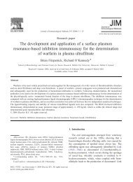
Figure 1. Library selection <strong>against</strong> <strong>the</strong> <strong>C5aR</strong> N-terminal peptide<br />
DX-1186 <strong>target</strong>. (A) Amino acid sequences <strong>of</strong> <strong>the</strong> <strong>target</strong> peptide<br />
DX-1186 and <strong>the</strong> peptide DX-1185 for elution. A lysine residue<br />
was added at <strong>the</strong> C-terminus <strong>of</strong> <strong>the</strong> peptide for labeling with<br />
dPEG4-biotin for DX-1186 peptide. DX-1185 was <strong>the</strong> unlabeled<br />
peptide for elution during selection. (B) Fraction <strong>of</strong> input (FOI)<br />
recovered after each round <strong>of</strong> selection. Three rounds <strong>of</strong> selection<br />
from <strong>the</strong> Fab phage library (Fab400) were carried out. FOI<br />
from each round was calculated as <strong>the</strong> total amount <strong>of</strong> output<br />
phage divided by <strong>the</strong> total amount <strong>of</strong> input phage. FOIs from<br />
both selections (liquid binding and bead binding) are shown.<br />
Copyright # 2005 John Wiley & Sons, Ltd. J. Mol. Recognit. 2005; 18: 327–333
330 L. HUANG ET AL.<br />
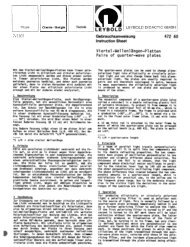
Figure 2. Representative phage ELISA results. Phage enriched from <strong>the</strong> third round <strong>of</strong> selection<br />
were screened for positive binders by phage ELISA. The <strong>target</strong> wells were sequentially coated<br />
with streptavidin and biotinylated DX-1186 peptide, and <strong>the</strong> background wells were coated only<br />
with streptavidin. Plotted in <strong>the</strong> histograms is absorbance at 630 nm (A630) <strong>of</strong> individual phage<br />
clone. Solid bar: DX-1186 <strong>target</strong> signal; open bar: streptavidin background signal. Shown here are<br />
representative ELISA data from two selections, liquid binding selection (A) and bead binding<br />
selection (B).<br />
selections, input phage were first incubated with <strong>the</strong> <strong>target</strong><br />
DX-1186 in solution, and <strong>the</strong>n immobilized to streptavidincoated<br />
magnetic beads via DX-1186. In <strong>the</strong> bead binding<br />
selection campaigns, input phage were allowed to bind <strong>the</strong><br />
immobilized <strong>target</strong> DX-1186 on streptavidin-coated magnetic<br />
beads. After removal <strong>of</strong> unbound phage by washing<br />
extensively with PBST, <strong>the</strong> bound phage were eluted with<br />
DX-1185, <strong>the</strong> non-biotinylated <strong>C5aR</strong> peptide [Fig. 1(A)].<br />
Eluted phage and <strong>the</strong> remaining phage on beads were<br />
subsequently amplified, and subjected to fur<strong>the</strong>r rounds <strong>of</strong><br />
selection. In rounds 2 and 3, depletion steps were carried out<br />
to remove non-specific phage binders. After three rounds <strong>of</strong><br />
selection, <strong>the</strong> fraction <strong>of</strong> input (FOI), which was calculated<br />
as <strong>the</strong> total amount <strong>of</strong> output phage divided by <strong>the</strong> total<br />
amount <strong>of</strong> input phage, increased from 10 7 –10 6 at <strong>the</strong><br />
first round to 10 3 –10 2 by <strong>the</strong> third round [Fig. 1(B)],<br />
suggesting enrichment <strong>of</strong> phage binders to <strong>the</strong> <strong>target</strong>.<br />
To identify positive phage binders, <strong>the</strong> eluted phage from<br />
<strong>the</strong> third round <strong>of</strong> selection were screened by phage ELISA.<br />
The <strong>target</strong> plates were sequentially coated with streptavidin<br />
and DX-1186; and <strong>the</strong> background plates were coated only<br />
with streptavidin. As shown in Fig. 2 for representative<br />
ELISA data and Table 1 for summary <strong>of</strong> results, <strong>the</strong> hit rates<br />
<strong>of</strong> positive isolates with ELISA signals greater than 5-fold<br />
over background levels were high from both selection<br />
campaigns: 44% for liquid binding selection and 53% for<br />
bead binding selection. If phage isolates with ELISA signals<br />
greater than 3-fold over background levels were considered<br />
positive, <strong>the</strong>n <strong>the</strong> hit rates were more than 80% from both<br />
selections. As noted in Fig. 2, a significant number <strong>of</strong> phage<br />
isolates showed no to very low ELISA signals (A630 < 0.2).<br />
These phage isolates were ei<strong>the</strong>r Fab-displaying phage that<br />
did not bind <strong>the</strong> <strong>target</strong> or phage with no Fab display, and thus<br />
served as good internal negative controls for <strong>the</strong> assay.<br />
Therefore, <strong>the</strong> selection campaigns on peptide <strong>target</strong> were<br />
successful in identifying Fab-phage binders to <strong>the</strong> <strong>target</strong><br />
peptide.<br />
Table 1. Summary <strong>of</strong> phage ELISA results<br />
Selection arm Liquid binding Bead binding<br />
Total isolates 384 384<br />
Positive isolates a 170 204<br />
Hit rate (%) 44 53<br />
a Isolates with ELISA signals greater than 5-fold over streptavidin<br />
background levels.<br />
Copyright # 2005 John Wiley & Sons, Ltd. J. Mol. Recognit. 2005; 18: 327–333
HUMAN ANTIBODIES AGAINST THE <strong>C5aR</strong> TARGET 331<br />
Sequencing <strong>of</strong> Fab-phage isolates and IgG reformatting<br />
Selected ELISA positive isolates were subsequently sequenced.<br />
Sequence alignment analysis <strong>of</strong> Fab amino acid<br />
sequences <strong>of</strong> 77 isolates with full Fab sequence data available<br />
resulted in <strong>the</strong> classification <strong>of</strong> <strong>the</strong> ELISA hits into five<br />
major groups, based on heavy chain (HC) CDR1–3 usage.<br />
Each major group has <strong>the</strong> same HC CDRs, but different light<br />
chain (LC) CDRs. Variations <strong>of</strong> LC usage fur<strong>the</strong>r divided<br />
each group into subgroups, with a total number <strong>of</strong> groups<br />
being 22. Besides <strong>the</strong> 22 subgroups, <strong>the</strong>re were seven unique<br />
sequences. Representative phage clones, a total <strong>of</strong> 29 isolates,<br />
were chosen from each subgroup and unique isolates,<br />
and reformatted into whole IgG molecules. Sixteen<br />
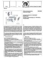
Figure 3. FACS analysis <strong>of</strong> reformatted IgGs. U-937 cells were differentiated into macrophages by treatment with Bt 2 cAMP at<br />
0.5 mM for 3 days. Differentiated cells were stained with IgGs by indirect immun<strong>of</strong>luorescence. Cells were first incubated with<br />
reformatted IgGs and control <strong>human</strong> IgG (hIgG) in staining buffer. S5/1, which binds to <strong>the</strong> <strong>C5aR</strong> N-terminal region, was used<br />
as a positive control. Mouse IgG (mIgG) was used as an isotype control for S5/1. After washing, cells were stained with a FITClabeled<br />
secondary antibody, and <strong>the</strong>n analyzed by flow cytometry. (A) Individual histograms <strong>of</strong> control hIgG, selected<br />
reformatted IgGs (IgG-4, 32, 15, 28, 31, 33, 38, 43), control mIgG and S5/1. The x-axis depicts <strong>the</strong> fluorescence intensity <strong>of</strong><br />
individual cells, and <strong>the</strong> y-axis represents <strong>the</strong> cell number. (B) Summary <strong>of</strong> percentage positive cells for all IgGs tested. The y-<br />
axis represents <strong>the</strong> relative percentages <strong>of</strong> positively stained cells, which were set based on negative controls. Data shown<br />
are representative <strong>of</strong> three independent experiments.<br />
Copyright # 2005 John Wiley & Sons, Ltd. J. Mol. Recognit. 2005; 18: 327–333
332 L. HUANG ET AL.<br />
reformatted clones were expressed as IgGs (5–15 mg/ml) in<br />
transiently transfected HEK293T cells. IgGs from <strong>the</strong>se 16<br />
clones were subsequently purified, and tested in cell binding<br />
assays.<br />
Testing reformatted IgGs for cell binding activities<br />
Since syn<strong>the</strong>tic peptides lack post-translational modifications<br />
such as glycosylation and may adopt conformations different<br />
to <strong>the</strong> same sequence in <strong>the</strong> native protein, <strong>antibodies</strong><br />
discovered from selection on peptide <strong>target</strong>s need to be<br />
fur<strong>the</strong>r evaluated in cell-based assays. Reformatted IgGs<br />
were thus tested by flow cytometric analysis for <strong>the</strong>ir cell<br />
binding activities on <strong>C5aR</strong>-expressing cells. <strong>C5aR</strong> was<br />
induced in differentiated promonocytic U-937 cells (Gerard<br />
and Gerard, 1990). U-937 cells were terminally differentiated<br />
by treatment with 0.5 mM Bt 2 cAMP for 3 days, and <strong>the</strong>n<br />
stained with IgGs by indirect immun<strong>of</strong>luorescence. Cells<br />
were first incubated with reformatted IgGs and control <strong>human</strong><br />
IgG (hIgG) in staining buffer. S5/1, amAb that recognizes<br />
<strong>the</strong> <strong>C5aR</strong> N-terminal region (Oppermann et al., 1993)<br />
was used as a positive control. Flow cytometric analysis<br />
demonstrated that six reformatted IgGs (IgG-15, 28, 31, 33,<br />
38 and 43) showed good binding to differentiated U-937<br />
cells, with percentages <strong>of</strong> positive cells ranging from 11–30%<br />
(Fig. 3). The positive control antibody S5/1 also showed<br />
binding to cells with <strong>the</strong> percentage <strong>of</strong> positive cells being<br />
approximately 17%. The o<strong>the</strong>r 10 reformatted IgGs (IgG-4,<br />
11, 12, 16, 17, 18, 25, 29, 32 and 37) did not show significant<br />
binding to cells, indicating that <strong>the</strong>y were not cell binders.<br />
Consistent with <strong>the</strong> <strong>C5aR</strong> expression pr<strong>of</strong>ile that this<br />
GPCR was not significantly expressed on undifferentiated<br />
U-937 cells but was induced in differentiated U-937 cells,<br />
one representative positive cell binder IgG-28 showed<br />
binding in differentiated U-937 cells but not in undifferentiated<br />
cells (Fig. 4), suggesting that IgG-28 is specific to<br />
differentiated U-937 cells.<br />
Figure 5. Specificity <strong>of</strong> positive cell binders to <strong>C5aR</strong>. Competition<br />
ELISA was carried out as described in <strong>the</strong> Experimental<br />
Procedures. The <strong>target</strong> wells were sequentially coated with<br />
streptavidin and biotinylated DX-1186 peptide (white and striped<br />
bars), and <strong>the</strong> background wells were coated just with streptavidin<br />
(black bars). Prior to <strong>the</strong> addition <strong>of</strong> hIgG control and<br />
positive IgGs, <strong>target</strong> wells were incubated with 10 mIgG (white<br />
bars) or 10 S5/1 (striped bars). Plotted in <strong>the</strong> histograms are<br />
ELISA signals as measured by A630.<br />
Testing <strong>the</strong> specificities <strong>of</strong> positive cell binding IgGs<br />
To fur<strong>the</strong>r determine if <strong>the</strong> positive cell binding IgGs were<br />
specific to <strong>C5aR</strong>, <strong>the</strong> positive IgGs (IgG-15, 28, 31, 33, 38<br />
and 43) were tested in a peptide-based ELISA. Each <strong>of</strong> <strong>the</strong> 6<br />
IgGs bound to DX-1186 (Fig. 5). Competition ELISA with<br />
mAb S5/1 known to bind <strong>the</strong> same <strong>C5aR</strong> N-terminal region<br />
showed that all six IgGs were competed with by S5/1<br />
(Fig. 5), indicating that <strong>the</strong>se positive IgG cell binders<br />
were specific to <strong>C5aR</strong>. This was fur<strong>the</strong>r confirmed by<br />
FACS analysis <strong>of</strong> <strong>C5aR</strong>-expressing cells showing that <strong>the</strong><br />
IgG binding to cells was competed by S5/1 (data not<br />
shown).<br />
DISCUSSION<br />
Figure 4. Specific binding <strong>of</strong> positive IgG binders to differentiated<br />
U-937 cells. U-937 cells treated with Bt 2 cAMP at 0.5 mM for<br />
3 days were used as differentiated cells, and untreated cells were<br />
used as undifferentiated cells. Both differentiated and undifferentiated<br />
cells were stained by indirect immun<strong>of</strong>luorescence and<br />
analyzed by flow cytometry. Shown here are histograms <strong>of</strong><br />
representative positive IgG binder, IgG-28.<br />
We have used phage display technology to identify <strong>human</strong><br />
<strong>antibodies</strong> to <strong>the</strong> chemoattractant receptor <strong>C5aR</strong>, a member<br />
<strong>of</strong> <strong>the</strong> GPCR superfamily. GPCRs have proved successful<br />
drug <strong>target</strong>s and a large proportion <strong>of</strong> existing drugs <strong>target</strong><br />
<strong>the</strong>se receptors. However, owing to <strong>the</strong> complex structures<br />
<strong>of</strong> <strong>the</strong>se seven transmembrane domain-based proteins,<br />
GPCRs are <strong>of</strong>ten considered difficult <strong>target</strong>s for antibodybased<br />
<strong>the</strong>rapeutics and <strong>target</strong>ing. We have designed a<br />
strategy <strong>using</strong> a syn<strong>the</strong>tic peptide encompassing <strong>the</strong> N-<br />
terminal region <strong>of</strong> <strong>C5aR</strong> as <strong>the</strong> selection <strong>target</strong>. After three<br />
rounds <strong>of</strong> selection with a Fab phage library, we were able to<br />
obtain very high hit rates (50% <strong>of</strong> isolates with ELISA<br />
signals greater than 5-fold over background levels) utilizing<br />
two selection strategies: liquid binding and bead binding. In<br />
liquid binding selection, <strong>the</strong> input phage were incubated<br />
with <strong>the</strong> biotin-conjugated <strong>target</strong> in solution before capture<br />
Copyright # 2005 John Wiley & Sons, Ltd. J. Mol. Recognit. 2005; 18: 327–333
HUMAN ANTIBODIES AGAINST THE <strong>C5aR</strong> TARGET 333<br />
on streptavidin beads; in bead binding selection, <strong>the</strong> input<br />
phage were incubated with <strong>the</strong> <strong>target</strong> immobilized on beads.<br />
There are advantages and disadvantages for both selection<br />
strategies. In liquid binding selection, high-affinity binders<br />
may be enriched due to <strong>the</strong> lack <strong>of</strong> avidity effect, but <strong>the</strong><br />
<strong>target</strong> concentration should be well controlled so that <strong>the</strong><br />
<strong>target</strong> is well below <strong>the</strong> saturating concentration and potential<br />
binders will not be lost after immobilizing to beads. In<br />
bead binding selection, low-affinity binders also may be<br />
enriched due to avidity effect, but <strong>the</strong> <strong>target</strong> concentrations<br />
did not need to be well-controlled as in liquid binding<br />
selection. In this study, we included both selection strategies<br />
to ensure maximum success. Our results showed that both<br />
selections worked very well, resulting in similar hit rates.<br />
Representative Fab clones were reformatted into IgGs, and<br />
tested for cell binding activities by flow cytometry. Approximately<br />
40% <strong>of</strong> <strong>the</strong> reformatted IgGs showed binding to<br />
differentiated U-937 cells in which <strong>C5aR</strong> expression was<br />
induced. Flow cytometric analysis <strong>of</strong> HepG2 cells known to<br />
express <strong>C5aR</strong> (Haviland et al., 1995) paralleled <strong>the</strong> results<br />
from U-937 cells: positive binders to differentiated U-937<br />
cells were also positive binders to HepG2 cells, and negative<br />
binders to differentiated U-937 cells were also negative<br />
binders to HepG2 cells (data not shown). These positive<br />
cell binders were specific for <strong>C5aR</strong> since <strong>the</strong>y bound to<br />
<strong>C5aR</strong> N-terminal peptide DX-1186 in peptide-based<br />
ELISA, and <strong>the</strong>ir binding was competed by S5/1, an<br />
anti-<strong>C5aR</strong> mAb known to bind <strong>the</strong> same <strong>C5aR</strong> N-terminal<br />
region (Oppermann et al., 1993). The cell binding activities<br />
<strong>of</strong> <strong>the</strong>se positive binders also were competed by<br />
S5/1, suggesting specific binding to <strong>the</strong> <strong>C5aR</strong> N-terminal<br />
region.<br />
The library used here for selection is a Fab phage display<br />
library with combined natural and syn<strong>the</strong>tic diversity (Hoet<br />
et al., 2004). The advantage <strong>of</strong> <strong>using</strong> a Fab phage library for<br />
selection is that <strong>the</strong>re is no need to do phagemid rescue<br />
during selection and phage ELISA, thus greatly reducing <strong>the</strong><br />
amount <strong>of</strong> time for selection and screening. The <strong>human</strong> Fab<br />
library has been used for selection with many soluble<br />
protein <strong>target</strong>s, yielding <strong>target</strong>-specific <strong>human</strong> <strong>antibodies</strong><br />
with high affinity (Hoet et al., 2004). With <strong>the</strong> Fab library<br />
and <strong>the</strong> IgG reformatting procedure (Jostock et al., 2004),<br />
we were able to obtain specific <strong>human</strong> IgGs to a GPCR<br />
within a short period <strong>of</strong> time (8 weeks), demonstrating<br />
that our phage display approach has utility for rapid development<br />
<strong>of</strong> antibody <strong>the</strong>rapeutic and imaging agents to<br />
GPCRs.<br />
Acknowledgments<br />
We thank Greg Conley for peptide syn<strong>the</strong>sis and Gary Bassil for DNA<br />
sequencing. We also thank Albert Edge for helpful discussions and Clive<br />
Wood for critical reading <strong>of</strong> <strong>the</strong> manuscript.<br />
REFERENCES<br />
Azzazy HME, Highsmith WE Jr. 2002. Phage display technology:<br />
clinical applications and recent innovations. Clin. Biochem.<br />
35: 425–445.<br />
Gerard C, Gerard NP. 1994. C5a anaphylatoxin and its seven transmembrane-segment<br />
receptor. A. Rev. Immunol. 12: 775–808.<br />
Gerard NP, Gerard C. 1990. The chemotactic receptor for <strong>human</strong><br />
C5a anaphylatoxin. Nature 349: 614–617.<br />
Haviland DL, McCoy RL, Whitehead WT, Akama H, Molmenti EP,<br />
Brown A, Haviland JC, Parks WC, Perlmutter DH, Wetsel RA.<br />
1995. Cellular expression <strong>of</strong> <strong>the</strong> C5a anaphylatoxin receptor<br />
(<strong>C5aR</strong>): demonstration <strong>of</strong> <strong>C5aR</strong> on nonmyeloid cells <strong>of</strong> <strong>the</strong><br />
liver and lung. J. Immunol. 154: 1861–1869.<br />
Hoet RM, Cohen EH, Kent RB, Rookey K, Schoonbroodt S, Hogan<br />
S, Rem L, Frans N, Daukandt M, Pieters H, van Hegelsom R,<br />
Coolen-van Neer N, Nastri HG, Rondon IJ, Leeds JA, Hufton<br />
SE, Huang L, Kashin I, Devlin M, Kuang G, Steukers M,<br />
Viswanathan M, Nixon AE, Sexton DJ, Hoogenboom HR,<br />
Ladner RC. 2004. High affinity <strong>human</strong> <strong>antibodies</strong> from<br />
combined syn<strong>the</strong>tic and captured diversity. Nat. Biotechnol.<br />
in press.<br />
Jostock T, Vanhove M, Brepoels E, van Gool R, Daukandt M,<br />
Wehnert A, van Hegelsom R, Dransfield D, Sexton D, Devlin<br />
M, Ley A, Mullberg J. 2004. Rapid generation <strong>of</strong> functional<br />
<strong>human</strong> IgG <strong>antibodies</strong> derived from Fab-on-phage display<br />
libraries. J. Immunol. Meth. 289: 65–80.<br />
Makrides SC. 1998. Therapeutic inhibition <strong>of</strong> <strong>the</strong> complement<br />
system. Pharmac. Rev. 50: 59–87.<br />
Oppermann M, Raedt U, Hebell T, Schmidt B, Zimmermann B,<br />
Gotze O. 1993. Probing <strong>the</strong> <strong>human</strong> receptor for C5a anaphylatoxin<br />
with site-directed <strong>antibodies</strong>. Identification <strong>of</strong> a potential<br />
ligand binding site on <strong>the</strong> NH2-terminal domain. J.<br />
Immunol. 151: 3785–3794.<br />
Pellas TC, Wennogle LP. 1999. C5a receptor antagonists. Curr.<br />
Pharm. Des. 5: 737–755.<br />
Stockwin LH, Holmes S. 2003. Antibodies as <strong>the</strong>rapeutic agents:<br />
vive la renaissance! Exp. Opin. Biol. Ther. 3: 1133–1152.<br />
Copyright # 2005 John Wiley & Sons, Ltd. J. Mol. Recognit. 2005; 18: 327–333