

Vascular calcification and osteoporosis—from clinical observation ...
Vascular calcification and osteoporosis—from clinical observation ...
Vascular calcification and osteoporosis—from clinical observation ...

You also want an ePaper? Increase the reach of your titles
YUMPU automatically turns print PDFs into web optimized ePapers that Google loves.
254 Osteoporos Int (2007) 18:251–259<br />
The passive hypothesis is based on the concept that—in<br />
an appropriate microenvironment—calcium <strong>and</strong> phosphate<br />
physiochemically precipitate in areas of advanced tissue<br />
degeneration or necrosis within the vascular wall when the<br />
physiological calcium phosphate solubility threshold is<br />
exceeded [11]. This largely acellular extracellular process<br />
that occurs in association with membrane debris is further<br />
enhanced by a deficient macrophage-mediated clearance<br />
mechanism [31]. There are several potential <strong>calcification</strong><br />
inhibitors that may act either locally or systemically [11].<br />
MGP is a vitamin K-dependent inhibitor of <strong>calcification</strong><br />
that acts locally, while fetuin-A is a circulating factor that<br />
prevents mineral precipitation <strong>and</strong> serves as an opsonin,<br />
thus enhancing phagocytosis of mineral precipitates [11]. In<br />
addition, pyrophosphate is a potent inhibitor of vascular<br />
<strong>calcification</strong>, as exemplified by a syndrome termed generalized<br />
arterial <strong>calcification</strong> of infancy that is caused by lossof-function<br />
mutations of ecto-nucleotide pyrophosphatase/<br />
phosphodiesterase-1 (ENPP1), the enzyme that generates<br />
pyrophosphate [32]. Several lines of evidence indicate that<br />
active <strong>and</strong> passive mechanisms occur in parallel <strong>and</strong> are not<br />
mutually exclusive [10].<br />
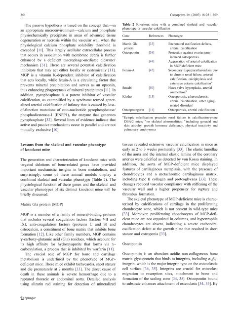
Table 2 Knockout mice with a combined skeletal <strong>and</strong> vascular<br />
phenotype or vascular <strong>calcification</strong><br />
Gene References Phenotype<br />
Matrix Gla<br />
protein<br />
[33] Enchondral ossification defects,<br />
arterial <strong>calcification</strong><br />
Osteopontin [39] Protection against ovariectomyinduced<br />
osteoporosis<br />
[44] Aggravation of arterial <strong>calcification</strong><br />
in MGP-deficient mice<br />
Fetuin-A [47] Secondary hyperparathyroidism due<br />
to chronic renal failure, arterial<br />
<strong>calcification</strong>, calciphylaxis <strong>and</strong><br />
extensive ectopic <strong>calcification</strong> a<br />
Smad6 [50] Heart valve hyperplasia, arterial<br />
ossification b<br />
Klotho [13] Osteoporosis, atherosclerosis,<br />
arterial <strong>calcification</strong>, other agingrelated<br />
disorders c<br />
Osteoprotegerin [14] Osteoporosis, arterial <strong>calcification</strong><br />
a Ectopic <strong>calcification</strong> precedes renal failure in <strong>calcification</strong>-prone<br />
DBA/2 mice; b no skeletal abnormalities; c including gonadal <strong>and</strong><br />
skin atrophy, growth hormone deficiency, physical inactivity <strong>and</strong><br />
pulmonary emphysema<br />
Lessons from the skeletal <strong>and</strong> vascular phenotype<br />
of knockout mice<br />
The generation <strong>and</strong> characterization of knockout mice with<br />
targeted deletions of bone-related genes have provided<br />
important mechanistic insights in bone metabolism, <strong>and</strong><br />
surprisingly, some of these animal models display a<br />
combined skeletal <strong>and</strong> vascular phenotype (Table 2). The<br />
physiological function of these genes <strong>and</strong> the skeletal <strong>and</strong><br />
vascular phenotypes of six distinct knockout mice will be<br />
briefly discussed.<br />
Matrix Gla protein (MGP)<br />
MGP is a member of a family of mineral-binding proteins<br />
that includes several coagulation factors (factors VII <strong>and</strong><br />
IX), anti-coagulation factors (proteins C <strong>and</strong> S) <strong>and</strong><br />
osteocalcin, a constituent of bone matrix that inhibits bone<br />
formation [12]. Like other family members, MGP contains<br />
γ-carboxy-glutamic acid (Gla) residues, which account for<br />
its high affinity for hydroxyapatite that forms via γ-<br />
carboxylation, a process that is inhibited by warfarin [11].<br />
The crucial role of MGP for bone <strong>and</strong> cartilage<br />
metabolism is underlined by the phenotype of MGPdeficient<br />
mice. These mice exhibit tachycardia, short stature<br />
<strong>and</strong> die prematurely at 2 months [33]. The direct cause of<br />
death in these animals is severe hemorrhage due to a<br />
ruptured thoracic or abdominal aorta. Detailed analysis<br />
using alizarin red staining for detection of mineralized<br />
tissues revealed extensive vascular <strong>calcification</strong> in mice as<br />
early as 2 to 3 weeks postnatally [33]. The elastic lamellae<br />
of the aorta <strong>and</strong> the internal elastic lamina of the coronary<br />
arteries were calcified as detected by von Kossa staining. In<br />
addition, the aorta of MGP-deficient mice displayed<br />
features of cartilaginous metaplasia, with the presence of<br />
chondrocytes <strong>and</strong> a metachromic cartilaginous matrix,<br />
including type II collagen <strong>and</strong> proteoglycans [33]. These<br />
changes reduced vascular compliance with stiffening of the<br />
vascular wall <strong>and</strong> a higher propensity for rupture <strong>and</strong><br />
thrombus formation.<br />
The skeletal phenotype of MGP-deficient mice is characterized<br />
by <strong>calcification</strong>s of cartilage in the proliferating<br />
chondrocyte zone, which is not present in wild-type mice<br />
[33]. Moreover, proliferating chondrocytes of MGP-deficient<br />
mice are not organized in columns, <strong>and</strong> hypertrophic<br />
chondrocytes are absent, indicating a severe enchondral<br />
ossification defect at the growth plate that resulted in short<br />
stature <strong>and</strong> osteopenia [33].<br />
Osteopontin<br />
Osteopontin is an abundant acidic non-collagenous bone<br />
matrix glycoprotein that binds to integrins, including α v β 3 -<br />
integrin, which is the major integrin type on the osteoclastic<br />
cell surface [34, 35]. Integrins are crucial for osteoclast<br />
migration to resorption sites, attachment to bone <strong>and</strong><br />
formation of the sealing zone [34, 35]. Osteopontin bound<br />
to substrate enhances attachment of osteoclasts [34, 35]. By
