The early years of radiation protection: a tribute to Madame Curie
The early years of radiation protection: a tribute to Madame Curie
The early years of radiation protection: a tribute to Madame Curie

You also want an ePaper? Increase the reach of your titles
YUMPU automatically turns print PDFs into web optimized ePapers that Google loves.
Coppes-Zantinga and Coppes<br />
X-rays, <strong>radiation</strong>, radioiso<strong>to</strong>pes<br />
and <strong>radiation</strong> therapy<br />
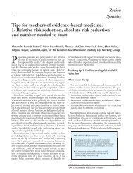
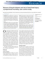
© Association <strong>Curie</strong> et Joliot <strong>Curie</strong>, Paris<br />
Mme <strong>Curie</strong> in her labora<strong>to</strong>ry at the Radium Institute, 1921<br />
Mor<strong>to</strong>n suggested that x-rays were the cause <strong>of</strong> their eye<br />
trouble. During the same period, Alan Archibald Campbell-Swin<strong>to</strong>n<br />
recorded that he and his associates had not<br />
experienced ill effects <strong>to</strong> their eyes after working with<br />
Crookes tubes (part <strong>of</strong> the apparatus used <strong>to</strong> generate<br />
x-rays) for many hours. 19 Nonetheless, as more powerful<br />
x-ray equipment was introduced, additional accounts <strong>of</strong><br />
complications began <strong>to</strong> appear. Several reports described<br />
skin reactions similar <strong>to</strong> sunburn.<br />
<strong>The</strong> American physicist Elihu Thomson was the first<br />
<strong>to</strong> prove a direct relation between exposure <strong>to</strong> x-rays and<br />
some <strong>of</strong> the reported effects. He deliberately exposed his<br />
left index finger <strong>to</strong> an x-ray tube for half an hour a day for<br />
several days. <strong>The</strong> resulting erythema, swelling and pain<br />
confirmed the suspected relation. 13 Unequivocal pro<strong>of</strong> <strong>of</strong><br />
the damaging effects <strong>of</strong> x-rays came with the reports <strong>of</strong><br />
William Rollins, who described the fatal results <strong>of</strong> prolonged<br />
x-ray exposure on guinea pigs. 14 On the basis <strong>of</strong> his<br />
observations, Rollins suggested that x-ray users wear radio-opaque<br />
glasses, that the x-ray tubes be enclosed in<br />
leaded housing and that only areas <strong>of</strong> interest be irradiated<br />
and adjacent areas covered with radio-opaque materials.<br />
From 1887 <strong>to</strong> 1904, Rollins, a true pioneer in <strong>radiation</strong><br />
<strong>protection</strong>, made many scientific contributions <strong>to</strong> the<br />
field and developed numerous devices <strong>to</strong> protect both patients<br />
and x-ray opera<strong>to</strong>rs. Unfortunately, his warnings<br />
<strong>The</strong> first x-rays were produced using a cathode x-<br />
ray tube <strong>of</strong> the type used by the English physicist<br />
William Crookes and other pioneers in their <strong>early</strong> experiments.<br />
Subsequently, many changes were made<br />
<strong>to</strong> improve the efficiency <strong>of</strong> generating x-rays. X-rays<br />
are produced when electrons are accelerated across<br />
a high potential difference, usually measured in kiloor<br />
mega-volts, and impinge on a suitable target. <strong>The</strong><br />
energy <strong>of</strong> the accelerated electrons is dissipated<br />
largely through heating <strong>of</strong> the target material (usually<br />
a heavy metal such as tungsten) and through the release<br />
<strong>of</strong> x-rays. <strong>The</strong>se are bundles <strong>of</strong> energy without<br />
mass or charge and are termed pho<strong>to</strong>ns. <strong>The</strong>re is a<br />
wide spectrum <strong>of</strong> pho<strong>to</strong>ns, their basic physical properties<br />
being the same, but their characteristics varying<br />
according <strong>to</strong> their inherent energy. Radio waves<br />
and visible light are made up <strong>of</strong> pho<strong>to</strong>ns, for example.<br />
When pho<strong>to</strong>ns, i.e., x-rays, are produced by<br />
kilo- or mega-voltage machines, they are very penetrating<br />
and can be used for both diagnostic and therapeutic<br />
purposes.<br />
Radiographs are produced using x-rays <strong>of</strong> relatively<br />
low voltage. When they strike a sensitive<br />
film emulsion they produce a latent image, which<br />
is brought out by developing and fixing the film in<br />
a process similar <strong>to</strong> that <strong>of</strong> black-and-white pho<strong>to</strong>graphy.<br />
X-rays used therapeutically have higher energy,<br />
usually in the megavoltage range. <strong>The</strong>se pho<strong>to</strong>ns can<br />
penetrate deeper in<strong>to</strong> the body, where their energy is<br />
released and biologic effects are produced through<br />
complex interactions with cells. <strong>The</strong> source <strong>of</strong> the <strong>radiation</strong><br />
is at a distance from the patient, and the x-ray<br />
beam is channelled so that only the cancer site receives<br />
the high-dose <strong>radiation</strong>. Because the source is<br />
at a distance, this technique is termed “teletherapy,”<br />
from the Greek tele, “far <strong>of</strong>f.”<br />
So far, we have only described x-rays produced<br />
by machines. Naturally or artificially produced radioactive<br />
materials (radioiso<strong>to</strong>pes) can also emit<br />
pho<strong>to</strong>ns. Radium and cobalt-60, respectively, are<br />
examples <strong>of</strong> these 2 types. <strong>The</strong> <strong>radiation</strong> they produce<br />
is <strong>of</strong>ten termed “gamma rays.” <strong>The</strong> physical<br />
properties <strong>of</strong> gamma rays are identical <strong>to</strong> those <strong>of</strong> x-<br />
rays. For practical as well as technical reasons,<br />
gamma rays are not used for diagnostic purposes but<br />
are employed only for therapy. <strong>The</strong>y are inserted<br />
in<strong>to</strong> or applied closely <strong>to</strong> the cancerous tissue. This<br />
technique is known as “brachytherapy,” from the<br />
Greek brachy, “short.”<br />
1390 JAMC • 1 er DÉC. 1998; 159 (11)