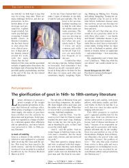
The early years of radiation protection: a tribute to Madame Curie
The early years of radiation protection: a tribute to Madame Curie
The early years of radiation protection: a tribute to Madame Curie

You also want an ePaper? Increase the reach of your titles
YUMPU automatically turns print PDFs into web optimized ePapers that Google loves.
<strong>The</strong> <strong>early</strong> <strong>years</strong> <strong>of</strong> <strong>radiation</strong><br />
<strong>protection</strong>: a <strong>tribute</strong> <strong>to</strong> <strong>Madame</strong> <strong>Curie</strong><br />
Arty R. Coppes-Zantinga, MA; Max J. Coppes, MD, PhD<br />
In 1936, almost 4 decades after the discovery <strong>of</strong> the x-ray and <strong>of</strong> radium, the<br />
German Röntgen Society erected a monument <strong>to</strong> commemorate all who<br />
had died as a consequence <strong>of</strong> exposure <strong>to</strong> x-rays or radium. George W.C.<br />
Kaye <strong>of</strong> the US National Physical Labora<strong>to</strong>ry wrote the inscription: “To the<br />
röntgenologists and radiologists <strong>of</strong> all nations, doc<strong>to</strong>rs, physicists, chemists,<br />
technical workers, labora<strong>to</strong>ry workers, and hospital sisters who gave their lives<br />
in the struggle against the diseases <strong>of</strong> mankind. <strong>The</strong>y were heroic leaders in the<br />
development <strong>of</strong> the successful and safe use <strong>of</strong> x-rays and radium in medicine.<br />
Immortal is the glory <strong>of</strong> the work <strong>of</strong> the dead.”<br />
One hundred <strong>years</strong> ago, on Dec. 26, 1898, Marie <strong>Curie</strong>, Pierre <strong>Curie</strong> and<br />
Gustave Bémont announced their discovery <strong>of</strong> a chemical element that would<br />
revolutionize medicine: “Les diverses raisons que nous venons d’énumérer nous<br />
portent à croire que la nouvelle substance radioactive renferme un élément nouveau,<br />
auquel nous proposons de donner le nom de radium. La nouvelle substance<br />
radioactive renferme certainement une très grande proportion de baryum: malgré<br />
cela, la radioactivité est considerable. La radioactivité du radium doit donc être<br />
enorme.” 1 <strong>The</strong> discovery <strong>of</strong> radium came only 5 months after the <strong>Curie</strong>s had announced<br />
the existence <strong>of</strong> another previously unknown element, which they<br />
named “polonium, du nom du pays d’origine de l’un de nous.” 2<br />
Four <strong>years</strong> after the discovery <strong>of</strong> radium, Marie <strong>Curie</strong> reported its a<strong>to</strong>mic<br />
weight. 3 This was the result <strong>of</strong> a very labour-intensive endeavour. <strong>The</strong> isolation <strong>of</strong><br />
1 gram <strong>of</strong> pure radium had required the handling and processing <strong>of</strong> 8 <strong>to</strong>ns <strong>of</strong><br />
pitchblende ore. In handling this enormous amount, Marie and Pierre <strong>Curie</strong> unknowingly<br />
exposed themselves continuously <strong>to</strong> radioactivity; they contaminated<br />
their food and clothes with radium and inhaled radon, the gaseous by-product <strong>of</strong><br />
decaying uranium and radium. It is therefore not surprising that they both complained<br />
<strong>of</strong> fatigue and ill health. In addition, Mme <strong>Curie</strong> grew thinner by several<br />
kilograms. <strong>The</strong>se changes did not go unnoticed by their friends: “J’ai été frappé,<br />
en voyant M me <strong>Curie</strong> à la Société de Physique, de l’altération de ses traits.” 4 Nevertheless,<br />
Mme <strong>Curie</strong> gave birth <strong>to</strong> 2 healthy daughters as well as leaving a remarkable<br />
scientific legacy. 5,6 She went on <strong>to</strong> receive 2 Nobel prizes — one in<br />
physics and one in chemistry — and received many honorary degrees from universities<br />
all over the world. She also con<strong>tribute</strong>d significantly <strong>to</strong> the development<br />
<strong>of</strong> radiology during World War I. 7 It is interesting that the <strong>Curie</strong>s initially chose<br />
<strong>to</strong> ignore exposure <strong>to</strong> radioactivity as a health hazard. In 1900, Pierre <strong>Curie</strong> voluntarily<br />
exposed his arm <strong>to</strong> radium for several hours and as a consequence developed<br />
a burn. 8 Eventually, though, Mme <strong>Curie</strong> not only recognized “that radium<br />
was dangerous in untrained hands” but went on <strong>to</strong> advocate specific training for<br />
those who worked with radioactive substances. 9<br />
On this, the 100th anniversary <strong>of</strong> the discovery <strong>of</strong> radium, it is fitting <strong>to</strong> review<br />
the first <strong>years</strong> <strong>of</strong> <strong>radiation</strong> <strong>protection</strong>, a process that started 3 <strong>years</strong> before the discovery<br />
<strong>of</strong> radium and that initially was focused on the health hazards <strong>of</strong> x-ray exposure.<br />
Within a few weeks after the discovery <strong>of</strong> x-rays by the German physicist Wilhelm<br />
Konrad Röntgen, 10 the first published reports <strong>of</strong> the ill effects <strong>of</strong> x-ray exposure<br />
began <strong>to</strong> appear. Thomas A. Edison and William J. Mor<strong>to</strong>n independently<br />
reported that their eyes were affected after exposure <strong>to</strong> x-rays. 11,12 It is unclear<br />
whether this was caused by x-ray exposure or simply by the strain <strong>of</strong> peering for<br />
prolonged periods at a dimly fluorescing screen. Indeed, neither Edison nor<br />
Education<br />
Éducation<br />
Mrs. Coppes-Zantinga is with<br />
the Department <strong>of</strong> Oncology,<br />
University <strong>of</strong> Calgary,<br />
Calgary, Alta. Dr. Coppes is<br />
with the Departments <strong>of</strong><br />
Oncology and Pediatrics,<br />
University <strong>of</strong> Calgary, the<br />
Tom Baker Cancer Centre<br />
and the Alberta Children’s<br />
Hospital, Calgary, Alta.<br />
Dr. Coppes is a Clinical<br />
Investiga<strong>to</strong>r with the Alberta<br />
Heritage Foundation for<br />
Medical Research, Calgary,<br />
Alta.<br />
CMAJ 1998;159:1389-91<br />
CMAJ • DEC. 1, 1998; 159 (11) 1389<br />
© 1998 Canadian Medical Association
Coppes-Zantinga and Coppes<br />
X-rays, <strong>radiation</strong>, radioiso<strong>to</strong>pes<br />
and <strong>radiation</strong> therapy<br />
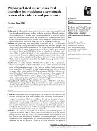
© Association <strong>Curie</strong> et Joliot <strong>Curie</strong>, Paris<br />
Mme <strong>Curie</strong> in her labora<strong>to</strong>ry at the Radium Institute, 1921<br />
Mor<strong>to</strong>n suggested that x-rays were the cause <strong>of</strong> their eye<br />
trouble. During the same period, Alan Archibald Campbell-Swin<strong>to</strong>n<br />
recorded that he and his associates had not<br />
experienced ill effects <strong>to</strong> their eyes after working with<br />
Crookes tubes (part <strong>of</strong> the apparatus used <strong>to</strong> generate<br />
x-rays) for many hours. 19 Nonetheless, as more powerful<br />
x-ray equipment was introduced, additional accounts <strong>of</strong><br />
complications began <strong>to</strong> appear. Several reports described<br />
skin reactions similar <strong>to</strong> sunburn.<br />
<strong>The</strong> American physicist Elihu Thomson was the first<br />
<strong>to</strong> prove a direct relation between exposure <strong>to</strong> x-rays and<br />
some <strong>of</strong> the reported effects. He deliberately exposed his<br />
left index finger <strong>to</strong> an x-ray tube for half an hour a day for<br />
several days. <strong>The</strong> resulting erythema, swelling and pain<br />
confirmed the suspected relation. 13 Unequivocal pro<strong>of</strong> <strong>of</strong><br />
the damaging effects <strong>of</strong> x-rays came with the reports <strong>of</strong><br />
William Rollins, who described the fatal results <strong>of</strong> prolonged<br />
x-ray exposure on guinea pigs. 14 On the basis <strong>of</strong> his<br />
observations, Rollins suggested that x-ray users wear radio-opaque<br />
glasses, that the x-ray tubes be enclosed in<br />
leaded housing and that only areas <strong>of</strong> interest be irradiated<br />
and adjacent areas covered with radio-opaque materials.<br />
From 1887 <strong>to</strong> 1904, Rollins, a true pioneer in <strong>radiation</strong><br />
<strong>protection</strong>, made many scientific contributions <strong>to</strong> the<br />
field and developed numerous devices <strong>to</strong> protect both patients<br />
and x-ray opera<strong>to</strong>rs. Unfortunately, his warnings<br />
<strong>The</strong> first x-rays were produced using a cathode x-<br />
ray tube <strong>of</strong> the type used by the English physicist<br />
William Crookes and other pioneers in their <strong>early</strong> experiments.<br />
Subsequently, many changes were made<br />
<strong>to</strong> improve the efficiency <strong>of</strong> generating x-rays. X-rays<br />
are produced when electrons are accelerated across<br />
a high potential difference, usually measured in kiloor<br />
mega-volts, and impinge on a suitable target. <strong>The</strong><br />
energy <strong>of</strong> the accelerated electrons is dissipated<br />
largely through heating <strong>of</strong> the target material (usually<br />
a heavy metal such as tungsten) and through the release<br />
<strong>of</strong> x-rays. <strong>The</strong>se are bundles <strong>of</strong> energy without<br />
mass or charge and are termed pho<strong>to</strong>ns. <strong>The</strong>re is a<br />
wide spectrum <strong>of</strong> pho<strong>to</strong>ns, their basic physical properties<br />
being the same, but their characteristics varying<br />
according <strong>to</strong> their inherent energy. Radio waves<br />
and visible light are made up <strong>of</strong> pho<strong>to</strong>ns, for example.<br />
When pho<strong>to</strong>ns, i.e., x-rays, are produced by<br />
kilo- or mega-voltage machines, they are very penetrating<br />
and can be used for both diagnostic and therapeutic<br />
purposes.<br />
Radiographs are produced using x-rays <strong>of</strong> relatively<br />
low voltage. When they strike a sensitive<br />
film emulsion they produce a latent image, which<br />
is brought out by developing and fixing the film in<br />
a process similar <strong>to</strong> that <strong>of</strong> black-and-white pho<strong>to</strong>graphy.<br />
X-rays used therapeutically have higher energy,<br />
usually in the megavoltage range. <strong>The</strong>se pho<strong>to</strong>ns can<br />
penetrate deeper in<strong>to</strong> the body, where their energy is<br />
released and biologic effects are produced through<br />
complex interactions with cells. <strong>The</strong> source <strong>of</strong> the <strong>radiation</strong><br />
is at a distance from the patient, and the x-ray<br />
beam is channelled so that only the cancer site receives<br />
the high-dose <strong>radiation</strong>. Because the source is<br />
at a distance, this technique is termed “teletherapy,”<br />
from the Greek tele, “far <strong>of</strong>f.”<br />
So far, we have only described x-rays produced<br />
by machines. Naturally or artificially produced radioactive<br />
materials (radioiso<strong>to</strong>pes) can also emit<br />
pho<strong>to</strong>ns. Radium and cobalt-60, respectively, are<br />
examples <strong>of</strong> these 2 types. <strong>The</strong> <strong>radiation</strong> they produce<br />
is <strong>of</strong>ten termed “gamma rays.” <strong>The</strong> physical<br />
properties <strong>of</strong> gamma rays are identical <strong>to</strong> those <strong>of</strong> x-<br />
rays. For practical as well as technical reasons,<br />
gamma rays are not used for diagnostic purposes but<br />
are employed only for therapy. <strong>The</strong>y are inserted<br />
in<strong>to</strong> or applied closely <strong>to</strong> the cancerous tissue. This<br />
technique is known as “brachytherapy,” from the<br />
Greek brachy, “short.”<br />
1390 JAMC • 1 er DÉC. 1998; 159 (11)
<strong>The</strong> <strong>early</strong> <strong>years</strong> <strong>of</strong> <strong>radiation</strong> <strong>protection</strong><br />
against the dangers <strong>of</strong> x-rays were initially characterized as<br />
overdramatic, 15 and many <strong>of</strong> his safety innovations went<br />
unnoticed. Looking back, it is apparent that Rollins was<br />
ahead <strong>of</strong> his time in the field <strong>of</strong> <strong>radiation</strong> <strong>protection</strong>.<br />
Once a direct relation between x-ray exposure and erythema<br />
<strong>of</strong> the skin was acknowledged, most x-ray opera<strong>to</strong>rs<br />
felt that protecting the skin by means <strong>of</strong> x-ray filters<br />
would likely also provide <strong>protection</strong> against delayed reactions.<br />
George E. Pfahler’s introduction <strong>of</strong> a novel filter<br />
that selectively strained the least penetrating rays was felt<br />
<strong>to</strong> be a huge step forward in the <strong>protection</strong> <strong>of</strong> patients and<br />
opera<strong>to</strong>rs. 16 Indeed, Pfahler’s simple disk <strong>of</strong> sole leather<br />
provided <strong>protection</strong> because it is the less penetrating rays<br />
that burn the skin. However, the filter did not provide adequate<br />
<strong>protection</strong> for those using the same device for<br />
therapeutic purposes. In that setting, many physicians<br />
used skin <strong>protection</strong> <strong>to</strong> increase the dose <strong>to</strong> the deeper tissues.<br />
As a consequence, the risk for delayed nonderma<strong>to</strong>logic<br />
effects increased. Unfortunately, the earliest therapeutic<br />
use <strong>of</strong> x-rays was in the treatment not <strong>of</strong> malignant<br />
conditions but <strong>of</strong> benign disorders such as tinea capitis,<br />
acne vulgaris, eczema, lupus, skin tuberculosis and so on.<br />
<strong>The</strong> issue <strong>of</strong> <strong>radiation</strong> <strong>protection</strong> had become a <strong>to</strong>pic<br />
<strong>of</strong> great concern internationally by 1907. <strong>The</strong> death <strong>of</strong><br />
several x-ray opera<strong>to</strong>rs revealed the serious risks associated<br />
with their pr<strong>of</strong>ession and led <strong>to</strong> recommendations<br />
with regard <strong>to</strong> the need for adequate training, knowledge<br />
and experience. 17 It wasn’t until 8 <strong>years</strong> later, 1 year after<br />
the start <strong>of</strong> World War I, that the Röntgen Society promulgated<br />
the first guidelines regarding <strong>radiation</strong> <strong>protection</strong><br />
for x-ray opera<strong>to</strong>rs. It <strong>to</strong>ok another 6 <strong>years</strong> before the<br />
British X-ray and Radium Protection Committee issued a<br />
preliminary report on <strong>radiation</strong> <strong>protection</strong> measures. 18<br />
<strong>The</strong> committee had been assigned the task <strong>of</strong> drawing up<br />
recommendations for the safe manufacture and use <strong>of</strong> radium<br />
and roentgen ray apparatuses. One year later, similar<br />
recommendations were published by the American<br />
Roentgen Ray Protection Committee. <strong>The</strong> recommendations<br />
<strong>of</strong> the British X-ray and Radium Protection Committee<br />
were accepted internationally in 1928 after the establishment<br />
<strong>of</strong> the International X-ray and Radium<br />
Protection Committee during the second International<br />
Congress <strong>of</strong> Radiology in S<strong>to</strong>ckholm, Sweden. Some radiologists<br />
and equipment makers continued <strong>to</strong> believe<br />
that the recommendations were unnecessarily stringent<br />
and burdensome, 19 but by the mid-thirties most, if not all,<br />
objections had been overcome. No one <strong>to</strong>day denies the<br />
need for the greatest care and strictest observance <strong>of</strong> the<br />
recommendations promulgated by international bodies<br />
charged with those responsibilities.<br />
Radiation safety measures evolved <strong>to</strong>o late <strong>to</strong> save the<br />
protagonist <strong>of</strong> this brief note. Upon the death <strong>of</strong> Mme<br />
<strong>Curie</strong> in 1934, Dr. Tobé reported: “<strong>Madame</strong> Pierre <strong>Curie</strong><br />
est décédée à Sancellemoz le 4 juillet 1934. La maladie est<br />
une anémie pernicieuse aplastique à marche rapide,<br />
fébrile. La moelle osseuse n’a pas réagi, probablement<br />
parce qu’elle était altérée par une longue accumulation de<br />
rayonnements.” 5 Until recently, it was generally believed<br />
that the extensive and prolonged exposure <strong>to</strong> radium<br />
caused her final illness. 20 This seems not <strong>to</strong> have been the<br />
case, however. In 1995, Mme <strong>Curie</strong>’s body was exhumed<br />
for reburial in France’s national mausoleum, the Panthéon.<br />
Scientists from the French Office de Protection<br />
contre les Rayonnements Ionisants found that the level <strong>of</strong><br />
radium emanations within the c<strong>of</strong>fin was significantly<br />
lower than the maximum accepted safe levels <strong>of</strong> public exposure.<br />
21 Given these low levels and the very long half-life<br />
<strong>of</strong> radium (1620 <strong>years</strong>), the Office concluded that Mme<br />
<strong>Curie</strong>’s final illness and death were probably not caused by<br />
extended exposure <strong>to</strong> radium. More likely, it was the direct<br />
result <strong>of</strong> her overexposure <strong>to</strong> x-rays during World<br />
War I, when she made significant contributions <strong>to</strong> military<br />
medicine through the establishment <strong>of</strong> mobile radiographic<br />
units. 7 Thus, ironically, Marie <strong>Curie</strong> became a<br />
martyr <strong>to</strong> the advances in radiography and not <strong>to</strong> <strong>radiation</strong><br />
therapy, the clinical specialty that developed from her<br />
epochal labora<strong>to</strong>ry research.<br />
References<br />
1. <strong>Curie</strong> P, <strong>Curie</strong> M, Bémont MG. Sur une nouvelle substance fortement radioactive<br />
contenue dans la pechblende. C R Acad Sci 1898;127:1215-7.<br />
2. <strong>Curie</strong> P, <strong>Curie</strong> M. Sur une substance nouvelle radio-active, contenue dans la<br />
pechblende. C R Acad Sci 1898;127:175-8.<br />
3. <strong>Curie</strong> M. Sur le poids a<strong>to</strong>mique du radium. C R Acad Sci 1902;135:161-3.<br />
4. Letter from George Sagnac <strong>to</strong> Pierre <strong>Curie</strong>, dated Apr. 23, 1903. Cited in:<br />
Giroud F. Une femme honorable. Paris: Fayard; 1981.<br />
5. <strong>Curie</strong> E. <strong>Madame</strong> <strong>Curie</strong> Paris: Gallimard; 1938.<br />
6. Quinn S. Marie <strong>Curie</strong>: a life. New York: Simon & Schuster; 1995.<br />
7. Coppes-Zantinga AR, Coppes MJ. Marie <strong>Curie</strong>’s contributions <strong>to</strong> radiology<br />
during World War I. Med Pediatr Oncol 1998;31:541-3.<br />
8. <strong>Curie</strong> M. Pierre <strong>Curie</strong>. New York: Macmillan; 1923.<br />
9. Mme <strong>Curie</strong> warns radium amateurs. New York Times 1929 Nov 1: 4.<br />
10. Röntgen WC. Ueber eine neue Art von Strahlen. Sitz Ber Phys Med Ges<br />
Wuerzb 1895;9:132-41.<br />
11. Notes. Nature 1896;53:421.<br />
12. Dyer FL, Martin TC, Meadocraft WH. Edison: his life and inventions.<br />
Harpers 1929;2:581.<br />
13. Thomson E. Roentgen ray burns. Am X-ray J 1898;3:451-3.<br />
14. Rollins W. X-light kills. Bos<strong>to</strong>n Med Surg J 1901;144:173.<br />
15. Codman EA. No practical danger from the x-ray. Bos<strong>to</strong>n Med Surg J<br />
1901;144:197.<br />
16. Pfahler GE. A roentgen filter and a universal diaphragm and protecting<br />
screen. Trans Am Roentgen Ray Soc 1906:217-24.<br />
17. Leonard CL. Protection <strong>of</strong> roentgenologists. Trans Am Roentgen Ray Soc<br />
1907:95-102.<br />
18. British X-ray and Radium Protection Committee. Preliminary report. J<br />
Roentgen Soc 1921;17:100-3.<br />
19. Eisenberg RL. Radiation injury and <strong>protection</strong>. In: Radiology: an illustrated his<strong>to</strong>ry.<br />
St. Louis: Mosby Year Book; 1992. p. 156-82.<br />
20. Eisenberg RL. Radium therapy. In: Radiology: an illustrated his<strong>to</strong>ry. St. Louis:<br />
Mosby Year Book; 1992. p. 511-26.<br />
21. Butler B. X-rays, not radium, may have killed <strong>Curie</strong>. Nature 1995;377:96.<br />
Reprint requests <strong>to</strong>: Mrs. Arty R. Coppes-Zantinga, Southern<br />
Alberta Children’s Cancer Program, Alberta Children’s Hospital,<br />
1820 Richmond Rd SW, Calgary AB T2T 5C7; fax 403 229-<br />
7684; acoppesz@ucalgary.ca<br />
CMAJ • DEC. 1, 1998; 159 (11) 1391