10 Conidiogenesis 2012 - CBS
10 Conidiogenesis 2012 - CBS
10 Conidiogenesis 2012 - CBS

You also want an ePaper? Increase the reach of your titles
YUMPU automatically turns print PDFs into web optimized ePapers that Google loves.
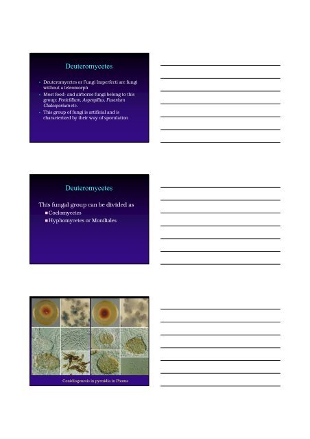
Deuteromycetes<br />
• Deuteromycetes or Fungi Imperfecti are fungi<br />
without a teleomorph<br />
• Most food- and airborne fungi belong to this<br />
group: Penicillium, Aspergillus, Fusarium<br />
Cladosporium etc.<br />
• This group of fungi is artificial and is<br />
characterized by their way of sporulation<br />
Deuteromycetes<br />
This fungal group can be divided as<br />
•Coelomycetes<br />
•Hyphomycetes or Moniliales<br />
<strong>Conidiogenesis</strong> in pycnidia in Phoma
<strong>Conidiogenesis</strong><br />
• <strong>Conidiogenesis</strong> = the mode of conidium<br />
formation<br />
• Conidia = specialized non-motile<br />
asexual spore<br />
• Conidiogenous cell = specialized cell<br />
which give rise to the conidia<br />
• Conidiophore = entire system of fertile<br />
hyphae<br />
Conidium<br />
Conidiogenous cell<br />
Vegetative cells of<br />
the conidiophore<br />
Conidiophore<br />
Penicillium<br />
Vegetative hypha
Important conidial types: Thallic<br />
conidia<br />
Arthroconidia<br />
Formed in<br />
chains =<br />
catenulate<br />
58
Thallic conidia in Geotrichum candidum<br />
Blastic conidiogenesis
Truncate conidia in Epicoccum<br />
Important conidial types: Tretic conidia<br />
= poroconidia<br />
• Formed on darkly pigmented,<br />
thick-walled conidiogenous<br />
cells<br />
• After conidium secession, a<br />
recognizable pore in the wall<br />
of the conidiogenous cell<br />
marks the place where the<br />
conidium was formed
Poroconidia in Alternaria<br />
Conidiogenous cell proliferation results in:<br />
Basipetal<br />
sequence<br />
Acropetal<br />
chains<br />
Sympodial<br />
sequence<br />
56f<br />
geniculate<br />
55d<br />
straight<br />
Conidia in<br />
unbranched chains<br />
or slimy heads<br />
Conidia in<br />
Branched or<br />
unbranched chains<br />
Conidia on a<br />
geniculate or<br />
straight rachis<br />
Basipetal<br />
sequence
Conidiogenous cell proliferation results in:<br />
Basipetal<br />
sequence<br />
Acropetal<br />
chains<br />
Sympodial<br />
sequence<br />
56f<br />
geniculate<br />
55d<br />
straight<br />
Conidia in<br />
unbranched chains<br />
or slimy heads<br />
Conidia in<br />
Branched or<br />
unbranched chains<br />
Conidia on a<br />
geniculate or<br />
straight rachis<br />
Acropetal chains
Important conidial types: Phialidic conidia<br />
• Formed on a<br />
phialide in basipetal<br />
succession<br />
Basipetal chains<br />
Important conidial types:<br />
Phialidic conidia<br />
56d,e<br />
Slimy heads<br />
• Formed on a phialide in<br />
basipetal succession<br />
• Arrangement:<br />
• Basipetal chains<br />
• slimy heads<br />
• Conidia usually with no<br />
distinct or a narrow<br />
base
Collarette
Conidiogenous cell proliferation<br />
Annellidic conidiogenesis<br />
• The conidiogenous cell =<br />
annellide<br />
• Annellides elongate as<br />
successive blastoconidia are<br />
formed<br />
• ring-like ‘annellations’ are<br />
formed on the apex of the<br />
annellide due to percurrent<br />
proliferation<br />
Foodb 22b
Scopulariopsis brevicaulis<br />
Annellated conidiogenous<br />
cell<br />
Conidia with truncate base<br />
Identification of Deuteromycetes<br />
• Microscopical examination of the<br />
conidiogenesis and characteristics of the<br />
conidiophore and conidia<br />
• Microscopical slides mostly prepared in lactic<br />
acid + aniline blue<br />
• Chain or slimy head formation must be<br />
examined with a dissecting microscope or<br />
under low power with a light microscope<br />
• In some cases adhesive tape (cellotape)<br />
preparation might be useful<br />
Cellotape preparation<br />
• This method for making microscopical<br />
slides is only recommended for fragile<br />
sporulating genera e.g. Botrytis,<br />
Cladosporium or making slides driectly<br />
from surfaces.
Transparent tape preparation<br />
cellotape preparation<br />
Press the adhesive side of the cellotape gently on the colony.<br />
Avoid finger prints!<br />
Cellotape preparation from a surface or material
The adhesive tape with fungus is transferred to the microscope slide<br />
with a drop of lactic acid (cotton blue)