View - Facultatea de Biologie
View - Facultatea de Biologie
View - Facultatea de Biologie

You also want an ePaper? Increase the reach of your titles
YUMPU automatically turns print PDFs into web optimized ePapers that Google loves.
Analele ştiinţifice ale Universităţii “Al. I. Cuza” Iaşi<br />
Tomul LIV, fasc. 2, s.II a. <strong>Biologie</strong> vegetală, 2008<br />
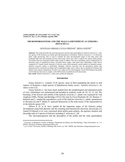
MICROSPOROGENESIS AND THE MALE GAMETOPHYTE AT EPHEDRA<br />
DISTACHYA L.<br />
GENŢIANA MIHAELA IULIA PREDAN * , IRINA GOSTIN **<br />
Abstract: This study <strong>de</strong>scribes the microsporogenesis and the male gametophyte in Ephedra distachya L., only<br />
one species of the Ephedraceae family growing spontaneous in Romania. Ephedra distachya L. is a dioecious<br />
plant. Light microscopy (lm) is used to examine the early <strong>de</strong>velopmental stages of the male cones and<br />
combined light (lm) and scanning electron microscopy (sem) were used to analyze the mature pollen grains.<br />
The pollen cones are clustered at no<strong>de</strong>s either sessile or stalked. They are oval-oblong, each is composed of 2-8<br />
<strong>de</strong>cussate pairs of membranous bracts, proximal bracts empty; each distal bract subtending a male flower<br />
composed of 2 basally fused, orbicular or obovate scales (false perianth); sessile anthers on staminal column.<br />
Ephedra distachya pollen is particularly abundant. Ephedra distachya has the polyplicate pollen type<br />
characterized by a pointed-oval shape and 6 longitudinal ridges and 6 valleys, due to alternation of thicker and<br />
thinner exine regions, inaperturate. In polar view, ridges are low and triangular. On the SEM analysis the pollen<br />
wall of Ephedra distachya is simple, so the SEM closely resembles the LM observation.<br />
Key words: Ephedra distachya L., male cones, pollen <strong>de</strong>velopment<br />
Introduction<br />
Genus Ephedra L. contains 35-45 species, most of them populating the <strong>de</strong>sert or arid<br />
regions. In Romania a single species of Ephedraceae family occurs - Ephedra distachya L. Its<br />
status is rare [12].<br />
Genus Ephedra L. has been much studied from the morphological and anatomical point<br />
of view, information was summarized and inclu<strong>de</strong>d in synthesis works [8, 10, 14, 15, 16]. The<br />
histology of the flowers and strobili of the Ephedra distachya L. plants was examined by Van<br />
Tieghem (1869), Thoday and Berridge (1912) [10], later by Favre-Duchartre M. [5]. Baranec T.<br />
Rehorek and V. studied the reproductive cycle of the Ephedra distachya L. plants, spontaneous<br />
in Slovakia [2] and P. Mehra N. analysed dimensions of the male nuclei of the representatives<br />
in the Ephedra genus [10].<br />
Allison S. D. et al. have studied all the important stages of the Ephedra pollen<br />
<strong>de</strong>velopment using both photonic and the scanning and transmission electronic microscope [3].<br />
Gamal El-Ghazaly et al. were concerned about pollen grain polarity, the aperture situation and<br />
the pollen tube at 5 species of Ephedra including E. distachya L. [6].<br />
The microsporogenesis and the <strong>de</strong>scription of the pollen and the male gametophyte<br />
* University of Bucharest, Faculty of Biology, Department <strong>de</strong> Botany and Microbiology, Aleea Portocalelor, nr. 1-3,<br />
060101, Bucureşti, Romania; ggentiana@yahoo.com<br />
** „Al. I. Cuza” University, Faculty of Biology, Bd. Carol I, no. 20A, 700506, Iaşi, Romania; irinagostin@yahoo.com<br />
24
<strong>de</strong>velopment in Ephedra distachya L. provi<strong>de</strong>s interesting characterization in this species.<br />
Material and methods<br />
Our study was carried out on samples of Ephedra distachya L. from different places in<br />
Romania: Agigea Marine Dune Reserve (Constanta County), Culmea Pricopanului (Tulcea<br />
County), the Botanical Gar<strong>de</strong>n of Iaşi (cultivated plants) and from Bulgaria (Balchik) [7, 8].<br />
Ephedra distachya L. is a dioecious plant. Male cones were collected during the three spring<br />
months (March, April and May), in successive phaenological phases, from the cones initiation<br />
up to the pollen shedding.<br />
The biological material (male cones) was fixed in 70% alcohol or FAA (formalin 40%, 5<br />
cc: alcohol 70%, 90 cc: glacial acetic acid 5 cc). After being fixed, the material has been<br />
processed in accordance with the paraffin embeding protocol [3], sectioned (transverselly and /<br />
or longitudinally) with a Minot type microtome at 12-15 μm thick, colored with Ehrlich’s<br />
haematoxylin and embed<strong>de</strong>d in Canada balm. To highlight microspores and young pollen grains<br />
microscopical preparations squash type have been ma<strong>de</strong>, that have been colored with acetic<br />
carmine.<br />
All obtained sections were analyzed using light microscope (LM) Docuval type and<br />
photographed with a digital photo camera slr Canon EOS 400D. For the analysis of the pollen<br />
grains at electronic scanning microscope (SEM) a Vega - TESCAN LSH microscope was used<br />
in The Faculty of Biology of The “Al I. Cuza” University from Iaşi [12].<br />
Male cones were consi<strong>de</strong>red from the morphological point of view with a Belphotonics<br />
stereomicroscope and photographed with the same digital camera.<br />
The <strong>de</strong>scription of the pollen grains has been ma<strong>de</strong> in accordance with the terminology<br />
adopted by Faegri [4].<br />
Results and discussions<br />
The male cones, either sessile or stalked, are found clustered at no<strong>de</strong>s (fig. 1). The male<br />
cones are green, oval-oblongue, each of them being composed of 2-8 <strong>de</strong>scussate pairs of<br />
membranous bracts, like cups (Fig. 2, 3). The lower pair bracts is sterile, each distal bract<br />
subtending a male flower composed of 2 basally fused, orbicular or obovate scales (primitive<br />
periant) (Fig. 4, 7), an axis (microsporangiophore or anterophore) terminal bearing a number of<br />
anthers, each anther with two micro sporangia (pollen sacs) (Fig. 4). A group of archespores<br />
subepi<strong>de</strong>rmal cells in the mature cones is observed in March (fig. 5).<br />
In April, following meiosis I, macrospore dyads have resulted. After meiosis II numerous<br />
tetrads of microspores appear, the wall formation taking place after each stage of meiosis. Soon,<br />
the wall surrounding the 4 microspore cells dissolve and the microspores in the pollen sac are<br />
25
eleased. The microsporangia wall is composed of three known layers: epi<strong>de</strong>rmis, the median<br />
layer and the tapetum layer. The median layer is composed of a single row of parenchimatic<br />
cells and, as the pollen sac grows, its cells are flattened so that they ultimately disappear. The<br />
tapetum has signs of <strong>de</strong>generation after the reductional division (Fig. 6) and no longer in the<br />
stage of mature pollen (Fig. 8). At mature pollen sac epi<strong>de</strong>rmis persists. Its cells have a regular<br />
appearance and their walls are thickened (Fig. 9). Only in a small portion of the pollen sac<br />
upper part a few parenchimatic cells with thin walls remain. By their breaking a hole will form<br />
that will be issued pollen (porici<strong>de</strong> opening).<br />
In May, the pollen is divi<strong>de</strong>d and forms a small prothalian cell and a large, central one<br />
(Fig. 10). The last divi<strong>de</strong> and formed the second prothalian cell and initial anteridial cell. The<br />
last divi<strong>de</strong> and give a vegetative larger cell, and a generative cell. The generative cell will form<br />
the stalk cell and spermatogene cell, the latter giving rise to gametes. Regarding pollen sacs, the<br />
pollen has 5 cells [8]. The mature pollen grains are yellow, fusiforme and obvious poliplicate:<br />
they have 6 longitudinal ridges (plicae) (sometimes slightly corrugated) and 6 valleys, formed<br />
due to the alternation of thicker and thinner exine regions (Fig. 9, 10). On a polar view, the<br />
ridges are short and triangular. Pollen grains have 53 μm long axis and 20 μm short axis. At the<br />
electronic scanning microscope (SEM), the pollen surface is no obvious regulate and no<br />
aperture was noticed eather, so the pollen is inaperturate (Fig. 11, 12).<br />
Conclusions<br />
In March, a group of archespore cells appears in the pollen sacs, subepi<strong>de</strong>rmaly.<br />
In April the anther wall is composed of an epi<strong>de</strong>rmis and a scrap tapetum and the<br />
microspores are free.<br />
In May the anther wall is only represented by the epi<strong>de</strong>rmis and numerous grains of<br />
fusiforme and yellow pollen are found insi<strong>de</strong> the pollen sac, in our observation (in 17 May 2007<br />
on Culmea Pricopanului) the pollen grains are uninucleate.<br />
According to the SEM observations, the pollen grains of Ephedra distachya L. have a<br />
simple structure, the SEM observations being very similar to those ma<strong>de</strong> on an optical<br />
microscope.<br />
REFERENCES<br />
1. ANDREI M., RĂDULESCU D., 1972 - Caiet pentru tehnica preparării şi conservării materialului biologic.<br />
Tipogr. Univ. din Bucureşti<br />
2. BARANEC, T., V. REHOREK, et al., 1994 - Generative reproduction of ephedra (Ephedra distachya L.) in<br />
Slovakia. Biologia Bratislava, 49 (1): 65-67<br />
3. DOORES ALLISON S., OSBORN J. M., GAMAL EL-GHAZALY., 2007 - Pollen Ontogeny in Ephedra<br />
americana (Gnetales). International Journal of Plant Sciences 168 (7): 985-997<br />
4. FAEGRI K., IVERSEN J., 1966 - Textbook of pollen analysis. 2nd Ed. Munksgaard, Copenhagen<br />
26
5. FAVRE-DUCHARTRE M., 1959 - Contribution à l’étu<strong>de</strong> <strong>de</strong> la reproduction sexuée chez Ephedra distachya. C.<br />
R. Acad. Sci. Paris. 249: 1551-1553<br />
6. GAMAL EL-GHAZALY, ROWLEY J., HESSE M., 1998 - Polarity, aperture condition and germination in pollen<br />
grains of Ephedra (Gnetales). Plant Systematics and Evolution, 213 (3-4): 217-231<br />
7. GRINŢESCU G. P., 1952 - Ephedra L. In: T. SĂVULESCU (red. princip.). Flora României. 1. Bucureşti: Edit.<br />
Aca<strong>de</strong>miei Române<br />
8. JOHRY B. M., BISWAS C., 1997 - The Gymnosperms. Narosa Publishing House, New – Delhi<br />
9. KRÜSSMANN G., 1985 - Manual of cultivated conifers. B.T. Batsford Ltd. London<br />
10. LEHMANN-BAERTS M., 1967 - La morphologie du sporophyte dans le genre Ephedra. La Cellule (Belg.), 67<br />
(5): 7-56<br />
11. MEHRA P. N., 1950 - Inequality in size of the male nuclei in the genus Ephedra. Ann. Bot., July 1950; 14: 331-<br />
339<br />
12. OLTEAN M., NEGREAN G., POPESCU A., ROMAN N., DIHORU G. SANDA V., MIHĂILESCU S., 1994 -<br />
Lista roşie a plantelor superioare din România. Studii, sinteze, documentaţii <strong>de</strong> ecologie, 1<br />
13. PLOAIE G. P., PETRE ZOE. 1979 - Introducere in microscopia electronica cu aplicatii in biologia celulara si<br />
moleculara. Edit. Acad. Romane, Bucuresti<br />
14. SCHNARF K., 1933 - Embryologie <strong>de</strong>r Gymnospermen. In: Linsbauer K. „Handbuch <strong>de</strong>r Pflanzenanatomie”, II<br />
Abt., 2 Teil, Bd. 2. Verlag Gebru<strong>de</strong>r Borntraeger, Berlin<br />
15. SINGH H., 1978. - Embryology of gymnosperms. Berlin-Stuttgart<br />
16. SMARANDACHE D., 2005 - Embriologia plantelor. I. Embriologia arhegoniatelor. Edit. Univ. din Bucureşti,<br />
Bucureşti.<br />
17. STEEVES M. W., BARGHOORN E. S., 1959 - The pollen of Ephedra. J. Arnold Arboretum, 40: 221-255<br />
The explanation of figures:<br />
Plate I<br />
Fig. 1 Ephedra distachya L. - Branch with male cones (1 May 2008; Orig)<br />
Fig. 2 Ephedra distachya L. – Male cone (20 May 2007) (Oc. 10x; Amplific. 2; Orig.)<br />
Fig. 3. Ephedra distachya L. - Longitudinal section of a male cone - fragment (13 April 2008) (Oc 12.5x; ob. 3.2x;<br />
Amplific. 8; Orig.)<br />
Fig. 4. Ephedra distachya L. - Male flower (20 May 2007) (Oc. 10x; Amplific. 3; Orig.)<br />
Fig. 5. Ephedra distachya L. - Longitudinal section through the anther that outlines the archaesporal cells (17 March<br />
2007) (Oc 12.5x.; ob. 25x; Amplific. 160; Orig.)<br />
Fig. 6. Ephedra distachya L. - Cross-section through the male flower (13 April 2008) (Oc. 12.5x; ob. 10x; Amplific.<br />
6.3; Orig.)<br />
Fig. 7. Ephedra distachya L. - Longitudinal section through the male flower (13 April 2008) (Oc. 12.5x; ob. 10x;<br />
Amplific. 6.3; Orig.)<br />
Plate II<br />
Fig. 8 Ephedra distachya L. - Longitudinal section through the male flower (29 May 2001) (Oc. 12.5x; ob. 3.2x;<br />
Amplific.16; Orig.)<br />
Fig. 9. Ephedra distachya L. - Longitudinal section through the anther - anteral wall and young pollen grains (29 May<br />
2001) (Oc. 12.5x; ob. 40x; Ampific. 16; Orig.)<br />
Fig. 10. Ephedra distachya L. - Unicellular pollen grain (17 May 2007) (Oc. 12.5x; ob. 40x; Amplific.16; Orig.)<br />
Fig. 11. SEM micrograph of Ephedra distachya L. poliplicate pollen (21 May 2008; Orig.)<br />
Fig 12. SEM micrograph of Ephedra distachya L. pollen (21 May 2008; Orig.)<br />
27
GENŢIANA PREDAN, IRINA GOSTIN<br />
PLATE I<br />
1 2 3<br />
4 5<br />
6<br />
7<br />
28
GENŢIANA PREDAN, IRINA GOSTIN<br />
PLATE II<br />
8<br />
9<br />
10<br />
11<br />
12<br />
29