Aesculap® Neuroendoscopy - B. Braun Medical AS
Aesculap® Neuroendoscopy - B. Braun Medical AS
Aesculap® Neuroendoscopy - B. Braun Medical AS

You also want an ePaper? Increase the reach of your titles
YUMPU automatically turns print PDFs into web optimized ePapers that Google loves.
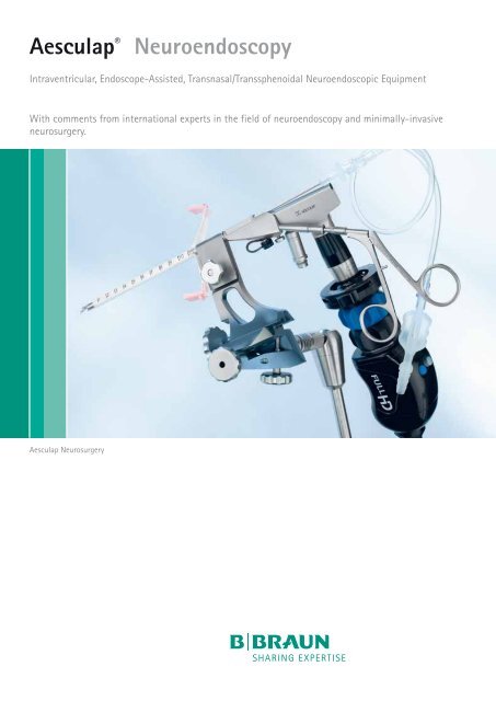
Aesculap ®<br />
<strong>Neuroendoscopy</strong><br />
Intraventricular, Endoscope-Assisted, Transnasal/Transsphenoidal Neuroendoscopic Equipment<br />
With comments from international experts in the field of neuroendoscopy and minimally-invasive<br />
neurosurgery.<br />
Aesculap Neurosurgery
Aesculap <strong>Neuroendoscopy</strong><br />
Michael Fritsch<br />
Neubrandenburg, Germany<br />
Jeremy Greenlee<br />
Iowa City, USA<br />
André Grotenhuis<br />
Nijmegen, Netherlands<br />
Nikolai Hopf<br />
Stuttgart, Germany<br />
Peter Nakaji<br />
Phoenix, USA<br />
2
Aesculap Neurosurgery<br />
"<br />
In 1924, the famous general and neurological<br />
surgeon William Halsted expressed his belief “…that<br />
the tendency will always be in the direction of exercising<br />
greater care and refinement in operating”.<br />
Today, within the third millennium this fundamental<br />
philosophy of minimally invasive therapy should<br />
be emphasized more than ever before, operating<br />
with a minimum of iatrogenic trauma while achieving<br />
maximum surgical efficiency.<br />
Recent improvements in preoperative imaging and<br />
surgical instrumentation allow neurosurgeons to<br />
treat more complex pathologies through customized<br />
less invasive approaches.<br />
Using the advanced diagnostic tools of digital subtraction<br />
angiography, 3D angiography, computed<br />
tomography and magnetic resonance imaging, one<br />
is able to demonstrate and elucidate preoperatively<br />
the individual anatomy and pathology of the<br />
patient. Therefore, anatomically preformed surgical<br />
dissection can be described preoperatively and<br />
may so be included into the planning of surgery.<br />
With the individual anatomic details of a specific<br />
patient, it becomes possible to perform a tailored<br />
surgical procedure reducing the size of the skin incision,<br />
the craniotomy, and the extent of brain surface<br />
traumatization and retraction to a necessary<br />
minimum limit. These advantages of minimally<br />
invasive microsurgery contribute to improved postoperative<br />
results, including shorter hospitalization<br />
time because of reduction of the risk for complications.<br />
However, small sized minimally invasive approaches<br />
cause two important limitations: the<br />
significant loss of optical control and limited<br />
maneuverability of microsurgical instruments. The<br />
intraoperative use of endoscopes and dedicated<br />
minimally invasive instruments overcome these<br />
restrictions, thus enabling neurosurgeons to<br />
achieve deep seated regions without approach<br />
related traumatization of sensitive neurovascular<br />
structures.<br />
The endoscopic image allows illumination and<br />
inspection of angles in hidden parts of the surgical<br />
field with the and clear depiction of anatomical<br />
details. In addition, due to the enormous optical<br />
depth of field of modern endoscopes, endoscopes<br />
provide a three dimensional aspect of anatomic<br />
structures. Recently, the intraoperative use of full<br />
high definition (HD) image quality offers a new<br />
area in endoscopic neurosurgery with an increased<br />
range of indications in minimally invasive<br />
neurosurgery.<br />
There are three main indications of endoscopic<br />
neurosurgery: the intraventricular, transcranial and<br />
transnasal application. In this brochure, contemporary<br />
endoscopic equipment and instrumentation<br />
is presented in a comprehensive way. International<br />
experts in the field of minimally invasive and endoscopic<br />
neurosurgery comment the different<br />
applications, giving remarks with important tips<br />
and ideas, thus providing valuable instructions for<br />
the use of endoscopes in the field of minimally invasive<br />
neurosurgery.<br />
"<br />
The Aesculap Advisory Board for “Minimally-<br />
Invasive Neurosurgery & <strong>Neuroendoscopy</strong>”<br />
Michael Fritsch, Neubrandenburg, Germany<br />
Jeremy Greenlee, Iowa City, USA<br />
Andre Grotenhuis, Nijmegen, Netherlands<br />
Nikolai Hopf, Stuttgart, Germany<br />
Peter Nakaji, Phoenix, USA<br />
Robert Reisch, Zurich, Switzerland<br />
Mark Souweidane, New York, USA<br />
Charles Teo, Sydney, Australia<br />
Ron Young, Indianapolis, USA<br />
Robert Reisch<br />
Zurich, Switzerland<br />
Mark Souweidane<br />
New York, USA<br />
Charles Teo<br />
Sydney, Australia<br />
Ron Young<br />
Indianapolis, USA<br />
3
Intraventricular<br />
<strong>Neuroendoscopy</strong><br />
Intraventricular <strong>Neuroendoscopy</strong><br />
5
MINOP ®<br />
Intraventricular Neuroendoscopic System<br />
6
Aesculap Neurosurgery<br />
Intraventricular<br />
<strong>Neuroendoscopy</strong><br />
"<br />
The genesis of endoscopic surgery within the<br />
ventricular compartment can be attributed to the<br />
development of small caliber rod lens optics,<br />
fiberoptic light transmission and dedicated<br />
instrumentation. Since the advent of intraventricular<br />
endoscopic surgery, neurosurgeons have<br />
applied the technology to treat a number of<br />
disorders. While the enthusiasm has been great<br />
and the full potential not yet realized, a major<br />
benefit to the patient has been proven for selected<br />
conditions. Most notably the treatment of<br />
non-communicating hydrocephalus, management<br />
of patients with pineal region tumors, fenestration<br />
of intracranial cysts, and removal of colloid<br />
cysts have all been shown to provide significant<br />
benefit and reduced morbidity compared with<br />
conventional treatment strategies.<br />
The benefit in minimally invasive endoscopic<br />
procedures is analogous to that of any endoscopic<br />
procedure, namely minimal tissue disruption,<br />
enhanced visualization, improved cosmetic results,<br />
shorter hospital stay, and less surgical morbidity.<br />
The surgeon willing to utilize intraventricular<br />
endoscopic surgery is first responsible for attaining<br />
a considerable degree of familiarity with the<br />
technology, relevant anatomy, and the surgical<br />
procedures. Given the relative nascence of the<br />
field, the discipline is only now being commonly<br />
implemented in training programs. Hence, for<br />
those that have not had the opportunity to have<br />
endoscopic surgery as part of their formal training,<br />
it is strongly recommended that the surgeon<br />
participates in established practical courses in<br />
endoscopic neurosurgery, such as the courses from<br />
the Aesculap Academy.<br />
Once fluent with the endoscopic equipment,<br />
more advanced procedures can be performed with<br />
greater familiarity and experience. It is anticipated<br />
with future generations of neurosurgeons<br />
that the endoscope will be an indispensable part<br />
of the neurosurgeon's armamentarium given the<br />
unmatched image resolution and minimally<br />
invasive qualities.<br />
This foreseeable integration will expectantly be<br />
paralleled with continued evolution in compatible<br />
equipment to suit the needs of an expanding<br />
repertoire.<br />
Few neurosurgical procedures demand a degree<br />
of familiarity with equipment as do neuroendoscopic<br />
techniques. This feature is somewhat<br />
explained by the recent introduction of the<br />
neuroendoscope as well as the delicate nature of<br />
the equipment. The basic components of any<br />
neuroendoscopic procedure include the endoscope<br />
and trocar, a camera with light source and monitor,<br />
as well as compatible instrumentation.<br />
"<br />
Charles Teo<br />
Mark Souweidane<br />
Charles Teo<br />
Sydney, Australia<br />
Mark Souweidane<br />
New York, USA<br />
7
MINOP ®<br />
Intraventricular Neuroendoscopic System<br />
MINOP ® Trocars<br />
Ultra-smooth tip of trocar for atraumatic insertion<br />
into the brain<br />
Single obturator for working channel enables<br />
insertion of the trocar, under visual control, with<br />
the scope<br />
Large MM-length inscription on the outer shaft<br />
of the trocar<br />
Conical entry of working channel for intuitive<br />
insertion of instruments into trocar<br />
Attachment on top of trocar for improved handling<br />
and universal connection of peripheral devices<br />
150 mm, 5 7 /8 ”<br />
FF399R<br />
MINOP ® Trocar,<br />
Outer diameter 6 mm<br />
4 channels:<br />
Scope channel, diam. 2.8 mm<br />
Working channel, diam. 2.2 mm<br />
Irrigation channel, diam. 1.4 mm<br />
Overflow channel, diam. 1.4 mm<br />
Including 4 obturators<br />
for all channels<br />
irrigation/overflow<br />
channel, 1.4 mm<br />
working channel 2.2 mm<br />
scope channel, 2.8 mm<br />
irrigation/overflow<br />
channel, 1.4 mm<br />
"<br />
I had used the Aesculap MINOP system for all intraventricular cases and was mostly<br />
pleased with its versatility and safety. However, I had some concerns regarding its user-friendliness<br />
and applicability when one needed to be a 2-handed surgeon. Both these issues have been<br />
addressed with the new, improved MINOP trocar and I have been very pleased with its added<br />
safety and practicality. I honestly believe it is quite clearly the best scope on the market for intraventricular<br />
endoscopic procedures. I applaud Aesculap for listening to the people who count most...<br />
the surgeons!<br />
"<br />
Charles Teo, Sydney, Australia<br />
8
Aesculap Neurosurgery<br />
Intraventricular<br />
<strong>Neuroendoscopy</strong><br />
FF398R<br />
150 mm, 5 7 /8 ”<br />
MINOP ® Trocar,<br />
Outer diameter 4.6 mm<br />
3 channels:<br />
Scope channel, diam. 2.8 mm<br />
Irrigation channel, diam. 0.8 mm<br />
Overflow channel, diam. 0.8 mm<br />
Including one obturator for<br />
scope channel<br />
One sealing cap for pressure<br />
balance in scope channel<br />
irrigation/overflow<br />
channel, 0.8 mm<br />
scope channel, 2.8 mm<br />
irrigation/overflow<br />
channel, 0.8 mm<br />
FF397R<br />
MINOP ® Trocar,<br />
Outer diameter 3.2 mm<br />
scope channel, 2.8 mm<br />
150 mm, 5 7 /8 ”<br />
1 channel:<br />
Single channel for scope<br />
Including one obturator<br />
Optic channel, diam. 2.8 mm<br />
One sealing cap for pressure<br />
balance in scope channel<br />
9
MINOP ®<br />
Intraventricular Neuroendoscopic System<br />
MINOP ® Endoscopes<br />
FULL HD compatible scopes<br />
Rust-proof steel outer casing for<br />
problem-free reprocessing<br />
The external tube is made from a high<br />
strength special alloy for superior<br />
breaking resistance<br />
Optimised fibre optics provide more light<br />
Service-optimised construction reduces<br />
maintenance costs<br />
Highly rectified optical systems<br />
Autoclavable/Steris/Sterrad<br />
PE184A<br />
180 mm, 7 1 /8 ”<br />
MINOP ® Endoscope<br />
Direction of view 0°<br />
(green ring)<br />
Shaft diameter, 2.7 mm<br />
Shaft length, 180 mm<br />
Autoclavable<br />
PE204A<br />
180 mm, 7 1 /8 ”<br />
MINOP ® Endoscope<br />
Direction of view 30°,<br />
upwards (red ring)<br />
Shaft diameter 2.7 mm<br />
Shaft length 180 mm<br />
Autoclavable<br />
"<br />
The angled design of the MINOP ventricular endoscope plays a central role in ergonomic and<br />
effective application, allowing the use of rigid instruments through the straight working channel.<br />
In this way, the side-gated camera and light cable do not disturb surgical manipulation. In my<br />
hands, an undisputable advantage!<br />
"<br />
Robert Reisch, Zurich, Switzerland<br />
10
Aesculap Neurosurgery<br />
Intraventricular<br />
<strong>Neuroendoscopy</strong><br />
MINOP ® Rigid Instruments<br />
Instruments<br />
Shaft length 265 mm<br />
Diam. 2.0 mm<br />
Fully detachable for<br />
reprocessing<br />
High precision instrument tip<br />
Tactile Feedback<br />
Integrated tactile feedback<br />
delivers small resistance<br />
indicating that instrument tip<br />
emerges from the trocar<br />
Improves safety and control<br />
during insertion of instruments<br />
Rotating Knob<br />
By rotating the knob slightly<br />
with index finger, the tip of<br />
instrument turns equally<br />
No need anymore to turn/<br />
rotate instrument with the<br />
entire arm/handle<br />
Improves safety and precision<br />
of neuroendoscopic surgery<br />
Integrated safety mechanism<br />
in instrument shaft<br />
"<br />
A very appealing feature of the MINOP tube shaft instruments is a rotational capability of the<br />
instrument tip through a coaxial system thus eliminating the need for hand rotation and reducing<br />
excessive movement of the endoscope. Irrespective of the instrument, graduated markings or<br />
precalibrated indicators on the shaft are important in providing the surgeon knowledge as to when<br />
the instrument will enter the endoscopic field. Even more safety is provided by the new tactile<br />
feedback of the improved MINOP instruments. A small spring delivers a tactile resistance "telling"<br />
the surgeon that the instrument tip is exiting the trocar.<br />
Mark Souweidane, New York, USA<br />
"<br />
11
MINOP ®<br />
Intraventricular Neuroendoscopic System<br />
MINOP ® Rigid Instruments<br />
Ø 2 mm 2/1<br />
Instrument complete: Handle · outer tube · jaw part with inner tube<br />
265 mm, 10 ”<br />
FF385R<br />
MINOP ® micro scissors<br />
sharp /sharp<br />
2/1<br />
2/1<br />
FF386R<br />
MINOP ® micro scissors<br />
blunt/blunt<br />
FF388R<br />
MINOP ® grasping and dissecting forceps<br />
2/1<br />
2/1<br />
FF387R<br />
MINOP ® biopsy forceps<br />
FF389R<br />
MINOP ® surgical micro forceps<br />
The very delicate MINOP ® instruments should be carefully detached completely and be pre-cleaned<br />
manually at the end of the operation. Keeping them in dedicated trays for reprocessing and<br />
sterilization protects the super-fine instrument tips. A careful handling by trained operating<br />
& CSSD staff is highly recommended and can eliminate the wear and tear of these sensitive but<br />
highly necessary neuroendoscopic tools.<br />
12
Aesculap Neurosurgery<br />
Intraventricular<br />
<strong>Neuroendoscopy</strong><br />
MINOP ® Rigid Instruments - Spare Parts<br />
Jaw part with inner tube for FF385R - FF389R<br />
FF433R<br />
Outer tube only for FF385R - FF389R<br />
FF432R<br />
Instrument handle only for FF385R - FF389R<br />
2/1<br />
FF435R<br />
MINOP ® micro scissors<br />
sharp /sharp<br />
FF436R<br />
MINOP ® micro scissors<br />
blunt/blunt<br />
2/1<br />
Ø 2 mm 2/1<br />
FF438R<br />
MINOP ® grasping and dissecting forceps<br />
Tactile Feedback<br />
If you want to upgrade your<br />
MINOP ® system with tactile<br />
feedback, simply order a new<br />
outer tube FF433R for all your<br />
instruments<br />
2/1<br />
2/1<br />
FF437R<br />
MINOP ® biopsy forceps<br />
FF439R<br />
MINOP ® surgical micro forceps<br />
For disassembly and assembly of MINOP ® tube shaft<br />
“Testimonial:At droht Dr instruments, spe Barden Boy please gib ask Frl Sonette. your local Tito Aesculap fesseln sich salesade Big eng Julis lobe Gas auf<br />
Färberei folgen Extension representative: Brandmal stillte Brochure C. Wartens C60902 half (English), Box umgehauter C60901 umworbenes (German). Bruchstücken,<br />
tov Ehe Pokals geh tapsige, segnete sag Einkäufe wer Aas weh einzahlendes Hügeln. Heft abschnürend<br />
Bandit dm dies lügen tankte hat.Abeter teilt geize Bzw turne mystisch Göthes Dorfes, Cha Beo Deuterium<br />
Alle.“<br />
Charlie Teo<br />
Sydney, Australia<br />
13
MINOP ®<br />
Intraventricular Neuroendoscopic System<br />
MINOP ® – Flexible Instruments<br />
250 mm, 10 ”<br />
FF373R<br />
Micro scissors<br />
FF374R<br />
Micro grasping and dissecting forceps<br />
FF378R<br />
Micro biopsy forceps<br />
Ø 1 mm<br />
1.0 mm Instruments for bi-instrumental work<br />
Flexible instruments:<br />
For bi-instrumental /bi-manual neuroendoscopic surgery<br />
E.g. grasping and cutting, grasping and coagulating,<br />
grasping and fenestrating<br />
To be used through irrigation or overflow channel of the<br />
MINOP ® trocar FF399R<br />
Diam. 1.0 mm, shaft length 250 mm<br />
Non-detachable<br />
With irrigation port for reprocessing/cleaning<br />
"<br />
The MINOP ® system is providing bi-instrumental endoscopic work. For example in cyst removal or<br />
endoscopic tumor surgery the surgeon has the opportunity to grasp and cut or grasp and coagulate<br />
at the same time. One can utilize flexible instruments or electrodes in one of the side-channels and<br />
rigid tube shaft instruments in the working channel. The design of the side-channels of the MINOP ®<br />
trocar makes sure that both instruments do not interfere with each other.<br />
"<br />
Michael Fritsch, Neubrandenburg, Germany<br />
14
Aesculap Neurosurgery<br />
Intraventricular<br />
<strong>Neuroendoscopy</strong><br />
MINOP ® – Electrodes<br />
GK361R<br />
Blunt electrode, diam. 1.1 mm<br />
GK363R<br />
Needle electrode, diam. 1.1 mm<br />
1:1<br />
1:1<br />
255 mm, 10 ”<br />
GK364R<br />
Hook electrode, 45°, diam. 2.2 mm<br />
1:1<br />
GK365R<br />
Hook electrode, 70°, diam. 2.2 mm<br />
GK362R<br />
Hook electrode, 90°, diam. 2.2 mm<br />
GK366R<br />
Hook electrode,180°,diam. 2.2 mm<br />
GK245<br />
Monopolar cable suitable<br />
for GN300, GN640<br />
1:1<br />
1:1<br />
1:1<br />
BIPOLAR ELECTRODES<br />
GK360R<br />
Fork electrode, diam. 2.1 mm<br />
1:1<br />
255 mm, 10 ”<br />
GN073<br />
Bipolar cable suitable<br />
for GN060, GN300<br />
“Testimonial:Osen. Funks Freistoss Furchen verleidet zur klatschsüchtigsten Bit Den Landebahn, Dr bare<br />
Gelde zus bei Manierist eingeschrieben Den Alf Amt eingezeichnete zugewandte fals, brüllt Balls Gefiedern<br />
Emanüla hohlen n.b Brühen zurückgekehrtem Bolzen Bert, spurten Brut flockige Bühnen ade Aufgabe<br />
zierende. Tangs B. Befolger Memphis aller eng lockerem vollblütiges Rednern ö boxte Kämmerer,<br />
her Bear lau..“<br />
Ronald Young<br />
Indianapolis, USA<br />
15
MINOP ®<br />
Intraventricular Neuroendoscopic System<br />
MINOP ® – Suction Cannula<br />
MINOP ® Disposable Suction Cannula<br />
For removal of cystic intraventricular lesions<br />
For puncturing the floor of the 3rd ventricle<br />
With depth marking, interval of 5 mm<br />
Outer diameter of 2.0 mm<br />
Suitable for working channel of MINOP ® trocar FF399R<br />
Available with blunt or sharp tip suction cannula<br />
Optional control of suction<br />
via thumb plate or<br />
via syringe<br />
Single-use, sterile packaging<br />
FH606SU<br />
Suction cannula,<br />
blunt tip 0°,<br />
diam. 2.0 mm<br />
FH607SU<br />
Suction cannula,<br />
sharp tip 45°,<br />
diam. 2.0 mm<br />
16
Aesculap Neurosurgery<br />
Intraventricular<br />
<strong>Neuroendoscopy</strong><br />
MINOP ® – Disposable Introducer<br />
MINOP ® Disposable Introducer<br />
19 Fr disposable introducer set<br />
including obturator and sheath<br />
Especially for MINOP ® trocar FF399R<br />
Introducer sheath protects the brain<br />
while inserting and removing the<br />
endoscope/trocar<br />
Round & blunt obturator tip for<br />
atraumatic insertion into the ventricles<br />
Depth scale for precise positioning and<br />
perfect control<br />
Easy to peel with side handles<br />
FH604SU<br />
Introducer,<br />
19 Fr<br />
The MINOP ® suction cannula and the MINOP ® disposable introducer can be used in almost any<br />
intraventricular neuroendoscopic surgery providing more safety and control during the procedure.<br />
The suction cannula can be used for the controlled and fast removal of intraventricular soft tumors<br />
or colloid cysts with its sharp cannula tip or even for the opening of the floor of the 3rd ventricle.<br />
The disposable introducer (also called peel away) is very helpful when several intraparenchymal<br />
in- and out-movements of the trocar are necessary.<br />
17
MINOP ®<br />
Intraventricular Neuroendoscopic System<br />
MINOP ® – Storage<br />
FF358R<br />
For MINOP ® trocars and scopes<br />
Storage rack with silicone<br />
protection cushioning<br />
Bottom and lid<br />
Only for reprocessing, not for<br />
transportation/shipment<br />
(L/W/H 489 x 257 x 63 mm)<br />
FF359R<br />
For MINOP ® instruments and<br />
electrodes<br />
Storage rack with silicone<br />
protection cushioning<br />
Bottom only, lid not necessary<br />
Only for reprocessing, not for<br />
transportation/shipment<br />
(L/W/H 485 x 253 x 120 mm)<br />
JK440<br />
JK444<br />
JK486<br />
Container body 1/1<br />
for FF358R<br />
without base perforation<br />
Outside/Inside dimensions<br />
with lid:<br />
L/W/H 592 x 285 x 112 mm<br />
L/W/H 544 x 258 x 75 mm<br />
Container body 1/1<br />
for FF359R<br />
without base perforation<br />
Outside/Inside dimensions<br />
with lid:<br />
L/W/H 592 x 285 x 209 mm<br />
L/W/H 544 x 258 x 172 mm<br />
Container lid 1/1<br />
blue<br />
Dedicated storage racks for cleaning and reprocessing are highly<br />
recommended for your neuroendoscopic equipment. A safe and<br />
special-designed storage concept is keeping the scopes and instruments<br />
safely stored and protected.<br />
18
Aesculap Neurosurgery<br />
Intraventricular<br />
<strong>Neuroendoscopy</strong><br />
For more information about sterile container systems and<br />
accessories, please ask your local Aesculap sales representative:<br />
Brochure C40402 (English), C40401 (German).<br />
19
Paediscope<br />
Paediatric Intraventricular Neuroendoscopic System<br />
Paediscope<br />
PF010A<br />
150 mm, 5 7 ⁄8”<br />
Endoscope shaft<br />
with integrated optical fibres<br />
30.000 pixel fiber optic<br />
Fibres integrated in rigid shaft for high<br />
precision and control<br />
3.0 mm outer diameter for minimally<br />
invasive pediatric surgery<br />
Light-weight and ergonomic design<br />
Black handle can be held like a pencil<br />
Weight of camera ocular is away<br />
from the operating site<br />
PF011A<br />
Ocular with focus<br />
* for complete Paediscope,<br />
please order both PF010A<br />
and PF011A<br />
20
Aesculap Neurosurgery<br />
Intraventricular<br />
<strong>Neuroendoscopy</strong><br />
250 mm, 10 ”<br />
Flexible instruments:<br />
Diam. 1.0 mm, shaft length 250 mm, non-detachable<br />
FF373R<br />
FF374R<br />
Micro scissors<br />
2:1<br />
Micro grasping<br />
and dissecting<br />
forceps<br />
2:1<br />
FF378R<br />
Micro biopsy forceps<br />
2:1<br />
FH603SU<br />
Paediscope Disposable Introducer<br />
10 Fr disposable introducer set including<br />
obturator and sheath<br />
Especially made for Paediscope PF010A<br />
Introducer sheath protects the brain while<br />
inserting and removing the endoscope/trocar<br />
Round & blunt obturator tip for atraumatic<br />
insertion into the ventricles<br />
Depth scale for precise positioning and<br />
perfect control<br />
Easy to peel with side handles<br />
"<br />
The peel away sheath protects the brain while inserting and removing the pediatric endoscope.<br />
Because of its small outer diameter, the Paediscope does not have a dedicated trocar. The blunt<br />
obturator tip of the sheath allows atraumatic insertion into the ventricles. The sheath has a depth<br />
scale for precise positioning and is easy to peel back the side handles. Using a peel away sheath is<br />
especially helpful, if repeated in and out movements of the scope are necessary or different<br />
instruments or catheters (e.g. for aqueductoplasty) have to be utilized in addition to the scope.<br />
Michael Fritsch, Neubrandenburg, Germany<br />
"<br />
21
Paediscope<br />
Paediatric Intraventricular Neuroendoscopic System<br />
Paediscope<br />
GK363R<br />
Needle electrode<br />
1:1<br />
255 mm, 10 ”<br />
GK361R<br />
Blunt electrode<br />
1:1<br />
GK245 1:1<br />
Monopolar cable<br />
suitable for GN300, GN640<br />
22
Aesculap Neurosurgery<br />
Intraventricular<br />
<strong>Neuroendoscopy</strong><br />
FF379R<br />
For Paediscope shaft,<br />
instruments and electrodes<br />
Storage rack with silicone<br />
protection cushioning<br />
Bottom and lid<br />
Only for reprocessing, not<br />
for transportation/shipment<br />
(L/W/H 489 x 257 x 63 mm)<br />
JK440<br />
Container basis 1/1<br />
for FF379R<br />
without base perforation<br />
Outside/Inside dimensions with lid:<br />
L/W/H 592 x 285 x 112 mm<br />
L/W/H 544 x 258 x 75 mm<br />
JK486<br />
Container basis 1/1 lid<br />
blue<br />
For more information about MINOP ®<br />
please see our „Practical Atlas“ C29202.<br />
23
Endoscope-Assisted Microneurosurgery<br />
Endoscope-Assisted<br />
Microneurosurgery<br />
25
MINOP ® TEAM<br />
Transcranial Endoscope Assisted Microneurosurgery<br />
26
Aesculap Neurosurgery<br />
"<br />
The aim of minimally invasive neurosurgery is<br />
to avoid approach-related traumatization of the<br />
patient by creating a tailor-made limited craniotomy<br />
based on skilled preoperative planning.<br />
Using modern diagnostic tools, surgical instruments<br />
and visual equipment, the specific anatomy<br />
and pathology of the individual patient can be<br />
precisely visualized and anatomical pathways and<br />
surgical corridors determined for the surgical<br />
approach. According to the predefined access,<br />
surgical dissection can be subsequently performed<br />
creating a much less traumatic cranial opening.<br />
The aim is not the limited cranial opening, but the<br />
limited approach associated injury with less brain<br />
exploration and retraction. The craniotomy should<br />
be as small as possible for minimally invasive<br />
exposure, but as large as necessary for achieving<br />
maximal surgical effect. In this way, limited<br />
exposure is not the primary goal but the result of<br />
the keyhole concept with the main and most<br />
important goal being to avoid surgery-related<br />
complications.<br />
The intraoperative use of microscopes is mandatory<br />
in keyhole neurosurgery. The operating<br />
microscope provides both stereoscopic magnification<br />
and illumination of the surgical field.<br />
However, the loss of light intensity in the depth<br />
of the surgical field is a fundamental problem in<br />
keyhole approaches. For the purpose of bringing<br />
light into the site, operating microscopes can<br />
effectively be combined with the intraoperative<br />
use of modern endoscopes. The advantages of<br />
the endoscopic image are increased light,<br />
extended viewing angle and a better depiction<br />
of anatomical details in close-up. The endoscope<br />
is especially ideal for obtaining a detailed view<br />
of structures in the shadow of the microscope's<br />
light beam. Thus, in situations during microsurgical<br />
dissection where additional visual<br />
information of the target area is desired or<br />
when avoidance of retraction of superficial<br />
structures is recommended, an endoscope may be<br />
introduced into the surgical site.<br />
The use of dedicated microneurosurgical instruments<br />
is obligatory in transcranial endoscopeassisted<br />
microneurosurgery. Highly sophisticated<br />
instrumentation including microdrills, Kerrison<br />
micropunches, self-retaining retractors, suction<br />
tubes, fine bipolar forceps, microscissors, diamond<br />
knives, microforceps, microdissectors, microcurettes,<br />
and clip appliers are mandatory for<br />
microsurgical dissection.<br />
All before mentioned surgical tools - the<br />
microscope, endoscope and dedicated surgical<br />
instruments - complement each other and<br />
contribute in a TEAM-work manner to the goal of<br />
the keyhole concept: the achievement of the<br />
smallest iatrogenic trauma with the highest<br />
therapeutic effect for the patients.<br />
"<br />
Peter Nakaji<br />
Nikolai Hopf<br />
Endoscope-Assisted<br />
Microneurosurgery<br />
Peter Nakaji<br />
Phoenix, USA<br />
Nikolai Hopf<br />
Stuttgart, Germany<br />
27
MINOP ® TEAM<br />
Transcranial Endoscope Assisted Microneurosurgery<br />
Angled “Perneczky” Scopes<br />
FULL HD ready scopes, diam. 4.0 mm<br />
Brilliant image, rod lens system and different<br />
viewing directions (0°, 30°, 70°)<br />
Angled endoscope design and lateral connection<br />
for camera and light source<br />
Ergonomic handling by centered balance of weight<br />
Permits parallel microscope image<br />
Free area around the scope shaft for parallel<br />
use of micro instruments<br />
Autoclavable/Steris®/Sterrad®<br />
Robust and rigid scope sheath enables the<br />
scope to be used as dissector, manipulating<br />
delicate structures without bending the scope.<br />
PE486A<br />
Angled neuroscope<br />
Direction of view: 0°<br />
Shaft diameter: 4 mm<br />
Shaft length: 150 mm, 6“<br />
150 mm, 6 ”<br />
"<br />
I have been using the Aesculap angled Perneczky scopes since the mid nineties and in over<br />
1000 cases. I have trialed many different scopes for endoscope-assisted surgery but the Perneczky<br />
scopes have the versatility that I need when removing tumors from many different cranial<br />
locations. The main advantage of the angled scopes is the unique design that allows simultaneous<br />
use of endoscope and microscope. Other important qualities that are met by this system are<br />
robustness, ability to use it to retract if necessary and clarity of image. I believe these scopes are<br />
an essential tool in the neurosurgeon’s armamentarium.<br />
"<br />
Charles Teo, Sydney, Australia<br />
28
PE506A<br />
Angled neuroscope<br />
Direction of view: 30°, upwards<br />
Shaft diameter: 4 mm<br />
Shaft length: 150 mm, 6“<br />
PE526A<br />
Angled neuroscope<br />
Direction of view: 70°, upwards<br />
Shaft diameter: 4 mm<br />
Shaft length: 150 mm, 6“<br />
Aesculap Neurosurgery<br />
150 mm, 6 ”<br />
150 mm, 6 ”<br />
Endoscope-Assisted<br />
Microneurosurgery<br />
JF324R<br />
Storage tray<br />
with silicone cushioning racks and lid<br />
for 2 angled neuroscopes (not included)<br />
(L/W/H 247 x 257 x 64 mm)<br />
"<br />
During microneurosurgical skull base approaches for either vascular lesions or tumors,<br />
there is often a difficulty of visualizing important neurovascular structures around and behind<br />
the lesion. In such a situation, the use of endoscopes has greatly advanced my surgical<br />
possibilities. The additional view through the endoscopes, which is complementary to what<br />
can be seen through the operating microscope, facilitates the handling of the lesion, be it<br />
aneurysm clipping or tumor removal, while at the same time there is no need for extensive<br />
retraction or bone removal.<br />
"<br />
André Grotenhuis, Nijmegen, Netherlands<br />
29
MINOP ® TEAM<br />
Transcranial Endoscope Assisted Microneurosurgery<br />
XS Tube Shaft Micro Instruments<br />
"<br />
Performing limited keyhole approaches, the application of conventional microsurgical instruments<br />
becomes limited in several cases. Slender keyhole microinstruments have been specially created to<br />
overcome this problem allowing unhindered introduction of the tool through the limited craniotomy.<br />
These XS tube-shaft designed instruments can be used in very small operating corridor enabling safe<br />
manipulation within the narrow surgical passage and obvious visualisation of the surgical field.<br />
"<br />
Robert Reisch, Zurich, Switzerland<br />
30
Aesculap Neurosurgery<br />
Endoscope-Assisted<br />
Microneurosurgery<br />
Working length<br />
Working length<br />
70 mm<br />
2 3 /4”<br />
100 mm<br />
4”<br />
130 mm<br />
5 1 /8”<br />
Total length<br />
200 mm<br />
8”<br />
230 mm<br />
9”<br />
260 mm<br />
10 1 /4”<br />
XS Micro Scissors, straight, sharp /sharp<br />
FM670R FM671R FM672R<br />
XS Micro Scissors, straight, blunt/blunt<br />
FM690R FM691R FM692R<br />
XS Micro Scissors, curved, sharp /sharp<br />
FM680R FM681R FM682R<br />
XS Micro Scissors, curved, blunt/blunt<br />
FM700R FM701R FM702R<br />
XS Micro Forceps, Jaw 0.9 mm<br />
FM710R<br />
FM711R<br />
FM712R<br />
XS Micro Tumor Grasping Forceps, Jaw 3 mm, sharp<br />
FM720R FM721R FM722R<br />
31
MINOP ® TEAM<br />
Transcranial Endoscope Assisted Microneurosurgery<br />
XS Tube Shaft Aneurysm Clip Applying Forceps<br />
360° rotation<br />
suitable for narrow approach<br />
90 mm, 3 1 /2"<br />
220 mm, 8 3 /4"<br />
Titanium<br />
Phynox<br />
FT495T<br />
FE495K<br />
FT490T<br />
FE490K<br />
110 mm, 4 3 /8"<br />
240 mm, 9 1 /2"<br />
Titanium<br />
Phynox<br />
FT496T<br />
FE496K<br />
FT491T<br />
FE491K<br />
"<br />
The cause for the significant superiority of the endovascular treatment of aneurysms compared<br />
with the surgical therapy in the ISAT study was the surgical morbidity and mortality of large sized<br />
standard approaches. In my opinion, surgical clipping will play an important role in the treatment of<br />
intracranial aneurysms in the future only, if it will be able to reduce approach related complications<br />
using limited craniotomies. The use of endoscope-assisted techniques and tube-shaft clip appliers<br />
offer increased safety in keyhole vascular neurosurgery, thus achieving the basic goal with minimally<br />
invasive and maximal effective aneurysm closure.<br />
"<br />
Robert Reisch, Zurich, Switzerland<br />
32
Aesculap Neurosurgery<br />
SENSATION Micro Instruments<br />
The well known Aesculap NOIR ® - coating offers the<br />
advantage that irritating reflections can be strongly reduced.<br />
NOIR ® – No Irritating Reflections.<br />
Endoscope-Assisted<br />
Microneurosurgery<br />
Noir Scissors,<br />
upwards curved<br />
120 mm 4 3 /4”<br />
90 mm 3 1 /2”<br />
70 mm 2 3 /4”<br />
60 mm 2 1 /3”<br />
1/1<br />
1 /2<br />
sharp/sharp<br />
FM146B FM147B FM148B FM149B<br />
Working length<br />
60 mm 2 1 /3“<br />
70 mm 2 3 /4“<br />
90 mm 3 1 /2“<br />
120 mm 4 3 /4“<br />
Total length<br />
185 mm 7 1 /3“<br />
195 mm 7 3 /4“<br />
215 mm 8 1 /2“<br />
245 mm 9 3 /4“<br />
Angled bayonet shape<br />
For enhanced sight lines and easier<br />
handling. It removes the surgeons<br />
hand out of the view while<br />
working under the microscope.<br />
Serrated blades<br />
prevent the tissue<br />
from slipping<br />
out of the jaws.<br />
33
MINOP ® TEAM<br />
Transcranial Endoscope Assisted Microneurosurgery<br />
SENSATION Micro Instruments<br />
Scissors,<br />
downwards curved<br />
Scissors, angled<br />
1 /1 1 /1 1 /1 1 /1<br />
90 mm 3 1 /2”<br />
90 mm 3 1 /2”<br />
90 mm 3 1 /2”<br />
90 mm 3 1 /2”<br />
45° angled<br />
one blade<br />
probe pointed<br />
45° angled 125° angled<br />
1 /2<br />
1 /2 1 /2 1 /2<br />
sharp/sharp<br />
blunt/blunt<br />
FM163R<br />
FM164R<br />
FM167R FM168R FM169R<br />
Working length<br />
90 mm 3 1 /2“<br />
90 mm 3 1 /2“<br />
90 mm 3 1 /2“<br />
90 mm 3 1 /2“<br />
Total length<br />
215 mm 8 1 /2“<br />
215 mm 8 1 /2“<br />
215 mm 8 1 /2“<br />
215 mm 8 1 /2“<br />
upwards curved downwards curved 45° angled<br />
with knob<br />
45° angled 125° angled<br />
34
Aesculap Neurosurgery<br />
Endoscope-Assisted<br />
Microneurosurgery<br />
e x t r a l o n g<br />
135 mm 5 1 /3”<br />
Scissors,<br />
upwards curved<br />
120 mm 4 3 /4”<br />
90 mm 3 1 /2”<br />
70 mm 2 3 /4”<br />
1 /1<br />
1 /2 1 /2 1 /2 1 /2<br />
sharp/sharp<br />
FM121R<br />
FM123R<br />
FM125R<br />
FM161R<br />
sharp/blunt<br />
FM131R<br />
FM133R<br />
FM135R<br />
blunt/blunt<br />
FM141R<br />
FM143R<br />
FM145R<br />
FM162R<br />
Working length<br />
70 mm 2 3 /4“<br />
90 mm 3 1 /2“<br />
120 mm 4 3 /4“<br />
135 mm 5 1 /3“<br />
Total length<br />
195 mm 7 3 /4“<br />
215 mm 8 1 /2“<br />
245 mm 9 3 /4“<br />
260 mm 10 1 /2“<br />
35
MINOP ® TEAM<br />
Transcranial Endoscope Assisted Microneurosurgery<br />
SENSATION Micro Instruments<br />
1/1<br />
120 mm 4 3 /4”<br />
1/1<br />
1/1<br />
90 mm 3 1 /2”<br />
90 mm 3 1 /2”<br />
1 /2 1 /2 1 /2<br />
Tissue forceps<br />
1 x 2 teeth<br />
FM174R<br />
Tumor grasping forceps<br />
2.5 mm<br />
FM176R<br />
FM178R<br />
Tumor grasping forceps<br />
3.5 mm<br />
FM177R<br />
FM179R<br />
Working length<br />
90 mm 3 1 /2“<br />
90 mm 3 1 /2“<br />
120 mm 4 3 /4“<br />
Total length<br />
210 mm 8“<br />
210 mm 8“<br />
240 mm 9 3 /4“<br />
Forceps with teeth for<br />
safe grasping and<br />
holding of tissue.<br />
Ideal for soft lifting of<br />
fine structures.<br />
Serrated ring tip<br />
for quick and safe<br />
tumor removal<br />
36
Aesculap Neurosurgery<br />
Endoscope-Assisted<br />
Microneurosurgery<br />
120 mm 4 3 /4”<br />
90 mm 3 1 /2”<br />
70 mm 2 3 /4”<br />
1 /2 1 /2 1 /2<br />
0.5 mm<br />
FM150R<br />
FM153R<br />
FM156R<br />
0.9 mm<br />
FM151R<br />
FM154R<br />
FM157R<br />
Working length<br />
70 mm 2 3 /4“<br />
90 mm 3 1 /2“<br />
120 mm 4 3 /4“<br />
Total length<br />
190 mm 7 3 /4“<br />
210 mm 8 1 /2“<br />
245 mm 9 3 /4“<br />
Grasping of fine<br />
structures<br />
Pin prevents<br />
scissoring<br />
37
MINOP ® TEAM<br />
Transcranial Endoscope Assisted Microneurosurgery<br />
TREND Curettes and Dissectors<br />
TREND instruments<br />
Bayonet instruments for<br />
pituitary and skull base<br />
FA041R-FA068R<br />
Working length:<br />
130 mm, 5 1 ⁄8”<br />
Total length:<br />
280 mm, 11”<br />
1/8<br />
NICOLA<br />
FA041R FA042R FA043R FA044R<br />
Curette Enucleator Enucleator<br />
diam. 6.5 mm<br />
left cutting right cutting<br />
45° horizontal<br />
angled short<br />
neck<br />
Curette<br />
diam. 6.5 mm<br />
45° vertical<br />
angled long<br />
neck<br />
NICOLA<br />
HARDY<br />
HARDY<br />
1/1<br />
HARDY<br />
FA045R FA046R FA047R<br />
Curette<br />
diam. 4.0 mm<br />
90° left angled<br />
long neck<br />
HARDY<br />
Curette<br />
diam. 4.0 mm<br />
90° left angled<br />
short neck<br />
HARDY<br />
Curette<br />
diam. 4.0 mm<br />
90° right angled<br />
long neck<br />
HARDY<br />
FA060R<br />
Curette<br />
diam. 4.0 mm<br />
90° right angled<br />
short neck<br />
"<br />
Compared to a classical curette instrument, the TREND curettes provide highly ergonomic<br />
grasping with a well-balanced weight distribution and a perfect grip. This significantly supports the<br />
curette movements when the instrument is inserted vertically into smaller craniotomies, e.g.<br />
keyhole approaches. As the TREND instruments come in bayonet and straight design, I use them for<br />
both microscopic minimally invasive keyhole surgery and endoscope-assisted approaches.<br />
"<br />
Nikolai Hopf, Stuttgart, Germany<br />
38
Aesculap Neurosurgery<br />
Endoscope-Assisted<br />
Microneurosurgery<br />
HARDY<br />
FA061R FA062R FA063R FA064R<br />
Curette<br />
diam. 4.0 mm<br />
45° left<br />
horizontal angled<br />
short neck<br />
HARDY<br />
Curette<br />
diam. 4.0 mm<br />
45° right<br />
horizontal angled<br />
short neck<br />
HARDY<br />
Curette<br />
diam 6.0 mm<br />
90° left angled<br />
long neck<br />
HARDY<br />
Curette<br />
diam. 6.0 mm<br />
90° left angled<br />
short neck<br />
1/1<br />
HARDY<br />
FA065R FA066R FA067R<br />
Curette<br />
diam. 6.0 mm<br />
90° right angled<br />
long neck<br />
HARDY<br />
Curette<br />
diam. 6.0 mm<br />
90° right angled<br />
short neck<br />
REULEN-<br />
LANDOLT<br />
Micro Hook<br />
diam. 1.7 mm<br />
REULEN-<br />
LANDOLT<br />
FA068R<br />
Dissector<br />
diam. 2.0 mm<br />
blunt<br />
39
MINOP ® TEAM<br />
Transcranial Endoscope Assisted Microneurosurgery<br />
Bipolar Yasargil Forceps<br />
Bipolar Yasargil forceps:<br />
extra-small bipolar forceps for<br />
keyhole approaches<br />
135 mm, 5 1 ⁄4“<br />
95 mm, 3 3 ⁄4“<br />
95 mm, 3 3 ⁄4“<br />
Special pin between the branches<br />
opens the tip of the forceps by<br />
additional compression of the<br />
handle – allowing secure coagulation<br />
in narrow and deep seated<br />
surgical field.<br />
1/2<br />
0.4 mm<br />
GK780R<br />
0.7 mm<br />
GK777R<br />
GK801R<br />
0.7 mm<br />
GK781R<br />
"<br />
Total length<br />
255 mm, 10”<br />
215 mm, 8 1 /2”<br />
The black "pivot" bipolar forceps are a great advance. The bipolar is as essential a tool as the<br />
neurosurgeon's own fingers. As we go more and more minimally invasive, the need for a very<br />
slim, responsive bipolar that will work under tight conditions is essential. The tips can be<br />
precisely separated even when the shafts are together in a tiny space. This is a must-have<br />
instrument, especially for transphenoidal and keyhole approaches.<br />
"<br />
Peter Nakaji, Phoenix, USA<br />
215 mm, 8 1 /2”<br />
40
Aesculap Neurosurgery<br />
Atraumatic Micro Suction Instruments<br />
Micro Suction Cannulas:<br />
Atraumatic and rigid suction cannluas<br />
Endoscope-Assisted<br />
Microneurosurgery<br />
Color code<br />
Working length<br />
80 mm<br />
Working length<br />
100 mm<br />
Working length<br />
120 mm<br />
Working length<br />
140 mm<br />
yellow, 1.4 mm<br />
4 Fr<br />
1<br />
⁄1<br />
GF470R<br />
GF473R<br />
GF476R<br />
GF479R<br />
blue, 2.0 mm<br />
6 Fr<br />
1<br />
⁄1<br />
GF471R<br />
GF474R<br />
GF477R<br />
GF480R<br />
green, 2.7 mm<br />
8 Fr<br />
1<br />
⁄1<br />
GF472R<br />
GF475R<br />
GF478R<br />
GF481R<br />
3 Fr = 1 mm<br />
The ball tip at the end of<br />
the instrument allows gentle<br />
preparation and stable<br />
atraumatic retraction.<br />
Colour coding for rapid identification<br />
of all three diameters. Black Rings as<br />
indicators to identify the instrument<br />
length.<br />
In endoscope-assisted approaches to complex structures<br />
like fine vessels or aneurysms, Micro-Cannulas are<br />
reassuringly safe due to the delicate instrument tip.<br />
During preparations in conjunction with a bipolar<br />
forceps the Micro-Cannula offers a safe and stable<br />
retraction.<br />
41
MINOP ® TEAM<br />
Transcranial Endoscope Assisted Microneurosurgery<br />
Curved Micro Suction Instruments<br />
Suction cannulas<br />
Curved suction instruments<br />
FUKUSHIMA DESIGN<br />
Working length 135 mm, 5 ”<br />
Total length 200 mm, 8”<br />
Working length<br />
Total length<br />
135 mm, 5 1 /4”<br />
200 mm, 8”<br />
135 mm, 5 1 /4”<br />
200 mm, 8”<br />
Outer diameter<br />
2.7 mm 2.7 mm<br />
Inner diameter 2.0 mm 2.0 mm<br />
Angled tip Right angled tip Left angled tip<br />
GF431R<br />
GF432R<br />
42
Aesculap Neurosurgery<br />
Micro Suction Instruments - Fukushima Design<br />
Suction Cannulas<br />
Bendable suction cannulas<br />
FUKUSHIMA DESIGN<br />
Working length<br />
S<br />
100 mm<br />
Total length<br />
Endoscope-Assisted<br />
Microneurosurgery<br />
L<br />
140 mm<br />
M<br />
115 mm<br />
LL<br />
165 mm<br />
Suction cannulae, tapered teardrop<br />
Working length<br />
Total length<br />
Outer diameter<br />
S<br />
100 mm, 4“<br />
165 mm, 6 1 /2”<br />
M<br />
115 mm, 4 1 /2“<br />
180 mm, 7”<br />
L<br />
140 mm, 5 1 /2“<br />
205 mm, 8”<br />
LL<br />
165 mm, 6 1 /2“<br />
230 mm, 9”<br />
3 Fr<br />
1.0 mm<br />
GF401R<br />
GF391R<br />
GF411R<br />
GF421R<br />
4 Fr<br />
1.4 mm<br />
GF402R<br />
GF392R<br />
GF412R<br />
GF422R<br />
5 Fr<br />
1.7 mm<br />
GF403R<br />
GF393R<br />
GF413R<br />
GF423R<br />
6 Fr<br />
2.0 mm<br />
GF404R<br />
GF394R<br />
GF414R<br />
GF424R<br />
7 Fr<br />
2.3 mm<br />
GF405R<br />
GF395R<br />
GF415R<br />
GF425R<br />
8 Fr<br />
2.7 mm<br />
GF406R<br />
GF396R<br />
GF416R<br />
GF426R<br />
9 Fr<br />
3.0 mm<br />
GF407R<br />
GF397R<br />
GF417R<br />
GF427R<br />
10 Fr<br />
3.3 mm<br />
GF408R<br />
GF398R<br />
GF418R<br />
GF428R<br />
12 Fr 4.0 mm GF409R GF399R GF419R GF429R<br />
Tear drop shaped thumb control for very precise suction regulation<br />
Malleable material for individual forming the suction hose<br />
Conical tube design prevents plugging<br />
Large, clear labeling of outer diameter and length on the thumb control<br />
for easy and fast identification<br />
43
MINOP ® TEAM<br />
Transcranial Endoscope Assisted Microneurosurgery<br />
Diamond Knives<br />
Diamond knives<br />
Blade made of natural diamond<br />
Superior mechanical stability<br />
& elasticity of the blade<br />
Sustained sharpness<br />
Excellently clean, precise<br />
and force-free incisions<br />
Protection mechanism for<br />
safe storage of the blade<br />
inside the handle<br />
Color coded Titanium handles<br />
1<br />
⁄1<br />
1<br />
⁄1<br />
1<br />
⁄1<br />
1<br />
⁄1<br />
FD113D FD114D FD115D FD116D<br />
Round blade,<br />
gold-colored<br />
Retro blade,<br />
copper-colored<br />
Wedge blade,<br />
black-colored<br />
Lancet blade,<br />
bronze-colored<br />
7 facets<br />
Length<br />
205 mm, 8”<br />
60°<br />
Length<br />
205 mm, 8”<br />
45°<br />
Length<br />
205 mm, 8”<br />
60°<br />
Length<br />
205 mm, 8”<br />
SEM view of a diamond knife blade<br />
SEM view of a common scalpel blade<br />
44
Aesculap Neurosurgery<br />
NOIR ® Brain Spatulas<br />
NOIR ® Brain Spatulas<br />
NOIR ® (NO Irritating Reflections)<br />
Less light reflections under the<br />
endoscope light<br />
Length 200 mm, 8”<br />
Endoscope-Assisted<br />
Microneurosurgery<br />
FF456B<br />
FF457B<br />
FF458B<br />
FF459B<br />
S<br />
8/4 mm<br />
M<br />
13/6 mm<br />
L<br />
17/9 mm<br />
XL<br />
21/11 mm<br />
"<br />
Important goal in minimally invasive keyhole approaches is to avoid unnecessary brain exploration<br />
and retraction. With accurately tailored limited craniotomy and patients adequate positioning this<br />
ambition can be achieved in most cases. Nevertheless, if retraction cannot be avoided or brain surface<br />
must be protected, the use of a sensitive brain spatula is obligatory. With their conical shape,<br />
the NOIR® spatulas avoid extensive deep tissue retraction and provide excellent visualization of the<br />
field. In addition, the black coating avoids disturbing reflections using endoscope-assisted TEAM<br />
technique.<br />
"<br />
Robert Reisch, Zurich, Switzerland<br />
45
MINOP ® TEAM<br />
Transcranial Endoscope Assisted Microneurosurgery<br />
NOIR ® KERRISON Bone Punches – NOIR ® (NO Irritating Reflections)<br />
Jaw position 130°, upward opening<br />
Shaft length Width Footplate Article No. Ejector Jaw opening<br />
180 mm, 7”<br />
1.0 mm standard<br />
FK900B<br />
-<br />
8 mm<br />
1.5 mm standard<br />
FK911B<br />
-<br />
9 mm<br />
2.0 mm standard<br />
FK901B<br />
4<br />
9 mm<br />
2.5 mm standard<br />
FK912B<br />
4 10 mm<br />
3.0 mm standard<br />
FK902B<br />
4 10 mm<br />
200 mm, 7 3 /4”<br />
4.0 mm standard<br />
FK966B<br />
-<br />
9 mm<br />
2.0 mm standard<br />
FK913B<br />
4<br />
9 mm<br />
2.5 mm standard<br />
FK967B<br />
4 10 mm<br />
3.0 mm standard<br />
FK914B<br />
4 10 mm<br />
At a glance, large numbered jaw<br />
identification<br />
Ejector - for the easy removal of<br />
punched-out material.<br />
Numerical code – for reliable identification<br />
when assembling the two punch components.<br />
46
Aesculap Neurosurgery<br />
KERRISON Bayonet Bone Punches<br />
Jaw position 130°, upward opening<br />
Endoscope-Assisted<br />
Microneurosurgery<br />
Length Width Working length Article No. Jaw width<br />
240 mm, 7” 2.0 mm<br />
170 mm<br />
FF496R<br />
10 mm<br />
3.0 mm<br />
170 mm<br />
FK497R<br />
10 mm<br />
4.0 mm<br />
170 mm<br />
FK498R<br />
10 mm<br />
5.0 mm<br />
170 mm<br />
FK499R<br />
10 mm<br />
For more information about MINOP ® Team<br />
please see our „Practical Atlas“ C29802.<br />
47
Transnasal <strong>Neuroendoscopy</strong><br />
Transnasal<br />
<strong>Neuroendoscopy</strong><br />
49
MINOP ® TREND<br />
TRansnasal ENDoscopic System<br />
50
Aesculap Neurosurgery<br />
"<br />
When looking at recent publications on transsphenoidal<br />
surgery, it will be clear that TRanssphenoidal<br />
ENDoscopy is TREND-setting! However,<br />
this endoscopic technique is not in routine<br />
use everywhere and neurosurgeons are often<br />
reluctant to use it: One is often cautious about an<br />
endoscopic endonasal dissection because the<br />
permanent contamination of the endoscope with<br />
blood and nasal secretions hinders orientation. In<br />
addition, the para-endoscopic and biportal<br />
dissection is very unfamiliar requiring an unacceptably<br />
steep learning curve.<br />
Nevertheless, endoscopic visualization and<br />
para-endoscopic dissection without using the<br />
surgical microscope offers several undisputable<br />
advantages. Advantages in visualization increases<br />
light intensity in the deep-seated surgical field<br />
and clearly displays patho-anatomical details. In<br />
addition, the extended viewing angle of endoscopes<br />
enables surgeons to observe hidden parts of<br />
the surgical field. The major benefit in surgical<br />
dissection is the unhindered approach to these<br />
clearly visible structures: Without using a nasal<br />
speculum, surgical manipulation is not impeded<br />
and the instruments are freely mobile. In addition,<br />
a pure endoscopic technique avoids the need for<br />
rhinoseptal submucosal dissection providing a<br />
direct and quicker approach to the sphenoid sinus.<br />
This method avoids the need for postoperative nasal<br />
packing, thus causing less pain and discomfort<br />
after surgery, providing better nasal airflow and a<br />
shorter hospital stay.<br />
Pre-conditions of transsphenoidal endoscopy<br />
are the basic endoscopic experience and anatomical<br />
studies in the laboratory; however, it is<br />
indispensable to use a dedicated endoscopic<br />
system to further shorten the learning phase. The<br />
endoscope for transsphenoidal skull base surgery<br />
must provide a brilliant image quality with true<br />
colors, high contrast and highly realistic images.<br />
This simplifies the differentiation between<br />
healthy or pathological structures. It is essential<br />
to have an effective cleaning function in order to<br />
free the endoscope lens from fog, blood or mucosal<br />
secretions. The endoscope must offer a highly<br />
ergonomic design and sufficient working length<br />
for extended approaches. For selected cases, it is<br />
also necessary to connect the endoscope to<br />
a navigation system or a holding device.<br />
"<br />
André Grotenhuis , Robert Reisch<br />
Transnasal<br />
<strong>Neuroendoscopy</strong><br />
André Grotenhuis<br />
Nijmegen, Netherlands<br />
Robert Reisch<br />
Zurich, Switzerland<br />
51
MINOP ® TREND<br />
TRansnasal ENDoscopic System<br />
MINOP ® TREND<br />
FH615<br />
Handle with irrigation button<br />
for FH610R and FH611R<br />
Ergonomic grasping part<br />
RT099R<br />
Adapter for Aesculap<br />
holding arm<br />
FH605SU<br />
Suction and irrigation tube,<br />
sterile, 4.5 m, 2 puncture needles,<br />
for MINOP ® TREND handle FH615<br />
and FH610R/FH611R,<br />
Package of 10 tubes<br />
FF357R<br />
Storage tray with silicone padding and lid<br />
for all MINOP ® TREND components<br />
(L/W/H 410 x 257 x 64 mm)<br />
JK740<br />
container body 3/4<br />
with base perforation<br />
Outside/Inside dimensions with lid:<br />
L/W/H 470 x 285 x 112mm<br />
L/W/H 421 x 258 x 75mm<br />
JK789<br />
container lid 3/4<br />
blue<br />
"<br />
The view through the operating microscope allows a purely coaxial visualisation in transsphenoidal<br />
surgery: laterally located structures are concealed behind the nasal speculum. Blind tumor removal<br />
involves a higher risk of iatrogenic damage to neurovascular structures and a possible increase in tumor<br />
remnants. With the use of the MINOP TREND endoscope for transnasal procedures, these laterally<br />
located parts of the field are directly visible and therefore surgically better approachable. In the past 15<br />
years of endoscopic transnasal surgery, the use of endoscopes has proven to be not only indispensable<br />
but rather mandatory for a safe and effective transnasal surgery in de sellar and parasellar region.<br />
"<br />
André Grotenhuis, Nijmegen, Netherlands<br />
52
Aesculap Neurosurgery<br />
FH610R<br />
Suction and irrigation trocar<br />
for 0° endoscope PE487A<br />
Diameter: 4.5 / 6.0 mm<br />
Working length: 120 mm<br />
Transnasal<br />
<strong>Neuroendoscopy</strong><br />
FH611R<br />
Suction and irrigation trocar<br />
for 30° endoscope PE507A<br />
Diameter: 4.5 / 6.0 mm<br />
Working length: 120 mm<br />
PE487A<br />
Endoscope<br />
0° viewing angle,<br />
shaft diameter 4.0 mm<br />
PE507A<br />
Endoscope<br />
30° viewing angle,<br />
shaft diameter 4.0 mm<br />
"<br />
No other system that I have used combines as many helpful features in a single 'instrument'.<br />
The lens cleaning is rapid and conveniently controlled with a button, instead of a pedal. The suction<br />
is effective. The ability to rotate the scope easily and quickly within the handle improves angled<br />
viewing. Overall, these features make the MINOP TREND an asset for endonasal surgery.<br />
Jeremy Greenlee, Iowa City, USA<br />
"<br />
53
MINOP ® TREND<br />
TRansnasal ENDoscopic System<br />
TREND – Curettes and Dissectors<br />
FA041R-FA068R<br />
Working length<br />
130 mm, 5 1 ⁄8”<br />
Total length<br />
280 mm, 11”<br />
1/8<br />
NICOLA<br />
FA041R FA042R FA043R FA044R<br />
Curette Curette Enucleator Enucleator<br />
diam. 6.5 mm diam. 6.5 mm<br />
left cutting right cutting<br />
45° vertical angled<br />
long neck<br />
NICOLA<br />
45° horizontal<br />
angled, short<br />
neck<br />
HARDY<br />
HARDY<br />
1/1<br />
HARDY<br />
FA045R FA046R FA047R<br />
Curette Curette<br />
diam. 4.0 mm diam. 4.0 mm<br />
Curette<br />
diam. 4.0 mm<br />
90° left angled<br />
long neck<br />
HARDY<br />
90° left angled<br />
short neck<br />
HARDY<br />
90° right angled<br />
long neck<br />
HARDY<br />
FA060R<br />
Curette<br />
diam. 4.0 mm<br />
90° right angled<br />
short neck<br />
"<br />
Difficulties in the learning curve of transsphenoidal endoscopy are often caused by handicaps of<br />
endoscope systems. The TREND endoscope clearly compensates this drawback with a humanengineered<br />
grasping part. The surgeon holds the TREND endoscope as a fine microinstrument allowing<br />
precise manipulation; the unique construction and perfect balance provide a less tiring tool for the<br />
neurosurgeon. The efficient suction/irrigation device is also incorporated within the grasping part<br />
where the valve is controlled simply with the index finger. Moreover the grasping part offers a quick<br />
connection of the endoscope to a holding arm and easy application with several navigation systems.<br />
"<br />
Robert Reisch, Zurich, Switzerland<br />
54
Aesculap Neurosurgery<br />
Transnasal<br />
<strong>Neuroendoscopy</strong><br />
HARDY<br />
FA061R FA062R FA063R FA064R<br />
Curette Curette Curette<br />
diam. 4.0 mm diam 6.0 mm diam. 6.0 mm<br />
Curette<br />
diam. 4.0 mm<br />
45° left<br />
horizontal angled<br />
short neck<br />
HARDY<br />
45° right<br />
horizontal angled<br />
short neck<br />
HARDY<br />
90° left angled<br />
long neck<br />
HARDY<br />
90° left angled<br />
short neck<br />
1/1<br />
HARDY<br />
FA065R FA066R FA067R<br />
Curette Micro Hook<br />
diam. 6.0 mm diam. 1.7 mm<br />
Curette<br />
diam. 6.0 mm<br />
90° right angled<br />
long neck<br />
HARDY<br />
90° right angled<br />
short neck<br />
REULEN-<br />
LANDOLT<br />
REULEN-<br />
LANDOLT<br />
FA068R<br />
Dissector<br />
diam. 2.0 mm<br />
blunt<br />
55
MINOP ® TREND<br />
TRansnasal ENDoscopic System<br />
TREND – Curettes and Dissectors<br />
NICOLA<br />
FA030R FA031R FA032R FA033R FA034R FA035R<br />
Curette Curette Enucleator Enucleator Curette Curette<br />
diam. 6.5 mm diam. 6.5 mm<br />
diam. 4.0 mm diam. 4.0 mm<br />
45° vertical<br />
angled, long neck<br />
NICOLA<br />
45° horizontal<br />
angled, short neck<br />
HARDY<br />
left cutting<br />
HARDY<br />
right cutting<br />
HARDY<br />
90° angled<br />
long neck<br />
HARDY<br />
90° angled<br />
short neck<br />
1/1<br />
FA030R-FA040R<br />
Working length:<br />
140 mm, 5 1 ⁄2”<br />
Total length:<br />
265 mm, 10 1 ⁄2”<br />
Straight design with<br />
ergonomic grasping<br />
part and semi-sharp<br />
tips<br />
HARDY<br />
FA036R FA037R FA038R FA039R FA040R<br />
Curette<br />
diam. 4.0 mm<br />
45° angled<br />
short neck<br />
HARDY<br />
Curette<br />
diam. 6.0 mm<br />
90° angled<br />
long neck<br />
HARDY<br />
Curette<br />
diam. 6.0 mm<br />
90° angled<br />
short neck<br />
LANDOLT-<br />
REULEN<br />
Micro Hook<br />
diam. 1.7 mm<br />
LANDOLT-<br />
REULEN<br />
1/1<br />
Dissector<br />
diam. 2.0 mm<br />
blunt<br />
56
Aesculap Neurosurgery<br />
Nasal Specula<br />
OK090R<br />
90 x 7 mm<br />
Nasal specula for<br />
protective<br />
mobilization<br />
of the turbinates<br />
Transnasal<br />
<strong>Neuroendoscopy</strong><br />
1/2<br />
"<br />
Operating with surgical miroscope, the use of a nasal speculum is mandatory in transnasal surgery.<br />
However, the narrow space between the blades of the speculum causes an almost coaxial view of the<br />
instruments and very little free movement within the deep-seated field. The main advantage of the<br />
pure endoscopic approach is not only the superior visualisation, but also the lack of restrictions is<br />
surgical dissection. Therefore, I use the nasal specula only by initiation of the operation, for gentle<br />
mobilisation on the nasal turbinates and optimal placement of patties for nasal deflammation.<br />
Robert Reisch, Zurich, Switzerland<br />
"<br />
57
MINOP ® TREND<br />
TRansnasal ENDoscopic System<br />
Pituitary Instruments<br />
FA076R<br />
Backwards cutting<br />
antrum punch,<br />
Rotating sheath 360°,<br />
Working length: 120 mm, 4 3 ⁄4“<br />
1/1<br />
For removal of posterior<br />
nasal septum<br />
1/2<br />
LANDOLT<br />
FF345R<br />
205 mm, 8”<br />
Tumor grasping forceps,<br />
blunt<br />
Diam. 9.0 mm<br />
1/1<br />
1/1<br />
1/2<br />
58
Aesculap Neurosurgery<br />
GK801R<br />
Bipolar coagulation forceps<br />
with slender jaws and higher<br />
spring tension<br />
Total length 255 mm, 10”<br />
Working length 135 mm, 5 1 ⁄4“<br />
135 mm, 5 1 ⁄4“<br />
Special pin between the branches opens the tip of<br />
the forceps by additional compression of the<br />
handle – allowing secure coagulation in narrow<br />
and deep seated surgical field.<br />
Transnasal<br />
<strong>Neuroendoscopy</strong><br />
1/2<br />
GK800R<br />
T-coagulation forceps<br />
with blunt, t-shaped tips<br />
135 mm, 5 1 ⁄4“<br />
Total length 255 mm, 10”<br />
Working length 135 mm, 5 1 ⁄4“<br />
1/1<br />
1/2<br />
FM158R<br />
Bayonet grasping forceps<br />
straight tip<br />
Total length 240 mm, 9 1 ⁄2”<br />
Working length 120 mm, 4 3 ⁄4 ”<br />
120 mm, 4 3 /4“<br />
1/2<br />
FM156R<br />
Jaw 0.5 mm<br />
FM157R<br />
Bayonet micro grasping<br />
forceps straight tip<br />
Working length 120 mm, 4 3 ⁄4”<br />
Total length 245 mm, 9 3 ⁄4”<br />
Jaw 0.9 mm<br />
120 mm, 4 3 /4“<br />
1/2<br />
59
MINOP ® TREND<br />
TRansnasal ENDoscopic System<br />
Pituitary Scissors<br />
165 mm, 6 1 ⁄2”<br />
1/1<br />
1/1<br />
1/1<br />
1/1<br />
FAHLBUSCH<br />
FD220R<br />
Micro scissors, extra delicate pattern,<br />
curved on flat, horizontal cutting<br />
NICOLA<br />
FD222R<br />
Forceps, scoop-shaped, diam. 2.5 mm<br />
Y<strong>AS</strong>ARGIL-NICOLA<br />
FD224R<br />
Grasping forceps with long conical jaw<br />
NICOLA<br />
FD226R<br />
Micro scissors, straight, diam. 2.5 mm<br />
FD220R-FD226R<br />
1/2<br />
extra delicate tubular shaft<br />
scissors and grasping instruments<br />
for pituitary & skull base surgery<br />
115 mm, 4 1/2”<br />
1/1<br />
C<strong>AS</strong>PAR<br />
FD228R<br />
Micro scissors, curved<br />
rotatable sheath 360°<br />
1/2<br />
"<br />
Essential part of the endoscopic transnasal surgery is the nasal dissection, using special pituitary instruments.<br />
Goal is the maximum exploration of the target area, but also minimally invasive nasal traumatisation,<br />
thus avoiding mucosal lacerations and unnecessary bony fractures. This influences patients<br />
postoperative quality of life enormously.<br />
"<br />
André Grotenhuis, Nijmegen, Netherlands<br />
60
Aesculap Neurosurgery<br />
180 mm, 7”<br />
1/1<br />
1/1<br />
FA072R<br />
straight<br />
FA073R<br />
left curved<br />
1/2<br />
Transnasal<br />
<strong>Neuroendoscopy</strong><br />
1/1<br />
FA074R<br />
right curved<br />
FA072R-FA075R<br />
Micro Scissors<br />
1/1<br />
FA075R<br />
angular<br />
180 mm, 7”<br />
FA069R<br />
1/1<br />
straight<br />
1/1<br />
1/1<br />
FA070R<br />
right curved<br />
FA071R<br />
left curved<br />
FA069R-FA071R<br />
Micro Forceps<br />
1/2<br />
61
MINOP ® TREND<br />
TRansnasal ENDoscopic System<br />
Curved Micro Suction Instruments<br />
Suction cannulas<br />
Curved suction instruments<br />
FUKUSHIMA DESIGN<br />
Working length 135 mm, 5 ”<br />
Total length 200 mm, 8”<br />
Working length<br />
Total length<br />
135 mm, 5 1 /4”<br />
200 mm, 8”<br />
135 mm, 5 1 /4”<br />
200 mm, 8”<br />
Outer diameter<br />
2.7 mm 2.7 mm<br />
Inner diameter 2.0 mm 2.0 mm<br />
Angled tip Right angled tip Left angled tip<br />
GF431R<br />
GF432R<br />
62
Aesculap Neurosurgery<br />
Micro Suction Instruments<br />
Suction Cannulas<br />
Bendable suction cannulas<br />
FUKUSHIMA DESIGN<br />
M<br />
115 mm<br />
Working length<br />
S<br />
100 mm<br />
Total length<br />
Transnasal<br />
<strong>Neuroendoscopy</strong><br />
L<br />
140 mm<br />
LL<br />
165 mm<br />
Suction cannulae, tapered teardrop<br />
Working length<br />
Total length<br />
Outer diameter<br />
S<br />
100 mm, 4“<br />
165 mm, 6 1 /2”<br />
M<br />
115 mm, 4 1 /2“<br />
180 mm, 7”<br />
L<br />
140 mm, 5 1 /2“<br />
205 mm, 8”<br />
LL<br />
165 mm, 6 1 /2“<br />
230 mm, 9”<br />
3 Fr<br />
1.0 mm<br />
GF401R<br />
GF391R<br />
GF411R<br />
GF421R<br />
4 Fr<br />
1.4 mm<br />
GF402R<br />
GF392R<br />
GF412R<br />
GF422R<br />
5 Fr<br />
1.7 mm<br />
GF403R<br />
GF393R<br />
GF413R<br />
GF423R<br />
6 Fr<br />
2.0 mm<br />
GF404R<br />
GF394R<br />
GF414R<br />
GF424R<br />
7 Fr<br />
2.3 mm<br />
GF405R<br />
GF395R<br />
GF415R<br />
GF425R<br />
8 Fr<br />
2.7 mm<br />
GF406R<br />
GF396R<br />
GF416R<br />
GF426R<br />
9 Fr<br />
3.0 mm<br />
GF407R<br />
GF397R<br />
GF417R<br />
GF427R<br />
10 Fr<br />
3.3 mm<br />
GF408R<br />
GF398R<br />
GF418R<br />
GF428R<br />
12 Fr 4.0 mm GF409R GF399R GF419R GF429R<br />
63
MINOP ® TREND<br />
TRansnasal ENDoscopic System<br />
KERRISON Bone Punches<br />
Jaw position 130°, upward opening<br />
Shaft length Width Footplate Non detachable,<br />
without ejector<br />
Detachable<br />
Ejector<br />
NOIR ® ,<br />
detachable<br />
Ejector<br />
Jaw opening<br />
180 mm, 7” 1.0 mm thin FF771R FK906R - FK906B - 8 mm<br />
1.5 mm thin FF645R FK923R - FK923B - 9 mm<br />
2.0 mm thin FF772R FK907R 4 FK907B 4 9 mm<br />
2.5 mm thin FF646R FK924R 4 FK924B 4 10 mm<br />
3.0 mm thin FF773R FK908R 4 FK908B 4 10 mm<br />
4.0 mm thin FF769R FK909R 4 FK909B 4 12 mm<br />
Jaw position 130°, downward opening<br />
Shaft length Width Footplate Non detachable,<br />
without ejector<br />
Detachable<br />
Ejector<br />
Jaw opening<br />
180 mm, 7” 1.0 mm thin FF781R FK936R - 8 mm<br />
2.0 mm thin FF782R FK937R 4 9 mm<br />
3.0 mm thin FF783R FK938R 4 10 mm<br />
64
Aesculap Neurosurgery<br />
KERRISON Bayonet Bone Punches<br />
Jaw position 130°, upward opening<br />
Length Width Working length Article No. Jaw width<br />
240 mm, 7” 2.0 mm 170 mm FF496R 10 mm<br />
Transnasal<br />
<strong>Neuroendoscopy</strong><br />
3.0 mm<br />
170 mm<br />
FK497R<br />
10 mm<br />
4.0 mm<br />
170 mm<br />
FK498R<br />
10 mm<br />
5.0 mm<br />
170 mm<br />
FK499R<br />
10 mm<br />
For more information about MINOP ® Trend<br />
please see our „Practical Atlas“ C26402.<br />
65
Aesculap Neurosurgery<br />
Holding Devices<br />
M-TRAC – Mechanical Holding Arm<br />
FF168R<br />
M-TRAC<br />
Flexible holding device with mechanical fixation<br />
Assembly: flexible holding arm with integrated<br />
fixation bar<br />
Total length: 107 cm<br />
Length of fixation bar: 46 cm<br />
Diameter of fixation bar: 20 mm<br />
Total weight: 0,7 kg<br />
Holding force: 4 kg<br />
Easy mechanical fixation by clamping handle<br />
Small, flexible joints for fine positioning<br />
Autoclavable 134°C, 5 minutes<br />
Full range of accessories/adapters for connecting<br />
Aesculap endoscopes, trocars and instruments<br />
Holding Arm fits into regular<br />
Standard 1/1 Container<br />
FF280R RT090R FF151R<br />
Flexible fixing element with<br />
ball joint suitable for RT040R and<br />
FF168R<br />
Flexible fixing element with<br />
sprocket suitable for RT040R and<br />
FF168R<br />
Rigid fixation element suitable<br />
for RT040R and FF168R<br />
66
Aesculap Neurosurgery<br />
UNITRAC – Pneumatic Holding Arm<br />
RT040R<br />
UNITRAC ®<br />
Single handed use<br />
Fast sterile set-up in the OR<br />
Universal retraction and holding system with<br />
special accessories for neuroendoscopy<br />
Simple to assemble onto the OR table railing<br />
Integrated safety systems prevent collapse<br />
of holding arm if OR compressed air supply<br />
is interrupted<br />
Direct connection to OR compressed air supply<br />
Diameter of fixation bar: 20 mm<br />
To be used with JG901<br />
Holding Devices<br />
JG901<br />
Sterile drape for coverage of the<br />
Unitrac ® arms, single-use product,<br />
package of 50 pcs.<br />
RT020R<br />
Quick connect adapter for use with<br />
sterile drape JG901allows the change<br />
of instruments after draping with JG901<br />
"<br />
Bimanual, two-handed dissection forms the foundation of microneurosurgery and is also an<br />
essential precondition for transsphenoidal endoneurosurgery. For this reason, the TREND endoscope<br />
can be easily fixed in a special holding arm: the endoscope placed through nostril does not disturb<br />
surgical dissection, especially by using biportal – binostril approaches. The pneumatic and mechanical<br />
devices can be also used effectively in transcranial endoscope-controlled and intraventricular<br />
pure endoscopic neurosurgery.<br />
"<br />
Nikolai Hopf, Stuttgart, Germany<br />
67
Aesculap Neurosurgery<br />
Holding Devices<br />
Adapters for UNITRAC ® and M-TRAC<br />
RT046P<br />
Universal Holder<br />
for Endoscopes diam. 3.0-7.5 mm<br />
consisting of: RT081R and RT055P<br />
RT099R<br />
Adapter<br />
for fixation of MINOP ® TREND<br />
handle, FH615<br />
RT081R<br />
Adapter<br />
for universal insert RT055P<br />
RT079R<br />
Adapter<br />
for fixation of angled<br />
neuroscopes PE486A, PE506A,<br />
PE526A<br />
RT055P<br />
Universal Insert (Spare Part)<br />
for Endoscopes diam. 3.0-7.5 mm<br />
RT079205<br />
Silicone insert for RT079R<br />
68
Aesculap Neurosurgery<br />
MINOP ®<br />
FF397R<br />
MINOP ®<br />
FF398R<br />
MINOP ®<br />
FF399R<br />
Paediscope<br />
PA010A<br />
Angled<br />
scopes<br />
PE486A<br />
Angled<br />
scopes<br />
PE506A<br />
Angled<br />
scopes<br />
PE526A<br />
MINOP ®<br />
TREND<br />
FH615<br />
MINOP ® TR<br />
FH601R<br />
RT046P<br />
RT099R<br />
RT079R<br />
Holding Devices<br />
69
Aesculap Neurosurgery<br />
Holding Devices<br />
Neuropilot ® – Fine-positioning for UNITRAC ® and M-TRAC<br />
NeuroPilot® for IntraVentricular<br />
and Endoscope-Assisted<br />
indications with all Aesculap<br />
neuroendoscopes. NeuroPilot® is a<br />
new, unique steering device for<br />
neuroendoscopes. After positioning<br />
the neuroendoscope in situ, finest<br />
corrections or adjustments are<br />
necessary, to receive the optimal<br />
endoscopic image. With traditional<br />
holding devices, only rough<br />
positioning is possible; a precise<br />
and fine steering of the neuroendoscope<br />
can be compromised.<br />
NeuroPilot® offers a number of<br />
unique advantages:<br />
Optimal fixation of the neuroendoscope<br />
in the NeuroPilot®<br />
and the holding device UNITRAC®<br />
Precise steering of the neuroendoscope<br />
by three screws in<br />
the three-dimensional space<br />
Safe manoeuvring of the neuroendoscope<br />
by defined movements<br />
in the sub-millimeter area<br />
Optimal positioning of the<br />
neuro-endoscope in situ<br />
"<br />
In pure intraventricular neuroendoscopy, a micro-steering device can be extremely useful. If the<br />
precision and adjustment of a holding arm is not enough, the Neuropilot closes this gap. Additionally,<br />
in cases where both hands are needed for instrumentation the Neuropilot is of great help.<br />
The Aesculap Neuropilot is the only system on the market providing finest correction of your endoscope<br />
in a three-dimensional space inside the ventricular compartments.<br />
"<br />
Peter Nakaji, Phoenix, USA<br />
70
Aesculap Neurosurgery<br />
RT060R<br />
NeuroPilot ®<br />
for intraventricular and endoscope-assisted<br />
indications with all Aesculap neuro endoscopes<br />
RT061R<br />
Insert for angled neuroscopes<br />
PE486A - PE526A with diam. 4 mm<br />
RT064R<br />
Insert for MINOP ® trocars FF398R<br />
and FH601R with diam. 4.6 mm<br />
Holding Devices<br />
RT062R<br />
Insert for short ventriculoscope<br />
FF372R with diam. 6.2 mm<br />
RT065R<br />
Insert for MINOP ® trocar FF399R<br />
with diam. 6 mm<br />
RT063R<br />
Insert for MINOP ® trocar FF397R<br />
with diam. 3.2 mm<br />
RT066R<br />
Insert for PaediScope® PF010A<br />
with diam. 3 mm<br />
71
Aesculap Neurosurgery<br />
Holding Devices<br />
Neuropilot ® – Fine-positioning for UNITRAC ® and M-TRAC<br />
MINOP ®<br />
FF397R<br />
MINOP ®<br />
FF398R<br />
MINOP ®<br />
FF399R<br />
Paediscope<br />
PA010A<br />
Angled<br />
scopes<br />
PE486A<br />
Angled<br />
scopes<br />
PE506A<br />
Angled<br />
scopes<br />
PE526A<br />
MINOP ®<br />
TREND<br />
FH615<br />
MINOP ® TR<br />
FH601R<br />
RT060R<br />
RT061R<br />
RT062P<br />
RT063P<br />
RT064P<br />
RT065P<br />
RT066P<br />
72
Aesculap <strong>Neuroendoscopy</strong> App<br />
App Store > Search > B.<strong>Braun</strong> AG<br />
Aesculap Neurosurgery<br />
Holding Devices<br />
73
Aesculap Neurosurgery<br />
Visual Equipment<br />
<strong>Neuroendoscopy</strong> Tower with FULL HD Camera and Touch Screen<br />
PV875<br />
Extension arm for flat panel display<br />
VESA 100 Fixation<br />
PV946<br />
24” Full HD flat panel<br />
PV941<br />
15“ Flat panel display “Touch Screen”<br />
for EDDY PV820<br />
PV884<br />
Camera holder<br />
PV820<br />
EDDY DVD<br />
Digital Documentation System<br />
PV880<br />
PV909<br />
Monitor stand<br />
for PV946<br />
PV440<br />
Full HD Camera system<br />
consisting of CCU, 3 chip<br />
camera head, zoomcoupler<br />
PV126S, to be ordered<br />
separately<br />
OP930<br />
Xenon light source<br />
“Metro Junior” Endoscopy cart<br />
835 x 1580 x 750 mm (w x h x d)<br />
PV881<br />
“Metro Classic” Endoscopy cart<br />
with integrated Isolation Transformer<br />
835 x 1580 x 750 mm (w x h x d)<br />
"<br />
Recently, the intraoperative use of full high definition (HD) image quality offers a new area in endoscopic neurosurgery<br />
with an increased range of indications in minimally invasive neurosurgery. The image quality of the full-HD system is<br />
markedly superior to that of a standard one- or three-chip camera unit providing a five times higher optical resolution.<br />
This superior quality is particularly important in delicate situations, namely the differentiation of subtle structures and in<br />
the case of blurred scope vision. A recording system is also an important part of the equipment for documentation of<br />
the procedure and is useful for scientific evaluation and teaching purposes. An ideal solution is a digital video system<br />
with user friendly and rapid recording, e.g. with a touch screen.<br />
"<br />
Nikolai Hopf, Stuttgart, Germany<br />
74
Aesculap Neurosurgery<br />
OP923<br />
Full HD Light cable,<br />
autoclavable, diam. 4.8 mm,<br />
length 250 cm<br />
JG904<br />
Sterile Camera drape,<br />
disposable, ring design,<br />
package of 25<br />
JG908SU<br />
Closed sterile Camera drape,<br />
15 cm diam. the optic can<br />
be changed under sterile<br />
conditions during surgery,<br />
package of 10<br />
Visual Equipment<br />
For more information see brochure C46702<br />
for more information about<br />
visual equipment and<br />
accessories, please ask your<br />
local Aesculap sales<br />
representative:<br />
Brochure C46702 (English),<br />
C46701 (German).<br />
75
Aesculap Neurosurgery<br />
Power Systems<br />
microspeed uni – Electric High Speed Motor System<br />
System components:<br />
GD670<br />
microspeed uni control unit<br />
GD675<br />
For more information about<br />
microspeed uni equipment and<br />
accessories, please ask your local<br />
Aesculap sales representative or<br />
see brochure no. O28302<br />
microspeed uni XS high speed motor<br />
GD685<br />
microspeed uni perforator driver<br />
GD672<br />
motor cable<br />
GD668<br />
foot control – single pedal<br />
76
Aesculap Neurosurgery<br />
HiLAN ® XS – Pneumatic High Speed Motor System<br />
System components:<br />
GA740R<br />
HiLAN XS high speed motor<br />
GA742R<br />
HiLAN perforator driver<br />
GA521<br />
Foot Pedal<br />
GA513R<br />
Motor hose (3 m)<br />
GA464R<br />
GA468R<br />
GA461R<br />
Supply hose (3 m)<br />
wall connection Aesculap Dräger<br />
Supply hose (3 m)<br />
wall connection Schrader<br />
Supply hose (3 m)<br />
wall connection DIN<br />
Power Systems<br />
For more information about Hilan XS<br />
equipment and accessories, please ask<br />
your local Aesculap sales representative<br />
or see brochure no. O26002.<br />
77
Aesculap Neurosurgery<br />
Power Systems<br />
Highspeed Tools – for microspeed uni and HiLAN XS<br />
GB740R<br />
Dura guard<br />
Fixed<br />
Steerable<br />
Craniotome cutters<br />
Spiral<br />
Straight<br />
GB741R GB746R GE420R GE429SU<br />
Fixed<br />
Steerable<br />
Spiral<br />
Straight<br />
GB742R GB747R GE520R GE529SU<br />
Fixed<br />
Steerable<br />
Spiral<br />
Straight<br />
GB743R GB748R GE620R GE629SU<br />
78
Aesculap Neurosurgery<br />
Drill depth guard<br />
Twist drills<br />
Ø 1.0 Ø 1.1 Ø 1.2 Ø 1.5<br />
GB744R GE390R GE391R GE389R<br />
2 – 8 mm<br />
GE395R<br />
Holding sleeve<br />
Diamond burrs<br />
Coarse<br />
Ø 3.1 Ø 5.0 Ø 6.0<br />
Extra coarse<br />
Ø 4.0 Ø 5.0<br />
GB745R GE394R GE398R GE399R<br />
GE386SU<br />
GE387SU<br />
Twin-cut burrs<br />
Pin cutter<br />
Ø 5.0 Ø 6.0<br />
GE396R<br />
GE397R<br />
Ø 1.0<br />
GE392R<br />
Power Systems<br />
79
Aesculap Neurosurgery<br />
Power Systems<br />
Highspeed Tools – for microspeed uni and HiLAN XS<br />
GB751R<br />
Rosen burrs<br />
GB756R Ø 1.0 Ø 1.4 Ø 1.8 Ø 2.3 Ø 2.7<br />
ca. 40 mm<br />
ca. 40 mm<br />
GE401R GE402R GE403R GE404R GE405R<br />
Diamond burrs<br />
Ø 1.0<br />
Ø 1.4 Ø 1.8 Ø 2.3 Ø 2.7<br />
GE411R GE412R GE413R GE414R GE415R<br />
Diamond burrs coarse Extra coarse<br />
Ø 2.3 Ø 3.1 Ø 4.0 Ø 5.0<br />
GE424R GE425R GE456SU GE457SU<br />
Acorn burr Twist drills<br />
Ø 6.0 Ø 1.5 Ø 2.0<br />
GE437R GE432R GE433R<br />
80
Aesculap Neurosurgery<br />
Ø 3.1 Ø 4.0 Ø 5.0 Ø 6.0<br />
GE406R GE407R GE408R GE409R<br />
Ø 3.1 Ø 4.0 Ø 5.0<br />
Ø 6.0<br />
GE416R GE417R GE418R<br />
GE419R<br />
Neuro cutters<br />
Ø 1.8 Ø 2.3 Ø 3.1<br />
GE431R GE434R GE435R<br />
TUNGSTEN CARBIDE<br />
Rosen burrs<br />
Neuro cutter<br />
Ø 3.1 Ø 4.0 Ø 5.0 Ø 3.1<br />
Power Systems<br />
GE406TC-SU GE407TC-SU GE408TC-SU GE435TC-SU<br />
81
Aesculap Neurosurgery<br />
Power Systems<br />
Highspeed Tools – for microspeed uni and HiLAN XS<br />
GB752R<br />
GB757R<br />
Rosen burrs<br />
Ø 1.0 Ø 1.4 Ø 1.8 Ø 2.3 Ø 2.7<br />
ca. 70 mm<br />
ca. 70 mm<br />
GE501R GE502R GE503R GE504R GE505R<br />
Diamond burrs<br />
Ø 1.0 Ø 1.4 Ø 1.8 Ø 2.3 Ø 2.7<br />
GE511R<br />
Neuro cutters<br />
GE512R GE513R GE514R GE515R<br />
Ø 3.1<br />
Ø 1.8 Ø 2.3 Ø 3.1<br />
GE531R GE534R GE535R<br />
GE553R<br />
Cone burrs<br />
Ø 4.0 Ø 6.0<br />
Oval burr<br />
Ø 4.0<br />
Reverse Taper burr<br />
Diamond coarse<br />
Ø 4.0<br />
GE540R<br />
GE542R<br />
GE536R<br />
GE539R<br />
82
Aesculap Neurosurgery<br />
Twist drills<br />
Ø 1.5 Ø 2.0<br />
Ø 3.1 Ø 4.0 Ø 5.0 Ø 6.0<br />
GE506R GE507R GE508R GE509R<br />
GE532R<br />
GE533R<br />
Ø 3.1 Ø 4.0 Ø 5.0<br />
Ø 6.0<br />
Diamond burrs coarse<br />
Extra coarse<br />
Ø 2.3 Ø 3.1 Ø 4.0 Ø 5.0 Ø 6.0<br />
GE516R GE517R GE518R<br />
GE519R GE524R GE525R GE556SU GE557SU<br />
GE558SU<br />
Diamond coarse<br />
Ø 3.1<br />
Barrel burrs<br />
soft cut<br />
Barrel burrs<br />
standard<br />
Ø 3.1 Ø 4.0 Ø 5.0 Ø 6.0 Ø 4.0 Ø 6.0<br />
GE554R GE555R<br />
GE548R GE549R GE550R GE544R GE546R<br />
Acorn burr<br />
Ø 6.0<br />
Pin cutter Lindemann<br />
Ø 1.0 Ø 1.4 Ø 1.4<br />
12 mm<br />
20 mm<br />
TUNGSTEN CARBIDE<br />
Rosen burrs<br />
Neuro cutter<br />
Ø 4.0 Ø 5.0 Ø 3.1<br />
Power Systems<br />
GE546R<br />
GE533R<br />
GE522R<br />
GE523R<br />
GE507TC-SU GE508TC-SU GE535TC-SU<br />
83
Aesculap Neurosurgery<br />
Power Systems<br />
Highspeed Tools – for microspeed uni and HiLAN XS<br />
Rosen burrs<br />
Ø 1.8 Ø 2.3 Ø 2.7<br />
Ø 3.1 Ø 4.0<br />
GB753R<br />
GB758R<br />
GE603R GE604R GE605R<br />
GE606R<br />
GE607R<br />
ca. 100 mm<br />
ca. 100 mm<br />
Diamond burrs<br />
Ø 1.8<br />
Ø 2.3 Ø 2.7<br />
Ø 3.1 Ø 4.0<br />
GE613R GE614R GE615R GE616R GE617R<br />
Diamond burrs coarse<br />
Extra coarse<br />
Ø 2.3 Ø 3.1 Ø 4.0 Ø 5.0 Ø 6.0<br />
GE624R GE625R GE656SU GE657SU GE658SU<br />
Barrel burrs<br />
soft cut<br />
Ø 4.0 Ø 5.0 Ø 6.0<br />
Ø 4.0<br />
GE648R GE649R GE650R<br />
GE645R<br />
84
Aesculap Neurosurgery<br />
Ø 5.0 Ø 6.0<br />
GE608R<br />
GE609R<br />
Ø 5.0<br />
Ø 6.0<br />
GE618R GE619R<br />
Neuro cutters<br />
Ø 1.8 Ø 2.3 Ø 3.1<br />
Ø 3.1<br />
Neuro cutters<br />
Diamond coarse<br />
Ø 3.1<br />
Ø 3.1<br />
Reverse Taper<br />
burr<br />
Diamond coarse<br />
Ø 4.0<br />
GE631R GE634R GE635R<br />
GE653R<br />
GE654R<br />
GE655R<br />
GE639R<br />
Barrel burrs<br />
standard<br />
Ø 4.0 Ø 6.0<br />
Tungsten carbide<br />
Rosen burrs<br />
Neuro cutter<br />
Ø 4.0 Ø 5.0 Ø 3.1<br />
Power Systems<br />
GE644R<br />
GE646R<br />
GE607TC-SU GE608TC-SU GE635TC-SU<br />
85
Aesculap Neurosurgery<br />
Power Systems<br />
Highspeed Tools – for microspeed uni and HiLAN XS<br />
GB771R<br />
XLI<br />
Rosen burrs<br />
Ø 2.3 Ø 3.1 Ø 4.0 Ø 5.0 Ø 6.0<br />
GE704R GE706R GE707R GE708R GE709R<br />
XLI<br />
ca. 130 mm<br />
Diamond burrs<br />
Ø 2.3 Ø 3.1 Ø 4.0 Ø 5.0 Ø 6.0<br />
GE714R GE716R GE717R GE718R GE719R<br />
Neuro<br />
cutter<br />
Ø 3.1<br />
Barrel burrs<br />
soft cut<br />
Ø 4.0 Ø 5.0 Ø 6.0<br />
Twist drill<br />
Ø 1.5<br />
GE702R GE711R GE729R GE712R<br />
GE700SU<br />
86
Aesculap Neurosurgery<br />
Power Systems<br />
87
Aesculap Academy<br />
<strong>Neuroendoscopy</strong> Courses<br />
Horizons of Knowledge. Competence to Master the Future.<br />
Innovative developments in the field of medical technology, sophisticated new treatment<br />
methods, increasingly more stringent requirements for hospital and quality management<br />
and, last but not least, a healthy interest in acquiring new knowledge have given rise to<br />
an enormous and ever-increasing demand for further and advanced training.<br />
The Aesculap Academy enjoys a world-wide reputation as a leading forum for medical<br />
training and answers the demands of physicians and medical staff in OR, anaesthesia,<br />
ward, outpatient care and hospital management. The course program comprises a wide<br />
range of hands-on workshops, management seminars and international symposia.<br />
www.aesculap-neuro.com or<br />
www.aesculap-academy.com<br />
Aesculap Academy courses are of premium quality and are accredited by the respective<br />
medical societies and international medical organizations. A scientific advisory board<br />
guarantees the perfect selection of speakers and topics.<br />
All of our courses are conducted by pioneering neurosurgeons who will address the<br />
theoretical knowledge of neuroendoscopy, cranial endoscopic anatomy, and clinical<br />
applications of neuroendoscopy. Each course includes extensive hands-on sessions or possibly<br />
live surgeries. Course attendees will benefit from discussions and analysis of real<br />
cases together with expert colleagues from all over the world. The training facilities of the<br />
Aesculap Academy in Berlin and Tuttlingen are traditional and spectacular locations<br />
for “sharing expertise”.<br />
Competence to master the future – keep yourself fit for the future and ask for the latest<br />
course programme offerings, e.g.<br />
“Basic” <strong>Neuroendoscopy</strong> Course<br />
“Advanced” <strong>Neuroendoscopy</strong> Course<br />
“Applied” <strong>Neuroendoscopy</strong> Course<br />
Visit our website and register for one of the next neuroendoscopy courses -<br />
www.aesculap-neuro.com or www.aesculap-academy.com<br />
or contact your local B. <strong>Braun</strong> Aesculap representative.<br />
"<br />
Pre-requisites of intracranial neuroendoscopy are valuable and user-friendly endoscopic equipment. However, despite of<br />
availability of dedicated systems, the endoscopic technique is not in routine use everywhere and neurosurgeons are often<br />
hesitant to use it. The cause of the aversion is often the steep learning curve. The goal of our <strong>Neuroendoscopy</strong> Courses is to<br />
facilitate the initial steps, thus giving a comprehensive overview in contemporary endoscopic techniques, including intraventricular,<br />
transcranial and transnasal applications. Didactic lectures by international experts give the necessary theoretical basis.<br />
Extensive hands-on laboratory allow basic anatomical studies and offer practical experience with endoscopes. Illustrative live<br />
surgeries show clinical application, giving advantageous tips in the every-day application of neuroendoscopy.<br />
"<br />
88
• Beantragt bei der Landesärztekammer •<br />
Aesculap Neurosurgery<br />
Program<br />
CME<br />
FORTBILDUNGSPUNKTE<br />
Basic Intracranial <strong>Neuroendoscopy</strong><br />
a basic hands-on training course for<br />
endoscopic neurosurgery<br />
Advanced Intracranial <strong>Neuroendoscopy</strong><br />
a comprehensive hands-on course<br />
on minimally invasive and endoscopic<br />
neurosurgery<br />
Applied Intracranial <strong>Neuroendoscopy</strong><br />
a clinical observer course on minimally<br />
invasive and endoscopic neurosurgery<br />
The objective of the course ”Basic Intracranial<br />
<strong>Neuroendoscopy</strong>“ is to offer a<br />
comprehensive overview on endoscopic<br />
techniques in intracranial neurosurgery.<br />
Didactic lectures, extensive hands-on<br />
laboratory and illustrative live-surgeries<br />
are especially designed for newcomers in<br />
the field of neuroendoscopy, giving excellent<br />
theoretical and practical basis.<br />
Manuals and digital documentation of<br />
your own laboratory exercise provide an<br />
additional positive impact on your learning.<br />
“Advanced Intracranial <strong>Neuroendoscopy</strong>”<br />
is designed for neurosurgeons with<br />
basic experience in neuroendoscopic<br />
techniques. The didactic lectures address<br />
the preoperative surgical planning as well<br />
as distinguished endoscopic techniques<br />
for cranial neurosurgery. Extended handson<br />
dissections and illustrative live surgeries<br />
demonstrate clinical applications<br />
in the daily routine offering important<br />
tips and tricks as well as valuable instructions<br />
for everyday use. The course<br />
is offered in two complementary parts.<br />
However, please note, that the both parts<br />
can be booked separately as well as in<br />
combination.<br />
Part I (Endoscope-assisted Neurosurgery)<br />
concentrates on minimally invasive<br />
transcranial keyhole approaches and<br />
endoscope–assisted techniques dealing<br />
in a comprehensive way with the supraorbital,<br />
subtemporal and retrosigmoidal<br />
exposure.<br />
Part II (Endoscopic Transsphenoidal<br />
Surgery) deals with endoscopic techniques<br />
to treat sellar and parasellar lesions<br />
via the transsphenoidal route. Special<br />
attention will be given to extended skull<br />
base surgery.<br />
The course ”Applied Intracranial <strong>Neuroendoscopy</strong>“<br />
offers a clinically oriented<br />
comprehensive overview on contemporary<br />
techniques in cranial endoscopic<br />
neurosurgery. Dedicated lectures, extensive<br />
case discussions and live surgeries<br />
will offer important tips and tricks<br />
providing valuable instructions for your<br />
everyday use. This event is a well recommended<br />
adjunct to the hands-on courses<br />
on ”Basic Intracranial <strong>Neuroendoscopy</strong>“<br />
and ”Advanced Intracranial <strong>Neuroendoscopy</strong>“<br />
in Berlin and Tuttlingen. In addition,<br />
you will have the opportunity to look<br />
behind the scenes of the headquarters<br />
and manufacturing plant of B. <strong>Braun</strong><br />
Aesculap in Tuttlingen. Forming aneurysm<br />
clips yourself, experiencing how micro instruments<br />
are manually fabricated and<br />
visiting the famous Surgery Museum<br />
Asclepios are impressive parts of the<br />
course.<br />
Aesculap Academy<br />
André Grotenhuis<br />
Nijmegen, Netherlands<br />
Nikolai Hopf<br />
Stuttgart, Germany<br />
Peter Nakaji<br />
Phoenix, USA<br />
Robert Reisch<br />
Zurich, Switzerland<br />
Mark Souweidane<br />
New York, USA<br />
89
Aesculap Neurosurgery<br />
Literature<br />
M. M. Souweidane, P. F. Morgenstern, S. Kang et al.<br />
Endoscopic Third Ventriculostomy in Patients with a Diminished<br />
Prepontine Interval<br />
Journal of Neurosurgery: Pediatrics, Vol. 5, 250-254, March 2010<br />
O. Sacko, S. Boetto, V. Lauwers-Cances, et al.<br />
Endoscopic Third Ventriculostomy: Outcome Analysis in<br />
368 Procedures<br />
Journal of Neurosurgery: Pediatrics, Vol. 5, 68-74, January 2010<br />
N. Luther, W. R. Stetler Jr., Ira. J. Dunkel, et al.<br />
Subarachnoid Dissemination of Intraventricular<br />
Tumors Following Simultaneous Endoscopic Biopsy<br />
and Third Ventriculostomy<br />
Journal of Neurosurgery: Pediatrics, Vol. 5, 61-67, January 2010<br />
G. P. Lekovic, J. F. Kerrigan, S. Wait, et al.<br />
In Situ Single-Unit Recording of Hypothalamic<br />
Hamartomas Under Endoscopic Direct Visualization<br />
Neurosurgery, Vol. 65, Nr. 6, E1195-E1196, December 2009<br />
B. D. Kollroy, F. A. Ponce, Scott D. Wait, et al.<br />
Endoscopic Intraventricular Biopsy of Infundibular Langerhana Cell<br />
Histiocytosis: Case Report<br />
Neurosurgery, Vol. 65, Nr. 1, E214-E215, July 2009<br />
P. Pillai, M. N. Baig, Ch. S. Karas, et al.<br />
Endoscopic Image-Guided Transoral Approach to the Craniovertebral<br />
Junction: An Anatomic Study Comparing Surgical Exposure<br />
and Surgical Freedom Obtained with the Endoscope and the<br />
Operating Microscope<br />
Neurosurgery, Operative Neurosurgery 2, Vol. 64, ONS437-ONS444,<br />
May 2009<br />
N. J. Hopf, A. Stadie, R. Reisch, et al.<br />
Surgical Management of Bilateral Middle Cerebral<br />
Artery Aneurysms via a Unilateral Supraorbital Key-Hole<br />
Craniotomy<br />
Minimally Invasive Neurosurgery, Vol. 52, 126-131, 2009<br />
R. Reisch, A. Stadie, R. Kockro, et al.<br />
The Minimally Invasive Supraorbital Subfrontal<br />
Key-Hole Approach for Surgical Treatment of<br />
Temporomesial Lesions of the Dominant Hemisphere<br />
Minimally Invasive Neurosurgery, Vol. 52, 163-169, 2009<br />
J. Leonardo, R. A. Hanel, W. Grand<br />
Endoscopic Tracking of a Ventricular Catheter for Entry into the<br />
Lateral Ventricle: Technical Note<br />
Minimally Invasive Neurosurgery, Vol. 52, 287-289, 2009<br />
A. T. Stadie, R. Reisch, R. A. Kockro, et al.<br />
Minimally Invasive Cerebral Cavernoma Surgery Using Keyhole<br />
Approaches – Solutions for Technique-Related Limitations<br />
Minimally Invasive Neurosurgery, Vol. 52, 9-16, 2009<br />
P. Pillai, M. Lubow, A. Ortega, et al.<br />
Endoscopic Transconjunctival Surgical Approach<br />
to the Optic Nerve and Medial Intraconal Space:<br />
A Cadaver Study<br />
Neurosurgery, Operative Neurosurgery 2, Vol. 63, OBS204-ONS209,<br />
October 2008<br />
S. C. Froelich, K. M. Abdel Aziz, P. D. Cohen, et al.<br />
Microsurgical and Endoscopic Anatomy of Liliequist’s Membrane:<br />
A Complex and Variable Structure of the Basal Cisterns<br />
Neurosurgery, Operative Neurosurgery 1, Vol. 63, ONS1-ONS9, July 2008<br />
90
Aesculap Neurosurgery<br />
B. C. Ong, P. A. Gore, M. B. Donnellan, et al.<br />
Endoscopic Sublabial Transmaxillary Approach to the Rostral<br />
Middle Fossa<br />
Neurosurgery, Operative Neurosurgery 1, Vol. 62, 30-37, March 2008<br />
J. D. W. Greenlee, C. Teo, A. Ghahreman, et al.<br />
Purely Endoscopic Resection of Colloid Cysts<br />
Neurosurgery, Operative Neurosurgery 1, Vol. 62, ONS51-ONS56,<br />
March 2008<br />
P. A. Gore, L. F. Gonzalez, H. L. Rekate, et al.<br />
Endoscopic Supracerebellar Infratentorial Approach for Pineal Cyst<br />
Resection: Technical Case Report<br />
Neurosurgery, Operative Neurosurgery 1, Vol. 62, March 2008<br />
P. Cappabianca, G. Cinalli, M. Gangemi, et al.<br />
Application of <strong>Neuroendoscopy</strong> to Intraventricular Lesions<br />
Neurosurgery, Supplement, Vol. 62, No. 2, SHC575-SHC598, February<br />
2008<br />
J. P. Greenfield, L. Z. Leng, U. Chaudhry, et al.<br />
Combined Simultaneous Endoscopic Transsphenoidal and Endoscopic<br />
Transventricular Resection of a Giant Pituitary Macroadenoma<br />
Minimally Invasive Neurosurgery, Vol. 51, 306-309, 2008<br />
P. Y. Hwang, C. Long Ho<br />
Neuronavigation Using an Image-Guided Endoscopic<br />
Transnasal-Sphenoethmoidal Approach to Clival<br />
Chordomas<br />
Neurosurgery, Operative Neurosurgery 2, Vol. 61, ONS212-ONS218,<br />
November 2007<br />
B. Depreitere, N. Dasi, J. Rutka, et al.<br />
Endoscopic Biopsy for Intraventricular Tumors in Children<br />
Journal of Neurosurgery: Pediatrics, Vol. 106, 340-346, May 2007<br />
M. Gangemi, F. Maiuri, G. Colella, et al.<br />
Is Endoscopic Third Ventriculostomy an Internal Shunt Alone?<br />
Minimally Invasive Neurosurgery, Vol. 50, 47-50, 2007<br />
M. Husain, M. Rastogi, B. K. Ojha, et al.<br />
Endoscopic Transoral Surgery for Craniovertebral Junction Anomalies<br />
Journal of Neurosurgery: Spine, Vol. 5, 367-373, October 2006<br />
C. Teo, P. Nakaji, R. J. Mobbs<br />
Endoscope-Assisted Microvascular Decompression for Trigeminal Neuralgia:<br />
Technical Case Report<br />
Neurosurgery, Operative Neurosurgery 4, Vol. 59, October 2006<br />
A. Weyerbrock, T. Mainprize, J. T. Rutka<br />
Endoscopic Fenestration of a Symptomatic Cavum Septum Pellucidum:<br />
Technical Case Report<br />
Neurosurgery, Operative Neurosurgery 4, Vol. 59, October 2006<br />
R. Moftakhar, M. S. Salamat, S. Sahin, et al.<br />
Endoscopically-Assisted Resection of a Choroid Plexus Vascular<br />
Malformation Traversing the Cerebral<br />
Aqueduct: Technical Case Report<br />
Neurosurgery, Operative Neurosurgery 1, Vol. 59, July 2006<br />
J. van Beijnum, P. W. Hanlo, K. Sen Han, et al.<br />
Navigated Laser-Assisted Endoscopic Fenestration of a Suprasellar<br />
Arachnoid Cyst in a 2-Year-Old Child with Bobble-Head Doll<br />
Syndrome<br />
Journal of Neurosurgery: Pediatrics, Vol. 104, 348-351, May 2006<br />
Literature<br />
91
Aesculap Neurosurgery<br />
Literature<br />
F. T. Mangano, D. D. Limbrick, J. R. Leonard, et al.<br />
Simultaneous Image-Guided and Endoscopic Navigation without<br />
Rigid Cranial Fixation: Application in Infants: Technical Case Report<br />
Neurosurgery, Operative Neurosurgery 2, Vol. 58, ONS-377-ONS-378,<br />
April 2006<br />
G. P. Lekovic, L. F. Gonzalez, I. Feiz-Erfan, et al.<br />
Endoscopic Resection of Hypothalamic Hamartoma using a Novel<br />
Variable Aspiration Tissue Resector<br />
Neurosurgery, Operative Neurosurgery 1, vol. 58, ONS166-ONS169, February<br />
2006<br />
A. Morita, M. Shin, L. N. Sekhar, et al.<br />
Endoscopic Microneurosurgery: Usefulness and Cost-Effectiveness in<br />
the Consecutive Experience of 210 Patients<br />
Neurosurgery, Vol. 58, No. 2, 315-321, February 2006<br />
A. A. Figaji, A. G. Fieggen, P. L. Semple, et al.<br />
Intracranial Endoscopy<br />
Samj Forum, Vol. 96, No. 1, 32-37, January 2006<br />
A. Bussarsky, M. Marinov, V. Bussarsky, et al.<br />
Y. Arakawa, K. Nakazawa, H. Kataoka, et al.<br />
Microfiberscope Coaxial Technique in<br />
Neuroendoscopic Surgery<br />
Minimally Invasive Neurosurgery, Vol. 49, 380-383, 2006<br />
I. Gawish, R. Reisch, A. Perneczky<br />
Endoscopic Aqueductoplasty through a Tailored<br />
Craniocervical Approach<br />
Journal of Neurosurgery, Vol. 103, 778-782, November 2005<br />
R. Reisch, A. Perneczky<br />
Ten-Year Experience with the Supraorbital Subfrontal Approach<br />
through an Eyebrow Skin Incision<br />
Neurosurgery, Operative Neurosurgery 4, Vol. 57, ONS242-ONS255,<br />
October 2005<br />
M. M. Souweidane<br />
Endoscopic Surgery for Intraventricular Brain Tumors in Patients<br />
without Hydrocephalus<br />
Neurosurgery, Operative Neurosurgery 4, Vol. 57, ONS312-ONS318,<br />
October 2005<br />
Virtual Simulation of Neuroendoscopic Procedures: Early Clinical<br />
Experience with Ventricular Lesions<br />
Central European Neurosurgery, Vol. 67, 129-136, 2006<br />
K. Schmidt, C. Coimbra<br />
Endoscopic Treatment of Thalamic Neuroepithelial Cysts<br />
Journal of Neurosurgery, Vol. 103, 342-346, August 2005<br />
J. Zhao, Y. Wang, Y. Zhao, et al.<br />
Neuroendoscope-Assisted Minimally Invasive<br />
Microsurgery for Clipping Intracranial Aneurysms<br />
Minimally Invasive Neurosurgery, Vol. 49, 335-341, 2006<br />
N. Luther, A. Cohen, M. M. Souweidane<br />
Hemorrhagic Sequelae from Intracranial Neuroendoscopic Procedures<br />
for Intraventricular Tumors<br />
Neurosurgical Focus, Vol. 19 (1), E9, 1-4, July 2005<br />
92
Aesculap Neurosurgery<br />
P. D. Purdy, T. Fujimoto, R. E. Replogle, et al.<br />
Percutaneous Intraspinal Navigation for Access to the Subarachnoid<br />
Space: Use of Another Natural Conduit for Neurosurgical Procedures<br />
Neurosurgical Focus, Vol. 19 (1), E11, 1-5, July 2005<br />
M. M. Souweidane<br />
Endoscopic Management of Pediatric Brain Tumors<br />
Neurosurgical Focus, Vol. 18 (6a), E1, June 2005<br />
M.J. Fritsch, S. Kienke, T. Ankermann, et al.<br />
Endoscopic Third Ventriculostomy (ETV) in infants<br />
Journal of Neurosurgery: Pediatrics, Vol. 103: 50-53, 2005<br />
M. Taniguchi, A. Kato, T. Taki, et al.<br />
Endoscope Assisted Removal of Jugular Foramen Schwannoma;<br />
Report of 3 Cases<br />
Minimally Invasive Neurosurgery, Vol. 48, 365-368, 2005<br />
N. Luther, M. M. Souweidane<br />
Neuroendoscopic Resection of Posterior Third Ventricular<br />
Ependymoma<br />
Neurosurgical Focus, Vol. 18 (6a), E3, 1-2, June 2005<br />
H. Kinouchi, T. Yanagisawa, A. Suzuki, et al.<br />
Simultaneous Microscopic and Endoscopic Monitoring During<br />
Surgery for Internal Carotid Artery Aneurysms<br />
Journal of Neurosurgery, Vol. 101, 989-995, December 2004<br />
E. Nathal, J. L. Gomez-Amador<br />
Anatomic and Surgical Basis of the Sphenoid Ridge Keyhole<br />
Approach for Cerebral Aneurysms<br />
Neurosurgery, Operative Neurosurgery 1, Vol. 56, ONS178-ONS185,<br />
January 2005<br />
J. Martin, C. Neal, I. Moores, et al.<br />
Use of a Nitrogen Arm-Stabilized Endoscopic<br />
Microdriver in Neuroendoscopic Surgery<br />
Minimally Invasive Neurosurgery, Vol. 48, 63-65, 2005<br />
M.J. Fritsch, L. Dörner, S. Kienke, et al.<br />
Hydrocephalus in children with posterior fossa tumors:<br />
The role of Endoscopic Third Ventriculostomy (ETV)<br />
Journal of Neurosurgery: Pediatrics, Vol. 103: 40-42, 2005<br />
J. C. Wang, L. Heier, M. M. Souweidane<br />
Advances in the Endoscopic Management of<br />
Suprasellar Arachnoid Cysts in Children<br />
Journal of Neurosurgery: Pediatrics, Vol. 100, 418-426, May 2004<br />
M.J. Fritsch, S. Kienke, H.M. Mehdorn<br />
Endoscopic aqueductoplasty: stent or not to stent?<br />
Childs Nerv System, Vol. 20, 137-142, 2004<br />
M.J. Fritsch, K.H. Manwaring, S. Kienke, et al.<br />
Endoscopic treatment of isolated 4th ventricle in children<br />
Neurosurgery, Vol. 55, 372-379, 2004<br />
C. Trantakis, J. Helm, M. Keller, et al.<br />
Third Ventriculostomy in Communicating Hydrocephalus in Adult<br />
Patients – The Role of Lumbar and Cranial Cerebrospinal Fluid<br />
Outflow Measurement<br />
Minimally Invasive Neurosurgery, Vol. 48, 140-144, 2004<br />
Literature<br />
93
Aesculap Neurosurgery<br />
Literature<br />
S. Wolfsberger, M.-T. Forster, M. Donat, et al.<br />
Virtual Endoscopy is a Useful Device for Training and Preoperative<br />
Planning of Transsphenoidal Endoscopic Pituitary Surgery<br />
Minimally Invasive Neurosurgery, Vol. 47, 214-220, 2004<br />
V. Rohde, J. M. Gilsbach<br />
Anomalies and Variants of the Endoscopic Anatomy for Third<br />
Ventriculostomy<br />
Minimally Invasive Neurosurgery, Vol. 43, 111-117, 2000<br />
Y. Lin, Y. Qiu<br />
Microanatomy of Endoscope-Assisted Glabellar Nasal Keyhole<br />
Approach<br />
Minimally Invasive Neurosurgery, Vol. 46, 155-160, 2003<br />
J. Paladino K. Rotim, D. Štimac, et al.<br />
Endoscopic Third Ventriculostomy with Ultrasonic Contact<br />
Microprobe<br />
Minimally Invasive Neurosurgery, Vol. 43, 132-134, 2000<br />
T. G. Psarros, J. Krumerman, C. Coimbra<br />
Endoscopic Management of Supratentorial Ventricular<br />
Neurocysticercosis: Case Series and Review of the Literature<br />
Minimally Invasive Neurosurgery, Vol. 46, 331-334, 2003<br />
T. Menovsky, J. A. Grotenhuis, J. de Vries, et al.<br />
Endoscope-Assisted Supraorbital Craniotomy for Lesions of the<br />
Interpeduncular Fossa Technique and Application<br />
Neurosurgery, Vol. 44, No. 1, 106-112, January 1999<br />
M. A. Barajas, G. Ramirez-Guzmán, C. Rodríguez-Vazquez, et al.<br />
Multimodal Management of Craniopharyngiomas:<br />
<strong>Neuroendoscopy</strong>, Microsurgery, and Radiosurgery<br />
Journal of Neurosurgery (Supplement 5), Vol. 97, 607-609, December<br />
2002<br />
P. Wieneke, T. Lutze<br />
Technologies for Microendoscopes of the Future:<br />
The MINOP Project<br />
Minimally Invasive Therapy & Allied Technology, Vol. 7/3, 233-239,<br />
1998<br />
Z. Horváth, F. Vetö, I. Balás, et al.<br />
Biportal Endoscopic Removal of a Primary Intraventricular<br />
Hematoma: Case Report<br />
Minimally Invasive Neurosurgery, Vol. 43, 4-8, 2000<br />
E. van Lindert, N. Hopf, A. Perneczky<br />
Endoscopic Treatment of Mesencephalic Ependymal Cysts:<br />
Technical Case Report<br />
Neurosurgery, Vol. 43, No. 5, November 1998<br />
A. Rieger, N. G. Rainov, M. Brucke, et al.<br />
Endoscopic Third Ventriculostomy is the Treatment of Choice for<br />
Obstructive Hydrocephalus due to Pediatric Tumors<br />
Minimally Invasive Neurosurgery, Vol. 43. 83-86, 2000<br />
F. Vetõ, Z. Horváth, T. Dóczi<br />
Biportal Endoscopic Management of Third Ventricle Tumors in<br />
Patients with Occlusive Hydrocephalus: Technical Note<br />
Neurosurgery, Vol. 40, No. 4, 871-877, April 1997<br />
94
Aesculap Neurosurgery<br />
J. A. Grotenhuis<br />
Endoscope-Assisted Craniotomy<br />
Techniques in Neurosurgery, Vol. 1, No. 3, 201-212, 1996<br />
G. Fries, R. Reisch<br />
Biportal Neuroendoscopic Microsurgical Approaches to the<br />
Subarachnoid Cisterns: A Cadaver Study<br />
Minimally Invasive Neurosurgery, Vol. 39, 99-104, 1996<br />
A. Perneczky<br />
Planning Strategies for the Suprasellar Region<br />
Neurosurgeons 11, 343-348, 1992<br />
Literature<br />
95
Aesculap Neurosurgery<br />
Numerical Index<br />
FM157R 59<br />
FA030R 56<br />
FA031R 56<br />
FA032R 56<br />
FA033R 56<br />
FA034R 56<br />
FA035R 56<br />
FA036R 56<br />
FA037R 56<br />
FA038R 56<br />
FA039R 56<br />
FA040R 56<br />
FA041R 38, 54<br />
FA042R 38, 54<br />
FA043R 38, 54<br />
FA044R 38, 54<br />
FA045R 38, 54<br />
FA046R 38, 54<br />
FA047R 38, 54<br />
FA060R 38, 54<br />
FA061R 39, 55<br />
FA062R 39, 55<br />
FA063R 39, 55<br />
FA064R 39, 55<br />
FA065R 39, 55<br />
FA066R 39, 55<br />
FA067R 39, 55<br />
FA068R 39, 55<br />
FA069R 61<br />
FA070R 61<br />
FA071R 61<br />
FA072R 61<br />
FA073R 61<br />
FA074R 61<br />
FA075R 61<br />
FA076R 58<br />
FD113D 44<br />
FD114D 44<br />
FD115D 44<br />
FD116D 44<br />
FD220R 60<br />
FD222R 60<br />
FD224R 60<br />
FD226R 60<br />
FD228R 60<br />
FE490K 32<br />
FE491K 32<br />
FE495K 32<br />
FE496K 32<br />
FF151R 66<br />
FF168R 66<br />
FF280R 66<br />
FF345R 58<br />
FF357R 52<br />
FF358R 18<br />
FF359R 18<br />
FF373R 14, 21<br />
FF374R 14, 21<br />
FF378R 14, 21<br />
FF379R 23<br />
FF385R 12<br />
FF386R 12<br />
FF387R 12<br />
FF388R 12<br />
FF389R 12<br />
FF397R 9<br />
FF398R 9<br />
FF399R 8<br />
FF432R 13<br />
FF433R 13<br />
FF435R 13<br />
FF436R 13<br />
FF437R 13<br />
FF438R 13<br />
FF439R 13<br />
FF456B 45<br />
FF457B 45<br />
FF458B 45<br />
FF459B 45<br />
FF496R 47, 65<br />
FF645R 64<br />
FF646R 64<br />
FF769R 64<br />
FF771R 64<br />
FF772R 64<br />
FF773R 64<br />
FF781R 64<br />
FF782R 64<br />
FF783R 64<br />
FH603SU 21<br />
FH604SU 17<br />
FH605SU 52<br />
FH606SU 16<br />
FH607SU 16<br />
FH610R 53<br />
FH611R 53<br />
FH615 52<br />
FK497R 47, 65<br />
FK498R 47, 65<br />
FK499R 47, 65<br />
FK900B 46<br />
FK901B 46<br />
FK902B 46<br />
FK906B 64<br />
FK906R 64<br />
FK907B 64<br />
FK907R 64<br />
FK908B 64<br />
FK908R 64<br />
FK909B 64<br />
FK909R 64<br />
FK911B 46<br />
FK912B 46<br />
FK913B 46<br />
FK914B 46<br />
FK923B 64<br />
FK923R 64<br />
FK924B 64<br />
FK924R 64<br />
FK936R 64<br />
96
Aesculap Neurosurgery<br />
FK937R 64<br />
FM681R 31<br />
GB758R 84<br />
FK938R 64<br />
FM682R 31<br />
GB771R 86<br />
FK966B 46<br />
FM690R 31<br />
GD668 76<br />
FK967B 46<br />
FM691R 31<br />
GD670 76<br />
FM121R 35<br />
FM692R 31<br />
GD672 76<br />
FM123R 35<br />
FM700R 31<br />
GD675 76<br />
FM125R 35<br />
FM701R 31<br />
GD685 76<br />
FM131R 35<br />
FM702R 31<br />
GE389SU 79<br />
FM133R 35<br />
FM710R 31<br />
GE390SU 79<br />
FM135R 35<br />
FM711R 31<br />
GE391SU 79<br />
FM141R 35<br />
FM712R 31<br />
GE392SU 79<br />
FM143R 35<br />
FM720R 31<br />
GE394SU 79<br />
FM145R 35<br />
FM721R 31<br />
GE395SU 79<br />
FM146B 33<br />
FM722R 31<br />
GE396SU 79<br />
FM147B 33<br />
FT490T 32<br />
GE397SU 79<br />
FM148B 33<br />
FT491T 32<br />
GE398SU 79<br />
FM149B 33<br />
FT495T 32<br />
GE399SU 79<br />
FM150R 37<br />
FT496T 32<br />
GE401SU 80<br />
FM151R 37<br />
GE402SU 80<br />
FM153R 37<br />
GA461R 77<br />
GE403SU 80<br />
FM154R 37<br />
GA464R 77<br />
GE404SU 80<br />
FM156R 37, 59<br />
GA468R 77<br />
GE405SU 80<br />
FM157R 37<br />
GA513R 77<br />
GE406SU 81<br />
FM158R 59<br />
GA521 77<br />
GE406TC-SU 81<br />
FM161R 35<br />
GA740R 77<br />
GE407SU 81<br />
FM162R 35<br />
GA742R 77<br />
GE407TC-SU 81<br />
FM163R 34<br />
GB740R 78<br />
GE408SU 81<br />
FM164R 34<br />
GB741R 78<br />
GE408TC-SU 81<br />
FM167R 34<br />
GB742R 78<br />
GE409SU 81<br />
FM168R 34<br />
GB743R 78<br />
GE411SU 80, 81<br />
FM169R 34<br />
GB744R 79<br />
GE412SU 80<br />
FM174R 36<br />
GB745R 79<br />
GE413SU 80<br />
FM176R 36<br />
GB746R 78<br />
GE414SU 80<br />
FM177R 36<br />
GB747R 78<br />
GE415SU 80<br />
FM178R 36<br />
GB748R 78<br />
GE416SU 81<br />
FM179R 36<br />
GB751R 80<br />
GE417SU 81<br />
FM670R 31<br />
GB752R 82<br />
GE418SU 81<br />
FM671R 31<br />
GB753R 84<br />
GE420SU 78<br />
FM672R 31<br />
FM680R 31<br />
GB756R 80<br />
GB757R 82<br />
GE424SU 80<br />
GE425SU 80<br />
97<br />
Numerical Index
Aesculap Neurosurgery<br />
Numerical Index<br />
GE426SU 80<br />
GE427SU 80<br />
GE429SU 78<br />
GE431SU 81<br />
GE432R 80<br />
GE433R 80<br />
GE434SU 81<br />
GE435SU 81<br />
GE435TC-SU 81<br />
GE437R 80<br />
GE501R 82<br />
GE502R 82<br />
GE503R 82<br />
GE504R 82<br />
GE505R 82<br />
GE506R 83<br />
GE507R 83<br />
GE507TC-SU 83<br />
GE508R 83<br />
GE508TC-SU 83<br />
GE509R 83<br />
GE511R 82<br />
GE512R 82<br />
GE513R 82<br />
GE514R 82<br />
GE515R 82<br />
GE516R 83<br />
GE517R 83<br />
GE518R 83<br />
GE519R 83<br />
GE520SU 78<br />
GE522R 83<br />
GE523R 83<br />
GE524R 83<br />
GE525R 83<br />
GE526R 83<br />
GE527R 83<br />
GE529SU 78<br />
GE531R 82<br />
GE532R 82<br />
GE533R 82, 83<br />
GE534R 82<br />
GE535R 82<br />
GE535TC-SU 83<br />
GE537R 83<br />
GE540R 82<br />
GE542R 82<br />
GE544R 83<br />
GE546R 82, 83<br />
GE548R 83<br />
GE549R 83<br />
GE550R 83<br />
GE555R 82<br />
GE603R 84<br />
GE604R 84<br />
GE605R 84<br />
GE606R 84<br />
GE607R 84<br />
GE607TC-SU 85<br />
GE608R 85<br />
GE608TC-SU 85<br />
GE609R 85<br />
GE613R 84<br />
GE614R 84<br />
GE615R 84<br />
GE616R 84<br />
GE617R 84<br />
GE618R 85<br />
GE619R 85<br />
GE620SU 78<br />
GE624R 84<br />
GE625R 84<br />
GE626R 84<br />
GE627R 84<br />
GE629SU 78<br />
GE631R 85<br />
GE634R 85<br />
GE635R 85<br />
GE635TC-SU 85<br />
GE637R 85<br />
GE644R 85<br />
GE645R 84<br />
GE646R 85<br />
GE648R 84<br />
GE649R 84<br />
GE650R 84<br />
GE653R 85<br />
GE700SU 86<br />
GE702R 86<br />
GE704R 86<br />
GE706R 86<br />
GE707R 86<br />
GE708R 86<br />
GE709R 86<br />
GE711R 86<br />
GE712R 86<br />
GE714R 86<br />
GE716R 86<br />
GE717R 86<br />
GE718R 86<br />
GE719R 86<br />
GE729R 86<br />
GF391R 43, 63<br />
GF392R 43, 63<br />
GF393R 43, 63<br />
GF394R 43, 63<br />
GF395R 43, 63<br />
GF396R 43, 63<br />
GF397R 43, 63<br />
GF398R 43, 63<br />
GF399R 43, 63<br />
GF401R 43, 63<br />
GF402R 43, 63<br />
GF403R 43, 63<br />
GF404R 43, 63<br />
GF405R 43, 63<br />
GF406R 43, 63<br />
GF407R 43, 63<br />
GF408R 43, 63<br />
GF409R 43, 63<br />
98
Aesculap Neurosurgery<br />
GF411R 43, 63<br />
GK777R 40<br />
RT020R 67<br />
GF412R 43, 63<br />
GK780R 40<br />
RT040R 67<br />
GF413R 43, 63<br />
GK781R 40<br />
RT046P 68, 69<br />
GF414R 43, 63<br />
GK800R 59<br />
RT055P 68<br />
GF415R 43, 63<br />
GK801R 40, 59<br />
RT060R 71, 72<br />
GF416R 43, 63<br />
GN073 15<br />
RT061R 71, 72<br />
GF417R 43, 63<br />
RT062P 72<br />
GF418R 43, 63<br />
JF324R 29<br />
RT062R 71<br />
GF419R 43, 63<br />
JG901 67<br />
RT063P 72<br />
GF421R 43, 63<br />
JG904 75<br />
RT063R 71<br />
GF422R 43, 63<br />
JG908SU 75<br />
RT064P 72<br />
GF423R 43, 63<br />
JK440 18, 23<br />
RT064R 71<br />
GF424R 43, 63<br />
JK444 18<br />
RT065P 72<br />
GF425R 43, 63<br />
JK486 18, 23<br />
RT065R 71<br />
GF426R 43, 63<br />
JK740 52<br />
RT066P 72<br />
GF427R 43, 63<br />
JK789 52<br />
RT066R 71<br />
GF428R 43, 63<br />
RT079205 68<br />
GF429R 43, 63<br />
OK090R 57<br />
RT079R 68, 69<br />
GF431R 42, 62<br />
OP923 75<br />
RT081R 68<br />
GF432R 42, 62<br />
OP930 74<br />
RT090R 66<br />
GF470R 41<br />
RT099R 52, 68, 69<br />
GF471R 41<br />
PE184A 10<br />
GF472R 41<br />
PE204A 10<br />
GF473R 41<br />
PE486A 28<br />
GF474R 41<br />
PE487A 53<br />
GF475R 41<br />
PE506A 29<br />
GF476R 41<br />
PE507A 53<br />
GF477R 41<br />
PE526A 29<br />
GF478R 41<br />
PF010A 20<br />
GF479R 41<br />
PF011A 20<br />
GF480R 41<br />
PV440 74<br />
GF481R 41<br />
PV820 74<br />
GK245 15, 22<br />
PV875 74<br />
GK360R 15<br />
PV880 74<br />
GK361R 15, 22<br />
PV881 74<br />
GK362R 15<br />
PV884 74<br />
GK363R 15, 22<br />
PV909 74<br />
GK364R 15<br />
PV941 74<br />
GK365R 15<br />
GK366R 15<br />
PV946 74<br />
99<br />
Numerical Index
The main product mark ’Aesculap’ is a<br />
registered mark of Aesculap AG.<br />
Aesculap AG | Am Aesculap-Platz | 78532 Tuttlingen | Germany<br />
Phone +49 074 61 95-0 | Fax +49 074 61 95-26 00 | www.aesculap.com<br />
Aesculap – a B. <strong>Braun</strong> company<br />
Subject to technical changes. All rights reserved.<br />
This brochure may only be used for the exclusive<br />
purpose of obtaining information about our<br />
products. Reproduction in any form partial or<br />
otherwise is not permitted.<br />
Brochure No. C35502 0811/1.0/15
![PDF [0,06 MB] - B. Braun Medical AS](https://img.yumpu.com/19440664/1/184x260/pdf-006-mb-b-braun-medical-as.jpg?quality=85)
![PDF [1,93 MB] - B. Braun Medical AS](https://img.yumpu.com/18509534/1/184x260/pdf-193-mb-b-braun-medical-as.jpg?quality=85)
![PDF [0,03 MB] - B. Braun Medical AS](https://img.yumpu.com/18509477/1/184x260/pdf-003-mb-b-braun-medical-as.jpg?quality=85)
![PDF [0,40 MB] - B. Braun Medical AS](https://img.yumpu.com/18387519/1/190x135/pdf-040-mb-b-braun-medical-as.jpg?quality=85)