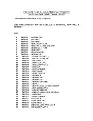
©
84803.pdf
84803.pdf

You also want an ePaper? Increase the reach of your titles
YUMPU automatically turns print PDFs into web optimized ePapers that Google loves.
<strong>©</strong><br />
A-<br />
o<br />
ro<br />
(X<br />
a-<br />
4:<br />
0<br />
-D<br />
Q<br />
r-<br />
0<br />
G><br />
t-<br />
Z<br />
do<br />
rO O ~<br />
cx
"• V, 01 <br />
-^<br />
i
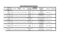
The Madras Bone Tumour Service i<br />
No. 4, Lakshmi Street, Kilpauk, Chenrtai - 600 010. INDIA<br />
, V<br />
E-mail : drmayil@bonetumor.org Website : www.bonetumour.org FAX : 91-44-6453365 PHONE : 91-144-6442279<br />
Chairman<br />
Prof : Mayil Vahanan Natarajan<br />
M.S.Orth. (Ma's) M.Ch.Trauma (L'pool) Ph.D. (Orth. Onco.)<br />
Scientist - State Council for Science & Technology<br />
Professor of Orthopaedic Surgery<br />
Madras Medical College & Research Institute<br />
Affiliated Hospitals<br />
f\l J^ Cancer Institute<br />
Govt. General Hospital<br />
M.N. Orthopaedic Hospital<br />
Apollo Cancer Hospital<br />
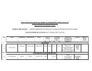
CUSTOM MEGA-PROSTHESIS<br />
Name.<br />
Age trfT...\, Sex.<br />
D.O.S.<br />
Region<br />
, 0 ,0 \.<br />
PROXIMAL FEMUR<br />
M n t e r i a i<br />
STAINLESS STEEL<br />
_<br />
Remarks<br />
Dor.iqn<br />
Rights Pending<br />
- MECHANICAL ENGINEERS ,<br />
Desmgers S Mr'rs. D' CUSTOM MEGA PROSTHESIS 4 BONE IMPLANTS<br />
*9, (Old No 4) Flag Staff Streei Royip-jram, Chennai 600 0'3 Phone QAi '>95
- HP - 7510<br />
Apollo Speciality Hospital<br />
towards greater strides in modern medicare<br />
320, Mount Road, Chennai 600 035.<br />
Phone : 24331740, 24331741, 24331712.<br />
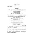
.i.ii.eiMR V K ABHINESH<br />
;ge : 23 Years Sex : Ha is<br />
tef.ByzDR hAYILvABANAN NATA<br />
Unit No:OTHERS Reed, on<br />
Ream s D.P.<br />
Rept. On<br />
oqy No :H03-2756<br />
23/06/2003<br />
30/06/2003 •0<br />
S LJ Ft O I OftL_ F* £% "T l~t O L_ O €3 V Ft EZ F" O R. "T<br />
Specimen :<br />
EXCISED MASS LEFT PROXIMAL FEMUR.<br />
Macroscopic Description :<br />
Received specimen of left proximal femur with attacned soft<br />
tissue, muscle measuring 12x9x6csn with head of femur measuring<br />
^N*^.4x4cm, Cut surface shows solid and cystic areas. Solio areas<br />
QI e grey white with areas of haemorrhage. The lesio^n is seen<br />
extended upto the soft tissue and eroding the cortex. Clearance<br />
from the distal resected margin is 2.5cm.<br />
A: Scoop margin == all.<br />
B: Lesion with lateral inked margin == 2 bits.<br />
C: Lesion with medial inked margin == 2. bits.<br />
D: Lesion == 2 bits.<br />
Microscopic Description _j_<br />
A: Sections show spicules of bone.<br />
E — 3: Shows bony trabeculaes with a neoplasm composea of spinal*<br />
cell stroma with numerous mui t inucleate osteoclastic giant cells,<br />
Foci of cystic Degeneration and haemorrnage is seen. There is nc<br />
evidence of anaplasia. The tumour is seen icm away from<br />
marg in .<br />
"3T<br />
Bjr<br />
5<br />
s<br />
o m<br />
o<br />
t<br />
CONSISTENT WITH GIANT CELL TUMOUR OF BONE; EXCISED MASS LEFT<br />
PROXIMAL FEMUR.<br />
Comments :<br />
Advised radiological correlation.<br />
?<br />
liii<br />
DR. K.R.AHESH,HD. ,<br />
PATHOLOSIST<br />
- r