Edwards et al., Curr Opin Struct Biol 2007
Edwards et al., Curr Opin Struct Biol 2007
Edwards et al., Curr Opin Struct Biol 2007

Create successful ePaper yourself
Turn your PDF publications into a flip-book with our unique Google optimized e-Paper software.
M<strong>et</strong>abolite recognition by riboswitches <strong>Edwards</strong>, Klein and Ferré-D’Amaré 277<br />
Figure 3<br />
thus far focused on isolated m<strong>et</strong>abolite-binding domains.<br />
Published studies include ensemble and single-molecule<br />
fluorescence resonance energy transfer (FRET) characterization<br />
of the adenine riboswitch (demonstrating the presence<br />
of Mg 2+ -dependent folding intermediates [41 ]),<br />
solution NMR experiments on the Mg 2+ and effector<br />
dependence of the termin<strong>al</strong> loop–loop interaction in the<br />
purine riboswitches [42 ], and sm<strong>al</strong>l-angle X-ray scattering<br />
an<strong>al</strong>ysis of the shape of the glycine riboswitch in the<br />
unliganded, Mg 2+ -induced folded and effector-bound<br />
states [43].<br />
With the exception of the glmS ribozyme, riboswitchmodulated<br />
gene regulation results from the sequestration<br />
of a switch segment (red in Figure 1) into either a<br />
m<strong>et</strong>abolite-bound or a m<strong>et</strong>abolite-free conformation<strong>al</strong><br />
fold. For riboswitches that exert gen<strong>et</strong>ic control at the<br />
transcription<strong>al</strong> level, co-transcription<strong>al</strong> mRNA folding is<br />
an essenti<strong>al</strong> component of the mechanism of action [44]<br />
and must be considered <strong>al</strong>ongside the thermodynamics<br />
and kin<strong>et</strong>ics of ligand binding [24 ,45], as well as the<br />
relative speed of transcription [46 ]. In a study of the<br />
flavin mononucleotide (FMN) riboswitch, it was shown<br />
that the speed of transcription relative to the kin<strong>et</strong>ics of<br />
FMN binding precludes this RNA from reaching thermodynamic<br />
equilibrium before the point at which a gen<strong>et</strong>ic<br />
decision must be made [46 ]. Consequently, m<strong>et</strong>abolite<br />
concentrations needed to trigger this riboswitch in vivo<br />
must be considerably higher than the in vitro dissociation<br />
constant measured with previously transcribed RNA.<br />
These studies <strong>al</strong>so implicate transcription<strong>al</strong> pause sites<br />
as critic<strong>al</strong> components of riboswitch-mediated regulation.<br />
Sever<strong>al</strong> ribosensors have been described that function by<br />
means other than direct m<strong>et</strong>abolite binding. These include<br />
non-coding mRNA elements that respond to the presence<br />
of uncharged tRNAs [47], the concentration of Mg 2+ [48]<br />
and temperature [49]. A series of ‘RNA thermom<strong>et</strong>ers’ in<br />
the 5 0 -UTRs of heat shock response genes unfold at<br />
elevated temperatures, releasing a Shine–D<strong>al</strong>garno sequence<br />
sequestered at lower temperatures (Figure 4a). A<br />
solution NMR structure of the lower temperature state<br />
has been d<strong>et</strong>ermined, in which sever<strong>al</strong> residues form noncanonic<strong>al</strong><br />
base pairs (Figure 4b) [49]. The imino proton<br />
resonances of these residues disappear at elevated temperatures,<br />
indicating that these pairs melt. The coupling of<br />
folding with the control of gene expression is a feature<br />
shared by these ribosensors and riboswitches.<br />
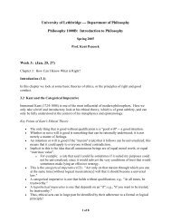
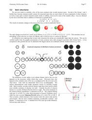
Sm<strong>al</strong>l-molecule binding by aptamers and riboswitches. (a) The<br />
solvent-accessible surface area of the ligand that is buried when in<br />
complex with aptamers or riboswitches correlates linearly with affinity,<br />
with a binding energy of 19 c<strong>al</strong>/mol per Å 2 of buried area. Red and<br />
blue represent riboswitches and aptamers, respectively; squares and<br />
circles denote structures solved by X-ray cryst<strong>al</strong>lography or NMR,<br />
respectively; 95% prediction interv<strong>al</strong>s are shown as dashed lines.<br />
The purine and theophylline complexes were excluded from the<br />
an<strong>al</strong>ysis. (b) Solvent-accessible surfaces of riboswitch- or aptamerbound<br />
ligands. Panels show the absolute solvent-accessible area of<br />
each ligand atom (dark blue 0–1 Å 2 , light blue 1–10 Å 2 , green<br />
10–20 Å 2 , yellow 20–30 Å 2 , orange 30–40 Å 2 , red 40–60 Å 2 ) mapped<br />
onto the molecular surface overlaying it. Buried surface areas were<br />
c<strong>al</strong>culated using a 1.4 Å radius probe [50]. Aptamer structures are<br />
reviewed in [32].<br />
www.sciencedirect.com <strong>Curr</strong>ent <strong>Opin</strong>ion in <strong>Struct</strong>ur<strong>al</strong> <strong>Biol</strong>ogy <strong>2007</strong>, 17:273–279