Edwards et al., Curr Opin Struct Biol 2007
Edwards et al., Curr Opin Struct Biol 2007
Edwards et al., Curr Opin Struct Biol 2007

You also want an ePaper? Increase the reach of your titles
YUMPU automatically turns print PDFs into web optimized ePapers that Google loves.
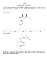
M<strong>et</strong>abolite recognition by riboswitches <strong>Edwards</strong>, Klein and Ferré-D’Amaré 275<br />
structure of the m<strong>et</strong>abolite-binding domain of the first<br />
class to be discovered (the SAM-I riboswitch) reve<strong>al</strong>s<br />
an architecture that is distinctly different from the<br />
inverted-h fold of the purine and TPP riboswitches<br />
(Figure 1e,f) [15 ]. The SAM-I riboswitch contains two<br />
stacks (P1-P4 and P2a-P3) that, rather than packing sideby-side,<br />
cross at an angle of 708. A pseudoknot coupled<br />
to a kink-turn [16] atop P2 appears to stabilize this<br />
fold. The SAM-binding site is located at the interface<br />
of the minor grooves of P1 and P3, and has features<br />
reminiscent of the m<strong>et</strong>abolite-binding sites of both the<br />
TPP and the purine riboswitches. Like TPP, SAM<br />
bridges two helic<strong>al</strong> stacks. Like the purine riboswitches,<br />
SAM makes van der Wa<strong>al</strong>s contact with the 3 0 strand of<br />
the P1 switch helix.<br />
In many Gram-positive bacteria, the glmS ribozyme-riboswitch<br />
is part of the 5 0 -UTR of the mRNA that encodes<br />
glucosamine-6-phosphate (GlcN6P) synth<strong>et</strong>ase [17]. This<br />
riboswitch has a self-cleavage activity that becomes activated<br />
when it binds GlcN6P. The structure of the glmS<br />
ribozyme [18 ,19 ] consists of three par<strong>al</strong>lel helic<strong>al</strong> stacks<br />
(Figure 1g,h). A doubly pseudoknotted core (P1-P2-P2.1-<br />
P2.2) is buttressed by a peripher<strong>al</strong> RNA domain (P4-P4.1).<br />
The solvent-exposed GlcN6P-binding pock<strong>et</strong> is composed<br />
of two highly distorted major grooves and abuts the site of<br />
self-cleavage, reflecting the coenzyme function of GlcN6P<br />
(see the review by Scott in this issue). Among currently<br />
characterized riboswitches, the glmS ribozyme is unique<br />
because it adopts its active structure in the absence of its<br />
m<strong>et</strong>abolite ligand [18 ,20 ]. As other ribozymes, such as<br />
the natur<strong>al</strong> hairpin ribozyme [21,22] and the in vitro<br />
selected Diels–Alderase [23], <strong>al</strong>so assemble rigid active<br />
sites, this disparity might reflect the different constraints<br />
under which ribozymes and RNAs that function by <strong>al</strong>ternative<br />
folding evolved.<br />
Ligand recognition by riboswitches<br />
The purine riboswitch recognizes its ligand <strong>al</strong>most exclusively<br />
through hydrogen-bonding interactions that satisfy<br />
nearly <strong>al</strong>l possible acceptors and donors of the purine<br />
(Figure 2a). Purine riboswitch structures have been<br />
solved bound to 2,6-diaminopurine [24 ] and 2,4,6-triaminopyrimidine<br />
[25], in addition to the biologic<strong>al</strong> activators<br />
hypoxanthine [6], guanine [7] and adenine [7]. The<br />
purine ligand is primarily recognized by residue 74 of the<br />
riboswitch, a pyrimidine, through Watson–Crick pairing<br />
[6,7,26]. In addition, U51 and the 2 0 -OH of U22 hydrogen<br />
bond to the N3/N9 edge (corresponding to the sugar edge<br />
of nucleotides) and the N7, respectively, of the purine.<br />
Reliance on Watson–Crick (as opposed to Hoogsteen)<br />
pairing for recognition enables the same RNA scaffold to<br />
regulate either adenine or guanine m<strong>et</strong>abolism by having<br />
U74 or C74, respectively. Gilbert <strong>et</strong> <strong>al</strong>.[24 ] noted that the<br />
purine ligand makes poor stacking interactions and proposed<br />
that this enhances the discriminatory role of Watson–Crick<br />
pairing with residue 74.<br />
The two helic<strong>al</strong> stacks of the thi-box riboswitch separately<br />
recognize the aminopyrimidine and pyrophosphate moi<strong>et</strong>ies<br />
of TPP. J3/2 of the ‘pyrimidine sensor helix’ (the P1-<br />
P2-P3 stack) adopts a canonic<strong>al</strong> T-loop fold [9 ,10 ,11 ].<br />
Binding of the aminopyrimidine ring of TPP to G40 of this<br />
T-loop (Figure 2b) mimics a tertiary interaction b<strong>et</strong>ween<br />
the D- and T-loops in the classic L-shaped fold of tRNA.<br />
The aminopyrimidine of TPP and G40 replace G18 and<br />
C55, respectively (purines and pyrimidines are reversed<br />
b<strong>et</strong>ween the riboswitch and the tRNA). Mimicry of an <strong>al</strong>l-<br />
RNA structure by an exogenous sm<strong>al</strong>l molecule is reminiscent<br />
of ATP binding by an in vitro selected aptamer<br />
RNA, whereby the ATP compl<strong>et</strong>es a GNRA t<strong>et</strong>r<strong>al</strong>oop<br />
[27,28]. Rather than directly binding to the negatively<br />
charged pyrophosphate of TPP, the ‘pyrophosphate sensor<br />
helix’ (the P4-P5 stack) of the riboswitch coordinates the<br />
pyrophosphate mostly through two solvated div<strong>al</strong>ent<br />
m<strong>et</strong><strong>al</strong>s ions (the exception is G78) [9 ,10 ,11 ,29]. Thus,<br />
the riboswitch effectively binds a positively charged TPP–<br />
cation complex. <strong>Struct</strong>ures of the TPP riboswitch bound to<br />
three m<strong>et</strong>abolite an<strong>al</strong>ogs suggest that the pyrimidine sensor<br />
helix is largely preformed, whereas the pyrophosphate<br />
sensor helix becomes organized concomitant with binding<br />
of the TPP–cation complex [11 ].<br />
The SAM-I riboswitch sandwiches its ligand b<strong>et</strong>ween two<br />
par<strong>al</strong>lel helices [15 ]. P1 recognizes the ribose–sulfur<br />
backbone of SAM primarily through van der Wa<strong>al</strong>s contacts<br />
(Figure 2c). By contrast, the P3 helix binds the<br />
adenine ring and the amino acid by making sever<strong>al</strong><br />
hydrogen bonds and stacking interactions. Interestingly,<br />
the RNA does not directly recognize the m<strong>et</strong>hionine<br />
e-m<strong>et</strong>hyl group. Rather, the positively charged sulfur atom<br />
makes a favorable electrostatic interaction with the parti<strong>al</strong><br />
negative charge on O2 of U7. This might explain the<br />
enhanced binding of SAM compared to S-adenosylhomocysteine<br />
and other non-positively charged an<strong>al</strong>ogs [12,30].<br />
Recognition of GlcN6P, a simple phosphorylated hexosamine<br />
sugar, by the glmS ribozyme-riboswitch presents a<br />
ch<strong>al</strong>lenge for RNA that is distinct from those posed by<br />
purines, TPP and SAM, <strong>al</strong>l of which have nucleotide-like<br />
substructures. Cryst<strong>al</strong> structures of the glmS ribozymeriboswitch<br />
were solved bound to glucose-6-phosphate<br />
(Glc6P), an isosteric comp<strong>et</strong>itive inhibitor (antagonist)<br />
of GlcN6P [18 ], and to the authentic activator GlcN6P<br />
([19 ]; DJ Klein and AR Ferré-D’Amaré, unpublished),<br />
reve<strong>al</strong>ing that GlcN6P and Glc6P are equiv<strong>al</strong>ently positioned<br />
in the glmS ribozyme-riboswitch active site. The<br />
sugar hydroxyl groups hydrogen bond to G1, C2, A50 and<br />
G65 (Figure 2d). The amine of GlcN6P hydrogen bonds<br />
to a water molecule, U51 and the 5 0 oxygen of G1; the last<br />
is the leaving group of the transesterification reaction<br />
cat<strong>al</strong>yzed by the ribozyme. As in the TPP riboswitch,<br />
the phosphate of the m<strong>et</strong>abolite interacts with the RNA<br />
through two solvated div<strong>al</strong>ent m<strong>et</strong><strong>al</strong> ions ([19 ]; DJ Klein<br />
and AR Ferré-D’Amaré, unpublished). TPP, SAM and<br />
www.sciencedirect.com <strong>Curr</strong>ent <strong>Opin</strong>ion in <strong>Struct</strong>ur<strong>al</strong> <strong>Biol</strong>ogy <strong>2007</strong>, 17:273–279