Lab 1 - Protists
Lab 1 - Protists
Lab 1 - Protists

Create successful ePaper yourself
Turn your PDF publications into a flip-book with our unique Google optimized e-Paper software.
LAB # 1. THE PROTOZOANS<br />
1. Overview<br />
Almost everything about Protozoan phylogeny and biology is controversial. No two<br />
biologists can agree whether this group should be alligned with the plants, the animals or<br />
even the fungi. We won’t go into the details of this controversy in the lab and we will<br />
adopt the view of Pechenik who simply refers to all organisms that are NOT plants,<br />
animals or fungi as Protozoans. They are truly a fascinating group, inhabiting all<br />
environments and displaying immense variety, especially in terms of their mechanisms to<br />
obtain food and to reproduce. As you look at the various species we have available,<br />
always remember that you are observing a single cell. Many of the processes that we<br />
have come to know in multicellular animals, occur within this cell. This cell must also<br />
avoid predators, compete, feed and avoid its own parasites and diseases. Thus, all the<br />
‘problems and constraints’ that we will talk about over the term, are solved within this<br />
single cell.<br />
The field of Protozoology is vast and we can only scratch the surface of this fascinating<br />
group. The main purpose of this lab is to introduce you to the diversity of selected<br />
Protozoans and to compare selected living material with preserved specimens. We will<br />
focus first on the free-living Protozoans and then shift to examples of parasitic types.<br />
This lab will tax your skills as a microscopist. Have patience and ask me for assistance if<br />
required. First, we will focus on the Ciliates, the Sarcodinids (Ameobas), the nonparasitic<br />
Flagellates and some other ‘misfits’. Concentrate on the living material, as we<br />
won’t have a chance to see much of it again this term. Make careful, labeled drawings<br />
where appropriate. Concentrate next on your slide-box material, and make careful<br />
comparisons to the appropriate figures in your text.<br />
Non-parasitic Protozoans<br />
1. Essential components of the lab<br />
• review set-up, cleaning and use of compound and dissecting microscopes<br />
• observe the locomotion, feeding behaviour and functional morphology of cultures of<br />
ciliates (especially Paramecium), amoebas, and perhaps others<br />
• observe samples of locally-collected pond water for diversity of commensal and freeliving<br />
ciliates (especially Stentor, Vorticella and perhaps Volvox)<br />
2. Classification<br />
We will follow the convention used by Pechenik who considers the Protozoa as<br />
comprising at least 16 separate phyla. We won’t be concerned with the taxonomy but<br />
you will need to be able to recognize selected major groups (e.g. ciliates, sarcodinids,<br />
dinoflagellates, choanoflagellates, radiolarians).<br />
Phylum Ciliophora<br />
Paramecium, Vorticella, Stentor
Phylum Rhizopoda (the ameba-like organisms)<br />
Amoeba, foraminiferans, radiolarians<br />
Phylum Euglenozoa<br />
Euglena, Volvox<br />
Phylum Dinozoa (the dinoflagellates)<br />
Phylum Choanozoa (the choanoflagellates)<br />
3. Protozoan ultrastructure and functional ecology<br />
Phylum Ciliophora<br />
We will have available free-living forms of a variety of ciliates. They are among the<br />
most structurally complex of the protozoans. Review your knowledge of Paramecium<br />
from Biol 1020 and from figures in your text. You should recognize unique features such<br />
as cilia, double nuclei, complex modes of reproduction, cytostome and at least one fixed<br />
contractile vacuole. Other structures such as toxicysts (eject toxins to subdue prey),<br />
haptocysts (prey capture) and trichocysts (probably defense) add to the complexity.<br />
The double nucleus distinguishes ciliates from other protists. The smaller of the two<br />
deals with reproduction; the larger with regulation of normal metabolism. Cilia usually<br />
covers most of the cell. Reproduction is typically by binary fission, with the plane of<br />
division along the long axis of the body.<br />
Place a drop of the Paramecium culture on a slide and observe for locomotion and<br />
functional morphology. Focus on mechanisms of locomotion and feeding. Note the<br />
forward, spiral movement (the anterior end is narrower than the posterior). Locate the<br />
oblique depression on the side called the oral groove, that then leads to the buccal cavity.<br />
Note the longitudinally-arranged cilia covering the entire body and lining the oral groove.<br />
Food trapped in the oral groove travels through the buccal cavity to the cytostome, where<br />
a food vacuole is formed. The<br />
outermost membrane is called the pellicle, which provides structure and support. A<br />
contractile vacuole with radiating canals occurs at each end. These are in fixed<br />
positions and open to the pellicle. Compare these features with stained specimens from<br />
your slide box and by comparison with the figures in your text. The nuclei should be<br />
more obvious in the stained specimens.<br />
Your slide box also contains an example of Paramecium conjugation. We will cover the<br />
details of this unique form of ‘sexual’ reproduction in class. Examine the specimen under<br />
the microscope and refer to fig. 3.17 in your text for comparison.<br />
Vorticella is common in our freshwater ponds. It is a solitary genus but often gregarious,<br />
attached to virtually any substrate. Take a sample of pond vegetation (or a snail shell)<br />
and observe under the scope. Note the inverted bell-shaped body and stalk. The stalk can<br />
retract and extend because of a large fiber that is composed of contractile filaments. Use<br />
fig. 3.19 in Pechenik to assist you in interpreting functional morphology.
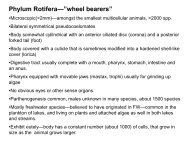
Stentor is a large, trumpet-shaped ciliate, often with delicate internal pigmentation (Fig.<br />
3.19). The anterior end is broadened. The macronucleus is slender and elongate and<br />
extends longitudinally. Micronuclei are present but hard to see. There should also be a<br />
rootlike holdfast at the tapered posterior end.<br />
Phylum Rhizopoda<br />
These Protozoans (commonly known as Amebas) should also be familiar to you from<br />
Biol 1020. Recall their use of pseudopodia, protoplasmic extensions of the motile cell,<br />
as their primary means of locomotion and feeding. The body can be naked (as in<br />
Amoeba) or with a test (shell) (e.g. Foramineferans). Almost all species are free-living<br />
and reproduction is by binary fission. They live in almost every conceivable habitat and<br />
are especially common in the soil.<br />
Amoeba proteus is a heterotrophic ameba. To view this species under the scope, try<br />
putting cracked pieces of a coverslip around the specimen before covering it (i.e. raise the<br />
coverslip off the surface of the slide). Take a sample from the bottom of the dish and<br />
find it under low power. Observe movement of the organism. This species has large<br />
pseudopodia used in food capture and locomotion. Refer to the figure 3.27 and to stained<br />
specimens to help find some of the important organelles. Try to observe a contractile<br />
vacuole discharging its liquid waste. You may also find food vacuoles.<br />
As you observe Amoeba movement, consider the mechanisms responsible. Based solely<br />
on your observations of pseudopod action, try to articulate this mechanism to one of your<br />
classmates. How does it compare to the mechanisms used by ciliates, flagellates,<br />
radiolarians and dinoflagellates?<br />
Phylum Euglenozoa (flagellated protozoans)<br />
These are the chlorophyll-containing, flagellated protists. Euglena should be familiar to<br />
you. Members of this phylum have a cell wall, cellulose and chloroplasts. Use your slide<br />
box and Fig. 3.30 in Pechenik to observe the functional morphology of Euglena (try to<br />
find chloroplasts, flagellum, eyespots, cytostome and pharynx). In addition to the basic<br />
functional morphology of this species, also note that in addition to obtaining nutrients via<br />
photosynthesis, Euglena can also absorb nutrients directly across the body wall (but it<br />
can’t capture its own prey).<br />
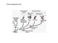
Volvox is a bizarre, colonial Euglenid. This is an extremely important group in<br />
phylogenetic terms because the more advanced species show division of the colony into<br />
specific groups of cells with specific functions (i.e. this is the first time we come across<br />
‘division of labor’). There are usually two flagella per cell. All are green with distinct<br />
cellulose cell walls. They live mostly in fresh water and both asexual and sexual<br />
reproduction is common. In Volvox, there is differentiation between somatic and<br />
reproductive cells within each colony. These large colonies also move with a particular<br />
pole consistently directed forward. The cells of the ‘anterior end’ bear well–developed<br />
eyespots and have significantly larger flagella than the trailing cells.
Phylum Dinozoa (the dinoflagellates)<br />
You might know the dinoflagellates for their ability to cause bioluminescence (the<br />
biochemical production of light) and also for their ability to cause pathogenic ‘red tides’.<br />
In class, we will talk about the recently-discovered Pfeisteria, arguably one of the most<br />
advanced protozoans. They are common in all aquatic habitats, especially in marine ones.<br />
Most are free-living. Many are odd-shaped indeed! There are two flagella used for<br />
locomotion and most species obtain their nutrition via photosynthesis. Most species have<br />
a distinctive ‘armor’ around the cell made of cellulose, thus providing nutrition, structure<br />
and protection. Possibly their most important ecological role is as intracellular symbionts<br />
in many marine invertebrates (we’ll see these live in two weeks in the tentacles of sea<br />
anenomes). These forms are known as zooxanthellae and they are known to play critical<br />
roles in the formation of coral reefs.<br />
5. Questions for discussion<br />
1. You’ve seen various ciliates and are hopefully impressed with their ability to move.<br />
Most have bodies completely covered by cilia. In order to chase prey, avoid predators<br />
and generally assess their environments, they must somehow coordinate the beating of<br />
countless numbers of cilia. Without a complex nervous system, how do they do this ?<br />
2. Paramecium and Amoeba are small, slow and full of protein, making them highly<br />
attractive to predators. What options to they have to avoid predators ? How has<br />
Vorticella and the dinoflagellates attempted to solve the problem ?<br />
3. What ecological roles do Amoebas play in the soil ? Ciliates in fresh water ?<br />
PART II: THE PARASITIC PROTOZOANS<br />
1. Overview<br />
Parasitism has evolved independently within almost every group of protozoans. Some<br />
groups (e.g. The sporozoans) are exclusively parasitic. You should not find it surprising<br />
that parasitism has evolved so often in these small, single-celled organisms. In this part<br />
of the lab, we will only be covering 2 examples (3 if we have time) of this immense<br />
diversity. There is no great logic in the 2 I have selected for study. Very simply, one is<br />
found locally in earthworms and represents a precursor to the evolution of a most<br />
important human parasite, Plasmodium. It is also among the largest protozoans known.<br />
The second is a species that is also a local problem, and one that many parasitologists<br />
consider will be one of our most important pathogens in the next century. As you go<br />
through these examples, pay attention to the morphological differences between them, the<br />
diversity of life-cycles, and their adaptations for parasitism. You will see that there are<br />
direct and indirect modes of transmission, sexual and asexual multiplicative phases, and<br />
various levels of pathology for the host.<br />
2. Essential features of the lab<br />
• observe the life-cycle stages of Monocystis from local earthworms
• understand the similarities and differences in life-histories between Monocystis,<br />
Plasmodium and Cryptosporidium and Giardia.<br />
Phylum Sporozoa<br />
Monocystis sp.<br />
Plasmodium spp.<br />
Cryptosporidium<br />
3. Classification<br />
Phylum Diplomonida<br />
Giardia lamblia<br />
4. Life-cycle of Monocystis in earthworms<br />
The Phylum Sporozoa is entirely parasitic and contains some of the most pathogenic<br />
parasites of humans. It also contains species that have incredibly complex life-cycles,<br />
often involving more than one host. The phylum is named after the presence of an<br />
‘apical complex’ that is typically used to penetrate host cells. The only other generalized<br />
characteristic is that all of the approximately 5,000 species typically alternate between<br />
sexual and asexual phases and often have complex life cycles. There are two major<br />
groups within the Sporozoa: The gregarines (e.g. Monocystis), and the coccidians (e.g.<br />
Plasmodium and Cryptosporidium).<br />
Monocystis lumbrici is an Apicomplexan parasite that lives in the seminal vesicles of<br />
terrestrial earthworms. The worm becomes infected when it ingests a spore containing<br />
several sporozoites. These hatch in the gizzard, where the released sporozoites penetrate<br />
the intestinal wall, enter the dorsal blood vessel, and then make their way to one of the<br />
host’s 5 or so ‘hearts’. From there they penetrate the seminal vesicle, where they enter the<br />
sperm-forming cells in the wall. At this point they ingest and destroy the developing<br />
spermocytes. Then they move into the lumen of the vesicle where they become mature<br />
trophozoites. After a period of feeding, two of these will come together, flatten against<br />
each other, and secrete a common cyst around each other. This is the gametocyst, usually<br />
containing 2 gamonts. Each now undergoes extensive division of their nuclei, pinches off<br />
a small portion of cell cytoplasm, which together then bud off to become the gametes.<br />
The fusion of a pair of gametes forms a zygote, each ultimately becoming a spore. Three<br />
cell divisions later forms 8 sporozoites. Thus, each gametocyst now contains many<br />
oocysts. New hosts become infected by ingesting gametocysts, or more commonly, by<br />
ingesting individual oocysts. Thus, meiosis is zygotic. Only the zygote is diploid, and<br />
reductional division in sporogony returns the sporozoites to the haploid condition.<br />
Proceedure:<br />
Dissect the anterior end of a freshly anesthetized worm. Remove the seminal vesicles and<br />
place in a drop of water. Take small pieces of seminal vesicle, squash under a cover slip<br />
and look for the different stages of Monocystis (see figures). Make drawings of each<br />
stage and construct an annotated life-cycle. Record as many different stages of infection<br />
as you can.
5. Life-cycle of Plasmodium spp.<br />
Plasmodium falciparum<br />
This intracellular parasite is the most dangerous of the 4 species that cause malaria in<br />
humans. This species will be the focus of our discussions in lecture. It is the most<br />
common and debilitating of human parasitic diseases. Over 2 million people each year<br />
die from the disease. Moreover, the disease has played a major role in shaping human<br />
history and civilizations. It is impossible for you to understand this disease without a<br />
thorough understanding of its life cycle. Please spend time before lab going over the lifecycle<br />
diagram in your text (Fig. 3.35) and the web site.<br />
Your slides only show a fraction of the various stages of the malaria life-cycle. One slide<br />
shows gametocytes inside host red-blood cells as deeper-staining structures (often<br />
crescent or bean-shaped). Using the oil immersion lens you should see that<br />
microgametocytes have a large nucleus and irregularly distributed granules.<br />
Macrogametocytes have a small compact nucleus with a dark red nucleolus. Further
development of these forms only continues inside a mosquito’s stomach. Sometimes, the<br />
gametocyte-infected cells have been distorted to such an extent that it ruptures during the<br />
fixing process.<br />
You also have a slide that shows trophozoites. They can be distinguished by the large<br />
food vacuole surrounded by a thin layer of cytoplasm and including a peripheral nucleus.<br />
This gives the characteristic ‘ring’ that forms the well-known ‘ring-stage’. Again, the<br />
infected RBC may appear distended or abnormal in shape. Use the demonstration slides<br />
of the various life-cycle stages and life-cycle diagrams to help you understand the<br />
biology of this important parasite.<br />
6. Life-cycle of Cryptosporidium parvum<br />
This waterborn parasite, together with Giardia, represents one of two major waterborn<br />
parasites of humans. It is generally a non-lethal disease but does cause severe diarrhea,<br />
vomiting, weight loss and cramping. People with normal immune systems expel the<br />
parasites in about 2 weeks, and are then immune to further infection. However, it is<br />
usually lethal in patients with compromised immune systems (especially the elderly and<br />
HIV-infected hosts). It is a major concern in the US, and now in Canada.<br />
The main concern is that cysts are highly resistant to standard disinfectants such as<br />
chlorine and chlorine dioxide. One instance in Milwaukee in 1994 led to an estimated<br />
370,000 illnesses (virtually the entire city); there was an outbreak in 1995 in Kelowna<br />
and in Medicine Hat in 2001. The Lethbridge area is considered one of the most likely<br />
areas in Canada for an outbreak of Cryptosporidiosis, due mostly to the large numbers of<br />
cattle. In the US, the main method of control is the conversion of water-treatment plants<br />
to use ozone rather than chlorine as a disinfectant. Costs run into the billions of dollars.<br />
This parasite is a recent problem, so it is still hard to buy specimens. We will sketch the<br />
generalized life cycle of this parasite in class.<br />
7. Giardia<br />
Your slides of Giardia lamblia contain the feeding form (trophozoite) and cysts of this,<br />
the most common intestinal parasite of humans. They are not easy to find on your slides;<br />
you will have to scan the slide at 40X to find the cysts. It is the causative agent of<br />
giardiasis or “beaver fever”. This is the disease most associated with wilderness campers<br />
and the parasite can be responsible for severe diarrhea. The parasite also matures in many<br />
other hosts (including beavers) which is a factor in the epidemiology of this disease. See<br />
also the SEM pictures from the website.<br />
Giardia has four pairs of flagella arising from a central pair of rod-like structures called<br />
axostyles (difficult to see as the flagella are usually lost during staining). Under oil<br />
immersion, you should be able to see two nuclei and a central pair of median bodies. The<br />
trophozoites are cup-shaped and the surface of the ventral side is concave and thickened<br />
to form a large adhesive disc (used for attachment). Locate a cyst at 40X (this will try
your patience as a microscopist!) and advance to oil immersion. Note the thick cyst wall,<br />
enclosed flagella, nuclei and median bodies (Fig. 3.30).<br />
8. Questions for discussion<br />
1. Other water-born diseases (such as Giardia) are killed by disinfectants such as<br />
chlorine. Cryptosporidium on the other hand has a notoriously thick cyst wall, which<br />
chlorine cannot penetrate. Why would Cryptosporidium evolve such a thick wall, while<br />
other water-born parasites do not ?<br />
2. What are the major differences in the life-cycles of the gregarines, Plasmodium and<br />
Cryptosporidium ? What is the advantage of incorporating a vector into the life-cycle ?<br />
3. What factors are leading to the worldwide resurgence in falciparum malaria ?