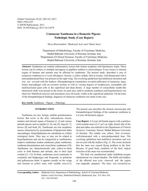
Cutaneous Xanthoma in a Domestic Pigeon: Pathologic ... - Idosi.org
Cutaneous Xanthoma in a Domestic Pigeon: Pathologic ... - Idosi.org
Cutaneous Xanthoma in a Domestic Pigeon: Pathologic ... - Idosi.org

You also want an ePaper? Increase the reach of your titles
YUMPU automatically turns print PDFs into web optimized ePapers that Google loves.
Global Veter<strong>in</strong>aria 10 (2): 140-143, 2013<br />
ISSN 1992-6197<br />
© IDOSI Publications, 2013<br />
DOI: 10.5829/idosi.gv.2013.10.2.6474<br />
<strong>Cutaneous</strong> <strong>Xanthoma</strong> <strong>in</strong> a <strong>Domestic</strong> <strong>Pigeon</strong>:<br />
<strong>Pathologic</strong> Study (Case Report)<br />
1 1 2<br />
Reza Kheirandish, Shahrzad Azizi and Omid Azari<br />
1<br />
Department of Pathobiology, Faculty of Veter<strong>in</strong>ary Medic<strong>in</strong>e,<br />
Shahid Bahonar University of Kerman, Kerman, Iran<br />
2<br />
Department of Cl<strong>in</strong>ical Sciences, Faculty of Veter<strong>in</strong>ary Medic<strong>in</strong>e,<br />
Shahid Bahonar University of Kerman, Kerman, Iran<br />
Abstract: <strong>Xanthoma</strong>s are nodular <strong>in</strong>flammatory lesions that imitate neoplasm with lipomatous orig<strong>in</strong>. These<br />
lesions can be solitary or multiple and appear as papillary nodules to raised plaques. Every tissues or <strong>in</strong>ternal<br />
<strong>org</strong>ans of humans and animals may be affected by xanthomas. The present study, described a case of<br />
cutaneous xanthoma <strong>in</strong> a 4 year old pigeon. Grossly, a yellow nodule, firm <strong>in</strong> texture, well-demarcated with a<br />
semi-pedunculated base was present on the right w<strong>in</strong>g. The overly<strong>in</strong>g epithelium had multifocal ulceration and<br />
was not covered with the feathers. Histopathological exam<strong>in</strong>ation revealed <strong>in</strong>filtration of numerous, large,<br />
foamy macrophages with an eccentric nucleus as well as vary<strong>in</strong>g degrees of lymphocytes, eos<strong>in</strong>ophils and<br />
mult<strong>in</strong>ucleated giant cells <strong>in</strong> the superficial and deep dermis. A large number of extracellular needle-like<br />
cholesterol clefts were present <strong>in</strong> the lesion. In some area, mild to moderate acanthosis and hyperkeratosis was<br />
observed. Multifocal necrosis and ulcerations were obviously visible at the superficial epidermis. On the basis<br />
of the histopathological f<strong>in</strong>d<strong>in</strong>gs, diagnosis of cutaneous xanthoma was made <strong>in</strong> this case.<br />
Key words: <strong>Xanthoma</strong> <strong>Pigeon</strong> Pathology<br />
INTRODUCTION<br />
<strong>Xanthoma</strong>s are rare, benign, nodular granulomatous<br />
lesions that occur <strong>in</strong> the sk<strong>in</strong>, subcutaneous tissues,<br />
tendons and <strong>in</strong>ternal <strong>org</strong>ans of humans [1, 2] and various<br />
animals species such as reptiles [3, 4], cats [5], dogs [6, 7],<br />
horses [8] and birds [9]. <strong>Xanthoma</strong>s are non neoplastic<br />
masses characterize by accumulation of lipoprote<strong>in</strong>-laden<br />
macrophages. Hyperlipidemias are considerate as a likely<br />
etiological factor. They may or may not be related to<br />
hyperlipidemias and can be categorized as xanthelasma,<br />
eruptive xanthomas, tendon xanthomas, plane xanthomas,<br />
xanthoma dissem<strong>in</strong>atum and verruciform xanthomas [10].<br />
<strong>Xanthoma</strong>s are characteristically pale yellow-to-white<br />
color <strong>in</strong> both humans and animals, due to their lipid<br />
content [11, 12]. In birds, xanthomas occur commonly <strong>in</strong><br />
cockatiels and budgerigars and frequently <strong>in</strong> psittac<strong>in</strong>e<br />
and gall<strong>in</strong>aceous birds. It appears usually on the w<strong>in</strong>gs<br />
and sternum as yellow mass with variable sizes [13].<br />
The present case describes the cl<strong>in</strong>ical, macroscopic and<br />
histopathological f<strong>in</strong>d<strong>in</strong>gs of the cutaneous xanthoma <strong>in</strong><br />
a 4-year old domestic pigeon.<br />
Case Report: A 4-year old female pigeon with a pa<strong>in</strong>less,<br />
2<br />
solid nodular mass (2×2 cm ) on right, lateral side of the<br />
ulna and radius was referred to the Department of Cl<strong>in</strong>ical<br />
Sciences, Veter<strong>in</strong>ary School, Shahid Bahonar University<br />
of Kerman. The nodule was yellow, firm <strong>in</strong> texture,<br />
well-demarcated with a semi-pedunculated base. The<br />
overly<strong>in</strong>g epithelium had multifocal ulceration and was<br />
not covered with the feathers (Fig. 1). The owner declared<br />
that the mass was caused fly<strong>in</strong>g problem <strong>in</strong> the bird.<br />
Because of good body condition of the bird, radical<br />
resection of the mass was recommended.<br />
The bird was anesthetized under halothane-oxygen<br />
adm<strong>in</strong>istered via a head chamber. The fluffs and feathers<br />
of the affected area were removed and the region<br />
was surgically prepared. The mass was excised by an<br />
Correspond<strong>in</strong>g Author:<br />
Shahrzad Azizi, Department of Pathobiology, School of Veter<strong>in</strong>aryMedic<strong>in</strong>e,<br />
Islamic Azad University, Shahrekord Branch, Shahrekord, Iran. P.O. Box 166.<br />
Tel: +3813361045.<br />
140
Global Veter<strong>in</strong>aria, 10 (2): 140-143, 2013<br />
electro-surgical unit. Oral consumption of oxytetracycl<strong>in</strong>e<br />
<strong>in</strong> dr<strong>in</strong>k<strong>in</strong>g water has been recommended for one week as<br />
a postoperative medication. The bird was discharged on<br />
day 14, when the wound was acceptably dim<strong>in</strong>ished <strong>in</strong><br />
size.<br />
For histopathological evaluation, the removed nodule<br />
by surgical excision was fixed <strong>in</strong> 10% buffered formal<strong>in</strong>.<br />
Embedded paraff<strong>in</strong> tissues were processed by us<strong>in</strong>g<br />
standard procedures. Sections <strong>in</strong> 5-µm thickness were<br />
sta<strong>in</strong>ed with hematoxyl<strong>in</strong>-eos<strong>in</strong> and exam<strong>in</strong>ed<br />
microscopically. Histopathological exam<strong>in</strong>ation revealed<br />
Fig. 1: <strong>Xanthoma</strong> <strong>in</strong> a pigeon. A yellow, sever <strong>in</strong>filtration of numerous, large, foamy macrophages<br />
semi-pedunculated nodule with multiple f<strong>in</strong>e (conta<strong>in</strong><strong>in</strong>g clear vacuoles <strong>in</strong> cytoplasm) with an eccentric<br />
ulcerations on its surface is visible <strong>in</strong> the nucleus as well as vary<strong>in</strong>g degrees of lymphocytes,<br />
featherless part of the w<strong>in</strong>g<br />
eos<strong>in</strong>ophils and mult<strong>in</strong>ucleated giant cells <strong>in</strong> the<br />
superficial and deep dermis. Free lipid-droplets <strong>in</strong> variable<br />
sizes were observed <strong>in</strong> lymphocytes and plasma cells.<br />
A large number of extracellular acicular cholesterol clefts<br />
were present <strong>in</strong> the lesion and some of them were<br />
visible <strong>in</strong> the cytoplasm of giant cells (Fig. 2 and 3).<br />
In some area, mild to moderate acanthosis and<br />
hyperkeratosis was observed. Multifocal necrosis and<br />
ulceration was observed at the superficial epidermis.<br />
The heterophils were <strong>in</strong>filtrated more near the ulcerated<br />
areas. The hemorrhage and necrosis were of the observed<br />
f<strong>in</strong>d<strong>in</strong>gs. On the basis of the histopathologic f<strong>in</strong>d<strong>in</strong>gs,<br />
diagnosis of cutaneous xanthoma was made.<br />
Fig. 2: Histopathologic section of xanthoma reveals<br />
numerous, large, foamy macrophages with an<br />
eccentric nucleus (arrows), lake of free lipid<br />
aggregation (asterisk) and extracellular needle-like<br />
cholesterol clefts (arrowhead) (H-&E, × 100)<br />
Fig. 3: A large number of extracellular cholesterol clefts<br />
(arrowheads) that some of them are visible as<br />
<strong>in</strong>tracellular <strong>in</strong> giant cells cytoplasm (arrow)<br />
(H-&E, × 400)<br />
DISCUSSION<br />
<strong>Xanthoma</strong>s are nodular <strong>in</strong>flammatory lesions that<br />
imitate neoplasms with lipomatous orig<strong>in</strong>. These lesions<br />
can be solitary or multiple and appear as papillary nodules<br />
to raised plaques. Every tissues or <strong>org</strong>ans such as<br />
subcutaneous, tendons, digestive system, kidney and<br />
conjunctiva <strong>in</strong> humans and animals may be affected by<br />
xanthoma [11, 12]. These lesions are not neoplastic but<br />
can <strong>in</strong>vade locally. <strong>Xanthoma</strong>s characterize by<br />
accumulation of lipid-laden macrophages, mult<strong>in</strong>ucleated<br />
giant cells and cholesterol clefts [14-16].<br />
The pathogenesis of xanthomas is not clearly<br />
understood. Disorders <strong>in</strong> lipid metabolism are presumed<br />
<strong>in</strong> the formation of xanthomas. Serum cholesterol or<br />
other lipids are often evaluated <strong>in</strong> affected cases [17, 18].<br />
In human, xanthomas are usually diagnosed as familiarly<br />
and <strong>in</strong> response to secondary hyperlipidemia <strong>in</strong> patients<br />
associated with diabetes mellitus, hypothyroidism,<br />
multiple myeloma, or cholestatic liver disease [19-21]. In<br />
animals affected to xanthomas different types of lipid<br />
components is frequently measured. Abnoemalities <strong>in</strong><br />
141
Global Veter<strong>in</strong>aria, 10 (2): 140-143, 2013<br />
lipid metabolism and metabolic diseases <strong>in</strong>clud<strong>in</strong>g REFERENCES<br />
hyperlipidemia, diabetes mellitus, hypothyroidism, or<br />
hyperadrenocorticism are found <strong>in</strong> different animal<br />
species [8, 22-24]. In experimental studies, xanthomatosis<br />
was <strong>in</strong>duced <strong>in</strong> Japanese quail by a diet conta<strong>in</strong><strong>in</strong>g<br />
cholesterol [25].<br />
The present study, described a case of cutaneous<br />
xanthoma <strong>in</strong> a 4 year old pigeon. <strong>Xanthoma</strong>s were<br />
reported <strong>in</strong> numerous avian species and various sites<br />
especially <strong>in</strong> psittac<strong>in</strong>e birds [26, 27] but it occur rarely <strong>in</strong><br />
the domestic pigeon. In this case, a firm, yellow color and<br />
semi-pedunculated on the w<strong>in</strong>g that composed of<br />
lipid-filled macrophages, mult<strong>in</strong>ucleated giant cells,<br />
cholesterol cleft as well as fewer <strong>in</strong>filtration of<br />
lymphocytes, plasma cells and eos<strong>in</strong>ophils <strong>in</strong> the stroma.<br />
Some gross and histopathological aspects of the<br />
cutaneous xanthomas <strong>in</strong> this case were similar to those<br />
reported <strong>in</strong> humans and different animal species. In<br />
contrast to our case that xanthoma was as a s<strong>in</strong>gle nodule<br />
with smooth surface, <strong>in</strong> most reports, xanthomas appeared<br />
grossly multicentric and papillary form.<br />
In previous studies, similar to present case,<br />
xanthomas were typically nodules of granulomatous<br />
<strong>in</strong>flammation compris<strong>in</strong>g of xanthoma cells (foamy<br />
macrophages), cholesterol clefts and giant cells which<br />
sometimes were associated with fewer numbers of<br />
lymphocytes, eos<strong>in</strong>ophils, neutrophils, mast cells and<br />
plasma cells [5, 6, 28].<br />
Jaensch et al. [28] reported cutaneous xanthomas <strong>in</strong><br />
a goose. Grossly, the multiple, papillary form masses<br />
were located on the feet-webb<strong>in</strong>g, per ocular sk<strong>in</strong> and<br />
submandibular space. In histopathological study, foamy<br />
cells conta<strong>in</strong><strong>in</strong>g <strong>in</strong>tracytoplamic vacuoles that sta<strong>in</strong>ed<br />
with oil-red O were observed. Also, lymphocytes and<br />
heterophils were <strong>in</strong>filtrated <strong>in</strong> the lesions.<br />
Vogelnest [5] described cutaneous xanthomas <strong>in</strong> a<br />
9 month old cat that appeared as multifocal papules and<br />
plaques on the head and neck. Microscopic evaluation<br />
was shown <strong>in</strong>filtration vacuolated macrophages,<br />
eos<strong>in</strong>ophils and neutrophils. Cholesterol clefts, giant cells<br />
and free lipids were absent.<br />
In birds, neoplasm’s related to fatty tissue and lipid<br />
deposition <strong>in</strong>clude lipoma, myelolipoma, liposarcoma<br />
and hibernoma. <strong>Xanthoma</strong>s and lipogranulomas may<br />
be confused with these tumors. Histopathological<br />
exam<strong>in</strong>ation is a best way for def<strong>in</strong>itive diagnosis of<br />
these neoplasm’s or tumor like masse. Further research<br />
is required to clarify the xanthomas pathogenesis and<br />
its relation with hyperlipidemia, detection of orig<strong>in</strong>al<br />
cells contributed <strong>in</strong> the lesion and their markers by<br />
immunohistochemistry.<br />
1. Herrera-Goepfert, R., M. Lizano-Soberon and<br />
M. Garcia-Perales, 2003. Verruciform xanthoma of<br />
the esophagus. Human Pathol., 34: 814-815.<br />
2. Philipsen, H.P., P.A. Reichart, T. Takata and I. Ogawa,<br />
2003. Verruciform xanthoma-biological profile of 282<br />
oral lesions based on a literature survey with n<strong>in</strong>e<br />
new cases from Japan. Oral Oncol., 39: 325-336.<br />
3. Ryan, M.J. and G.D. Whitney, 1980. <strong>Xanthoma</strong> <strong>in</strong><br />
a gopher snake. Vet. Med. Small. Anim. Cl<strong>in</strong>.,<br />
3: 503-507.<br />
4. Garner, M.M., N.P. Lung and S. Murray, 1999.<br />
<strong>Xanthoma</strong>tosis <strong>in</strong> geckos: five cases. J. Zoo. Wildl.<br />
Med., 30: 443-447.<br />
5. Vogelnest, L.J., 2001. <strong>Cutaneous</strong> xanthomas with<br />
concurrent demodicosis and dermatophytosis <strong>in</strong> a<br />
cat. Aust. Vet. J., 79(7): 470-475.<br />
6. Romanucci, M., D. Malatesta, P. Guardiani,<br />
P. Frescura and L. Della Salda, 2008.<br />
Xanthogranulomatous <strong>in</strong>flammation of the small<br />
bowel <strong>in</strong> a dog. Vet. Pathol., 45: 207-211.<br />
7. Balme, E., C. Thuilliez, T. Lejeune, L. Chateau-<br />
Escoffier and F. Bernex, 2009. Multiple atypical<br />
mucosal xanthomas <strong>in</strong> a dog similar to human<br />
verruciform xanthoma. J. Vet. Diagn. Invest.,<br />
21: 124-128.<br />
8. Hargis, A.M. and P.E. G<strong>in</strong>n, 2007. The <strong>in</strong>tegument.<br />
In: <strong>Pathologic</strong> basis of veter<strong>in</strong>ary disease, McGav<strong>in</strong><br />
th<br />
MD, Zachary JF, 4 edition, Mosby Elsevier, St.<br />
Louis, MO, pp: 1236-1238.<br />
9. Souza, M.J., N.S. Johnstone-McLean, D. Ward and<br />
K. Newkirk, 2009. Conjunctival xanthoma <strong>in</strong> a blue<br />
and gold macaw (Ara ararauna). Vet. Ophthalmol.,<br />
12(1): 53-55.<br />
10. Rekha, A. and D.K. Rai, 2010. Tendon xanthomas.<br />
The Foot, 20: 85-86.<br />
11. Grundy, S.M., 1999. <strong>Xanthoma</strong>toses and lipoprote<strong>in</strong><br />
disorders. In: I.M. Freedberg, A.Z. Eisen, K. Wolff,<br />
et al. eds. Fitzpatrick’s dermatology <strong>in</strong> general<br />
th<br />
medic<strong>in</strong>e. 5 edition, McGraw-Hill, New York,<br />
pp: 1804-1811.<br />
12. Scott, D.W., W.H. Miller and C.E. Griff<strong>in</strong>, 2001.<br />
Endocr<strong>in</strong>e and metabolic diseases. In: Muller and<br />
th<br />
Kirk’s small animal dermatology, 6 edition.<br />
Philadelphia, WB. Saunders, 873-874, 1148-1153.<br />
13. Lightfoot, T.L., 2006. Overview of tumors: Section I.<br />
In: Harrison GJ, Lightfoot TL, eds. Cl<strong>in</strong>ical avian<br />
medic<strong>in</strong>e. Spix Publish<strong>in</strong>g, Inc., Palm Beach, FL,<br />
pp: 560-565.<br />
142
Global Veter<strong>in</strong>aria, 10 (2): 140-143, 2013<br />
14. Campbell, T.C., 1994. Cytology. In: B.W. Ritchie, 22. Gumbrell, R.C., 1972. A case of multiple<br />
G.J. Harrison and L.R. Harrison, eds. Avian medic<strong>in</strong>e xanthomatosis and diabetes mellitus <strong>in</strong> a dog. N. Z.<br />
and surgery: Pr<strong>in</strong>cipals and Application. W<strong>in</strong>gers Vet. J., 20: 240-242.<br />
Publish<strong>in</strong>g, Lake Worth, pp: 200-222.<br />
23. Chasta<strong>in</strong>, C.B. and C.L. Graham, 1978. <strong>Xanthoma</strong>tosis<br />
15. Jones, T.C., R.D. Hunt and N.W. K<strong>in</strong>g, 1997. The sk<strong>in</strong> secondary to diabetes mellitus <strong>in</strong> a dog. J. Am. Vet.<br />
and its appendages. In: Jones TC, Hunt RD, K<strong>in</strong>g Med. Assoc., 172: 1209-1211.<br />
NW. Veter<strong>in</strong>ary pathology. Williams and Wilk<strong>in</strong>s, 24. Jones, B.R., W.S. Hancock and C.H. Campbell,<br />
Baltimore. 1983. Occurrence of idiopathic, familial<br />
16. Reavill, D.R., 2004. Tumors of pet birds. In: J.E. hyperchylomicronaemia <strong>in</strong> a cat. Vet. Rec.,<br />
Graham, ed. Veter<strong>in</strong>ary cl<strong>in</strong>ics of North America 112: 543-547.<br />
Exotic Animal Practice, 7: 537-560. 25. Hoekstra, K.A., C.R. Nichols, M.E. Garnett,<br />
17. Wissel<strong>in</strong>k, M.A., J.P. Koeman, T.H. Wens<strong>in</strong>g, J. De D.V. God<strong>in</strong> and K.M. Cheng, 1998. Dietary<br />
Bruijne and T. Willemse, 1994. Hyperlipoprote<strong>in</strong>aemia cholesterol-<strong>in</strong>duced xanthomatosis <strong>in</strong><br />
associated with atherosclerosis and cutaneous atherosclerosis-susceptible Japanese quail<br />
xanthomatosis <strong>in</strong> a cat. Vet. Q., 16: 199-202. (Cotunix japonica). J. Com. Pathol., 119: 419-27.<br />
18. Chanut, F., M.A. Colle, J.Y. Deschamps, O. Albaric 26. Schmidt, R.E., 1997. Neoplastic diseases. In: R.B.<br />
and M. Wyers, 2005. Systemic xanthomatosis Altman, S.L. Clubb, G.M. Dorreste<strong>in</strong> and K.<br />
associated with hyperchylomicronaemia <strong>in</strong> a cat. Quesenberry, editors. Avian medic<strong>in</strong>e and surgery.<br />
J. Vet. Med. A., 52: 272-274. Saunders, Philadelphia, pp: 590-599.<br />
19. Nayak, K.R. and R.G. Daly, 2004. Eruptive xanthomas 27. Monks, D.J., H.P. Zsivanovits, J.E. Cooper and<br />
associated with hypertriglyceridemia and new-onset N.A. Forbes, 2006. Successful treatment of<br />
diabetes mellitus. N. Engl. J. Med., 350: 1235.<br />
tracheal xanthogranulomatosis <strong>in</strong> a red-tailed hawk<br />
20. Sibley, C. and N.J. Stone, 2006. Familial (Buteo jamaicensis) by tracheal resection and<br />
hypercholesterolemia: a challenge of diagnosis and anastamosis. J. Avian. Med. Surg., 20: 247-252.<br />
therapy. Cleve. Cl<strong>in</strong>. J. Med., 73(1): 57-64.<br />
28. Jaensch, S.M., R. Butler, A. O’Hara, S.R. Raidal and<br />
21. White, L.E., 2009. <strong>Xanthoma</strong>toses and lipoprote<strong>in</strong> K. Wyatt, 2002. Atypical multiple, papilliform,<br />
disorders. In: Freedberg et al. Eds. Fitzpatrick’s xanthomatous, cutaneous neoplasia <strong>in</strong> a goose<br />
th<br />
dermatology <strong>in</strong> general medic<strong>in</strong>e, 7 edition, McGraw- (Anser anser). Aust. Vet. J., 80(5): 277-280.<br />
Hill Inc., pp: 1272-1281.<br />
143