Turtles as hopeful monsters
Turtles as hopeful monsters
Turtles as hopeful monsters
You also want an ePaper? Increase the reach of your titles
YUMPU automatically turns print PDFs into web optimized ePapers that Google loves.
What the papers say<br />
<strong>Turtles</strong> <strong>as</strong> <strong>hopeful</strong> <strong>monsters</strong><br />
Olivier Rieppel<br />
Summary<br />
A recently published study on the development of the<br />
turtle shell (1) highlights the important role that development<br />
plays in the origin of evolutionary novelties (1) . The<br />
evolution of the highly derived adult anatomy of turtles is<br />
a prime example of a macroevolutionary event triggered<br />
by changes in early embryonic development. Early<br />
ontogenetic deviation may cause patterns of morphological<br />
change that are not compatible with scenarios<br />
of gradualistic, stepwise transformation. BioEssays<br />
23:987±991, 2001. ß 2001 John Wiley & Sons, Inc.<br />
Introduction<br />
<strong>Turtles</strong> are a very popular group of reptiles that have played an<br />
important role in the folklore and mythology of many cultures<br />
through the ages. Recently, turtles have gained new prominence<br />
in the study of amniote relationships and evolution.<br />
What makes turtles so special is their peculiar appearance.<br />
Their body is ``boxed in'' between the carapace that covers<br />
the back of the trunk, and the pl<strong>as</strong>tron, which covers the belly.<br />
The turtle shell is also referred to <strong>as</strong> dermal armor, in reference<br />
to the fact that the bones that make up the turtle shell ossify in<br />
the dermis, the deep layer of the skin.<br />
Department of Geology, The Field Museum, 1400 S Lake Shore Drive,<br />
Chicago, IL 60605-2496.<br />
Correspondence to: Olivier Rieppel, Department of Geology, The Field<br />
Museum, 1400 S Lake Shore Drive, Chicago, IL 60605-2496.<br />
E-mail: rieppel@fieldmuseum.org<br />
The turtle shell<br />
The capacity to form bone and other hard tissues such <strong>as</strong><br />
dentine and enamel in the skin, at the interface between the<br />
dermis and the superficial epidermis, w<strong>as</strong> realized in the<br />
earliest vertebrates, jawless fishes whose fossil record dates<br />
back some 450 Mio years. Well-known examples of dermal<br />
ossifications among modern animals are the bony scales of<br />
fishes, or the bony plates embedded in the skin of crocodiles<br />
and many lizards. Such bony plates, developing in the dermis,<br />
are referred to <strong>as</strong> osteoderms. The scales of fishes or the<br />
osteoderms of crocodiles and lizards, are always arranged in a<br />
single layer. In most turtles, the carapace also forms a single<br />
layer of bone, but there are some species in which the<br />
carapace consists of two superimposed sets of ossifications.<br />
This then requires the distinction of deeper thecal and<br />
superficial epithecal ossifications. (2±7) The thecal ossifications<br />
are always arranged in a very regular, strictly defined geometry<br />
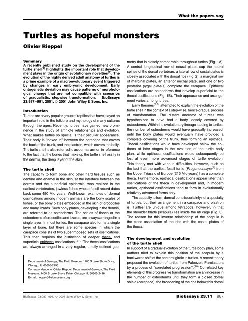
that is closely comparable throughout turtles (Fig. 1A).<br />
A central longitudinal row of neural plates cap the neural<br />
spines of the dorsal vertebrae; a lateral row of costal plates is<br />
closely <strong>as</strong>sociated with the dorsal ribs (Fig. 2); a marginal row<br />
of marginal plates, an anterior nuchal plate, and one or two<br />
posterior pygal plate(s) complete the carapace. Epithecal<br />
ossifications are osteoderms that develop superficial to the<br />
thecal ossifications (Fig. 1B). Their appearance and arrangement<br />
varies among turtles.<br />
Early theories (2,8) attempted to explain the evolution of the<br />
turtle shell in the context of a step-wise, hence gradual process<br />
of transformation. The distant ancestor of turtles w<strong>as</strong><br />
hypothesized to have had a body loosely covered by<br />
osteoderms. Within the evolutionary lineage leading to turtles,<br />
the number of osteoderms would have gradually incre<strong>as</strong>ed,<br />
until the bony plates would eventually have provided a<br />
complete covering of the trunk, thus forming an epitheca.<br />
Thecal ossifications would have developed below the epitheca<br />
at later stages in the evolution of the turtle body<br />
plan, while epithecal ossifications would subsequently be<br />
lost at even more advanced stages of turtle evolution.<br />
This theory met with various difficulties, however, such <strong>as</strong><br />
the fact that the earliest fossil turtle (Proganochelys) (9) from<br />
the Upper Tri<strong>as</strong>sic of Europe (215 Mio years) h<strong>as</strong> a complete<br />
theca. Furthermore, epithecal ossifications appear later than<br />
ossifications of the theca in development and, in modern<br />
turtles, epithecal ossifications tend to form in evolutionarily<br />
relatively advanced forms only.<br />
The capacity to form dermal bone is certainly not a specialty<br />
of turtles, but their arrangement in a carapace and pl<strong>as</strong>tron<br />
is. <strong>Turtles</strong> are unique among tetrapods, however, in that<br />
the shoulder blade (scapula) lies inside the rib cage (Fig. 3).<br />
The re<strong>as</strong>on for this inverse relationship of the scapula is<br />
the close <strong>as</strong>sociation of the ribs with the costal plates of<br />
the theca.<br />
The development and evolution<br />
of the turtle shell<br />
In support of a gradual evolution of the turtle body plan, some<br />
authors tried to explain this position of the scapula by a<br />
backwards shift of the pectoral girdle in turtles. A recent theory<br />
proposed the evolution of turtles from Paleozoic Parei<strong>as</strong>aurs<br />
by a process of ``correlated progression''. (10) Correlated key<br />
elements of this progressive transformation are an incre<strong>as</strong>e in<br />
the number of osteoderms until they form a closed dorsal<br />
shield (carapace), the broadening of the ribs below this dorsal<br />
BioEssays 23:987±991, ß 2001 John Wiley & Sons, Inc. BioEssays 23.11 987
What the papers say<br />
Figure 1. Schematic representation of the turtle carapace in<br />
dorsal view (A) and in a transverse section (B) (A: after A.F.<br />
Carr, Handbook of <strong>Turtles</strong>, Cornell University Press, 1952,<br />
Fig. 2c).<br />
shield, the shortening of the trunk, the immobilization of the<br />
dorsal vertebral column and a backwards shift of the pectoral<br />
girdle.<br />
This evolutionary scenario (10) not only requires that the<br />
turtle carapace be derived from a fusion of ancestral osteoderms,<br />
but also encounters the problem that there is no, or only<br />
a very minor, backward shift of the pectoral girdle in turtles. (11)<br />
Instead, the scapula of turtles comes to lie inside the rib cage<br />
because of a deflection of rib growth to a more superficial<br />
position. (12) Recent developmental work (1,13,14) h<strong>as</strong> identified<br />
inductive interaction generated by the carapacial ridge <strong>as</strong><br />
probable cause of this deflection of rib growth. Here is how it<br />
works.<br />
The vertebrate embryo is organized in three germ layers,<br />
the outer ectoderm, the inner entoderm, and the mesoderm<br />
between the two. During development, that part of the<br />
mesoderm lying alongside the notochord (paraxial mesoderm)<br />
becomes compartmentalized into segments, discrete blocks<br />
of cells or somites. From the somites originate, by embryonic<br />
cell proliferation and migration, the endoskeleton, the voluntary<br />
body musculature, and the dermis, the deep layer of<br />
the skin. The somites define the primary segmentation of the<br />
vertebrate body, which in the adult is still reflected in the<br />
segmented body musculature of fishes or salamanders, for<br />
example. To bend the body into a lateral curve during<br />
locomotion, the segmented body muscles must bridge the<br />
intervertebral joint. This means that the joint between<br />
vertebrae must lie within the primary body segments, which<br />
also allows the spinal nerves to exit between two vertebrae on<br />
their way to the body muscles. By that arrangement, the<br />
vertebral body itself, and the rib attached to it, come to lie in<br />
between the primary body segments. This repositioning of the<br />
vertebrae relative to the primary body segments is achieved by<br />
resegmentation of the somites. Each somite splits in half, and<br />
the posterior part of one somite recombines with the anterior<br />
part of the succeeding somite to form a vertebra.<br />
As a turtle embryo grows and develops, the contours of the<br />
future carapace are soon mapped out by an accelerated<br />
growth and a thickening of the skin on its back. The carapacial<br />
disk is formed. The margin of the carapacial disk shares<br />
histological and chemical properties with the marginal apical<br />
ridge of the early limb bud, a region that w<strong>as</strong> long known to be<br />
an important site for inductive tissue interaction in the<br />
formation of the limb skeleton. Somite extirpation experiments<br />
confirmed a somitic origin for those cells that form the ribs <strong>as</strong><br />
well <strong>as</strong> the thickened dermis of the carapacial disk in the<br />
snapping turtle. (14) Extirpation experiments performed on<br />
the margin of the carapacial disk, referred to <strong>as</strong> carapacial<br />
ridge, confirmed its role in the deflection of rib growth to a more<br />
superficial position in turtles. (13)<br />
Exoskeleton versus endoskeleton<br />
By definition, the endoskeleton of vertebrates develops deep<br />
to the dermis of the skin, mostly preformed in cartilage that is<br />
later replaced by bone in a process called ossification. For<br />
most elements, ossification first forms a bony layer around the<br />
cartilage (perichondral bone), before bone replaces the<br />
degenerating cartilage inside the skeletal element (endochondral<br />
bone). All this is also true for most of the endoskeleton of<br />
turtles, but the ribs of turtles are unique among vertebrates in<br />
that they chondrify within the deep layers of the thickened<br />
dermis of the carapacial disk. Perichondral ossification starts<br />
at the point of entry of the rib into the dermis, and from there<br />
spreads laterally. Once the whole cartilage of the embryonic<br />
rib is surrounded by perichondral bone, trabecular bone starts<br />
to spread from the rib through the dermis of the carapacial disk,<br />
and the costal plate is formed. In a similar pattern, the<br />
cartilaginous tips of the neural spines of the dorsal vertebrae<br />
pierce the deep layers of the dermis of the carapacial disk.<br />
Following their perichondral ossification, trabecular bone<br />
988 BioEssays 23.11
What the papers say<br />
Figure 2. The carapace of a marine turtle (Caretta<br />
caretta) in ventral view, showing the close <strong>as</strong>sociation of<br />
the ribs with the costal plates.<br />
spreads from them through the dermis to form the neural<br />
plates.<br />
The unique pattern of development of the ribs at a level<br />
outside the shoulder blade is not the end of the story, however.<br />
The amniote vertebra is composed of two parts that originally<br />
develop independently, but most of the time fuse in the adult<br />
(Fig. 1B). One part is the centrum, which replaces the embryonic<br />
notochord. Above the centrum develops the neural arch,<br />
which together with the centrum forms the neural canal for the<br />
spinal cord. In turtles, the neural arches of the dorsal vertebrae<br />
shift forward by half a segment, carrying the ribs with them,<br />
again a unique condition in amniotes. (15) The neural arches of<br />
successive vertebrae consequently meet each other above<br />
the midpoint of the centrum, where<strong>as</strong> the ribs come to lie lateral<br />
to the no longer functional intervertebral joints. The myomeric<br />
and neuromeric segmentation is thus secondarily established<br />
in the dorsal region of turtles, (16) <strong>as</strong> w<strong>as</strong> already the c<strong>as</strong>e in<br />
Proganochelys. (9) The functional re<strong>as</strong>on for this anterior shift<br />
of the neural arches is not clear, other than that it may<br />
contribute to the mechanical strength of the carapace, <strong>as</strong> the<br />
neural plates come to alternate with the costal plates (Fig. 1A).<br />
The ossification of endoskeletal structures preformed in<br />
cartilage, such <strong>as</strong> the ribs, within the dermis poses special<br />
problems for the comparative anatomist. Three types of bone<br />
have cl<strong>as</strong>sically been recognized: dermal bone, which develops<br />
in the dermis and typically characterizes the exoskeleton,<br />
chondral bone, which replaces cartilage during ossification of<br />
the endoskeleton, and membrane bone, which ossifies in deep<br />
laying membranes <strong>as</strong> part of the endoskeleton without<br />
cartilaginous preformation. The costal and neural plates of<br />
the turtle carapace start to develop <strong>as</strong> perichondral bone, <strong>as</strong> is<br />
typical for the initial ph<strong>as</strong>e of ossification of endoskeletal<br />
elements. But this perichondral bone develops in the dermis,<br />
and from it trabecular bone spreads through the dermis during<br />
BioEssays 23.11 989
What the papers say<br />
Figure 3. The relationship of the shoulder blade to the carapace in turtles (Graptemys geographica).<br />
later developmental stages. In the turtle carapace, therefore,<br />
the distinction of endoskeleton versus exoskeleton becomes<br />
problematic. It had been recognized earlier that the endoskeleton<br />
versus exoskeleton cannot be distinguished on the b<strong>as</strong>is<br />
of histogenesis, but must be defined with reference to a<br />
phylogenetic framework. (17,18) Exoskeletal elements are<br />
homologous to structures that in the ancestral condition<br />
combine bone, dentine and enamel, i.e., develop at the<br />
ectoderm±mesoderm interface. Thus, the bony scales of a<br />
trout, or the osteoderms of a crocodile, are exoskeletal,<br />
because these are structures that ultimately can be traced<br />
back (are homologous) to the heavy scales of early fishes that<br />
combine bone, dentine and enamel in a three-layered scale.<br />
By contr<strong>as</strong>t, endoskeletal elements are elements that in the<br />
ancestral condition are preformed in cartilage, while the<br />
cartilaginous stage may be deleted in the descendant<br />
(membrane bone). In the turtle carapace, the neural and<br />
costal plates ossify from, and in continuity with, the periost of<br />
their endoskeletal component. This pattern of ossification<br />
corresponds to the definition of Zuwachsknochen, (18) i.e.,<br />
bone that complements an endoskeletal element that is itself<br />
preformed in cartilage. As such, neural and costal plates are<br />
endoskeletal components of the turtle carapace, and cannot<br />
be derived from a hypothetical ancestral condition by fusion of<br />
exoskeletal osteoderms. All other parts of the turtle carapace<br />
are exoskeletal, however.<br />
The origin of a new body plan<br />
The turtle body plan is evidently highly derived, indeed unique<br />
among tetrapods. The problem for an evolutionary biologist is<br />
to explain these transformations in the context of a gradualistic<br />
process. Given the recently obtained developmental evidence,<br />
(1,11,13) the theory of ``correlated progression'' (10) presents<br />
an incomplete explanation of the turtle body plan:<br />
formation of the carapace is not simply the consequence of a<br />
fusion of osteoderms, and the location of the scapula inside the<br />
rib cage is not the result of a backward migration of the pectoral<br />
girdle.<br />
Early in the 19th century, EÂ tienne Geoffroy Saint-Hilaire<br />
raised the question of how the lung of a reptile could be<br />
transformed into the lung of a bird? He found it impossible to<br />
postulate that the lung of a reptile, specialized in its own way,<br />
could transform into an even more specialized lung of a bird.<br />
But he recognized that both reptiles and birds share similar<br />
early embryonic rudiments of the lung, and he hypothesized<br />
that these could develop along different trajectories in the<br />
two groups due to a minor change in early ontogenetic<br />
development: ``It only required an `accident' [a change] that<br />
990 BioEssays 23.11
What the papers say<br />
is viable and of small magnitude in its origin, but which<br />
could be of unpredictable importance with respect to its<br />
effects''. (19)<br />
The same can be said with respect to the axial skeleton of<br />
turtles. The initial segmentation of the paraxial mesoderm is<br />
the same in turtles <strong>as</strong> in all other tetrapods, but further<br />
development of structures derived from the somites (dermis,<br />
vertebrae and ribs) proceeds along a different trajectory in<br />
turtles compared to all other tetrapods. Ribs can only be<br />
located either deep to, or superficial to, the scapula. There are<br />
no intermediates, and there is only one way to get from one<br />
condition to the other, which is the redirection of the migration,<br />
through the embryonic body, of the precursor cells that will<br />
form the ribs. The study of such complex problems <strong>as</strong> the<br />
evolution of the turtle body plan highlights the important role<br />
that development can play in the origin of evolutionary<br />
novelties.<br />
References<br />
1. Gilbert SF, Loredo GA, Brukman A, Burke AC. Morphogenesis of the<br />
turtle shell: the development of a novel structure in tetrapod evolution.<br />
Evol Dev 2001;3:47±58.<br />
2. Hay OP. On Protostega, the systematic position of Dermochelys, and the<br />
morphogeny of the chelonian carapace and pl<strong>as</strong>tron. Amer Nat<br />
1898;32:929±948.<br />
3. Kaelin J. Zur Morphogenese des Panzers bei den SchildkroÈten. Acta<br />
Anat 1945;1:144±176.<br />
4. ValleÂn E. BeitraÈge zur Kenntnis der Ontogenie und der vergleichenden<br />
Anatomie des SchildkroÈtenpanzers. Acta Zool Stockholm 1942;23:1±<br />
127.<br />
5. VoÈlker H. UÈ ber d<strong>as</strong> Stamm- Gliedm<strong>as</strong>sen-, und Hautskelett von<br />
Dermochelys coriacea L. Zool Jb Anat 1913;33:431±552.<br />
6. Zangerl R. The homology of the shell elements in turtles. J Morphol<br />
1939;65:383±406.<br />
7. Zangerl R. The turtle shell. In: Gans C, Bellairs Ad'A, Parsons TS, editors.<br />
Biology of the Reptilia Vol. 1, London: Academic Press; 1969.p. 311±<br />
339.<br />
8. Versluys J. UÈ ber die Phylogenie des Panzers der SchildkroÈten und uÈber<br />
die Verwandtschaft der LederschildkroÈte (Dermochelys coriacea).<br />
PalaÈont. Z 1914;1:321±347.<br />
9. Gaffney ES. The comparative osteology of the Tri<strong>as</strong>sic turtle Proganochelys.<br />
Bull. Amer Mus Nat Hist 1990;194:1±263.<br />
10. Lee MSY. Correlated progression and the origin of turtles. Nature<br />
1996;379:811±815.<br />
11. Burke AC. The development and evolution of the turtle body plan:<br />
inferring intrinsic <strong>as</strong>pects of the evolutionary process from experimental<br />
embryology. Amer Zool 1991;31:616±627.<br />
12. Ruckes H. Studies in chelonian osteology. Part II. The morphological<br />
relationships between girdles, ribs and carapace. Ann NY Acad Sci<br />
1929;31:81±120.<br />
13. Burke AC. Development of the turtle carapace: implications for the<br />
evolution of a novel bauplan. J. Morphol 1989;199:363±378.<br />
14. Yntema CL. Extirpation experiments on the embryonic rudiments of the<br />
carapace of Chelydra serpentina. J Morphol 1970;132:235±244.<br />
15. Goette A. UÈ ber die Entwicklung des knoÈchernen RuÈckenschildes<br />
(Carapax) der SchildkroÈten. Z wiss Zool 1899;66:407±434.<br />
16. Hoffstetter R, Rage J-C. Vertebrae and ribs of modern reptiles. In: Gans<br />
C, Bellairs Ad'A, Parsons TS, editors. Biology of the Reptilia Vol. 1<br />
London: Academic Press; 1969. p. 201±310.<br />
17. Patterson C. Cartilage bones, dermal bones and membrane bones, or<br />
the exoskeleton versus the endoskeleton. In: Andrews SM, Miles RS,<br />
Walker AD, editors. Problems in Vertebrate Evolution. London: Academic<br />
Press; 1977. p. 77±121.<br />
18. Starck D. Vergleichende Anatomie der Wirbeltiere auf evolutionsbiologischer<br />
Grundlage, Vol 2 Berlin: Springer Verlag; 1979.<br />
19. Geoffroy Saint-Hilaire E. Le deÂgre d'influence du monde ambiant pour<br />
modiifier les formes animales; question inteÂressant l'origine des espeÂces<br />
teÂleÂosauriennes et successivement celle des animaux de l'eÂpoque<br />
actuelle. MeÂm Acad R Sci Inst. France 1833;12:63±92. (The quote is<br />
from p. 80).<br />
BioEssays 23.11 991


