genomewide characterization of host-pathogen interactions by ...
genomewide characterization of host-pathogen interactions by ...
genomewide characterization of host-pathogen interactions by ...
Create successful ePaper yourself
Turn your PDF publications into a flip-book with our unique Google optimized e-Paper software.
Maren Depke<br />
Results<br />
Kidney Gene Expression Pattern in an in vivo Infection Model<br />
An essential part <strong>of</strong> the immune response to infection is the presentation <strong>of</strong> antigens to<br />
immune cells to direct the defense against the specific <strong>pathogen</strong> in the adaptive part <strong>of</strong> the<br />
immune system. Antigens belong to two main groups, extracellular and intracellular antigens,<br />
which are processed in separate pathways. Extracellular antigens are taken up <strong>by</strong><br />
phagocytosis/endocytosis and digested in vesicles after fusion with lysosomes. Finally, peptides<br />
are bound to MHC-II molecules originating from exocytic Golgi vesicles and presented on the cell<br />
surface. Intracellular antigens are digested <strong>by</strong> cytosolic proteasome with sequential transport <strong>of</strong><br />
peptides into the endoplasmatic reticulum (ER). These peptides are loaded to MHC-I molecules<br />
and transported via the Golgi to the cell surface for presentation. Presentation <strong>of</strong> extracellular<br />
antigens preferentially occurs <strong>by</strong> pr<strong>of</strong>essional antigen presenting cells (APC) like dendritic cells<br />
(DC), macrophages and B cells. The peptide-MHC-II complex is recognized <strong>by</strong> CD4 + helper T cells,<br />
which leads to initiation <strong>of</strong> mechanisms fighting extracellular infection or disposing extracellular<br />
antigens. Immune cells and other cells <strong>of</strong> the body are capable <strong>of</strong> presenting intracellular<br />
antigens via MHC-I. They present the antigenic peptide to CD8 + cytotoxic T cells and there<strong>by</strong><br />
activate processes to destroy cells and kill <strong>pathogen</strong> in case <strong>of</strong> intracellular infection.<br />
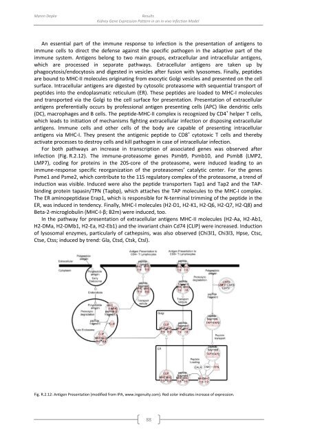
For both pathways an increase in transcription <strong>of</strong> associated genes was observed after<br />
infection (Fig. R.2.12). The immune-proteasome genes Psmb9, Psmb10, and Psmb8 (LMP2,<br />
LMP7), coding for proteins in the 20S-core <strong>of</strong> the proteasome, were induced leading to an<br />
immune-response specific reorganization <strong>of</strong> the proteasomes’ catalytic center. For the genes<br />
Psme1 and Psme2, which contribute to the 11S regulatory complex <strong>of</strong> the proteasome, a trend <strong>of</strong><br />
induction was visible. Induced were also the peptide transporters Tap1 and Tap2 and the TAPbinding<br />
protein tapasin/TPN (Tapbp), which attaches the TAP molecules to the MHC-I complex.<br />
The ER aminopeptidase Erap1, which is responsible for N-terminal trimming <strong>of</strong> the peptide in the<br />
ER, was induced in tendency. Finally, MHC-I molecules (H2-D1, H2-K1, H2-Q6, H2-Q7, H2-Q8) and<br />
Beta-2-microglobulin (MHC-I-β; B2m) were induced, too.<br />
In the pathway for presentation <strong>of</strong> extracellular antigens MHC-II molecules (H2-Aa, H2-Ab1,<br />
H2-DMa, H2-DMb1, H2-Ea, H2-Eb1) and the invariant chain Cd74 (CLIP) were increased. Induction<br />
<strong>of</strong> lysosomal enzymes, particularly <strong>of</strong> cathepsins, was also observed (Chi3l1, Chi3l3, Hpse, Ctsc,<br />
Ctse, Ctss; induced <strong>by</strong> trend: Gla, Ctsd, Ctsk, Ctsl).<br />
Fig. R.2.12: Antigen Presentation (modified from IPA, www.ingenuity.com). Red color indicates increase <strong>of</strong> expression.<br />
88


