genomewide characterization of host-pathogen interactions by ...
genomewide characterization of host-pathogen interactions by ...
genomewide characterization of host-pathogen interactions by ...
Create successful ePaper yourself
Turn your PDF publications into a flip-book with our unique Google optimized e-Paper software.
Maren Depke<br />
Discussion and Conclusions<br />
superfamilies <strong>of</strong> TNF ligands and receptors were identified (Hehlgans/Pfeffer 2005). TNF is<br />
associated with several functions linked to pro-inflammatory effects, which involve a<br />
multiprotein signaling complex at the cell membrane and MAP-kinase signaling and which are<br />
finally mediated via NFκB and c-Jun (Chen G/Goeddel 2002, Wajant et al. 2003). Rarely, TNF can<br />
induce apoptosis via caspase activation, depending on the cellular balance <strong>of</strong> antiapoptosis/proliferation<br />
signals mediated <strong>by</strong> NFκB (Gaur/Aggarwal 2003, Wajant et al. 2003).<br />
Furthermore, TNF is involved in the regulation <strong>of</strong> fever with both pyrogenic and antipyretic<br />
effects depending on physiological or experimental conditions (Leon 2002). In macrophages, TNF<br />
has an important function in controlling intracellular mycobacterial growth (Bekker et al. 2001).<br />
TNF deficient mice are susceptible to Listeria monocytogenes infections, exhibit reduced contact<br />
hypersensitivity and impairment <strong>of</strong> splenic follicular architecture and <strong>of</strong> maturation <strong>of</strong> the<br />
humoral immune response (Pasparakis et al. 1996). Knockout <strong>of</strong> TNF and lymphotoxin-α (TNF-β)<br />
leads to an increased susceptibility to S. aureus infections, but also to a reduced frequency <strong>of</strong><br />
arthritis indicating a damaging influence <strong>of</strong> the cytokine during the inflammatory reaction<br />
(Hultgren et al. 1998). Reflecting the central position <strong>of</strong> TNF in the regulation <strong>of</strong> the immune<br />
response to infection, several strategies <strong>of</strong> <strong>pathogen</strong>s to influence the TNF activity were<br />
reported. The interference can lead to diverse and contrary effects depending on the <strong>pathogen</strong>,<br />
like inhibition <strong>of</strong> apoptosis, enhancement <strong>of</strong> apoptosis, inhibition <strong>of</strong> TNF production or, in case <strong>of</strong><br />
S. aureus, the induction <strong>of</strong> a TNF-like response <strong>by</strong> interaction <strong>of</strong> protein A with the TNF receptor<br />
TNFR1 (Rahman/McFadden 2006).<br />
Beside other functions, TNF was described to further enhance the expression <strong>of</strong> the<br />
immunoproteasome (Loukissa et al. 2000). In non-pr<strong>of</strong>essional antigen-presenting cells, the<br />
induction <strong>of</strong> immunoproteasome, TAP peptide transporters, and MHC class I complexes <strong>by</strong> TNF<br />
independent <strong>of</strong> IFN-γ activity was observed and in addition increased stability <strong>of</strong> the MHC class I<br />
complexes at the cell surfaces (Hallermalm et al. 2001). Thus, induction <strong>of</strong> TNF in BMM after<br />
IFN-γ treatment might intensify the effect on antigen processing and presentation after a second<br />
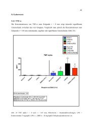
stimulus. Also in the serum-free system, the inducibility <strong>of</strong> TNF in IFN-γ and LPS treated BMM was<br />
described (Eske et al. 2009). IL-6, IL-10, and IL-12, which were also inducible <strong>by</strong> IFN-γ and LPS<br />
(Eske et al. 2009), were not expressed in IFN-γ treated BMM (this study), and the IFN-γ and LPS<br />
inducible chemokine Ccl2 (MCP-1; Eske et al. 2009) was expressed but not differentially regulated<br />
in IFN-γ treated BMM (this study). This strongly hints for the explanation that the induction only<br />
occurs when a second stimulus like LPS is given.<br />
After IFN-γ treatment, the induction <strong>of</strong> anti-inflammatory IL-10 receptor (Moore et al. 2001)<br />
as well as the induction <strong>of</strong> Il18bp coding for an IL-18 binding protein, which is a natural<br />
endogenous inhibitor <strong>of</strong> the proinflammatory IL-18 (McInnes et al. 2000, Gracie et al. 2003,<br />
Novick D et al. 1999), was observed (this study). Not only Stat1 and Irf1 <strong>of</strong> the positive IFN-γ<br />
feedback were induced, but also Stat3 and Socs1 which are implicated in negative feedback<br />
(Schroder et al. 2004, Hu et al. 2008). This might indicate the possible reactivity to antiinflammatory<br />
stimuli and a confinement <strong>of</strong> inflammatory response.<br />
After the priming signal <strong>of</strong> IFN-γ, the BMM are not fully activated and are not expected to<br />
secrete cytokines, but rather after a second stimulus (Hu et al. 2008). Analysis <strong>of</strong> cytokine<br />
secretion <strong>of</strong> BMM beyond the examples analyzed until now will be <strong>of</strong> interest, either after<br />
stimulation with LPS, or after stimulation with molecules <strong>of</strong> Gram-positive bacteria or even<br />
infection e. g. with S. aureus, as it will be in focus <strong>of</strong> research for follow-up experiments to this<br />
study.<br />
185


