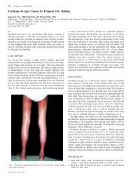
Coexistence of Epilepsy and Myasthenia Gravis: Report of Four ...
Coexistence of Epilepsy and Myasthenia Gravis: Report of Four ...
Coexistence of Epilepsy and Myasthenia Gravis: Report of Four ...

You also want an ePaper? Increase the reach of your titles
YUMPU automatically turns print PDFs into web optimized ePapers that Google loves.
182<br />
to find the association between these two neurological<br />
disorders.<br />
CASE REPORTS<br />
Case 1<br />
SLC, a 46-year-old woman, had several episodes <strong>of</strong><br />
generalized tonic-clonic convulsions at the age <strong>of</strong> 4. She<br />
received anticonvulsant therapy for 1 year <strong>and</strong> had had<br />
no seizures until the age <strong>of</strong> 16 when she had recurrence.<br />
The seizures were described as sudden head turning <strong>and</strong><br />
eye deviation to the right side for 15-25 seconds, followed<br />
by loss <strong>of</strong> consciousness <strong>and</strong> generalized tonicclonic<br />
convulsions for 1-2 minutes. On neurological<br />
examination, there were no abnormal findings. An<br />
awake EEG revealed focal epileptiform discharges in the<br />
left frontal region but isotope brain scans failed to disclose<br />
abnormal uptake. She began to receive combined<br />
phenobarbital (40~80 mg/day) <strong>and</strong> phenytoin (100~200<br />
mg/day) therapy. Her compliance was not adequate so<br />
that she had occasional attacks, about once or twice a<br />
year between the ages <strong>of</strong> 16 <strong>and</strong> 33 years. She had complained<br />
<strong>of</strong> mild intermittent weakness <strong>of</strong> the upper limbs<br />
for 2 years since she was 33 years old. The weakness<br />
was found most frequently during hair combing <strong>and</strong><br />
occasionally h<strong>and</strong> grasping. However, there was neither<br />
muscle atrophy nor hyporeflexia. Cranial nerves, coordination,<br />
sensations, <strong>and</strong> sphincter functions were normal,<br />
whereas repetitive 3-Hz electric stimulation revealed<br />
decremental responses at the right deltoid muscle. The<br />
chest computed tomography (CT) showed thymic<br />
enlargement. Treatment with pyridostigmine bromide<br />
alleviated her weakness. She then received thymectomy,<br />
<strong>and</strong> the pathology revealed lymphoid hyperplasia. After<br />
operation, her weakness disappeared gradually <strong>and</strong> pyridostigmine<br />
was discontinued within 3 months. She<br />
refused to take anticonvulsants in spite <strong>of</strong> occasional<br />
epileptic attacks. The muscle weakness has since not<br />
recurred.<br />
Case 2<br />
SCC, a 57-year-old man, suffered from a severe head<br />
injury in a traffic accident when he was 26 years old. At<br />
that time, he remained unconscious for 1 week <strong>and</strong><br />
stayed in a hospital for 3 weeks. Seven months later, the<br />
patient developed several episodes <strong>of</strong> nocturnal tonicclonic<br />
convulsions <strong>and</strong> was brought to our hospital for<br />
further work-up. On examination, there were no abnormal<br />
physical or neurological signs. An awake EEG<br />
revealed an epileptogenic focus at the right anterior temporal<br />
region. Under the diagnosis <strong>of</strong> post-traumatic<br />
epilepsy, anticonvulsant therapy with phenobarbital<br />
(80mg/day) <strong>and</strong> phenytoin (200 mg/day) was started. A<br />
follow-up EEG at the age <strong>of</strong> 34 still disclosed focal<br />
spikes in the bilateral temporal regions, although his<br />
epilepsy was kept under control. Anticonvulsants were<br />
gradually tapered 7 years later <strong>and</strong> totally discontinued<br />
when he was 37 years old.<br />
At the age <strong>of</strong> 34, the patient suffered from intermittent<br />
bilateral ptosis, which could be alleviated by an<br />
intramuscular injection <strong>of</strong> neostigmine methylsulfate. A<br />
clinical diagnosis <strong>of</strong> ocular myasthenia gravis was made.<br />
However, his ptosis did not respond to pyridostigmine<br />
<strong>and</strong> double vision occasionally supervened. At his age <strong>of</strong><br />
35, the patient received an alternate-day high-dose<br />
(100mg) prednisolone. The ptosis <strong>and</strong> double vision disappeared<br />
completely within 1 month but he refused further<br />
steroid therapy because <strong>of</strong> the its side effects.<br />
Intermittent ptosis <strong>and</strong> double vision recurred half a year<br />
later. He gradually adapted to the ocular problems <strong>and</strong><br />
gave up all anti-MG medications at the age <strong>of</strong> 37.<br />
At the age <strong>of</strong> 38, about one <strong>and</strong> a half years after discontinuing<br />
anticonvulsant therapy, the generalized tonicclonic<br />
convulsions recurred. The patient was again prescribed<br />
phenytoin (300 mg/day) for seizure control. At<br />
the same time, his eye problems remained stationary for<br />
2 years. However, ptosis <strong>and</strong> double vision worsened<br />
when he was 41 years <strong>of</strong> age. Repetitive electric stimulations<br />
at the right median <strong>and</strong> right facial nerves did not<br />
show significant decremental responses. Single fiber<br />
EMG study at the right extensor digitorum communis<br />
muscle showed a mean consecutive difference (MCD) <strong>of</strong><br />
97.7 µsec (normal