Transfer of foreign gene to giant freshwater prawn (Macrobrachium ...
Transfer of foreign gene to giant freshwater prawn (Macrobrachium ...
Transfer of foreign gene to giant freshwater prawn (Macrobrachium ...

You also want an ePaper? Increase the reach of your titles
YUMPU automatically turns print PDFs into web optimized ePapers that Google loves.
MOLECULAR REPRODUCTION AND DEVELOPMENT 56:149–154 (2000)<br />
<strong>Transfer</strong> <strong>of</strong> Foreign Gene <strong>to</strong> Giant Freshwater<br />
Prawn (<strong>Macrobrachium</strong> rosenbergii) by<br />
Sperma<strong>to</strong>phore-Microinjection<br />
SI-SHEN LI AND HUAI-JEN TSAI*<br />
Institute <strong>of</strong> Fisheries Science, National Taiwan University, Taipei, Taiwan<br />
ABSTRACT We developed a sperma<strong>to</strong>phoremicroinjection<br />
(SMI) technique that allows exogenous<br />
DNA fragments <strong>to</strong> be transferred easily in<strong>to</strong> the <strong>giant</strong><br />
<strong>freshwater</strong> <strong>prawn</strong> (<strong>Macrobrachium</strong> rosenbergii), an important<br />
aquacultural shellfish and aquatic invertebrate<br />
model. From 28 <strong>to</strong> 1,000 ng <strong>of</strong> the circular plasmid<br />
pGL, in a <strong>to</strong>tal volume <strong>of</strong> 1 ml, were directly microinjected<br />
in<strong>to</strong> sperma<strong>to</strong>phores. Fertilization and hatching<br />
<strong>of</strong> <strong>prawn</strong>s created with SMI were completed in vivo.<br />
Fertilization and hatching rates in the SMI treatments<br />
did not differ from those <strong>of</strong> the untreated control group.<br />
The genomes <strong>of</strong> free swimming, SMI-created larvae (21<br />
days after fertilization) were analyzed using PCR and<br />
Southern blot analyses. A product with a molecular<br />
mass <strong>of</strong> 680 bp was amplified. It corresponded <strong>to</strong><br />
amplifications <strong>of</strong> pGL, and Southern blot analysis revealed<br />
that the amplified band was positive. The <strong>gene</strong><br />
transfer rate was primarily dependent on the concentration<br />
<strong>of</strong> DNA during SMI. The higher the concentration <strong>of</strong><br />
pGL, the higher the rate <strong>of</strong> <strong>gene</strong> transfer. PCR and<br />
Southern blot analyses detected the existence <strong>of</strong><br />
<strong>foreign</strong> DNA in 16 <strong>of</strong> 23 samples (70%) <strong>of</strong> genomic DNA<br />
isolated from hatched larvae in the 750 ng pGL SMI<br />
treatment. SMI, described here for the first time, is the<br />
simplest and most efficient method for mass producing<br />
transgenic <strong>giant</strong> <strong>freshwater</strong> <strong>prawn</strong>s. Mol. Reprod. Dev.<br />
56:149–154, 2000 r 2000 Wiley-Liss, Inc.<br />
Key Words: <strong>gene</strong> transfer; microinjection; <strong>prawn</strong>;<br />
sperma<strong>to</strong>phore<br />
INTRODUCTION<br />
r 2000 WILEY-LISS, INC.<br />
Transgenic animals provide a powerful system for in<br />
vivo study <strong>of</strong> <strong>gene</strong> regulation, expression, and function,<br />
and make possible the production <strong>of</strong> transgenic varieties<br />
having special <strong>gene</strong>tic traits. Many approaches have<br />
been developed <strong>to</strong> introduce <strong>foreign</strong> DNA molecules in<strong>to</strong><br />
zygotes. In fish, methods for <strong>gene</strong> transfer include<br />
microinjection <strong>of</strong> <strong>foreign</strong> DNA in<strong>to</strong> oocyte nuclei (Oza<strong>to</strong><br />
et al., 1986; Tsai et al., 1995b), the cy<strong>to</strong>plasm <strong>of</strong><br />
developing embryos (Chourrout et al., 1986; Dunham et<br />
al., 1987), and fertilized eggs (Fletcher et al., 1988;<br />
Dunham et al., 1992; Lu et al., 1992). Electroporation <strong>of</strong><br />
exogenous DNA fragments in<strong>to</strong> fertilized eggs (Inoue et<br />
al., 1990; Powers et al., 1992; Tsai and Tseng, 1994) is a<br />
popular technique because, compared with the traditional<br />
microinjection technique, electroporation is<br />
simple, convenient, and efficient. However, the efficacy<br />
<strong>of</strong> <strong>gene</strong> transfer by electroporation <strong>of</strong> fertilized eggs is<br />
still not high enough <strong>to</strong> treat the tremendously large<br />
number <strong>of</strong> eggs spawned within a very short time by<br />
cultured finfish and shellfish species.<br />
Thus, for aquatic animals, interest in sperm-mediated<br />
<strong>gene</strong> transfer has increased because this technique<br />
has several advantages over other methods <strong>of</strong><br />
<strong>gene</strong> transfer applied <strong>to</strong> finfish and shellfish. First,<br />
sperm-mediated <strong>gene</strong> transfer can be applied <strong>to</strong> huge<br />
numbers <strong>of</strong> oocytes. Second, sperm-mediated <strong>gene</strong> transfer<br />
overcomes some <strong>of</strong> the disadvantages <strong>of</strong> conventional<br />
<strong>gene</strong> transfer systems resulting from egg characteristics<br />
such as opaqueness, stickiness, buoyancy,<br />
invisible pronuclei, and a <strong>to</strong>ugh chorion. Third, <strong>foreign</strong><br />
DNA carried by sperm is transferred in<strong>to</strong> the nucleus;<br />
with electroporation, the probability that DNA fragments<br />
will be transferred in<strong>to</strong> the blas<strong>to</strong>disc, the volume<br />
<strong>of</strong> which is extremely small in a fertilized egg, is<br />
very low. Fourth, finfish and shellfish sperm can be<br />
activated by simply adding water, and they can be<br />
cryopreserved. Thus, treated sperm is ready for use at<br />
any time.<br />
A number <strong>of</strong> researchers have attempted <strong>to</strong> use<br />
sperm as a vec<strong>to</strong>r for introducing <strong>foreign</strong> DNA and<br />
producing transgenic finfish and shellfish. Chourrout<br />
and Perrot (1992) reported failure using a spermincubation<br />
technique with rainbow trout. However,<br />
some success has been achieved using sperm-incubation<br />
with zebrafish (Khoo et al., 1992), and spermelectroporation<br />
with common carp, catfish, tilapia<br />
(Muller et al., 1992), salmon (Symonds et al., 1994),<br />
loach (Tsai et al., 1995a), and abalone (Tsai et al., 1997).<br />
There have been few attempts <strong>to</strong> apply <strong>gene</strong> transfer<br />
techniques <strong>to</strong> an important group <strong>of</strong> shellfish: crustaceans.<br />
Gendreau and colleagues (1995) successfully<br />
used a ballistic technique <strong>to</strong> transfer <strong>gene</strong>s in<strong>to</strong> brine<br />
shrimp (Artemia). However, the facilities and DNA<br />
Grant sponsor: National Science Council; Grant sponsor: Council <strong>of</strong><br />
Agriculture, Republic <strong>of</strong> China.<br />
*Correspondence <strong>to</strong>: Huai-Jen Tsai, Institute <strong>of</strong> Fisheries Science,<br />
National Taiwan University, Taipei, Taiwan, 106.<br />
E-mail: hjtsai@ccms.ntu.edu.tw<br />
Received 15 November 1999; Accepted 20 January 2000
150 S.-S. LI AND H.-J. TSAI<br />
preparations required for ballistic transfer are extremely<br />
expensive, and the work is tedious and unsuitable<br />
for field use. Bensheg and Khoo (1997) used<br />
microinjection <strong>to</strong> transfer a reporter <strong>gene</strong> in<strong>to</strong> the<br />
fertilized eggs <strong>of</strong> <strong>freshwater</strong> shrimp, <strong>Macrobrachium</strong><br />
lanchesteri. Again, traditional microinjection is laborious,<br />
time-consuming, and can only treat a limited<br />
number <strong>of</strong> eggs. In this communication, we describe a<br />
simple and effective way <strong>to</strong> transfer <strong>foreign</strong> DNA fragments<br />
in<strong>to</strong> <strong>giant</strong> <strong>freshwater</strong> <strong>prawn</strong> on a massive scale<br />
using sperma<strong>to</strong>phore-microinjection (SMI). SMI may<br />
be quite useful for studying <strong>gene</strong> regulation and for<br />
creating new crustacean strains having special traits.<br />
MATERIALS AND METHODS<br />
Experimental Animals<br />
Nine- <strong>to</strong> twelve-month-old, aquacultured, <strong>giant</strong> <strong>freshwater</strong><br />
<strong>prawn</strong>s were purchased locally. They averaged<br />
16.6 6 4.8 cm (males) and 11.5 6 2.9 cm (females) in<br />
length, 3.7 6 0.6 cm (males) and 2.5 6 0.4 cm (females)<br />
in width, and 61.4 6 11.7 g (males) and 33.6 6 7.4 g<br />
(females) in weight. Males and females were maintained<br />
in separate aquarium tanks at a density <strong>of</strong> 1<br />
(males) or 2 (females) per 10 liters at 28°C on a natural<br />
light cycle.<br />
Sperma<strong>to</strong>phore Extrusion and In Vitro<br />
Fertilization<br />
Sperma<strong>to</strong>phores were extruded from male <strong>prawn</strong>s<br />
using electric shock (36–10 V, DC, model PS-304,<br />
Shin-Ray Electronic, Taiwan). The positive pole was<br />
attached <strong>to</strong> the fifth swimming pod and the negative<br />
pole was placed on the ventral ganglia. Thirteen mature<br />
males were used <strong>to</strong> examine the degree <strong>of</strong> sperma<strong>to</strong>phore<br />
extrusion caused by voltages ranging from 0 <strong>to</strong> 10<br />
Vat10A.<br />
Foreign DNA, at concentrations <strong>of</strong> 28, 250, 400, 750,<br />
and 1,000 ng/µl in a <strong>to</strong>tal volume <strong>of</strong> 1 µl, was directly<br />
microinjected in<strong>to</strong> the extruded sperma<strong>to</strong>phore. For the<br />
mock-treated control group, we microinjected 1 µl <strong>of</strong><br />
double-distilled water in<strong>to</strong> the extruded sperma<strong>to</strong>phore.<br />
For in vitro fertilization, a treated sperma<strong>to</strong>phore<br />
was placed for 2–3 min in the spermatheca,<br />
located on the thorax between the third and fifth<br />
swimming pods, <strong>of</strong> a female <strong>prawn</strong> that had just<br />
finished molting and had well-developed ovaries. In<br />
<strong>gene</strong>ral, treated females began <strong>to</strong> fertilize their eggs in<br />
vivo 2–6 hr after sperma<strong>to</strong>phores were attached. When<br />
their eggs were close <strong>to</strong> hatching, these females were<br />
moved <strong>to</strong> a 100 liter tank containing 12 ppt salt water.<br />
DNA Preparation<br />
pGreen Latern-1 (pGL, Gibco BRL), with a molecular<br />
mass <strong>of</strong> 5 kb, consists <strong>of</strong> an immediate early-<strong>gene</strong><br />
promoter and enhancer <strong>of</strong> cy<strong>to</strong>megalovirus, fused with<br />
the cDNA <strong>of</strong> a green fluorescence protein (GFP) and a<br />
38-untranslated region <strong>of</strong> SV40. They were prepared by<br />
the cesium chloride-ethidium bromide ultra-centrifugation<br />
method (Sambrook et al., 1989). The circular form<br />
<strong>of</strong> pGL was resuspended in double-distilled water (sterilized)<br />
at concentrations <strong>of</strong> 28, 250, 400, 750, and 1,000<br />
ng per µl.<br />
DNase Digestion<br />
In order <strong>to</strong> ensure PCR detection <strong>of</strong> <strong>foreign</strong> DNA<br />
molecules in embryos, we had <strong>to</strong> avoid contamination <strong>of</strong><br />
the DNA fragments that remained outside the embryos.<br />
First, we determined the conditions that allowed DNase<br />
I (Sigma Chemical Co., St. Louis, MO) <strong>to</strong> completely<br />
digest the greatest amount <strong>of</strong> DNA employed in trans<strong>gene</strong>sis.<br />
pGL <strong>of</strong> 1,000 ng was resuspended in 100 µl <strong>of</strong><br />
digestion buffer (10 mM MgCl 2 and 20 mM TrisCl, pH<br />
8), then 0.5, 0.75, 1, or 10 µg <strong>of</strong> DNase I was added, and<br />
the solution was incubated at 37°C for 1 hr. Each<br />
treatment was duplicated. One-hundredth <strong>of</strong> the reaction<br />
volume was analyzed by PCR <strong>to</strong> determine the<br />
concentrations <strong>of</strong> DNase capable <strong>of</strong> completely digesting<br />
DNA molecules.<br />
Genomic DNA Extraction<br />
Five embryos that had developed <strong>to</strong> the zoea-I-stage<br />
were pooled and collected in an eppendorf tube, washed<br />
three times with double-distilled water, and resuspended<br />
in a 500 µl <strong>of</strong> digestion buffer. Then, DNase I<br />
was added until a final concentration <strong>of</strong> 100 µg/ml was<br />
achieved. The solution was incubated at 37°C for 1 hr.<br />
After the reaction, the embryos were centrifuged at 76g<br />
for 3 min. The supernatant was decanted, and samples<br />
were boiled for 10 min <strong>to</strong> halt DNase activity. A volume<br />
<strong>of</strong> 400 µl lysis solution (6 M guanidine hydrochloride<br />
and 0.1 M sodium acetate, pH 5.5) was added <strong>to</strong> the<br />
embryos, which were briefly crushed and then incubated<br />
at 37°C for 1 hr in a roller shaker. Then the<br />
genomic DNA <strong>of</strong> embryos was obtained by phenolchlor<strong>of</strong>orm<br />
extraction and ethanol precipitation.<br />
Twenty-three free-swimming larvae, randomly selected<br />
from the experimental and control groups, were<br />
examined for <strong>foreign</strong> DNA. Genomic DNA was extracted<br />
from each larva and prepared as described<br />
above for embryo DNA, except that DNase was not<br />
added. Each larva was washed three times in doubledistilled<br />
water, resuspended in 400 µl lysis buffer,<br />
crushed slightly, and incubated at 37°C for 1 hr in a<br />
roller shaker. The genomic DNA was obtained by<br />
phenol-chlor<strong>of</strong>orm extraction and ethanol precipitation.<br />
PCR Analysis<br />
Two oligonucleotide primers, EGFP-F and GFP-R,<br />
were synthesized for detection <strong>of</strong> GFP cDNA by PCR<br />
analysis. The nucleotide sequences for forward and<br />
reverse primers were ATGGTGAGCAAGGGCGAGGA<br />
(EGFP-F) and CAGCTCGTCCATGCCATGTG (GFP-<br />
R), respectively. The primers for detection <strong>of</strong> the endogenous<br />
b-tubulin <strong>gene</strong>, which served as an internal<br />
control, were CCCTTCCCTCGTCTCCAC (forward, tubf6)<br />
and GCCAGTGTACCAGTGAAGGGA (reverse, tubr7).<br />
Each PCR sample consisted <strong>of</strong> 20 µl <strong>of</strong> solution<br />
containing 10–20 ng <strong>of</strong> templates, 5–10 pmol <strong>of</strong> each
primer, 25 µM <strong>of</strong> each dNTP, 20 µg <strong>of</strong> bovine serum<br />
albumin (Giambernardi et al., 1998), and five units <strong>of</strong><br />
ProZyme II (Protech) in a 13 PCR buffer (Protech).<br />
Amplification was performed with a DNA Thermal<br />
Cycler (Perkin-Elmer Cetus, Norwalk, CT). PCR consisted<br />
<strong>of</strong> 25 cycles <strong>of</strong> denaturing at 94°C for 1 min,<br />
annealing at 60°C for 1 min and extension at 72°C for 2<br />
min, followed by 10 min extension at 72°C. Each PCR<br />
sample (10 µl) was subjected <strong>to</strong> electrophoresis on a 3%<br />
NuSieve GTG agarose gel (FMC BioProducts, Rockland,<br />
ME).<br />
TRANSFER OF FOREIGN GENE TO FRESHWATER PRAWN 151<br />
Probe Preparation<br />
The 0.6 kb fragment containing GFP cDNA was<br />
recovered from the agarose gel using Jetsorb Gel Extraction<br />
(Genomed, Inc., Research Triangle Park, NC) after<br />
plasmid pGL was restricted by NotI and run on the gel.<br />
Three micrograms <strong>of</strong> the 0.6 kb DNA fragment were<br />
DIG-labeled by the random priming method at 37°C for<br />
20 hr, according <strong>to</strong> the manufacturer’s recommended<br />
procedures (Boehringer Mannheim).<br />
Southern Blot Analysis<br />
PCR products were analyzed on an agarose gel and<br />
transferred on<strong>to</strong> a nitrocellulose membrane (Amersham,<br />
Pharmacia Biotech, Uppsala, Sweden). After air<br />
drying, the DNA was cross-linked <strong>to</strong> the membrane by<br />
UV irradiation and then hybridized <strong>to</strong> a probe. Hybridization<br />
was carried out overnight, at 42°C, in a standard<br />
buffer solution (Boehringer Mannheim) containing<br />
50% formamide and 10 ng/ml <strong>of</strong> denatured probe.<br />
After the membranes were washed, colorimetric detection<br />
was carried out with nitroblue tetrazolium and<br />
5-bromo-4-chloro-3-indolyl phosphate reagents, as recommended<br />
by the manufacturer (Boehringer Mannheim).<br />
Fertilization Rate, Hatching Rate, and Gene<br />
<strong>Transfer</strong> Rate<br />
The fertilization rate was calculated as the proportion<br />
<strong>of</strong> 40 randomly selected eggs that developed one<br />
day after spawning. The hatching rate was calculated<br />
as the proportion <strong>of</strong> 40 randomly selected eggs that<br />
hatched three weeks after spawning. The <strong>gene</strong> transfer<br />
rate was the number <strong>of</strong> positive PCR and Southern blot<br />
analyses divided by the <strong>to</strong>tal number <strong>of</strong> hatched larvae<br />
examined.<br />
Green Fluorescence Detection<br />
To detect green fluorescence, hatched larvae were<br />
examined under a microscope (Olympus BH-2, Tokyo,<br />
Japan) using an excitation filter (BP 490 nm) and a<br />
dichron mirror (DM 500 nm).<br />
RESULTS<br />
Effect <strong>of</strong> Voltage on Sperma<strong>to</strong>phore Extrusion<br />
In Vitro<br />
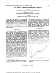
Eleven <strong>of</strong> 13 (85%) male <strong>giant</strong> <strong>freshwater</strong> <strong>prawn</strong>s<br />
extruded their sperma<strong>to</strong>phore in vitro when shocked<br />
with 7.4 V (Fig. 1). No sperma<strong>to</strong>phores were extruded<br />
Fig. 1. Effect <strong>of</strong> voltage (4–10 V at 10 A) on sperma<strong>to</strong>phore<br />
extrusion. The cumulative percentage was the cumulative number <strong>of</strong><br />
males, out <strong>of</strong> 13, whose sperma<strong>to</strong>phore was extruded at or below a<br />
given voltage.<br />
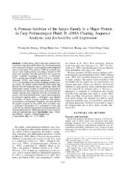
Fig. 2. Effect <strong>of</strong> DNase concentration on pGL digestion. An aliquot<br />
<strong>of</strong> 1 µg pGL was incubated with 0.5, 0.75, 1, or 10 µg DNase in a 100 µl<br />
solution at 37°C for 1 hr. After the DNase was denatured by boiling,<br />
samples were analyzed by PCR without (A) or with (B) an additional 1<br />
µg <strong>of</strong> pGL. M, molecular marker <strong>of</strong> phage fX174 digested with HaeIII.<br />
The arrow indicates the 680 bp PCR-product.<br />
at voltages less than 7 V, and two male <strong>prawn</strong>s failed <strong>to</strong><br />
extrude sperma<strong>to</strong>phores even at voltages above 7.4 V.<br />
Presence <strong>of</strong> Foreign DNA in Embryos<br />
Before we could ascertain whether <strong>foreign</strong> DNA was<br />
present in the embryos in the SMI treatments, we had<br />
<strong>to</strong> determine the conditions under which PCR sensitivity<br />
was sufficient <strong>to</strong> detect DNA molecules that had not<br />
been completely digested by DNase I. In the control and<br />
0.5 µg DNase I treatments, a 680-bp band appeared on<br />
the gel, but this band did not form in treatments using<br />
0.75 µg, or more, DNase I (Fig. 2). In the next experiment,<br />
we determined whether the trans<strong>gene</strong> was present<br />
in <strong>prawn</strong> embryos under a more stringent condition:<br />
embryos were treated with 10 µg DNase I.<br />
After DNase I digestion, genomic DNA was extracted<br />
from four samples, each consisting <strong>of</strong> five, pooled,<br />
zoea-I-stage embryos from the 400 ng/µl pGL SMI<br />
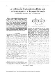
treatment. Three <strong>of</strong> the four samples <strong>of</strong> embryonic<br />
genomes yielded a PCR product with a molecular mass<br />
<strong>of</strong> 680 bp (Fig. 3, lanes 8, 9, and 11). The positive control<br />
group, for which pGL was used as a template, also<br />
<strong>gene</strong>rated a PCR product with the same size (Fig. 3,<br />
lane 3). Positive signal occurrence was at least 15%,<br />
that is, at least 3 <strong>of</strong> the 20 larvae in the four samples<br />
tested positive.
152 S.-S. LI AND H.-J. TSAI<br />
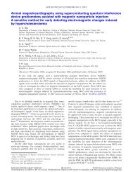
Fig. 3. Detection <strong>of</strong> pGL in <strong>giant</strong> <strong>freshwater</strong> <strong>prawn</strong> embryos using<br />
PCR analysis. Genomic DNAs were isolated from five pooled, zoea-Istage<br />
embryos developed from SMI treatment using 400 ng pGL. All<br />
embryos were incubated with 10 µg DNase at 37°C for 1 hr before their<br />
genomes were extracted and analyzed. Lane 1, molecular markers,<br />
HaeIII-digested fX-174-RF DNA; lane 2, 0.5 ng pGL as a template<br />
(positive control); lane 3, no template; lanes 4–7, from an SMI<br />
treatment using a solution lacking pGL (mock-treatment group); lanes<br />
8–11, from an SMI treatment using a solution containing pGL<br />
(experimental group). Primers added <strong>to</strong> amplify a 680-bp fragment<br />
from transgenic GFP cDNA by using GFP primer, and a 425-bp from<br />
endogenous b-tubulin <strong>gene</strong> by using Tub primer, were indicated.<br />
Analysis <strong>of</strong> Transgenic Larva by PCR<br />
and Southern Blotting<br />
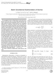
Genomic DNA was extracted from each hatched larva<br />
from the 750 ng pGL SMI treatment. Larval genomes<br />
were analyzed by PCR and Southern blot analyses. A<br />
single, positive band, with a molecular mass <strong>of</strong> 680 bp,<br />
was observed exclusively in the experimental group<br />
(lanes 5–14 <strong>of</strong> Fig. 4). Based on positive signal appearance<br />
in PCR and Southern blot analyses, the SMI <strong>gene</strong><br />
transfer success rate for <strong>prawn</strong>s was about 70% (16 <strong>of</strong><br />
23 larvae were positive).<br />
Effect <strong>of</strong> SMI on Fertility, Hatching,<br />
and Trans<strong>gene</strong>sis<br />
Egg fertilization and embryo hatching rates for the<br />
untreated group and the four SMI treatments (28, 250,<br />
750, and 1,000 ng <strong>of</strong> pGL) did not differ (Fig. 5).<br />
However, trans<strong>gene</strong>sis increased as the concentration<br />
<strong>of</strong> pGL used in SMI was increased, with the greatest<br />
increase occurring from 250 <strong>to</strong> 750 ng pGL.<br />
Green Fluorescence<br />
Green fluorescence was used <strong>to</strong> detect the expression<br />
<strong>of</strong> the GFP reporter <strong>gene</strong> in hatched larvae. Unexpectedly,<br />
green fluorescence was not seen in any larvae from<br />
the SMI with plasmid (experimental) treatments, nor<br />
in larvae from the SMI with water (mock-treated<br />
control) treatment.<br />
DISCUSSION<br />
Giant <strong>freshwater</strong> <strong>prawn</strong>s are one <strong>of</strong> the most valuable<br />
aquacultural crustaceans, and they are also suitable<br />
for indoor, labora<strong>to</strong>ry-scale culture. In addition,<br />
because embryo<strong>gene</strong>sis can be achieved in vitro under<br />
Fig. 4. PCR (A) and Southern blot (B) analyses <strong>of</strong> genomic DNA<br />
isolated from hatched larvae using GFP cDNA as a probe. Lane 1, 0.1<br />
ng pGL as a template (positive control); lane 2, molecular markers,<br />
HaeIII-digested fX-174-RF DNA; lane 3, no template added (negative<br />
control); lane 4, larva from an SMI treatment using a solution lacking<br />
pGL (mock-treatment group); lanes 5–14, larvae from an SMI treatment<br />
using a solution containing 750 ng pGL (experimental group). A<br />
680-bp fragment amplified from GFP cDNA was indicated.<br />
Fig. 5. Effect <strong>of</strong> the amount <strong>of</strong> microinjected DNA on the rates <strong>of</strong><br />
fertilization, hatching, and trans<strong>gene</strong>sis <strong>of</strong> <strong>giant</strong> <strong>freshwater</strong> <strong>prawn</strong>s.<br />
Plasmid DNA prepared at the concentrations <strong>of</strong> 28, 250, 750, and 1,000<br />
ng per µl <strong>of</strong> water were respectively microinjected in a volume <strong>of</strong> 1 µl<br />
in<strong>to</strong> each sperma<strong>to</strong>phore prior <strong>to</strong> in vivo fertilization. The number <strong>of</strong><br />
genomes from hatched larvae that were examined <strong>to</strong> determine the<br />
<strong>gene</strong> transfer rate is indicated above each solid bar.<br />
controlled conditions, this valuable decapod is used as<br />
an invertebrate model. Unfortunately, it is difficult <strong>to</strong><br />
transfer <strong>gene</strong>s in<strong>to</strong> the fertilized eggs <strong>of</strong> M. rosenbergii<br />
using electroporation because they are retained in a<br />
clump on the abdomen and cannot be detached until 16<br />
hr postfertilization (Caceci et al., 1996). Separating<br />
individual eggs from the clump and placing each fertilized<br />
egg in a container for electroporation is very<br />
tedious work. Consequently, only a small number <strong>of</strong><br />
fertilized <strong>prawn</strong> eggs can be treated. In contrast,<br />
SMI provides a fast, efficient, and easy method for<br />
transferring <strong>gene</strong>s <strong>to</strong> fertilized, <strong>giant</strong> <strong>freshwater</strong> <strong>prawn</strong><br />
eggs.<br />
We demonstrated that at least 1 µg <strong>of</strong> <strong>foreign</strong> DNA<br />
fragments (pGL) was completely digested by 0.75 µg<br />
DNase kept at 37°C for at least 1 hr. However, PCR
TRANSFER OF FOREIGN GENE TO FRESHWATER PRAWN 153<br />
analysis <strong>of</strong> genomic DNA extracted from zoea-I-stage<br />
embryos in the 10 µg DNase SMI treatment still found<br />
a 680-bp band. Moreover, the oligonucleotide primers<br />
chosen for PCR amplification <strong>of</strong> the GFP cDNA (trans<strong>gene</strong>)<br />
and b-tubulin <strong>gene</strong> (internal control <strong>gene</strong>) <strong>gene</strong>rated<br />
680 bp and 425 bp DNA fragments, respectively. A<br />
680-bp PCR product was observed when pGL fragments<br />
were present in transgenic, zoea-I-stage embryos and<br />
hatched larvae. Only nontransgenic, SMI-treated<br />
<strong>prawn</strong>s and <strong>prawn</strong>s from the control group contained a<br />
425-bp PCR product. The 680-bp PCR product from the<br />
SMI treated samples was positive for Southern blot<br />
hybridization using a labeled, GFP cDNA probe. All this<br />
evidence indicates that the exogenous plasmid DNA in<br />
embryos and larvae were introduced by SMI. Our<br />
findings concur with those <strong>of</strong> Shamila and Mathavan<br />
(1998), who demonstrated that a <strong>foreign</strong> plasmid could<br />
be introduced in<strong>to</strong> V instar silkworm larvae by testes<br />
microinjection.<br />
Although we have not yet analyzed all the experimental<br />
larvae, results from the positive samples <strong>of</strong> PCR and<br />
Southern blot analyses from hatched larvae indicate<br />
that SMI achieved a <strong>gene</strong> transfer rate <strong>of</strong> about 70%.<br />
This success rate is far superior <strong>to</strong> that reported for<br />
microinjection <strong>of</strong> finfish, which ranged from 16% <strong>to</strong> 27%<br />
for medaka (Oza<strong>to</strong> et al., 1986; Inoue et al., 1990; Lu et<br />
al., 1992) and 0% <strong>to</strong> 35% for zebrafish, common carp,<br />
and channel catfish (Powers et al., 1992). It is also much<br />
better than the success rates for electroporation <strong>of</strong><br />
fertilized finfish eggs, which ranged from 4% <strong>to</strong> 20% for<br />
medaka (Inoue et al., 1990; Lu et al., 1992), and<br />
electroporation <strong>of</strong> finfish sperm, which were only 2.6%<br />
<strong>to</strong> 4.2% for carp, tilapia, and catfish (Muller et al.,<br />
1992). The SMI <strong>gene</strong> transfer rate for <strong>giant</strong> <strong>prawn</strong>s is<br />
comparable <strong>to</strong> that obtained by electroporation <strong>of</strong> zebrafish<br />
eggs (35% <strong>to</strong> 75%, Powers et al., 1992) and the<br />
electroporation <strong>of</strong> loach (50%, Tsai et al., 1995a) and<br />
abalone sperm (65%, Tsai et al., 1997). SMI probably<br />
achieved a high rate <strong>of</strong> <strong>gene</strong> transfer in <strong>giant</strong> <strong>freshwater</strong><br />
<strong>prawn</strong>s because the microinjected plasmids are<br />
retained in the sperma<strong>to</strong>phore for 2–6 hr before in vivo<br />
fertilization begins. The longer the <strong>foreign</strong> plasmids<br />
remain in the sperma<strong>to</strong>phore, the greater the probability<br />
they will enter sperm.<br />
Green fluorescence, which was presumptively expressed<br />
by the transgenic GFP cDNA, was not detectable<br />
in hatched larvae. This may be attributed <strong>to</strong><br />
transient expression <strong>of</strong> the GFP cDNA and <strong>to</strong> the<br />
functionality <strong>of</strong> the CMV promoter. It is also possible<br />
that the exoskele<strong>to</strong>n somehow blocks visual detection <strong>of</strong><br />
GFP <strong>gene</strong> expression. Gendreau and colleagues (1995)<br />
reported that the transient expression <strong>of</strong> a luciferase<br />
<strong>gene</strong> transferred by ballistics in<strong>to</strong> brine shrimp was<br />
detectable only at 12 hr after bombardment. Problems<br />
detecting expression <strong>of</strong> the GFP <strong>gene</strong> could be overcome<br />
in future experiments by using <strong>prawn</strong>-specific promoter<br />
fused with GFP cDNA.<br />
Gendreau and colleagues (1995) reported that ballistic<br />
shots decreased survival <strong>of</strong> treated brine shrimp.<br />
Bensheng and Khoo (1997) reported that the early<br />
developmental stage <strong>of</strong> <strong>freshwater</strong> shrimp embryos is<br />
extremely vulnerable <strong>to</strong> microinjection, and recommended<br />
that the intermediate stage be used for microinjection.<br />
However, when a <strong>gene</strong> is transferred <strong>to</strong> the<br />
intermediate stage, the trans<strong>gene</strong> will have a mosaic<br />
distribution. In this study, we demonstrated several<br />
advantages <strong>of</strong> SMI <strong>gene</strong> transfer system, including: (1)<br />
a high <strong>gene</strong> transfer rate; (2) no effect on fertility,<br />
survival, and hatching rates; and (3) easy introduction<br />
<strong>of</strong> <strong>foreign</strong> DNA in<strong>to</strong> oocytes, which definitely helps<br />
<strong>to</strong> minimize transgenic mosaicism. In our opinion,<br />
SMI is the most promising technology for <strong>prawn</strong> trans<strong>gene</strong>sis.<br />
REFERENCES<br />
Besheng J, Khoo HW. 1997. Transient expression <strong>of</strong> two luciferase<br />
reporter <strong>gene</strong> constructs in developing embryos <strong>of</strong> <strong>Macrobrachium</strong>m<br />
lanchesteri (De Man). Aquaculture Res 28:183–190.<br />
Caceci T, Calson CB, Toth TE, Smith SA. 1996. In vitro embryo<strong>gene</strong>sis<br />
<strong>of</strong> <strong>Macrobrachium</strong> rosenbergii larvae following in vivo fertilization.<br />
Aquaculture 147:169–175.<br />
Chourrout D, Perrot E. 1992. No transgenic rainbow trout produced<br />
with sperm incubated with linear DNA. Mol Marine Biol Biotechnol<br />
1:282–285.<br />
Chourrout D, Guyomard R, Houdebine LM. 1986. High-efficiency <strong>gene</strong><br />
transfer in rainbow trout (Salmo gairdneri) by microinjection in<strong>to</strong><br />
egg cy<strong>to</strong>plasm. Aquaculture 51:143–150.<br />
Dunham RA, Eash J, Askins J, Townes TM. 1987. <strong>Transfer</strong> <strong>of</strong> the<br />
metallothionein-human growth hormone fusion <strong>gene</strong> in<strong>to</strong> channel<br />
catfish. Trans Am Fish Soc 116:87–91.<br />
Dunham RA, Ramboux AC, Duncan PL, Hayat M, Chen TT, Lin CM,<br />
Knight K, Gonzalez-Villasenor I, Powers DA. 1992. <strong>Transfer</strong>, expression,<br />
and inheritance <strong>of</strong> salmonid growth hormone <strong>gene</strong>s in channel<br />
catfish, Ictalurus punctatusa, and effects on performance traits. Mol<br />
Marine Biol Biotechnol 1:380–389.<br />
Fletcher GL, Shears MA, King MJ, Davies PL, Hew CL. 1988.<br />
Evidence for antifreeze protein <strong>gene</strong> transfer in Atlantic salmon<br />
(Salmo salar). Can J Fish Aquat Sci 45:352–357.<br />
Gedreau S, Lardans V, Cadoret JP, Mialhe E. 1995. Transient<br />
expression <strong>of</strong> a luciferase reporter <strong>gene</strong> after ballistic introduction<br />
in<strong>to</strong> Artemia franciscana (Crustacea) embryos. Aquaculture 33:199–<br />
205.<br />
Giambernardi TA, Rodeck U, Klebe RJ. 1998. Bovine serum albumin<br />
reverses inhibition <strong>of</strong> RT-PCR by melanin. BioTechniques 25:564-<br />
566.<br />
Inoue K, Yamashita S, Hata J, Kabeno S, Asada S, Nagahisa E, Fujita<br />
T. 1990. Electroporation as a new technique for producing transgening<br />
fish. Cell Differ Dev 29:123–128.<br />
Khoo HW, Ang LH, Lim HB, Wong Y. 1992. Sperm cells as vec<strong>to</strong>rs for<br />
introducing <strong>foreign</strong> DNA in<strong>to</strong> zebrafish. Aquaculture 107:1–19.<br />
Lu JK, Chen TT, Chrisman CL, Andrisani OM, Dixon JE. 1992.<br />
Integration, expression, and germ-line transmission <strong>of</strong> <strong>foreign</strong> growth<br />
hormone <strong>gene</strong>s in medaka (Oryzias latipes). Mol Marine Biol<br />
Biotechnol 1:366–375.<br />
Muller E, Ivics Z, Erdelyi F, Papp T, Varadi L, Horvath L, MacLean N,<br />
Orban L. 1992. Introducing <strong>foreign</strong> <strong>gene</strong>s in<strong>to</strong> fish eggs with<br />
electroporated sperm as a carrier. Mol Marine Biol Biotechnol<br />
1:276–281.<br />
Oza<strong>to</strong> K, Kondoh H, Inohara H, Iwamatsu T, Wakamatsu Y, Okada TS.<br />
1986. Production <strong>of</strong> transgenic fish: introduction and expression <strong>of</strong><br />
chicken d-crystallin <strong>gene</strong> in medaka embryos. Cell Differ 19:234–<br />
237.<br />
Powers DA, Hereford L, Cole T, Chen TT, Lin CM, Knight K, Creech K,<br />
Dunham R. 1992. Electroporation: a method for transferring <strong>gene</strong>s<br />
in<strong>to</strong> the gametes <strong>of</strong> zebrafish (Brachydanio rerio) channel catfish
154 S.-S. LI AND H.-J. TSAI<br />
(Ictalurus punctatus), and common carp (Cyprinus carpio). Mol<br />
Marine Biol Biotechnol 1:301–308.<br />
Sambrook J, Fritsch EF, Maniatis T. 1989. Large-scale preparations <strong>of</strong><br />
plasmid DNA. In: Nolan C, edi<strong>to</strong>r. Molecular cloning: a labora<strong>to</strong>ry<br />
manual. New York: Cold Spring Harbor Labora<strong>to</strong>ry Press. p 1.33–1.46.<br />
Shamila Y, Mathavan S. 1998. Sperm-mediated <strong>gene</strong> transfer in the<br />
silkworm Bombyx mori. Arch Insect Biochem Physiol 37:168–177.<br />
Symonds JE, Walker SP, Sin FYT, Sin I. 1994. Development <strong>of</strong> a mass<br />
<strong>gene</strong> transfer method in chinook salmon: optimization <strong>of</strong> <strong>gene</strong><br />
transfer by electroporated sperm. Mol Marine Biol Biotechnol<br />
3:104–111.<br />
Tsai HJ, Tseng FS. 1994. Electroporation <strong>of</strong> a <strong>foreign</strong> <strong>gene</strong> in<strong>to</strong> black<br />
porgy (Acanthopagrus schlegeli) embryos. Fish Sci 60:787–788.<br />
Tsai HJ, Tseng FS, Liao IC. 1995a. Electroporation <strong>of</strong> sperm <strong>to</strong><br />
introduce <strong>foreign</strong> DNA in<strong>to</strong> the genome <strong>of</strong> loach (Misgurnus anguillicaudatus).<br />
Can J Fish Aquat Sci 52:776–787.<br />
Tsai HJ, Wang SH, Inoue K, Kimura M, Wakamatsu Y, Oza<strong>to</strong> K.<br />
1995b. Initiation <strong>of</strong> the transgenic lacZ <strong>gene</strong> expressed in medaka<br />
(Oryzias latipes) embryos. Mol Marine Biol Biotechnol 4:1–9.<br />
Tsai HJ, Lai CH, Yang HS. 1997. Sperm as a carrier <strong>to</strong> introduce an<br />
exogenous DNA fragment in<strong>to</strong> the oocyte <strong>of</strong> Japanese abalone<br />
(Haliotis diversicolor supertexta). Transgenic Res 6:85–95.