il\VOLVEMENT OF RETII\OIC ACID II{ - MSpace at the University of ...
il\VOLVEMENT OF RETII\OIC ACID II{ - MSpace at the University of ...
il\VOLVEMENT OF RETII\OIC ACID II{ - MSpace at the University of ...
You also want an ePaper? Increase the reach of your titles
YUMPU automatically turns print PDFs into web optimized ePapers that Google loves.
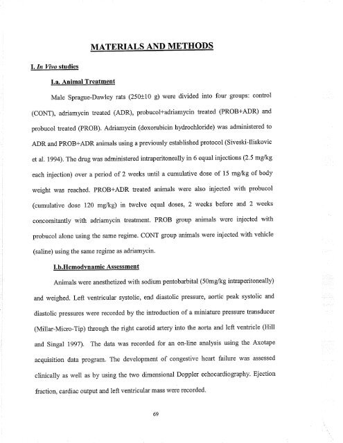
MATERIALS AND METHODS<br />
l.In Vivo studíes<br />
I.a. Animal Tre<strong>at</strong>ment<br />
Male Sprague-Dawley r<strong>at</strong>s (250t10 g) were divided into four gloups: control<br />
(CONT), adriamycin tre<strong>at</strong>ed (ADR), probucol+adriamycin tre<strong>at</strong>ed (PROB+ADR) and<br />
probucol tre<strong>at</strong>ed (PROB). Adriamycin (doxorubicin hydrochloride) was administered to<br />
ADR and PROB+ADR animals using a previously established protocol (Siveski-Iliskovic<br />
et al. 1994). The drug was administered intraperitoneally in 6 equal injections (2.5 mgkg<br />
each injection) over a period <strong>of</strong> 2 weeks until a cumul<strong>at</strong>ive dose <strong>of</strong> 15 mg/kg <strong>of</strong> body<br />
weight was reached. PROB+ADR tre<strong>at</strong>ed animals were also injected with probucol<br />
(cumul<strong>at</strong>ive dose 120 mglkg) in twelve equal doses, 2 weeks before and 2 weeks<br />
concomitantly with adriamycin tre<strong>at</strong>ment. PROB group animals were injected with<br />
probucol alone using <strong>the</strong> same regime. CONT group animals were injected with vehicle<br />
(saline) using <strong>the</strong> same regime as adriamycin.<br />
I.b.Hemodvnamic Assessment<br />
Animals were anes<strong>the</strong>tizedwith sodium pentobarbital (5Omglkg intraperitoneally)<br />
and weighed. Left ventricular systolic, end diastolic pressure, aortic peak systolic and<br />
diastolic pressures were recorded by <strong>the</strong> introduction <strong>of</strong> a mini<strong>at</strong>ure pressure transducer<br />
(Mitlar-Micro-Tip) through <strong>the</strong> right carotid artery into <strong>the</strong> aorta and left ventricle (Hill<br />
and Singal lggT). The d<strong>at</strong>a was recorded for an on-line analysis using <strong>the</strong> Axotape<br />
acquisition d<strong>at</strong>a program. The development <strong>of</strong> congestive heart failure was assessed<br />
clinically as well as by using <strong>the</strong> two dimensional Doppler echocardiography. Ejection<br />
fraction, cardiac output and left ventricular mass were recorded.<br />
69


![an unusual bacterial isolate from in partial fulf]lment for the ... - MSpace](https://img.yumpu.com/21942008/1/190x245/an-unusual-bacterial-isolate-from-in-partial-fulflment-for-the-mspace.jpg?quality=85)
![in partial fulfil]ment of the - MSpace - University of Manitoba](https://img.yumpu.com/21941988/1/190x245/in-partial-fulfilment-of-the-mspace-university-of-manitoba.jpg?quality=85)
