combined spinal-epidural anaesthesia techniques.......... a ... - medIND
combined spinal-epidural anaesthesia techniques.......... a ... - medIND
combined spinal-epidural anaesthesia techniques.......... a ... - medIND

You also want an ePaper? Increase the reach of your titles
YUMPU automatically turns print PDFs into web optimized ePapers that Google loves.
450 Indian J. Anaesth. 2005; 49 (6) : 450 - 458<br />
INDIAN JOURNAL OF ANAESTHESIA, DECEMBER 2005 450<br />
REVIEW ARTICLE<br />
SUMMARY<br />
COMBINED SPINAL-EPIDURAL ANAESTHESIA<br />
TECHNIQUES.......... A REVIEW<br />
Dr. Chandola H.C. 1 Dr. Zubair Umer Mohamed 2 Dr. Asok Jayaraj Pullani 3<br />
Combined Spinal Epidural (CSE) <strong>anaesthesia</strong> commands a unique place among the various <strong>techniques</strong> used for neuraxial blockade. In<br />
this article we delve into the history of CSE and try to enumerate the types of specialised needles devised for this technique. The<br />
<strong>spinal</strong> needles that are, and in the future may well be associated with this technique are also discussed. The methods used to locate<br />
the <strong>epidural</strong> space have been researched exhaustively and an attempt has been made to classify the various options available for CSE.<br />
Controversial aspects such as the preferred approach, catheter threading prior to or after subarachnoid block and the need for test dosing<br />
have also been addressed.<br />
Keywords : Combined Spinal Epidural, CSE, Tuohy Needle, Spinal needle, Catheter.<br />
Introduction<br />
Among the various <strong>techniques</strong> used for neuraxial<br />
blockade, CSE commands a unique place. The singularity<br />
lies in its ability to combine the rapidity, density, and<br />
reliability of the subarachnoid block with the flexibility of<br />
continuous <strong>epidural</strong> block to titrate a desired sensory level,<br />
vary the intensity of the block, control the duration of<br />
<strong>anaesthesia</strong>, and deliver postoperative analgesia. Of late,<br />
this procedure has come back in a big way, the fact justified<br />
by its use extended to paediatric and even infant<br />
laparotomies, apart from its use in orthopedic surgery,<br />
obstetrics, very old patients and in other high-risk patients. 1<br />
Although at first sight the CSE technique appears to be<br />
more complicated than either <strong>epidural</strong> or <strong>spinal</strong> block,<br />
intrathecal drug administration and placement of the <strong>epidural</strong><br />
catheter are both facilitated by the various modifications of<br />
the <strong>combined</strong> <strong>spinal</strong>-<strong>epidural</strong> technique. 1,2 In this article<br />
we try to follow the evolution and also summarise the<br />
various developments in this technique described by various<br />
authors over the years.<br />
History<br />
Leafing through the archives of <strong>anaesthesia</strong>, we find<br />
that CSE was first described in 1937 by Soresi 3 , who used<br />
the “episubdural” technique by first injecting a dose of<br />
local anesthetic <strong>epidural</strong>ly and, after advancing the needle<br />
inside the dural cavity, injecting the <strong>spinal</strong> dose. Curelaru 4<br />
1. M.D., Asso. Prof.<br />
2. M.B.B.S., PG Student<br />
3. M.B.B.S. PG Student<br />
M.L.N. Medical College, Allahabad, UP.<br />
Department of Anaesthesiology,<br />
M.L.N. Medical College, Allahabad, Uttar Pradesh - 211001.<br />
Correspond to :<br />
Dr. H. C. Chandola<br />
E-mail : chandola@sancharnet.in<br />
(Accepted for publication on 30 - 08 - 2005)<br />
performed the first <strong>combined</strong> <strong>spinal</strong> anesthesia and<br />
catheter-based <strong>epidural</strong> <strong>anaesthesia</strong> in 1979. He introduced<br />
an <strong>epidural</strong> catheter through a Tuohy needle, administered<br />
a test dose, and then performed a traditional dural puncture<br />
1–2 lumbar segments distally using a 26-gauge <strong>spinal</strong><br />
needle. CSE was performed in 1982 on a single <strong>spinal</strong><br />
segment for lower limb surgery by Coates 5 and Mumtaz 6<br />
et al. Epidural <strong>anaesthesia</strong> was performed using a 16-gauge<br />
Tuohy needle, after which a 25-to 26-gauge <strong>spinal</strong><br />
needle, approximately 1 cm longer than the <strong>epidural</strong><br />
needle, was introduced with the Tuohy needle as introducer.<br />
After the injection of local anesthetic, the <strong>spinal</strong> needle<br />
was withdrawn and the <strong>epidural</strong> catheter inserted. In<br />
1986, Rawal described a single segment sequential CSE<br />
for caesarean delivery. (see below “Sequential CSE”).<br />
Over the years, a variety of <strong>spinal</strong>, <strong>epidural</strong> and specialised<br />
<strong>combined</strong> <strong>spinal</strong>-<strong>epidural</strong> needles have been devised and a<br />
myriad of <strong>techniques</strong> described to refine this procedure.<br />
Classification<br />
The various options in CSE can be classified broadly<br />
according to the number of interspaces used for performing<br />
the procedure (table 1). If both the <strong>epidural</strong> and the <strong>spinal</strong><br />
punctures are performed in the same interspace it is called<br />
single segment technique (SST) or single interspace CSE<br />
technique and the others are named double segment <strong>techniques</strong><br />
(DST) or double interspace CSE technique. These can be<br />
further classified according to the type of needle used and<br />
the approach.<br />
Single and double segment <strong>techniques</strong><br />
The reduced number of skin punctures in SST<br />
decreases the incidence of patient discomfort, trauma, pain,<br />
infection at puncture sites, <strong>epidural</strong> venous puncture and<br />
formation of hematomas. 1 The use of an <strong>epidural</strong> needle as<br />
an introducer for the <strong>spinal</strong> needle lessens the likelihood of<br />
contamination of the <strong>spinal</strong> needle with skin-borne
CHANDOLA, MOHAMED, PULLANI : COMBINED SPINAL-EPIDURAL ANAESTHESIA 451<br />
microorganisms 7 . However, no quantitative studies have been<br />
performed to confirm these assumptions. In DST, there is<br />
always a theoretical possibility of damaging the <strong>epidural</strong><br />
catheter with the <strong>spinal</strong> needle if the catheter is threaded<br />
before subarachnoid block. The advantage of DST is that it<br />
does not require specialised CSE needles that are more<br />
expensive than the usual <strong>epidural</strong> and <strong>spinal</strong> needles.<br />
Table - 1 : Classification of CSE Techniques<br />
Single Segment<br />
Needle-Through-Needle<br />
Without Spinal Needle Support<br />
Median Approach<br />
Paramedian Approach<br />
With Spinal Needle Support<br />
Spinal Needle Sleeve<br />
Median Approach<br />
Paramedian Approach<br />
Spinal Needle Lock<br />
Median Approach<br />
Paramedian Approach<br />
Needle-Beside-Needle (Double Barrel/Lumen)<br />
Without Attached Spinal Needle Guide/Separate Needle<br />
Median Approach<br />
aramedian Approach<br />
Combined Median/Paramedian Approach<br />
With Attached Spinal Needle Guide<br />
Guide alongside Epidural Needle<br />
Median Approach<br />
Paramedian Approach<br />
Guide incorporated within Epidural Needle<br />
Median Approach<br />
Paramedian Approach<br />
Double Segment<br />
Median Approach<br />
Paramedian Approach<br />
Each of these can be performed with or with out <strong>epidural</strong> and/or <strong>spinal</strong><br />
catheters<br />
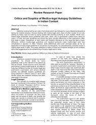
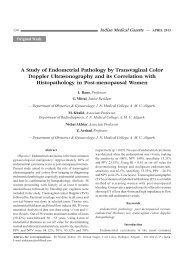
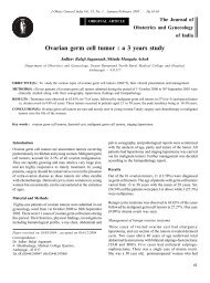
Fig. 1: Different types of needles used for CSE technique.<br />
A : needle through needle, B: Huber and Hanaoka needle,<br />
C: Eldor needle, D: Eldor, Coombs and Torrieri needle.<br />
Needles<br />
Single Lumen Epidural Needles (Needle-Through-<br />
Needle technique)<br />
1. Epidural Needle with Single Aperture: This most<br />
commonly used set consists of a conventional 16-18<br />
gauge <strong>epidural</strong> needle (Fig. 1A) to locate the <strong>epidural</strong><br />
space. A <strong>spinal</strong> needle is then introduced through the<br />
<strong>epidural</strong> needle to exit it through the Huber eye and<br />
proceed to puncture the dural wall and enter the<br />
subarachnoid space.<br />
2. CSE Needle with Additional Aperture (Huber and<br />
Hanaoka Needles): This is basically an <strong>epidural</strong> needle<br />
with an additional aperture (‘back eye’) situated at<br />
the end of the longitudinal axis. 8,9 (Fig. 1B)<br />
The usual technique is used for identification of<br />
the <strong>epidural</strong> space. The <strong>spinal</strong> needle is then introduced<br />
through the proximal port of the <strong>epidural</strong> needle to exit<br />
at the ‘back eye’ and after dural puncture and drug<br />
administration the <strong>spinal</strong> needle is withdrawn and the<br />
catheter inserted. The larger diameter of the catheter<br />
causes it to pass through the usual Huber opening. These<br />
modified needles function as a suitable introducer for even<br />
the thinnest <strong>spinal</strong> needle, as kinking and friction in the<br />
Huber eye of the <strong>epidural</strong> needle is avoided. Advancing the<br />
<strong>spinal</strong> needle through the “back eye” may offer a more<br />
distinct sensation of dural puncture. 10 To accomplish<br />
proper passage through the “back eye,” however, the distal<br />
end of the <strong>spinal</strong> needle must face the same way as the<br />
Huber opening of the <strong>epidural</strong> needle; otherwise the <strong>spinal</strong><br />
needle may pass through the Huber eye. 11 But a traditional<br />
insertion of the <strong>epidural</strong> needle with the Huber eye facing<br />
cephalad will then cause the cutting bevel of the <strong>spinal</strong><br />
needle to cut the dural fibers perpendicular to its alignment,<br />
which may increase the risk of postdural puncture<br />
headache. 12 With the needle through needle technique, there<br />
is a concern about the tip of the <strong>spinal</strong> needle scraping the<br />
inner wall of the <strong>epidural</strong> needle and thereby leading to<br />
deposition of metal particles in the <strong>epidural</strong> and/or<br />
subarachnoid spaces and possible, subsequent neurological<br />
sequelae. 13 However, studies by Herman et al 14 failed to<br />
find any evidence of additional metal particles produced by<br />
the needle-through-needle technique and puts these doubts<br />
to rest. The risk of damage to the bevel of the <strong>spinal</strong> needle<br />
has been circumvented by the introduction of a plastic sleeve<br />
for the <strong>spinal</strong> needle by the Espocan® system. 1 The plastic<br />
sleeve keeps the <strong>spinal</strong> needle centrally in the <strong>epidural</strong><br />
needle and guides it through the back eye.<br />
In this technique, the <strong>spinal</strong> needle is in contact<br />
with only dura and the <strong>epidural</strong> needle, which may increase<br />
the risk of displacement of the <strong>spinal</strong> needle at the time of
452<br />
INDIAN JOURNAL OF ANAESTHESIA, DECEMBER 2005<br />
syringe connection, aspiration of CSF or injection of local<br />
anaesthetic. 1 A study conducted by Tanaka N and coworkers<br />
15 and Hoffmann et al 16 clearly demonstrated that<br />
the lockable CSE provided safe and stable conditions and<br />
a good success rate of subarachnoid block. But the feel of<br />
the “dural click” was diminished when lockable CSE was<br />
used.<br />
larger distal side port of the Sprotte needle, as well as its<br />
greater distance from the tip, may account for the slightly<br />
greater deflection observed with Sprotte needles compared<br />
to Whitacre needles. (Fig. 2)<br />
Double-Lumen Needles: (Needle-Beside-Needle<br />
technique)<br />
In this technique, an <strong>epidural</strong> needle and <strong>spinal</strong> needle<br />
are introduced through separate lumen or barrels. The <strong>spinal</strong><br />
needle guide may or may not be attached to the <strong>epidural</strong><br />
needle.<br />
Separate Needle<br />
In the technique described by Eldor and Olshwang,<br />
the <strong>epidural</strong> needle is first placed in the <strong>epidural</strong> space by<br />
the usual method followed by the introduction of the <strong>spinal</strong><br />
needle beside the <strong>epidural</strong> needle to perform the subarachnoid<br />
puncture. 17<br />
A recent article by Cook described a variation from<br />
this sequence by first locating the <strong>spinal</strong> needle in the<br />
subarachnoid space and observing the free flow of CSF.<br />
The stylet of the <strong>spinal</strong> needle is then replaced. The <strong>epidural</strong><br />
needle is then introduced via the median or paramedian<br />
approach and catheter threaded, following which the<br />
intrathecal drug is administered. 18<br />
Attached Needle<br />
A parallel tube is attached to the <strong>epidural</strong> needle,<br />
which acts as the introducer for the <strong>spinal</strong> needle. The<br />
parallel tube may be either bent at the proximal end or<br />
straight through out. The Eldor Needle (Fig. 1C) has a<br />
straight tube attached while the Eldor, Coombs and Torrieri<br />
Needle (Fig. 1D) has a bent <strong>spinal</strong> needle attached. 7,9,19<br />
The T-A needle is a further modification where both the<br />
needles are welded together and the distal ends of the<br />
needles make a common tip. The <strong>epidural</strong> needle points<br />
cephalad and the <strong>spinal</strong> needle points caudad. 20 The more<br />
recently introduced E-SP needle has a needle wall<br />
configuration that is “over-under” rather than parallel. 1<br />
Spinal Needles<br />
The gauge and type of the <strong>spinal</strong> needle influences<br />
the success of the procedure and the incidence of outcome.<br />
The use of smaller gauge needles and ones with a conical<br />
tip reduces the incidence of post dural puncture headache. 21,22<br />
Lipov 23 et al. demonstrated that the axial force needed to<br />
deform the Sprotte needle tip was less than that needed for<br />
the Whitacre and Quincke needles of similar size. The<br />
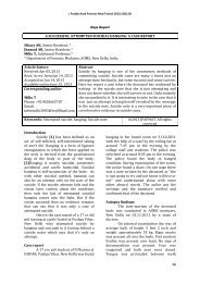
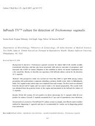
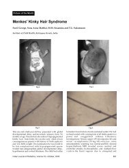
Fig. 2 : Tip of different types of <strong>spinal</strong> needles.<br />
A: Sprotte, B: Whitacre, C: modified pencil-point,<br />
D: ball pen, E: double-hole pencil-point, F: Quincke, G: Atraucan.<br />
Newer needles have been designed to improve<br />
efficacy. The Atraucan needle has a double bevel with the<br />
distal cutting bevel making a cutting incision in the dura.<br />
The second part of this needle dilates this incision rather<br />
than cutting through the fibres leaving only a small hole in<br />
the dura. The sharp tip however is prone to damage. 24 A<br />
new pencil-point <strong>spinal</strong> needle described is the double-hole<br />
pencil-point (DHPP) <strong>spinal</strong> needle, and is composed of a<br />
blunt ogival tip and two circular holes opposing each other<br />
just proximal to the tip. The area of the two windows is<br />
almost the same as of the single holed Sprotte needle’s<br />
area, which enables more rapid CSF reflux. The anesthetic<br />
solution injection spreads through both windows. This allows<br />
a more diffuse anesthetic distribution and less anesthetic<br />
solution dosage. The DHPP <strong>spinal</strong> needle allows anesthetic<br />
solution injection even when a tissue fragment obstructs<br />
one of the holes. More recently, the ball pen needles have<br />
been marketed in France. The tip of this needle is actually<br />
the tip of the stylet leaving a hollow cannula when the<br />
stylet is removed. The proposed advantages of this needle<br />
are that the tip of the needle is always completely in the<br />
subarachnoid space and on removal of the stylet there is no<br />
needle tip projecting beyond the orifice causing damage to<br />
the neurological tissue. There is no mechanical weakening<br />
at the tip caused by the presence of a lateral orifice. The<br />
open end of the needle allows laminar flow of cerebro<strong>spinal</strong><br />
fluid, which results in faster identification of the subarachnoid<br />
space. 24 Optimal Length of the Spinal Needle Beyond the<br />
Epidural Needle Tip<br />
The distance from the tip of the <strong>epidural</strong> needle to<br />
the posterior wall of the dural sac in the midline varies<br />
considerably among patients (0.3-1.03 cm). 25,26 Further, the
CHANDOLA, MOHAMED, PULLANI : COMBINED SPINAL-EPIDURAL ANAESTHESIA 453<br />
anteroposterior diameter of the dural sac varies considerably<br />
during flexion and extension of the <strong>spinal</strong> column. At L3-<br />
L4, the diameter increases from a range of 9-20 mm in<br />
extension to a range of 11-25 mm in flexion. 27 Additionally,<br />
these findings are only valid when the <strong>epidural</strong> puncture is<br />
performed in midline. This is because the dural sac is<br />
triangular with its base resting on the vertebral body and<br />
the triangle top points posteriorly to ligamentum flavum. 28<br />
A minimum of 13 mm length of the <strong>spinal</strong> needle protrusion<br />
beyond the <strong>epidural</strong> needle tip is recommended for the CSE<br />
sets for a reasonably high success rate. 10 Also, because of<br />
needle design, the length of protrusion for needles with side<br />
orifice should be greater than that for end orifice needles. 29<br />
Techniques<br />
The first neuraxial blockade performed by Corning<br />
must have been an <strong>epidural</strong> rather than a <strong>spinal</strong> block. 30<br />
The earlier methods to identify the <strong>epidural</strong> space were the<br />
feel and hear method of Pages, positive plunger pressure by<br />
Dogliotti, “hanging drop” method by Gutierrez and the<br />
balloon deflation technique by Macintosh and Oxford. 31 Most<br />
of these <strong>techniques</strong> were based on the concept of “negative”<br />
pressure in the extradural space. However, the experiments<br />
of Urayama 32 and Shah JL 33 showed that 77% of the lumbar<br />
cerebro<strong>spinal</strong> fluid pulse wave, which is directly transmitted<br />
to the extradural space as an extradural pressure wave<br />
originates in the vascular system (arteries and veins), and<br />
not in the brain. So, any rise in the blood pressure, which<br />
is not an infrequent observation during <strong>epidural</strong> needle<br />
insertion, can give a concomitant rise in the extradural<br />
pressure with the loss of the “negative” pressure in the<br />
extradural space and “unreliable” hanging drop and balloon<br />
indicator <strong>techniques</strong>. Many feel the loss of resistance to<br />
saline or demonstration of a free flow of the saline column<br />
is a superior method for properly identifying the <strong>epidural</strong><br />
space. However, when administering CSE, the use of air<br />
has been advocated so that there is no confusion that the<br />
fluid returning is CSF when the <strong>spinal</strong> needle is subsequently<br />
placed. These <strong>techniques</strong> have the disadvantage of identifying<br />
only the <strong>epidural</strong> space and not the subarachnoid space.<br />
Kopacz et al 34 describes a further modification of this<br />
technique which may increase the success rate and ease<br />
with which it is performed. The modification described<br />
allows the continued use of saline for loss of resistance<br />
using an <strong>epidural</strong> needle with a transparent hub. First,<br />
close attention is paid to the location of the saline meniscus<br />
left within the <strong>epidural</strong> hub after demonstrating the loss of<br />
resistance using saline. If a loss of resistance to air technique<br />
is used, a small amount of saline can be introduced to<br />
create this meniscus. The <strong>spinal</strong> needle is advanced in the<br />
usual fashion until it is felt to exit the <strong>epidural</strong> needle. As<br />
the <strong>spinal</strong> needle is advanced further to contact and indent<br />
the dura, the tenting of the dura generates a negative pressure<br />
within the <strong>epidural</strong> space, causing the meniscus to be drawn<br />
in. This is similar to what is seen when performing the<br />
“hanging drop” technique of identifying the <strong>epidural</strong> space.<br />
As the <strong>spinal</strong> needle is advanced further, the dura is punctured<br />
and the meniscus returns to near its original location as the<br />
pressure within the <strong>epidural</strong> space returns to baseline.<br />
Finally, CSF is observed to flow into the <strong>spinal</strong> needle to<br />
ultimately drip from its hub. The point at which the meniscus<br />
starts to advance is dependent on the dimensions of each<br />
particular patient’s <strong>epidural</strong> space and to what distance the<br />
needle has traversed the <strong>epidural</strong> space. This phenomenon<br />
is reported to be a very specific indicator of needle location<br />
and is most easily seen when a Whitacre <strong>spinal</strong> needle is<br />
used. When a Quincke or Sprotte needle is used, the<br />
movements of the meniscus may occur so suddenly that<br />
often only a “pop” or “splash” of the fluid is detected, or<br />
no movement is seen at all. These observations agree with<br />
the studies showing that more force is required to penetrate<br />
the dura with a Whitacre needle compared to a Quincke<br />
needle. 35 The ‘membrane in syringe’ technique is, in principle,<br />
a modification of the loss of resistance technique for<br />
identifying the <strong>epidural</strong> space. 36 A novel method to identify<br />
the <strong>epidural</strong> space using an acoustic device was described<br />
by Jacob S and Tierney E. 37 The traditional loss of resistance<br />
technique is <strong>combined</strong> with amplification of the sound made<br />
by the <strong>epidural</strong> needle as it traverses the interspinous<br />
ligament and the ligamentum flavum and then enters the<br />
<strong>epidural</strong> space. Lechner TJ and associates 38 described a<br />
variant of the previous method. This consisted of translating<br />
the pressure generated during the <strong>epidural</strong> puncture procedure<br />
into a corresponding acoustic signal. Both these methods<br />
combine the loss of resistance <strong>techniques</strong> with a newer<br />
approach to improve the success rate by giving an early<br />
warning of entry into <strong>epidural</strong> space. In addition, these can<br />
be excellent teaching aids for beginners.<br />
A still newer method to locate the <strong>epidural</strong> space<br />
using real-time ultrasonic guidance has been described by<br />
Grau T and colleagues. 39 Epidural space localization has<br />
also been reported by using an electronic device known as<br />
Episensor but with limited success. 40<br />
Sequential CSE Technique<br />
Rawal first described this variation of the standard<br />
CSE technique. 1 The <strong>spinal</strong> block is performed in the sitting<br />
position and a low dose of hyperbaric bupivacaine is<br />
administered to achieve a S5 to T8-T9 block. The patient<br />
is placed supine with a left lateral tilt. When the <strong>spinal</strong><br />
block is “fixed” it is extended to T4 by injecting fractionated<br />
doses of local anaesthetic into the <strong>epidural</strong> catheter.
454<br />
INDIAN JOURNAL OF ANAESTHESIA, DECEMBER 2005<br />
Two Catheter CSE Technique<br />
A technique of providing CSE <strong>anaesthesia</strong> using<br />
both <strong>spinal</strong> and <strong>epidural</strong> catheters in the same and<br />
different interspace was described by Dahl et al 41 and<br />
Vercauteren et al 42 respectively. The main advantage was<br />
the possibility of titrating the intrathecal dose of the local<br />
anaesthetic to the desired dermatomal level and to test the<br />
correct position of <strong>epidural</strong> catheter before injecting the<br />
drugs. Because of the requirement of a large diameter<br />
<strong>spinal</strong> needle (22-gauge), the risk of catheter penetration<br />
may be high; knotting of the catheters is also a theoretical<br />
risk. 42<br />
Approach<br />
Blomberg et al, 43 Gaynor A 44 and Hatfalvi BI 45<br />
compared the midline and paramedian approaches for lumbar<br />
<strong>epidural</strong> block and concluded that the paramedian approach<br />
was superior due to the following reasons.<br />
1. Easier to locate the <strong>epidural</strong> space and reduced<br />
incidence of technical and catheter-related problems -<br />
extreme flexion of the back to open up the interlaminar<br />
space is not required;<br />
2. Reduced risk of trauma to the <strong>epidural</strong> veins and<br />
lower incidence of <strong>epidural</strong> vein cannulation;<br />
3. Easier catheter insertion - the needle is angulated<br />
substantially more cephalad resulting in less “tenting”<br />
of the dura and a straighter catheter path;<br />
4. Lower incidence of dural puncture;<br />
5. Less paresthesia on catheter insertion and fewer<br />
traumas to the ligaments of the back, with fewer<br />
complaints of postpartum backache;<br />
Muranaka K and colleagues 46 concluded that though<br />
the paramedian approach may be extremely useful for<br />
patients with a midline scar or for those with a rigid spine,<br />
it requires a longer <strong>spinal</strong> needle “compared to the median<br />
approach.<br />
Rotation of Epidural Needle<br />
Rotation of the <strong>epidural</strong> needle by 180° between<br />
administration of the subarachnoid injection and threading<br />
of the <strong>epidural</strong> catheter had been advocated so that the site<br />
of the dural puncture is away from the point at which the<br />
catheter impinges. 47,48,49 This was presumed to decrease the<br />
chance of subarachnoid placement of the catheter. But, in<br />
fact, rotation of the <strong>epidural</strong> needle increased the chance<br />
of inadvertent dural tear or puncture and subarachnoid<br />
catheter placement. 10,50 This practice has hence been<br />
condemned. 1<br />
Catheter<br />
Holmström et al 51 states that the distribution of<br />
fat, rather than any dorso median connective tissue band,<br />
influences the course of <strong>epidural</strong> catheter in <strong>epidural</strong><br />
space. Single-holed, open-end (uniport) and multiple<br />
lateral holed, closed-end (multiport) <strong>epidural</strong> catheters are<br />
commonly used; however, the risks and benefits associated<br />
with each are controversial. Advantages cited in favor of<br />
the blunt-tipped multiport <strong>epidural</strong> catheter include a reduced<br />
likelihood of intravenous (IV) cannulation, since the catheter<br />
tip is less likely to traumatize an <strong>epidural</strong> vein during<br />
insertion, 52 and a reduced incidence of inadequate analgesia,<br />
since local anesthetic distribution may be enhanced by the<br />
total separation of the three lateral ports. In contrast,<br />
multiport <strong>epidural</strong> catheters have the theoretical disadvantage<br />
of multicompartment placement when at least one port is<br />
intrathecal, subdural, or IV, while the other port lies within<br />
the <strong>epidural</strong> space. Multicompartment placement increases<br />
the risk of high <strong>spinal</strong> blockade or local anesthetic toxicity<br />
when local anesthetics are rapidly administered. But<br />
D’Angelo et al 53 found that multiport <strong>epidural</strong> catheters<br />
were associated with less inadequate analgesia and required<br />
manipulation less often than uniport <strong>epidural</strong> catheters.<br />
A newer version of <strong>epidural</strong> catheter is the <strong>combined</strong><br />
end-multiple lateral hole (CEMLH) catheter. It has 7 holes<br />
within its 1.5 cm head: One at the tip; the first 3 lateral<br />
holes are arranged circumferentially at 1 mm from each<br />
other; the other 3 holes have a 4 mm distance from one to<br />
the other.<br />
Beilin 54 et al found that women in labor, who had<br />
multi-orifice <strong>epidural</strong> catheters threaded 5 cm into the<br />
<strong>epidural</strong> space through a cranially directed <strong>epidural</strong> needle<br />
orifice at the L2-3 or L3-4 interspace, had the highest<br />
incidence of successful analgesia with 0.25% bupivacaine,<br />
as compared to women with catheters threaded 3 or 7 cm<br />
into the <strong>epidural</strong> space. The studies of Holmström et al 51<br />
indicate that the risk of <strong>epidural</strong> catheter migration through<br />
the dural hole during uncomplicated <strong>combined</strong> <strong>spinal</strong> <strong>epidural</strong><br />
block is very small.<br />
Among the blind end catheters the softer tip ones<br />
were least likely to cause transient paresthesias during<br />
<strong>epidural</strong> catheter placement but the wire reinforced ones<br />
were easier to insert and maneuver. 55,56,57,58 Threading of<br />
the <strong>epidural</strong> catheter may at times be a tricky affair and<br />
consume a considerable amount of time. There are two<br />
schools of thought regarding whether the catheter should be<br />
threaded before or after the subarachnoid block. If catheter<br />
insertion is attempted after <strong>spinal</strong> drug administration,<br />
1. This may lead to the <strong>spinal</strong> drug “fixing” before the<br />
patient is positioned.
CHANDOLA, MOHAMED, PULLANI : COMBINED SPINAL-EPIDURAL ANAESTHESIA 455<br />
2. There is also a theoretical risk of the <strong>spinal</strong> anaesthetic<br />
obscuring paresthesia during the <strong>epidural</strong> catheter<br />
insertion.<br />
3. It may be difficult to verify the position of the catheter<br />
because of difficulty identifying unintentional<br />
subarachnoid injections in the presence of existing<br />
<strong>spinal</strong> <strong>anaesthesia</strong>. 1<br />
These problems can be overcome if the double<br />
segment technique is used and the <strong>epidural</strong> space location<br />
and catheter threading is performed prior to subarachnoid<br />
puncture, by using Cook’s technique or by using the double<br />
lumen needles.<br />
However the problems of prior placement of the<br />
<strong>epidural</strong> catheter has been enumerated by Rawal. 1<br />
1. Epidural test dose may make differentiation between<br />
<strong>spinal</strong> and <strong>epidural</strong> block difficult.<br />
2. A portion of <strong>epidural</strong> test dose may appear in the hub<br />
of the <strong>spinal</strong> needle and cause confusion.<br />
3. The final direction of <strong>epidural</strong> catheter is unpredictable.<br />
And may even tie itself in a knot.<br />
4. Because, <strong>epidural</strong> catheter migration can occur over<br />
time, only a recent test dose holds significance.<br />
5. The incidence of <strong>epidural</strong> catheter penetration through<br />
the dura has been reported to be more common with<br />
double segment technique than the single segment<br />
technique.<br />
Tseng CH et al 59 found that injection of 10 ml saline<br />
into the <strong>epidural</strong> space before catheter insertion could<br />
significantly diminish the incidence of <strong>epidural</strong> venous<br />
puncture by the catheter. Another method to ease the insertion<br />
of the catheter into the <strong>epidural</strong> space is by asking the<br />
patient to hold breath in deep inspiration so as to expand the<br />
<strong>epidural</strong> space cross-sectional area. 60 Withdrawing the<br />
catheter 1-2 cm, rotating it along the long axis and<br />
re-advancing may also help in some cases of difficult<br />
catheterisation. 57<br />
Long term indwelling <strong>epidural</strong> catheters are best<br />
cleaned with pledgets impregnated with 10% povidoneiodine.<br />
61 The removal of catheter in the lateral position is<br />
reported to have a higher success rate than removing in the<br />
sitting position. 62 Blackshear et al reported that catheters<br />
of high tensile strength should be used to minimize the<br />
chance of catheter tip breaking in the <strong>epidural</strong> space. 63<br />
Position<br />
Yun et al 64 found that hypotension was more severe<br />
and more difficult to treat when CSE for cesarean section<br />
was induced in the sitting versus the lateral position.<br />
Test Dose<br />
The location of the <strong>epidural</strong> catheter cannot be tested<br />
by injecting the usual volume of local anesthetic 65 , because<br />
of the risk of total <strong>spinal</strong> anesthesia if the catheter is<br />
placed <strong>spinal</strong>ly. The conventional <strong>epidural</strong> catheter test<br />
dose consists of either 1 ml of hyperbaric lidocaine with<br />
adrenaline or 2 ml of 0.5% bupivacaine with adrenaline. 66<br />
With a correctly inserted <strong>epidural</strong> catheter, the block height<br />
will increase only approximately two segments. If the<br />
catheter has entered the <strong>spinal</strong> canal, the resulting block<br />
will not extend to a level causing diaphragmatic paralysis.<br />
Crawford in his review of 27,000 lumbar <strong>epidural</strong> blocks<br />
concluded that “the requirements for safety are that all top<br />
ups are given in divided dose, with an interval of<br />
approximately 5 minutes between two increments”. 67<br />
When CSE is used in labour analgesia, a traditional<br />
<strong>epidural</strong> test dose causes unwanted loss of motor and<br />
proprioceptive functions, defeating the whole purpose of<br />
ambulatory analgesia or walking <strong>epidural</strong> in labour. In<br />
addition, traditional test doses frequently fail to detect<br />
subdural catheter placement. 68 These concerns have urged<br />
some workers to advocate that when low-dose mixtures of<br />
opioid and local anesthetic are used, as in the CSE technique<br />
or for <strong>epidural</strong> analgesia alone for labour, <strong>epidural</strong> test<br />
doses are unnecessary. 69<br />
The debate on test dosing the <strong>epidural</strong> catheter with<br />
local anaesthetic may well be put to rest if the method<br />
described by Tsui BC et al 70 and Ozawa T et al 71 becomes<br />
simpler and more applicable. This method was to electrically<br />
stimulate the catheter tip and see the neuromuscular response<br />
and was reported to be more sensitive and specific than the<br />
standard test dose (3 ml lidocaine 1.5% with 1:200,000<br />
epinephrine). The main drawbacks of this method being that<br />
the <strong>epidural</strong> catheter has already been placed and this method<br />
only verifies whether the catheter tip is in contact with a<br />
nerve or not. It cannot differentiate between an <strong>epidural</strong>ly<br />
and a subdurally placed catheter.<br />
Dosage in CSE<br />
CSE often produces a more extensive block than<br />
expected. The following possible mechanisms have been<br />
postulated for the reduced <strong>epidural</strong> drug requirement in<br />
CSE.<br />
1. Leakage of <strong>epidural</strong> local anaesthetic through the dural<br />
hole into the subarachnoid space. 72<br />
2. Continuing spread of initial subarachnoid block<br />
(unrelated to the <strong>epidural</strong> injection). 1<br />
3. Existence of “subclinical” analgesia at a higher level<br />
which is enhanced and becomes evident by perineural<br />
or transdural spread of <strong>epidural</strong> local anaesthetic. 73
456<br />
INDIAN JOURNAL OF ANAESTHESIA, DECEMBER 2005<br />
4. Change in <strong>epidural</strong> pressure. The pressure becomes<br />
atmospheric which may result in better spread of<br />
local anaesthetic because of an effect on volume and<br />
circulation of CSF. 74<br />
5. Epidural Volume Extension (EVE): Compression of<br />
subarachnoid space by the presence of <strong>epidural</strong> catheter<br />
and by the volume of local anaesthetic, resulting in a<br />
“squeezing” of CSF and more extensive spread of<br />
local anaesthetic. 50,65,75,76,77,78<br />
The necessary <strong>epidural</strong> dose for extension of a <strong>spinal</strong><br />
block varies between 1.5 and 3 ml per added segment, less<br />
than needed for extending a conventional <strong>epidural</strong><br />
anesthesia. 50<br />
Various aspects of the <strong>combined</strong> <strong>spinal</strong> <strong>epidural</strong><br />
<strong>anaesthesia</strong>, discussed above have been incorporated in many<br />
of the commercially available kits. Research is on and<br />
further improvements are expected in the near future.<br />
References<br />
1. Rawal N, Van Zundert A, Holmstrom B, Crowhurst JA.<br />
Combined Spinal-<strong>epidural</strong> Technique. Reg Anesth 1997; 22(5):<br />
406-23.<br />
2. Rawal N, Holmstrom B, Crowhurst JA et al. The <strong>combined</strong><br />
<strong>spinal</strong>-<strong>epidural</strong> technique. Anesthesiol Clin North America.<br />
2000; 18(2): 267-95.<br />
3. Soresi AL. Episubdural anesthesia. Anesth Analg 1937; 16:<br />
306-10.<br />
4. Curelaru I. Long duration subarachnoid anesthesia with<br />
continuous <strong>epidural</strong> blocks. Praktische Anaesthesie<br />
Wiederbelebung und Intensivtherapie 1979; 14: 71-78.<br />
5. Coates MB. Combined subarachnoid and <strong>epidural</strong> <strong>techniques</strong><br />
[letter] Anaesthesia 1982; 37: 89-90.<br />
6. Mumtaz MH, Daz M, Kuz M. Another single space technique<br />
for orthopedic surgery [letter] Anaesthesia 1982; 37: 90.<br />
7. Eldor J, Chaimsky G. Combined <strong>spinal</strong>-<strong>epidural</strong> needle (CSEN)<br />
[letter] Can J Anaesth 1988; 35: 537-39.<br />
8. Lifschitz R, Jedeikin R. Spinal <strong>epidural</strong> <strong>anaesthesia</strong>.<br />
Anaesthesia 1992; 47: 503-5.<br />
9. The Global Regional Anaesthesia. CSEN Website<br />
www.csen.com.<br />
10. Joshi GP, McCaroll MC. Evaluation of <strong>combined</strong> <strong>spinal</strong><strong>epidural</strong><br />
anesthesia using two different <strong>techniques</strong>. Reg Anesth<br />
1994; 19:169-74.<br />
11. Joshi GP. Combined <strong>spinal</strong>-<strong>epidural</strong> technique using the needle<br />
through-needle technique [letter] Anesthesiology 1993; 78: 406-7.<br />
12. Lybecker H, Moller JT, May O et al. Incidence and prediction<br />
of postdural puncture headache. A prospective study of 1021<br />
<strong>spinal</strong> anesthesias. Anesth Analg 1990; 70(4): 389-94.<br />
13. Eldor J, Brodsky V. Danger of mattalic particles in the <strong>spinal</strong><strong>epidural</strong><br />
spaces using the needle-through-needleapproach<br />
(letter).Acta Anaesthesiol Scand 1991; 35: 461.<br />
14. Herman N, Molin J, Knape KG. No additional metal particle<br />
formation using the needle-through-needle <strong>combined</strong><br />
<strong>spinal</strong>/<strong>epidural</strong> technique. Acta Anaesthesiol Scand 1996; 40:<br />
227-31.<br />
15. Tanaka N, Ohkubo S, Takasaki M. Evaluation of a lockable<br />
<strong>combined</strong> <strong>spinal</strong>-<strong>epidural</strong> device for use with needle-throughneedle<br />
technique. Masui. 2004; 53(2): 173-77.<br />
16. Hoffman VLH, Vercauteren MP, Buczkowski PW et al. A<br />
new ombined <strong>spinal</strong>-<strong>epidural</strong> apparatus: Measurement of the<br />
distance to the <strong>epidural</strong> and the subarachnoid spaces.<br />
Anaesthesia 1997; 52: 325-55.<br />
17. Eldor J. Combined <strong>spinal</strong>-<strong>epidural</strong> needle (abstract). Reg<br />
Anesth 1988; 15: 89.<br />
18. Cook TM. 210 Combined <strong>spinal</strong>-<strong>epidural</strong>s for <strong>anaesthesia</strong><br />
using a separate needle technique. Eur J Anaesthesiol 2004;<br />
21(9): 679-83.<br />
19. Eldor J, Guedj P, Gozal Y. Combined <strong>spinal</strong>-<strong>epidural</strong> anesthesia<br />
using the CSEN [letter] Anesth Analg 1992; 74: 169-70.<br />
20. Torrieri A, Aldrete JA. The T-A pair needle [letter] Acta<br />
Anaesth Belg 1988; 39: 65-66.<br />
21. Moore DC, Bridenbaugh LD. Spinal (subarachnoid) block- A<br />
review of 11,574 cases. JAMA 1966; 195: 907.<br />
22. Ready LB, Cuplin S, Haschke LH et al. Spinal needle<br />
determinants of rate of transdural fluid leak. Anesth Analg<br />
1969; 69: 457.<br />
23. Lipov EG, Sosis MB, McCarthy RJ et al. Does the design<br />
of the Sprotte <strong>spinal</strong> needle reduce the force needed to deform<br />
the tip? J Clin Anesth 1994; 6: 411-13.<br />
24. Calthorpe N. The history of <strong>spinal</strong> needles : Getting to the<br />
point. Anesthesia 2004; 59: 1231-41.<br />
25. Nikalls RWD, Dennison B. A modification of the <strong>combined</strong><br />
<strong>spinal</strong> and <strong>epidural</strong> technique. Anaesthesia 1984; 39:<br />
935-36.<br />
26. Katz J. Spinal and Epidural anatomy. In: Katz J ed. Atlas of<br />
Regional Anaesthesia. Norwalk: Appleton-Century-Crofts,<br />
1985; 168-69.<br />
27. Slattery PJ, Rosen M, Rees GAD. An aid to identification of<br />
the subarachnoid space with a 25 gauge needle. Anaesthesia<br />
1980; 35: 391.<br />
28. Husemeyer RP, White DC. Topography of the lumbar <strong>epidural</strong><br />
space. Anaesthesia 1980; 35: 7-11.<br />
29. Urmey WF, Stanton J, Sharrock NE. Combined <strong>spinal</strong>/<strong>epidural</strong><br />
anesthesia for outpatient surgery-in reply. Anesthesiology<br />
1996; 84: 481-82.<br />
30. Marx GF. The first <strong>spinal</strong> <strong>anaesthesia</strong>. Who deserves the<br />
laurels. Reg Anesth 1994; 19: 429-30.<br />
31. Miller’s anesthesia 6th ed Elsevier / Churchil living stone<br />
2004; 27.<br />
32. Urayama K. Origin of lumbar cerebro<strong>spinal</strong> fluid pulse wave.<br />
Spine 1994; 19: 441-45.<br />
33. Shah JL. Positive lumbar extradural space pressure. Br J<br />
Anaesth 1994; 73: 309-14.
CHANDOLA, MOHAMED, PULLANI : COMBINED SPINAL-EPIDURAL ANAESTHESIA 457<br />
34. Kopacz, Dan J., MD; Bainton, Bruce G., MD. Combined<br />
Spinal Epidural Anesthesia: A New “Hanging Drop”. Anesth<br />
Analg 1996; 82: 2.<br />
35. Westbrook JL, Uncles DR, Sitzman BT, et al. Comparison of<br />
the force required for dural puncture with different needles<br />
and subsequent leakage of cerebro<strong>spinal</strong> fluid. Anesth Analg<br />
1994; 79: 769-72.<br />
36. Lin BC, Chen KB, Chang CS et al. A ‘membrane in syringe’<br />
technique that allows identification of the <strong>epidural</strong> space with<br />
saline while avoids injection of air into the <strong>epidural</strong> space.<br />
Acta Anaesthesiol Sin 2002; 40(2): 55-60.<br />
37. Jacob S, Tierney E. A dual technique for identification of the<br />
<strong>epidural</strong> space. Anaesthesia 1997; 52(2): 141-43.<br />
38. Lechner TJ, van Wijk MG, Maas AJ. Clinical results with a<br />
new acoustic device to identify the <strong>epidural</strong> space. Anaesthesia.<br />
2002; 57(8): 768-72.<br />
39. Grau T, Leipold RW, Fatehi S. Real-time ultrasonic observation<br />
of <strong>combined</strong> <strong>spinal</strong>-<strong>epidural</strong> <strong>anaesthesia</strong>. Eur J Anaesthesiol.<br />
2004; 21(1): 25-31.<br />
40. de Andres J, Gomar C, Calatrava P, et al. Localization of<br />
lumbar <strong>epidural</strong> space by loss of resistance and using the<br />
Episensor: a comparative study. Rev Esp Anestesiol Reanim.<br />
1990; 37(1): 19-22.<br />
41. DahlJB, Rosenberg J, Dirkes WE, Mogensen T, Kehlet H.<br />
Prevention of postoperative pain by balanced analgesia. Br J<br />
Anaesth 1990; 64: 518-20.<br />
42. Vercauteren MP, Geernaert K, Vandeput DM, Adriansen H.<br />
Combined continuous <strong>spinal</strong>-<strong>epidural</strong> <strong>anaesthesia</strong> with a single<br />
interspace, double-catheter technique. Anaesthesia 1993; 48:<br />
1002-1004.<br />
43. Blomberg RG, Jaanivald A, Walther S. Advantages of the<br />
paramedian approach for lumbar <strong>epidural</strong> analgesia with<br />
catheter technique. A clinical comparison between midline and<br />
paramedian approaches. Anaesthesia 1989; 44(9): 742-46.<br />
44. Gaynor A. The <strong>epidural</strong> region: anatomy and approach. In;<br />
Reynolds F, ed. Epidural and <strong>spinal</strong> blockade in obstetrics.<br />
London: Balliere Tindall 1990: 3-18.<br />
45. Hatfalvi BI. The dynamics of post-<strong>spinal</strong> headache. Headache<br />
1977; 17: 64-67.<br />
46. Muranaka K, Mizutani H, Seo K, Yoshida M. comparison<br />
between midline and paramedian approaches for <strong>combined</strong><br />
<strong>spinal</strong>-<strong>epidural</strong> anesthesia. Masui 2001; 50(10): 1085-88.<br />
47. Hughes JA, Oldroyd GJ. A technique to avoid dural puncture<br />
by the <strong>epidural</strong> catheter. (Letter). Anaesthesia 1991; 46: 802.<br />
48. Ferguson DJM. Dural punctre and <strong>epidural</strong> catheters (Lettter).<br />
Anaesthesia. 1992; 47: 272.<br />
49. Rawal N. Single segment <strong>combined</strong> <strong>spinal</strong> <strong>epidural</strong> block for<br />
caesarean section. Can Anaesth Soc J 1986; 33: 254-55.<br />
50. Rawal N, Schollin J, Wesström G. Epidural versus <strong>combined</strong><br />
<strong>spinal</strong> <strong>epidural</strong> block for cesarean section. Acta Anaesthesiol<br />
Scand 1988; 32: 61-66.<br />
51. Holmström et al. Risk of Catheter Migration During Combined<br />
Spinal Epidural Block: Percutaneous Epiduroscopy Study.<br />
Anesth Analg 1995; 80:4.<br />
52. Lee JA. A new catheter for continuous extradural analgesia.<br />
Anaesthesia 1962; 17: 654-57.<br />
53. D’Angelo, Robert, MD et al. A Comparison of Multiport and<br />
Uniport Epidural Catheters in Laboring Patients. Anesth Analg<br />
1997; 6: 84.<br />
54. Beilin Y, Bernstein HH, Zucker-Pinchoff B. The optimal<br />
distance that a multiorifice <strong>epidural</strong> catheter should be threaded<br />
into the <strong>epidural</strong> space. Anesth Analg 1995; 81: 301-04.<br />
55. Juneja M, Kargas GA, Miller DL, Perry EA, Gupta B, Garcia<br />
E, Pajel V, Botic Z, Rigor B. Comparison of <strong>epidural</strong> catheter‘s<br />
induced paresthesias in parturients. Reg Anesth 1995;<br />
20(Suppl): 152-53.<br />
56. Amar AP, Wang MY, Larsen DW et al. Microcatheterization<br />
of the cervical <strong>epidural</strong> space via lumbar puncture: technical<br />
note. Neurosurgery. 2001; 48(5): 1183-87.<br />
57. Thoracic <strong>epidural</strong> anesthesia via the modified Taylor approach<br />
in infants. Gunter JB. Reg Anesth Pain Med. 2000; 25(6):<br />
561-65.<br />
58. Spriggs LE, Vasdev GM, Leicht CH. Clinical evaluation of<br />
wire impregnated <strong>epidural</strong> catheters with standard <strong>epidural</strong><br />
catheters for labor analgesia. Reg Anesth 1995; 20(Suppl):<br />
154.<br />
59. Tseng CH, Li AH, Kuo-Sheng H, Wu RS, Tan PP. Prior<br />
<strong>epidural</strong> injection of 10 ml normal saline reduces the incidence<br />
of inadvertant venous puncture in <strong>epidural</strong> catheterization.<br />
Acta Anaesthesiol Sin 1995; 33(1): 27-30.<br />
60. Igarashi T, Hirabayashi Y, Shimizu R et al. The <strong>epidural</strong><br />
structure changes during deep breathing. Can J Anaesth<br />
1999; 46(9): 850-55.<br />
61. Paice JA, DuPen A, Schwertz D. Catheter port cleansing<br />
<strong>techniques</strong> and the entry of povidone-iodine into the <strong>epidural</strong><br />
space. Oncol Nurs Forum. 1999; 26(3): 603-05.<br />
62. Blackshear RH, Gravenstein N, Radson E. Tension applied<br />
to lumbar <strong>epidural</strong> catheters during removal is much greater<br />
with patient sitting versus lying. Anesthesiology 1991; 75:<br />
A833<br />
63. Blackshear RH et al. Comparison of tensile strength of seven<br />
types of <strong>epidural</strong> catheters. Anesthesiology 1990; 73(3A):<br />
A961.<br />
64. Yun, Esther M., Marx, Gertie F. The Effects of Maternal<br />
Position During Induction of Combined Spinal-Epidural<br />
Anesthesia for Cesarean Delivery. Anesth Analg 1998; 84: 3.<br />
65. Blumgart CH, Ryall D, Dennison B et al. Mechanism of<br />
extension of <strong>spinal</strong> anesthesia by extradural injection of local<br />
anaesthetic. Br J Anaesth 1992; 69: 457-60.<br />
66. Moore DC, Batra MS. The components of an effective test<br />
dose prior to <strong>epidural</strong> block. Anesthesiology 1981; 55:<br />
693-96.<br />
67. Crawford JS. Some maternal complications of <strong>epidural</strong> analgesia<br />
for labour. Anaesthesia 1985; 40: 1219-25.
458<br />
INDIAN JOURNAL OF ANAESTHESIA, DECEMBER 2005<br />
68. Reynolds F, Speedy H. The subdural space: The third place<br />
to go astray. Anaesthesia 1990; 45: 120-23.<br />
69. Morgan BM. Is an <strong>epidural</strong> test dose necessary? Eur J Obstet<br />
Gynecol 1995; 59: 559-60.<br />
70. Tsui BC, Gupta S, Finucane B. Confirmation of <strong>epidural</strong><br />
catheter placement using nerve stimulation. Can J Anaesth.<br />
1998; 45(7): 640-44.<br />
71. Ozawa T, Kaneko H, Nomura T, Asano M. Usefulness of<br />
<strong>epidural</strong> nerve stimulation to confirm <strong>epidural</strong> catheter<br />
placement. Masui 2002; 51(4): 400-04.<br />
72. Bernards CM, Kopacz DJ, Michel MZ. Effect of needle<br />
puncture on morphine and lidocaine flux through the <strong>spinal</strong><br />
meninges of the monkey invitro. Implications for <strong>combined</strong><br />
<strong>spinal</strong>-<strong>epidural</strong> anesthesia. Anesthesiology 1994; 80L: 853-58.<br />
73. Zaric D, Axelsson K, Hallgren S, Nydahl PA, Philipson L,<br />
Samuelssson L. Evaluation of <strong>epidural</strong> sensory blockade by<br />
thermostimulation, laser stimulation and recording of somatosensory<br />
evoked potentials Reg Anesth 1996; 21: 124-30.<br />
74. Kumar CM. Combined subarachnoid and <strong>epidural</strong> block for<br />
caesarean section [letter] Can J Anaesth 1987; 34: 329-30.<br />
75. Bromage P. Mechanism of action of extradural <strong>anaesthesia</strong>. Br<br />
J Anaesth 1975; 47: 199-12.<br />
76. Carrie LES. Epidural versus <strong>combined</strong> <strong>spinal</strong>-<strong>epidural</strong> block<br />
for caesarean section. Acta Annaesthesiol Scand 1998; 32:<br />
595-596.<br />
77. Lew E, Yeo SW, Thomas E. Combined <strong>spinal</strong>-<strong>epidural</strong><br />
anesthesia using <strong>epidural</strong> volume extension leads to faster<br />
motor recovery after elective cesarean delivery: A prospective,<br />
randomized, double-blind study. Anesth Analg 2004; 98(3):<br />
810-44.<br />
78. Steinstra R, Dahan A, Alhadi ZRB, van Kleef JW, Burm<br />
AGL. Mechanism of action of an <strong>epidural</strong> top-up in <strong>combined</strong><br />
<strong>spinal</strong>-<strong>epidural</strong> <strong>anaesthesia</strong>. Anesth Analg 1996; 83: 382-86.<br />
CAUSES OF DIFFICULT INTUBATION<br />
(PNEUMONICS/ACRONYMS FOR EASY REMEMBRANCE)<br />
A – Ankylosis of TM joint, Acromegaly,<br />
Adentulous.<br />
B – Bull neck, Beard individual, Big breasts, Burns<br />
contracture.<br />
C – Collapsible pharynx, Cervical spine injuries,<br />
Cystic Hygroma, Cleft Lip, Cleft palate.<br />
D – Down syndrome, Dentures.<br />
E – Epiglotitis, Excessive pharynzeal tissue.<br />
F – Fascio-maxillary injuries/abnormalities, F.B.<br />
G – Goitre, Golden har’s Syndrome.<br />
H – Hydrocephalus, High arched palate.<br />
I - Inter Incissor Distance > 3.8 cm., Irradiation<br />
causing fixity of Larynx.<br />
J – Junior (Improperly trained) Anaesthesiologist.<br />
K – Kippel – Feil Syndrome.<br />
L – Laryngeal oedema, Ludwigs Angina, Large<br />
Tonsills, Large tongue, Lock jaw, Large<br />
Incissors.<br />
M – Micrognathia, macroglossia, Mallampati score<br />
> 3.<br />
N – Neck mobility, Neoplasms, No teeth.<br />
O – Obesity, Odema in Ac. Burns, Over confidence<br />
of Anaesthesiologist.<br />
P – Peritonsillar abscess, Pierre Robbinson<br />
Syndrome, Poor illumination of laryngoscope.<br />
Q – Quinsy.<br />
R – Retrognathia, Rheumatoid arthritis of TMJ/<br />
Cervical spine, Retropharyngeal abscess.<br />
S – SAS, Short Neck, Snorer.<br />
T – Tongue tie, Tracheal stenosis, Trismus,<br />
Treacher – collin syndrome.<br />
U – Uvulitis/abscess.<br />
V – Visualisation of only Epiglotis/only soft palate.<br />
W – Wrong tube size, wrong laryngoscope blade,<br />
wrong assistant, wrong position and place,<br />
Webbed neck.<br />
X – X-ray evidence of increased in mandibulo –<br />
hyoid distance. X – ray evidence of decreased<br />
space between C1 and occiput.<br />
Y – Younger children – infants, new borns/<br />
Neonates.<br />
Z – Zygomatic bone # and/dislocation.<br />
Dr.I.Rathnakar<br />
Karimnagar (AP)