Elements of Instruction for Functional Endoscopic Sinus Surgery
Elements of Instruction for Functional Endoscopic Sinus Surgery
Elements of Instruction for Functional Endoscopic Sinus Surgery

You also want an ePaper? Increase the reach of your titles
YUMPU automatically turns print PDFs into web optimized ePapers that Google loves.
<strong>Elements</strong> <strong>of</strong> <strong>Instruction</strong> <strong>for</strong> <strong>Functional</strong> <strong>Endoscopic</strong> <strong>Sinus</strong> <strong>Surgery</strong><br />
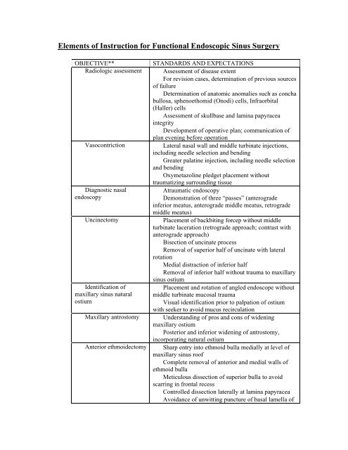
OBJECTIVE**<br />
Radiologic assessment<br />
Vasocontriction<br />
Diagnostic nasal<br />
endoscopy<br />
Uncinectomy<br />
Identification <strong>of</strong><br />
maxillary sinus natural<br />
ostium<br />
Maxillary antrostomy<br />
Anterior ethmoidectomy<br />
STANDARDS AND EXPECTATIONS<br />
Assessment <strong>of</strong> disease extent<br />
For revision cases, determination <strong>of</strong> previous sources<br />
<strong>of</strong> failure<br />
Determination <strong>of</strong> anatomic anomalies such as concha<br />
bullosa, sphenoethomid (Onodi) cells, Infraorbital<br />
(Haller) cells<br />
Assessment <strong>of</strong> skullbase and lamina papyracea<br />
integrity<br />
Development <strong>of</strong> operative plan; communication <strong>of</strong><br />
plan evening be<strong>for</strong>e operation<br />
Lateral nasal wall and middle turbinate injections,<br />
including needle selection and bending<br />
Greater palatine injection, including needle selection<br />
and bending<br />
Oxymetazoline pledget placement without<br />
traumatizing surrounding tissue<br />
Atraumatic endoscopy<br />
Demonstration <strong>of</strong> three “passes” (anterograde<br />
inferior meatus, anterograde middle meatus, retrograde<br />
middle meatus)<br />
Placement <strong>of</strong> backbiting <strong>for</strong>cep without middle<br />
turbinate laceration (retrograde approach; contrast with<br />
anterograde approach)<br />
Bisection <strong>of</strong> uncinate process<br />
Removal <strong>of</strong> superior half <strong>of</strong> uncinate with lateral<br />
rotation<br />
Medial distraction <strong>of</strong> inferior half<br />
Removal <strong>of</strong> inferior half without trauma to maxillary<br />
sinus ostium<br />
Placement and rotation <strong>of</strong> angled endoscope without<br />
middle turbinate mucosal trauma<br />
Visual identification prior to palpation <strong>of</strong> ostium<br />
with seeker to avoid mucus recirculation<br />
Understanding <strong>of</strong> pros and cons <strong>of</strong> widening<br />
maxillary ostium<br />
Posterior and inferior widening <strong>of</strong> antrostomy,<br />
incorporating natural ostium<br />
Sharp entry into ethmoid bulla medially at level <strong>of</strong><br />
maxillary sinus ro<strong>of</strong><br />
Complete removal <strong>of</strong> anterior and medial walls <strong>of</strong><br />
ethmoid bulla<br />
Meticulous dissection <strong>of</strong> superior bulla to avoid<br />
scarring in frontal recess<br />
Controlled dissection laterally at lamina papyracea<br />
Avoidance <strong>of</strong> unwitting puncture <strong>of</strong> basal lamella <strong>of</strong>
Suture medialization <strong>of</strong><br />
middle turbinate<br />
Posterior<br />
ethmoidectomy<br />
middle turbinate<br />
Understanding <strong>of</strong> alternatives to suture medialization<br />
Atraumatic introduction <strong>of</strong> needle into middle<br />
meatus<br />
Placement <strong>of</strong> circular stitch through middle<br />
turbinates and membranous columella<br />
Sharp entry through basal lamella <strong>of</strong> the middle<br />
turbinate medially and inferiorly, at the level <strong>of</strong> the<br />
maxillary sinus ro<strong>of</strong><br />
Thorough removal <strong>of</strong> basal lamella inferiorly to<br />
avoid superiorly directed dissection<br />
Complete dissection <strong>of</strong> posterior ethmoid cells,<br />
avoiding a conical dissection<br />
Identification <strong>of</strong> superior turbinate in medial portion<br />
<strong>of</strong> posterior ethmoid<br />
. Sphenoidotomy Identification <strong>of</strong> anterior and inferior edges <strong>of</strong><br />
superior turbinate<br />
Sharp removal <strong>of</strong> inferior 3-4 mm <strong>of</strong> superior<br />
turbinate<br />
Visual identification <strong>of</strong> sphenoid natural ostium, if<br />
possible<br />
Complete removal <strong>of</strong> anterior wall <strong>of</strong> sphenoid sinus<br />
superior to ostium and lateral to superior turbinate<br />
attachment<br />
. Completion <strong>of</strong> superior<br />
portion <strong>of</strong> ethmoidectomy<br />
. Frontal sinus<br />
exploration<br />
Posterior to anterior dissection <strong>of</strong> partitions along<br />
skull base<br />
Palpation behind partitions prior to removal<br />
Identification <strong>of</strong> bulla lamella<br />
Identification <strong>of</strong> agger nasi cell and its posterior wall<br />
Identification <strong>of</strong> remaining border structures <strong>of</strong><br />
frontal recess<br />
Observation <strong>of</strong> frontal sinus natural ostium<br />
Widening <strong>of</strong> frontal sinus drainage pathway as<br />
needed<br />
**For each objective, knowledge <strong>of</strong> anatomy and <strong>of</strong> equipment is expected prior to attempting<br />
objective.