Notes on the Development, Structure, and Origin of the Median and ...
Notes on the Development, Structure, and Origin of the Median and ...
Notes on the Development, Structure, and Origin of the Median and ...
Create successful ePaper yourself
Turn your PDF publications into a flip-book with our unique Google optimized e-Paper software.
NOTES ON MEDIAN AND PAIRED WNS OF FISH. 333<br />
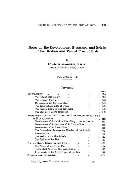
<str<strong>on</strong>g>Notes</str<strong>on</strong>g> <strong>on</strong> <strong>the</strong> <strong>Development</strong>, <strong>Structure</strong>, <strong>and</strong> <strong>Origin</strong><br />
<strong>of</strong> <strong>the</strong> <strong>Median</strong> <strong>and</strong> Paired Fins <strong>of</strong> Fish.<br />
By<br />
Edwin S. Oootii-icli, F.R.S.,<br />
Fellow <strong>of</strong> Mert<strong>on</strong> College, Oxford.<br />
With Plat.es 10—14.<br />
CONTENTS.<br />
PAGE<br />
INTRODUCTION . . . . . . 334<br />
The Lateral Fold Theory . . . .335<br />
The Gill-arch Theory . . . . .340<br />
Objecti<strong>on</strong>s to <strong>the</strong> Gill-arch Theory . . . 342<br />
The Apparent Migrati<strong>on</strong> <strong>of</strong> Fins . . . . 343<br />
The Faithfulness <strong>of</strong> Muscle <strong>and</strong> Nerve . . . 348<br />
The Shifting <strong>of</strong> Limbs Explained . . .349<br />
OBSERVATIONS ON THE STRUCTURE AND DEVELOPMENT OF THE FINS<br />
OF ELASMOBRANCHS . . . . . 352<br />
<strong>Development</strong> <strong>of</strong> <strong>the</strong> <strong>Median</strong> Fins <strong>of</strong> Scyllium canicula . 353<br />
<strong>Development</strong> <strong>of</strong> <strong>the</strong> Skelet<strong>on</strong> <strong>of</strong> <strong>the</strong> <strong>Median</strong> Fins . . 355<br />
<strong>Development</strong> <strong>of</strong> <strong>the</strong> Paired Fins . . . . 356<br />
The C<strong>on</strong>cordance between <strong>the</strong> Muscles <strong>and</strong> <strong>the</strong> Radials . 357<br />
C<strong>on</strong>centrati<strong>on</strong> . . . . . 358<br />
The Fusi<strong>on</strong> <strong>of</strong> <strong>the</strong> Muscle-buds . . . . 359<br />
The Positi<strong>on</strong> <strong>of</strong> <strong>the</strong> Fins . . . .360<br />
ON THE NERVE SUPPLY OF THE FINS . . . . 363<br />
The Plexus <strong>of</strong> <strong>the</strong> Dorsal Fins . . . .364<br />
On <strong>the</strong> Real Nature <strong>of</strong> <strong>the</strong> Nerve-plexus . . . 366<br />
Experiments <strong>on</strong> <strong>the</strong> Nerve Supply <strong>of</strong> <strong>the</strong> Fins . . 369<br />
SUMMARY AND CONCLUSION . . . . . 371<br />
VOL. 50, PART 2.—NEW SERIES. 24
334 EDWIN S. GOODRICH.<br />
INTRODUCTION.<br />
A GREAT deal has been written in <strong>the</strong> last few years about<br />
<strong>the</strong> structure, development, <strong>and</strong> origin <strong>of</strong> <strong>the</strong> paired fins <strong>of</strong><br />
fish, yet two rival <strong>and</strong> incompatible <strong>the</strong>ories are still prevalent.<br />
According to <strong>the</strong> <strong>the</strong>ory put forth by Gegenbaur (14,<br />
15,16), <strong>the</strong> paired fins have been derived from gill structures,<br />
<strong>the</strong> gill-arch having been modified into <strong>the</strong> limb-girdle, <strong>and</strong><br />
<strong>the</strong> fin itself, with its skelet<strong>on</strong>, having been derived from <strong>the</strong><br />
gill-flap or septum, <strong>and</strong> its supporting gill-rays. This may<br />
shortly be called <strong>the</strong> "gill-arch <strong>the</strong>ory." The sec<strong>on</strong>d <strong>the</strong>ory,<br />
that <strong>of</strong> Balfour (1, 2), Thacher (35), <strong>and</strong> Mivart (23), holds<br />
that <strong>the</strong> paired fins are <strong>of</strong> <strong>the</strong> same nature as <strong>the</strong> unpaired<br />
median fins. According to this view, <strong>the</strong> limbs have been<br />
derived from paired l<strong>on</strong>gitudinal fin-folds, in which skeletal<br />
supports, <strong>the</strong> radials, or somactidia (Lankester), became<br />
developed as in <strong>the</strong> median fins, <strong>and</strong> subsequently gave rise<br />
to <strong>the</strong> limb-girdles. This may be called <strong>the</strong> "lateral fin-fold<br />
<strong>the</strong>ory." Each <strong>of</strong> <strong>the</strong>se <strong>the</strong>ories may claim to have am<strong>on</strong>g<br />
its numerous supporters <strong>the</strong> names <strong>of</strong> some <strong>of</strong> <strong>the</strong> most<br />
eminent exp<strong>on</strong>ents <strong>of</strong> <strong>the</strong> morphology <strong>of</strong> <strong>the</strong> vertebrates.<br />
Dohrn (10), Hasvvell (20), Eabl (31), Mollier (24, 25,, 26),<br />
Harris<strong>on</strong> (19), Wiedersheim (36), A. Smith Woodward (37),<br />
<strong>and</strong> Dean (9) have written in favour <strong>of</strong> <strong>the</strong> lateral fold <strong>the</strong>ory;<br />
David<strong>of</strong>f (8), Fiirbringer (12), Braus (3-7), <strong>and</strong> o<strong>the</strong>rs have<br />
supported its rival. It is unnecessary for me in <strong>the</strong>se notes<br />
to give a history <strong>of</strong> <strong>the</strong> discussi<strong>on</strong>s to which <strong>the</strong> questi<strong>on</strong> has<br />
given rise; <strong>the</strong> literature has been recently reviewed by<br />
Mollier <strong>and</strong> Braus, <strong>and</strong> <strong>the</strong> whole subject is familiar to<br />
zoologists. But <strong>the</strong>re are certain essential points which seem<br />
to be in danger <strong>of</strong> being obscured from view in a cloud <strong>of</strong><br />
c<strong>on</strong>troversy, <strong>and</strong> it is in <strong>the</strong> hope <strong>of</strong> clearing up some <strong>of</strong> <strong>the</strong>se<br />
points <strong>and</strong> <strong>of</strong> filling up certain gaps in <strong>the</strong> evidence that<br />
<strong>the</strong>se notes have been published.<br />
As I am anxious to keep this paper within reas<strong>on</strong>able limits<br />
<strong>and</strong> not to overburden <strong>the</strong> already very bulky literature <strong>on</strong><br />
<strong>the</strong> subject <strong>of</strong> <strong>the</strong> origin <strong>of</strong> <strong>the</strong> paired limbs, <strong>on</strong>ly some aspects
NOTES ON MEDIAN AND I'ATBKD FINS OP FISH. 335<br />
<strong>of</strong> <strong>the</strong> problem will be dealt with in detail. A. brief <strong>and</strong> somewhat<br />
dogmatic statement <strong>of</strong> <strong>the</strong> case is made at <strong>the</strong> beginning,<br />
followed by a descripti<strong>on</strong> <strong>of</strong> my own observati<strong>on</strong>s, <strong>and</strong> ending<br />
with a short summary.<br />
THE LATERAL FOLD THEOUY.<br />
Balfour's c<strong>on</strong>cepti<strong>on</strong> <strong>of</strong> an originally c<strong>on</strong>tinuous fin-fold,<br />
reaching from <strong>the</strong> pectoral to <strong>the</strong> pelvic regi<strong>on</strong> (1) is discredited<br />
because it has been found <strong>on</strong>ly (as an epidermal fold)<br />
in those forms, like Torpedo, in which <strong>the</strong> pectoral fins reach<br />
<strong>the</strong> pelvic fins in <strong>the</strong> adult, a c<strong>on</strong>diti<strong>on</strong> which is probably<br />
rightly c<strong>on</strong>sidered as sec<strong>on</strong>dai'y. Moreover, <strong>the</strong> appearance<br />
<strong>of</strong> an epidermal l<strong>on</strong>gitudinal fold, as a first indicati<strong>on</strong> <strong>of</strong> <strong>the</strong><br />
development <strong>of</strong> <strong>the</strong> paired fins, is c<strong>on</strong>sidered to be <strong>of</strong> little<br />
importance, <strong>and</strong> its presence between <strong>the</strong> paired fins is denied<br />
in sharks (Mollier 24, Braus 4, etc.).<br />
Now, <strong>the</strong> c<strong>on</strong>tinuity <strong>of</strong> <strong>the</strong> pectoral with <strong>the</strong> pelvic finfold<br />
is not an essential point. The important thing is to<br />
recognise that <strong>the</strong> paired fins always arise as a l<strong>on</strong>gitudinal<br />
fold or ridge, similar to that which gives rise to<br />
<strong>the</strong> median fins. That this is really <strong>the</strong> case is now admitted<br />
by all (Braus 7). Even in Ceratodus, where <strong>the</strong> paired fins in<br />
<strong>the</strong> adult are set at a pr<strong>on</strong>ounced angle to <strong>the</strong> l<strong>on</strong>g axis <strong>of</strong><br />
<strong>the</strong> body, <strong>the</strong>y make <strong>the</strong>ir first appearance as l<strong>on</strong>gitudinal<br />
ridges (Sem<strong>on</strong> 33).<br />
• Possibly from <strong>the</strong> very first, in phylogeny, <strong>the</strong> paired fins<br />
were disc<strong>on</strong>tinuous, <strong>and</strong> differentiated into pectoral <strong>and</strong><br />
pelvic. For c<strong>on</strong>clusive evidence <strong>on</strong> this point we must look<br />
to palae<strong>on</strong>tology; <strong>and</strong> it has not yet been obtained. But<br />
<strong>the</strong>re is some evidence to be ga<strong>the</strong>red from comparative<br />
anatomy <strong>and</strong> embryology in favour <strong>of</strong> Balfour's view, as has<br />
frequently been pointed out (Dohrn 10, Mollier 24).<br />
For instance, <strong>the</strong> musculature <strong>of</strong> <strong>the</strong> fins is developed in<br />
Elasmobranchs, from buds given <strong>of</strong>f from <strong>the</strong> ventral ends <strong>of</strong><br />
<strong>the</strong> myotomes, <strong>and</strong> <strong>the</strong>se buds have been shown to be produced<br />
not <strong>on</strong>ly <strong>on</strong> <strong>the</strong> myotomes in <strong>the</strong> regi<strong>on</strong> <strong>of</strong> <strong>the</strong> fins, but
336 EDWIN S. GOODRICH.<br />
<strong>on</strong> all <strong>the</strong> trunk myotomes situated between <strong>the</strong> pectoral<br />
<strong>and</strong> <strong>the</strong> pelvic fins in such sharks as Pristiurus <strong>and</strong> Scyllium,<br />
in which <strong>the</strong>se fins are widely separated in <strong>the</strong> adult (Dohrn<br />
10, Rabl 31, Braus 4, <strong>and</strong> p. 343 below). Many <strong>of</strong> <strong>the</strong> intermediate<br />
buds seem to disappear entirely during development.<br />
In those segments which are near <strong>the</strong> fins <strong>the</strong> buds become<br />
better developed <strong>and</strong> more persistent, <strong>and</strong> a large number<br />
pass into <strong>the</strong> fin-fold. Muscle buds are also found in fr<strong>on</strong>t <strong>of</strong><br />
<strong>the</strong> pectoral fin <strong>and</strong> behind <strong>the</strong> pelvic fin, dwindling in size as<br />
<strong>the</strong>y are far<strong>the</strong>r removed from <strong>the</strong> fin-base. Thus, in <strong>the</strong>se<br />
sharks, <strong>the</strong> muscles <strong>of</strong> <strong>the</strong> paired fins are formed by <strong>the</strong> great<br />
development in two regi<strong>on</strong>s <strong>of</strong> a c<strong>on</strong>tinuous series <strong>of</strong> muscle<br />
buds, vanishing posteriorly behind <strong>the</strong> cloaca. The manner<br />
in which <strong>the</strong>se vestigial buds disappear by reducti<strong>on</strong> at<br />
ei<strong>the</strong>r end <strong>of</strong> <strong>the</strong> fin rudiment, <strong>and</strong> in which <strong>the</strong> persistent<br />
buds become c<strong>on</strong>centrated at <strong>the</strong> relatively narrowing<br />
base <strong>of</strong> <strong>the</strong> fin, has been admirably described <strong>and</strong> figured by<br />
Mollier (24) <strong>and</strong> Braus.<br />
The fin-base <strong>of</strong> <strong>the</strong> adult occupies much less space relatively<br />
than <strong>the</strong> fin-fold <strong>of</strong> <strong>the</strong> embryo.<br />
Now, <strong>the</strong> radial fin-muscles being developed from buds <strong>of</strong><br />
<strong>the</strong> myotomes, naturally receive <strong>the</strong>ir motor nerve-supply<br />
from <strong>the</strong> ventral roots <strong>of</strong> <strong>the</strong> spinal nerves, <strong>and</strong> <strong>the</strong>se corresp<strong>on</strong>d<br />
in number to <strong>the</strong> myotomes which share in <strong>the</strong> formati<strong>on</strong><br />
<strong>of</strong> <strong>the</strong> musculature. Owing to c<strong>on</strong>centrati<strong>on</strong>, <strong>the</strong> nerves<br />
are found to c<strong>on</strong>verge toward <strong>the</strong> base <strong>of</strong> <strong>the</strong> fin. In fr<strong>on</strong>t<br />
<strong>and</strong> behind <strong>the</strong> nerves may be drawn toge<strong>the</strong>r so as to form<br />
a "collector" nerve or compound stem.<br />
It is part <strong>of</strong> <strong>the</strong> lateral fold <strong>the</strong>ory to suppose that <strong>the</strong><br />
endoskelet<strong>on</strong> <strong>of</strong> <strong>the</strong> paired fins has been derived from a series<br />
<strong>of</strong> cartilaginous rods, radials, or somactidia, similar to those<br />
<strong>of</strong> <strong>the</strong> median fins (Thacher 35, Mivart 23). The various<br />
types <strong>of</strong> fin-skelet<strong>on</strong>, with <strong>the</strong>ir cartilage rays <strong>and</strong> basal pieces,<br />
would have been developed from such originally segmental<br />
radials by c<strong>on</strong>centrati<strong>on</strong> <strong>and</strong> fusi<strong>on</strong>. To this c<strong>on</strong>tenti<strong>on</strong> it is<br />
objected that in development <strong>the</strong> radials <strong>of</strong> Elasniobranchs<br />
arise in a c<strong>on</strong>tinu<strong>on</strong>s rudiment—a plate <strong>of</strong> procartilaginous
NOTES ON MEDIAN AND PAlllliD FINS Oi' PISH. 337<br />
mesenchyme (Balf<strong>on</strong>r 2, Mollier 24, Ruge 32 ; <strong>and</strong> p. 357 below).<br />
It may be answered (Dohrn 10, Mollier 24) that, <strong>the</strong> radials<br />
being closely approximated, <strong>the</strong>ir procartilaginous rudiments<br />
with indefinite borders necessarily merge toge<strong>the</strong>r to a c<strong>on</strong>siderable<br />
extent. As a matter <strong>of</strong> fact, <strong>the</strong> cartilage pieces<br />
appear as isl<strong>and</strong>s in <strong>the</strong> vaguely-defined rudiment, which<br />
corresp<strong>on</strong>d closely in positi<strong>on</strong> <strong>and</strong> number with <strong>the</strong> separate<br />
elements <strong>of</strong> <strong>the</strong> adult fin-skelet<strong>on</strong>. Some slight indicati<strong>on</strong>s<br />
<strong>of</strong> recapitulati<strong>on</strong>, some fusi<strong>on</strong> <strong>of</strong> neighbouring radials, may be<br />
detected, which bears out <strong>the</strong> views so c<strong>on</strong>vincingly advocated<br />
by Thacher <strong>and</strong> Mivart. But it cannot be claimed that recapitulati<strong>on</strong><br />
is complete in this respect in <strong>the</strong> development <strong>of</strong><br />
<strong>the</strong> paired fins. It is obvious, however, that if its absence is<br />
c<strong>on</strong>sidered as evidence against <strong>the</strong> lateral fold <strong>the</strong>ory it tells<br />
with equal force against <strong>the</strong> gill-arch <strong>the</strong>ory, since<br />
<strong>the</strong> skelet<strong>on</strong> is, according to this view, also derived from<br />
originally separate (branchial) rays.<br />
But <strong>the</strong> whole argument against <strong>the</strong> lateral fold <strong>the</strong>ory<br />
collapses when we find that, as Balfour l<strong>on</strong>g ago showed,<br />
<strong>the</strong> radials <strong>of</strong> <strong>the</strong> median fins likewise arise in a<br />
c<strong>on</strong>tinuous proch<strong>on</strong>dral plate, in <strong>the</strong> median fins <strong>of</strong><br />
Elasmobranchs, even "when <strong>the</strong>y are separate in <strong>the</strong> adult<br />
(p. 355 below). These median fins are much c<strong>on</strong>centrated,<br />
<strong>and</strong> nothing proves so clearly that <strong>the</strong> early c<strong>on</strong>tinuity <strong>of</strong> <strong>the</strong><br />
rudiments is due to <strong>the</strong>ir approximati<strong>on</strong>, for here <strong>the</strong> original<br />
metameric nature <strong>of</strong> <strong>the</strong> radials will not be denied. The<br />
most enthusiastic supporter <strong>of</strong> <strong>the</strong> gill-arch <strong>the</strong>ory would<br />
not suppose that <strong>the</strong> c<strong>on</strong>tinuous plate represents an early<br />
stage in <strong>the</strong> phylogenetic history <strong>of</strong> <strong>the</strong> skelet<strong>on</strong> <strong>of</strong> median<br />
fins! Unfortunately, we know but little c<strong>on</strong>cerning <strong>the</strong><br />
development <strong>of</strong> <strong>the</strong> skelet<strong>on</strong> in unc<strong>on</strong>centrated median fins.<br />
Doubtless, in such cases <strong>the</strong> radials arise separately; Harris<strong>on</strong>,<br />
indeed, has shown this in his valuable paper <strong>on</strong> <strong>the</strong> salm<strong>on</strong> (19).<br />
Yet o<strong>the</strong>r objecti<strong>on</strong>s have been brought forward by Braus,<br />
in <strong>the</strong> elaborate <strong>and</strong> beautiful memoirs which have <strong>of</strong> late<br />
c<strong>on</strong>tributed so much to our knowledge <strong>of</strong> <strong>the</strong> structure <strong>and</strong><br />
development <strong>of</strong> fins (3, 4, 6, 7). It has been shown that two
338 EDWIN S. GOODRICH.<br />
muscle-buds are given <strong>of</strong>f by each myotome to <strong>the</strong> paired fins<br />
in Blasmobranchs; that <strong>the</strong>se pass outwards into <strong>the</strong> fin-fold,<br />
dividing into upper <strong>and</strong> lower halves, which give rise to <strong>the</strong><br />
dorsal <strong>and</strong> ventral radial muscles. Between each pair <strong>of</strong><br />
corresp<strong>on</strong>ding upper <strong>and</strong> lower buds develops a cartilaginous<br />
radial. Thus, as Rabl showed, since two radial muscles <strong>and</strong><br />
cartilages corresp<strong>on</strong>d to each segment, <strong>the</strong> relati<strong>on</strong> between<br />
tlie number <strong>of</strong> radials in <strong>the</strong> fin-skelet<strong>on</strong>, <strong>and</strong> <strong>the</strong> number <strong>of</strong><br />
trunk vertebras bel<strong>on</strong>ging to those segments which c<strong>on</strong>tributed<br />
to <strong>the</strong> formati<strong>on</strong> <strong>of</strong> <strong>the</strong> fin, may be expressed in <strong>the</strong> formula<br />
—-^—? = vertebrae. Braus has endeavoured to prove that<br />
this formula does not hold good (p. 444, 3). But it is quite<br />
obvious that, although in tlie main correct, it can <strong>on</strong>ly be<br />
intended to give approximate results when applied to whole<br />
fins. In most paired fins <strong>of</strong> Elasmobranchs <strong>the</strong> anterior <strong>and</strong><br />
posterior regi<strong>on</strong>s are much modified by excessive c<strong>on</strong>centrati<strong>on</strong><br />
<strong>and</strong> reducti<strong>on</strong>, <strong>and</strong> here <strong>the</strong> corresp<strong>on</strong>dence between muscles<br />
<strong>and</strong> radials becomes much disturbed. The formula applies<br />
perfectly over <strong>the</strong> greater part <strong>of</strong> a fin which is normally<br />
developed, as is seen in Braus's own figures (4, 6). More important<br />
is <strong>the</strong> c<strong>on</strong>tenti<strong>on</strong> that <strong>the</strong> adult radial fin-muscles<br />
do not corresp<strong>on</strong>d to <strong>the</strong> muscle-buds in <strong>the</strong> embryo. It is<br />
urged that <strong>the</strong> muscle-buds become mixed <strong>and</strong> that <strong>the</strong> adult<br />
muscles are no l<strong>on</strong>ger unisegmental <strong>and</strong> hapl<strong>on</strong>eurous, but<br />
are compound <strong>and</strong> polyneurous, <strong>and</strong>, in fact, bear no definite<br />
relati<strong>on</strong> to <strong>the</strong> segments from which <strong>the</strong>y arose.<br />
It is true that, as Mollier has shown (24), <strong>the</strong> muscle-buds<br />
in Elasmobranch fins may be c<strong>on</strong>nected toge<strong>the</strong>r at <strong>the</strong>ir base,<br />
at all events temporarily, by str<strong>and</strong>s <strong>of</strong> tissue. It is also true<br />
that <strong>the</strong> mixed motor <strong>and</strong> sensory nerves form a complicated<br />
plexus at <strong>the</strong> base <strong>of</strong>, <strong>and</strong> round about, <strong>the</strong> radial muscles.<br />
But it does not follow that <strong>the</strong>se muscles are ei<strong>the</strong>r compound<br />
or polyneurous. So far as I am aware, it has never been proved<br />
that muscle-forming substance actually passes from <strong>on</strong>e bud<br />
to ano<strong>the</strong>r (p. 359); nor has it ever been proved that <strong>on</strong>e<br />
radial muscle is really innervated by more than <strong>on</strong>e motor
NOTES ON MEDIAN AND PAIRED FINS OF FLSH. 339<br />
root. In fact, it seems to be very probable indeed that, even<br />
in <strong>the</strong> adult, <strong>the</strong> radial muscles are strictly segmental <strong>and</strong><br />
haplorieurous (see below, pp. 364-371). Some fusi<strong>on</strong>s may<br />
take place, some disturbances <strong>of</strong> <strong>the</strong> nietameric order may<br />
occur, especially at <strong>the</strong> extreme anterior <strong>and</strong> posterior ends <strong>of</strong><br />
<strong>the</strong> fins; but it is quite firmlyestablished that each adult radial<br />
muscle develops from, <strong>and</strong> corresp<strong>on</strong>ds in positi<strong>on</strong> to, a single<br />
muscle-bud. It may be asserted with c<strong>on</strong>fidence that a radial<br />
muscle is derived, at least mainly, from that bud<br />
whose positi<strong>on</strong> it later occupies; <strong>and</strong> that <strong>the</strong> radial<br />
muscles in <strong>the</strong> normally developed regi<strong>on</strong> <strong>of</strong> <strong>the</strong><br />
paired fin <strong>of</strong> an Elasmobranch corresp<strong>on</strong>ds accurately<br />
in number <strong>and</strong> positi<strong>on</strong> to <strong>the</strong> group <strong>of</strong> primitive<br />
buds from which <strong>the</strong>y have been formed. 1<br />
There is a last objecti<strong>on</strong> which Braus persistently reiterates<br />
in his papers, <strong>and</strong> <strong>of</strong> which he makes a great deal. He alleges<br />
that <strong>the</strong> " c<strong>on</strong>cordance " which exists in <strong>the</strong> adult between <strong>the</strong><br />
radial muscles <strong>and</strong> <strong>the</strong> radial cartilages is not primitive, but<br />
sec<strong>on</strong>daiy. He states that in <strong>the</strong> early stages <strong>of</strong> development<br />
<strong>the</strong>re are " discrepancies" between <strong>the</strong>se elements, that <strong>the</strong><br />
muscle-buds do not corresp<strong>on</strong>d exactly with <strong>the</strong> rudiments <strong>of</strong><br />
<strong>the</strong> radials, <strong>and</strong> that <strong>the</strong> perfect corresp<strong>on</strong>dence, or c<strong>on</strong>cordance,<br />
is gradually established in later stages. This subject<br />
will be dealt with later <strong>on</strong> in greater detail (p. 357); but it<br />
may here be said that <strong>the</strong> evidence <strong>on</strong> which Braus bases his<br />
argument seems to be <strong>of</strong> <strong>the</strong> slenderest <strong>and</strong> most unc<strong>on</strong>vincing<br />
nature. Not even in <strong>the</strong> adult is <strong>the</strong> c<strong>on</strong>cordance perfect;<br />
marked disturbances occur at both <strong>the</strong> anterior <strong>and</strong> <strong>the</strong> posterior<br />
extremities <strong>of</strong> <strong>the</strong> fins. The peripheral ends <strong>of</strong> <strong>the</strong> adult<br />
muscles corresp<strong>on</strong>d exactly with <strong>the</strong> radials in <strong>the</strong> middle<br />
1 If it is objected that in Ceratodus, where <strong>the</strong> adult paired fin lias about<br />
thirty radials <strong>and</strong> radial muscles, <strong>on</strong>ly about three segments have been shown<br />
to c<strong>on</strong>tribute muscle-buds in <strong>the</strong> embryo (Sem<strong>on</strong> 33), it must be answered<br />
that, this result is not trustworthy. David<strong>of</strong>f (8) <strong>and</strong> Braus (3) have found<br />
twelve spinal nerves c<strong>on</strong>tributing to <strong>the</strong> limb-plexus. It is probable that<br />
llabl's formula holds good in Ceratodus (Mollier 24), <strong>and</strong> that a large <strong>and</strong><br />
sufficient number <strong>of</strong> segments really c<strong>on</strong>tribute muscle-forming cells to <strong>the</strong><br />
limb, but not in <strong>the</strong> form <strong>of</strong> distinct buds.
340 EDWIN S. GOODRICH.<br />
regi<strong>on</strong> ; but as <strong>the</strong>y pass inwards to become attached to <strong>the</strong><br />
base, or <strong>the</strong> girdle, <strong>the</strong> muscles no l<strong>on</strong>ger preserve <strong>the</strong> " c<strong>on</strong>cordance."<br />
On <strong>the</strong> o<strong>the</strong>r h<strong>and</strong>, nothing is so striking <strong>on</strong><br />
examining secti<strong>on</strong>s through <strong>the</strong> developing fins <strong>of</strong> Elasmobranchs,<br />
whe<strong>the</strong>r paired or unpaired, as <strong>the</strong> extraordinarily<br />
regular "c<strong>on</strong>cordance"; it is obvious <strong>on</strong> <strong>the</strong> very first appearance<br />
<strong>of</strong> <strong>the</strong> procartilaginous radial (p. 358, figs. 5, 8, 9, 18).<br />
These attempts to undermine <strong>the</strong> lateral fold <strong>the</strong>ory, by<br />
showing that <strong>the</strong> adult muscles are compound <strong>and</strong> polyneurous,<br />
<strong>and</strong> that <strong>the</strong> c<strong>on</strong>cordance is sec<strong>on</strong>dary, are not borne out by<br />
<strong>the</strong> evidence. Moreover, even if it could be proved that <strong>the</strong><br />
metamerism <strong>of</strong> <strong>the</strong> fin elements has been lost, <strong>the</strong> lateral fold<br />
<strong>the</strong>ory would scarcely be affected, since it <strong>on</strong>ly claims that<br />
<strong>the</strong> muscles <strong>and</strong> skeletal radials formed a l<strong>on</strong>gitudinal series<br />
<strong>of</strong> metameric origin in <strong>the</strong> beginning. No <strong>on</strong>e doubts that<br />
<strong>the</strong> metamerism has been obscured, or lost, in <strong>the</strong> higher<br />
vertebrates; it matters little, <strong>the</strong>oretically, whe<strong>the</strong>r it still<br />
persists in modern fisb.<br />
THE GILL-ARCH THEORY.<br />
Let us now pass to <strong>the</strong> rival <strong>the</strong>ory. It is claimed that <strong>the</strong><br />
initial stages in <strong>the</strong> phylogenetic history <strong>of</strong> <strong>the</strong> paired fins<br />
are more easily accounted for <strong>on</strong> <strong>the</strong> gill-arch <strong>the</strong>ory <strong>of</strong><br />
<strong>the</strong>ir origin. Now, according to <strong>the</strong> lateral fold <strong>the</strong>ory<br />
<strong>the</strong> paired fins appeared, as <strong>the</strong>y do in <strong>on</strong>togeny, as l<strong>on</strong>gitudinal<br />
ridges, which, from <strong>the</strong>ir very first appearance, may<br />
have been useful as balancing <strong>and</strong> directing organs. Even<br />
in modern fish <strong>the</strong> paired fins are used not so much for progressi<strong>on</strong><br />
as for guidance <strong>and</strong> balancing.<br />
On Gegenbaur's <strong>the</strong>ory <strong>the</strong> directi<strong>on</strong> <strong>of</strong> <strong>the</strong> paired fins must<br />
at first have been dorso-ventral across <strong>the</strong> l<strong>on</strong>g axis <strong>of</strong> <strong>the</strong><br />
body ; such folds would probably be a hindrance to progressi<strong>on</strong>,<br />
<strong>and</strong> both <strong>the</strong> pectoral <strong>and</strong> pelvic fins would have been<br />
placed close toge<strong>the</strong>r behind <strong>the</strong> head in a most unfavourable<br />
situati<strong>on</strong>.<br />
The positi<strong>on</strong> <strong>of</strong> <strong>the</strong> pelvic fin is accounted for by supposing
NOTES ON MEDIAN AND PAIRED FINS .OF FISH. 341<br />
that it has migrated backwards from <strong>the</strong> head regi<strong>on</strong>. Now,<br />
<strong>the</strong>re is no evidence <strong>of</strong> a more anterior positi<strong>on</strong> <strong>of</strong> <strong>the</strong> pelvics<br />
in primitive fishes generally, ei<strong>the</strong>r living or extinct. Indeed,<br />
<strong>the</strong> <strong>on</strong>ly known fish in which <strong>the</strong> pelvics are far forward<br />
(some Teleostei) are acknowledged to be specialised in this<br />
respect. The presence in <strong>on</strong>togeny <strong>of</strong> rudimentary musclebuds<br />
in fr<strong>on</strong>t <strong>of</strong> <strong>the</strong> pelvic tins, is supposed to indicate<br />
backward migrati<strong>on</strong>. This is negatived by <strong>the</strong> fact that<br />
similar rudimentary buds are found behind <strong>the</strong> pelvic fin<br />
(Braus 4, PI. 22, <strong>and</strong> in this paper, figs. 1, 4, 25). The fins<br />
could not have migrated both ways at <strong>on</strong>ce, <strong>and</strong> <strong>the</strong>re is<br />
no reas<strong>on</strong> to believe that <strong>the</strong>y first migrated backwards to a<br />
point behind <strong>the</strong> cloaca, <strong>and</strong> <strong>the</strong>n forwards towards <strong>the</strong> head.<br />
David<strong>of</strong>f (8), Gegenbaur (15), <strong>and</strong> o<strong>the</strong>rs have held that<br />
<strong>the</strong> presence in fr<strong>on</strong>t <strong>of</strong> <strong>the</strong> pelvic fin <strong>of</strong> a collector nerve,<br />
composed <strong>of</strong> branches <strong>of</strong> a number <strong>of</strong> spinal nerves, <strong>and</strong> <strong>the</strong><br />
greater extent <strong>of</strong> this plexus iu <strong>the</strong> young than in <strong>the</strong> adult<br />
(Punnett 29, 30), indicates backward migrati<strong>on</strong>. But, again,<br />
both a similar plexus <strong>and</strong> extensi<strong>on</strong> are found <strong>on</strong> <strong>the</strong> posterior<br />
side <strong>of</strong> <strong>the</strong> fin.<br />
The questi<strong>on</strong> <strong>of</strong> <strong>the</strong> nerve supply <strong>of</strong> <strong>the</strong> fins will be discussed<br />
in greater detail later (p. 363); but in describing <strong>the</strong><br />
general nerve-plexus at <strong>the</strong> base <strong>of</strong> <strong>the</strong> fins <strong>on</strong>e must be<br />
caref-ul to distinguish between <strong>the</strong> collector nerve formed<br />
by <strong>the</strong> c<strong>on</strong>vergence <strong>and</strong> combinati<strong>on</strong> <strong>of</strong> branches <strong>of</strong> a series<br />
<strong>of</strong> spinal uerves <strong>and</strong> <strong>the</strong> plexus proper, due to intertwining<br />
sec<strong>on</strong>dary branches, made up chiefly, if not entirely, <strong>of</strong><br />
sensory nerve-fibres. The formati<strong>on</strong> <strong>of</strong> a collector nerve<br />
is simply <strong>and</strong> easily explained as <strong>the</strong> result <strong>of</strong> c<strong>on</strong>centrati<strong>on</strong>.<br />
The mere presence <strong>of</strong> a c<strong>on</strong>necting plexus (mainly<br />
l<strong>on</strong>gitudinal) is due nei<strong>the</strong>r to c<strong>on</strong>centrati<strong>on</strong> nor to migrati<strong>on</strong><br />
(p. 367).<br />
Moreover, both <strong>the</strong>se arguments in support <strong>of</strong> <strong>the</strong> <strong>the</strong>ory<br />
<strong>of</strong> migrati<strong>on</strong> are sufficiently answered by <strong>the</strong> fact that<br />
rudimentary buds are found both iu fr<strong>on</strong>t <strong>of</strong> <strong>and</strong> behind<br />
<strong>the</strong> median fins (Mayer 22 <strong>and</strong> p. 353 below), <strong>and</strong> that a l<strong>on</strong>gitudinal<br />
nerve-plexus may extend al<strong>on</strong>g <strong>the</strong>ir base even when
342 EDWIN S. GOODKICH.<br />
<strong>the</strong> fin is c<strong>on</strong>tinuous <strong>and</strong> <strong>the</strong>re is no possibility <strong>of</strong> migrati<strong>on</strong>.<br />
L<strong>on</strong>gitudinal c<strong>on</strong>necting nerves have l<strong>on</strong>g been known to<br />
exist at <strong>the</strong> base <strong>of</strong> <strong>the</strong> unc<strong>on</strong>centrated fins <strong>of</strong> Teleostean<br />
fish; I find <strong>the</strong>m also at <strong>the</strong> base <strong>of</strong> <strong>the</strong> dorsal fin <strong>of</strong><br />
Chiinaera, which is scarcely, if at all, c<strong>on</strong>centrated.<br />
OBJECTIONS TO THE G-ILL-AKCH THEOEY.<br />
We may now deal with some very serious difficulties in <strong>the</strong><br />
way <strong>of</strong> <strong>the</strong> gill-arch <strong>the</strong>ory. Firstly, it <strong>of</strong>fers no intelligible<br />
explanati<strong>on</strong> <strong>of</strong> <strong>the</strong> participati<strong>on</strong> <strong>of</strong> a large number <strong>of</strong> segments<br />
in <strong>the</strong> formati<strong>on</strong> <strong>of</strong> <strong>the</strong> paired fins. Yet it is always<br />
<strong>the</strong> case that a c<strong>on</strong>siderable, <strong>and</strong> sometimes a very large,<br />
number <strong>of</strong> spinal nerves <strong>and</strong> myotomes c<strong>on</strong>tribute towards<br />
its development.<br />
Sec<strong>on</strong>dly, if <strong>the</strong> skelet<strong>on</strong> <strong>of</strong> <strong>the</strong> paired fins were derived<br />
from gill-rays we should expect <strong>the</strong> muscle supply to be<br />
drawn, not from <strong>the</strong> myotomes at all, but from <strong>the</strong> unsegmented<br />
"lateral-plate," or visceral, musculature, which is<br />
innervated by <strong>the</strong> dorsal roots <strong>of</strong> <strong>the</strong> spinal nerves. It is true<br />
that <strong>the</strong> trapezius muscle attached to <strong>the</strong> scapula is <strong>of</strong><br />
lateral-plate origin, <strong>and</strong> is supplied from <strong>the</strong> vagus nerve;<br />
yet it does not enter into <strong>the</strong> fin, does not, in fact, bel<strong>on</strong>g to<br />
<strong>the</strong> fin musculature. At all events, in <strong>the</strong> pelvic regi<strong>on</strong><br />
<strong>the</strong>re is no trace whatever <strong>of</strong> o<strong>the</strong>r than segmented muscles.<br />
A third, <strong>and</strong> perhaps still more important, objecti<strong>on</strong> to<br />
Gegenbaur's <strong>the</strong>ory is this: <strong>the</strong> positi<strong>on</strong> <strong>of</strong> <strong>the</strong> limb-girdles<br />
in relati<strong>on</strong> to <strong>the</strong> nerves, blood-vessels, coelom, etc., is<br />
exactly <strong>the</strong> reverse <strong>of</strong> what it should be if <strong>the</strong>y<br />
were derived from visceral arches. The coelom, <strong>the</strong> subintestinal<br />
vessel (heart, etc.), <strong>the</strong> myotomes <strong>and</strong> <strong>the</strong>ir nerves,<br />
all pass outside <strong>the</strong> visceral arches. The limb-girdles, <strong>on</strong> <strong>the</strong><br />
c<strong>on</strong>trary, lie morphologically outside <strong>the</strong>se structures, so that<br />
<strong>the</strong> nerves frequently pass through <strong>the</strong> girdles to reach <strong>the</strong><br />
fins. In fact, <strong>the</strong> girdles lie in <strong>the</strong> outer body-wall, while <strong>the</strong><br />
visceral arches lie in <strong>the</strong> wall <strong>of</strong> <strong>the</strong> alimentary canal. No<br />
mere superficial resemblance in shape <strong>of</strong> <strong>the</strong> girdle to <strong>the</strong>
NOTES ON MEDIAN AND PA1K15D FINS OF FISH. 343<br />
arch in a developing Elasmobranch, such as is insisted up<strong>on</strong><br />
by Braus (5), no mere opini<strong>on</strong>, unsupported by evidence, that<br />
<strong>the</strong> relative positi<strong>on</strong> <strong>of</strong> <strong>the</strong> girdle has been altered, such as is<br />
expressed by Eiirbringer (12), can outweigh <strong>the</strong>se facts.<br />
The fourth <strong>and</strong> last objecti<strong>on</strong> which we shall urge against<br />
<strong>the</strong> gill-arch <strong>the</strong>ory is <strong>on</strong>e which will probably seem to most<br />
zoologists to be <strong>the</strong> most fatal <strong>of</strong> all: <strong>the</strong> <strong>the</strong>ory gives no<br />
explanati<strong>on</strong> <strong>of</strong> <strong>the</strong> remarkable resemblance borne<br />
by <strong>the</strong> paired fins to <strong>the</strong> unpaired fins. The resemblance<br />
is not vague <strong>and</strong> indefinite, it is minute; it can be<br />
followed out in every detail both <strong>of</strong> <strong>the</strong>ir structure <strong>and</strong> <strong>of</strong> <strong>the</strong>ir<br />
development. In no respect is this more striking than in <strong>the</strong><br />
development <strong>and</strong> differentiati<strong>on</strong> <strong>of</strong> <strong>the</strong> dermal fin-rays in <strong>the</strong><br />
various groups <strong>of</strong> fishes.<br />
All <strong>the</strong>se facts, which clearly support <strong>the</strong> lateral fold<br />
<strong>the</strong>ory, are so many deadly blows aimed at <strong>the</strong> rival gillarch<br />
<strong>the</strong>ory. Far from being difficulties which have to be<br />
explained away, <strong>the</strong>y become evidence actually in favour <strong>of</strong><br />
<strong>the</strong> fundamental likeness <strong>of</strong> <strong>the</strong> paired <strong>and</strong> unpaired fins.<br />
THE APPARENT MIGRATION OF FINS.<br />
We have now to account for <strong>the</strong> apparent migrati<strong>on</strong> <strong>of</strong><br />
limbs from <strong>on</strong>e place to ano<strong>the</strong>r <strong>on</strong> <strong>the</strong> body <strong>of</strong> vertebrates.<br />
Every trunk segment may be said to be capable <strong>of</strong><br />
producing muscular, nervous, <strong>and</strong> skeletal "limb<br />
elements" <strong>of</strong> a paired character. This "potentiality" is<br />
actually called into force in <strong>the</strong> case <strong>of</strong> <strong>the</strong> Rajidte throughout<br />
<strong>the</strong> trunk regi<strong>on</strong>, with <strong>the</strong> excepti<strong>on</strong> <strong>of</strong> a few anterior segments<br />
(see Rabl 31, Mollier 24, <strong>and</strong> especially Braus 3). In<br />
Torpedo, for instance, <strong>the</strong> 4th to <strong>the</strong> 30th spinal nerves<br />
supply <strong>the</strong> pectoral fin, <strong>and</strong> <strong>the</strong> 31st to <strong>the</strong> 42nd <strong>the</strong> pelvic<br />
fin. In Tryg<strong>on</strong> <strong>the</strong> 3rd to <strong>the</strong> 59th supply <strong>the</strong> pectoral, <strong>and</strong><br />
<strong>the</strong> 60th to <strong>the</strong> 71st <strong>the</strong> pelvic fin (Braus). The same c<strong>on</strong>clusi<strong>on</strong><br />
is indicated in <strong>the</strong> case <strong>of</strong> forms like Pristiurus <strong>and</strong><br />
Scylliuni, where <strong>the</strong> paired fins are widely separated, by <strong>the</strong><br />
development <strong>of</strong> muscle-buds <strong>on</strong> all <strong>the</strong> trunk segments (see
344 EDWIN S. GOODRICH.<br />
figs. 1, 25). It is also borne out by a comparis<strong>on</strong> <strong>of</strong> <strong>the</strong> range<br />
<strong>of</strong> extensi<strong>on</strong> <strong>of</strong> <strong>the</strong> fins in various genera; for instance, whilst<br />
<strong>the</strong> paired fins occupy segments 5-23 <strong>and</strong> 47-65 in Zygcena,<br />
<strong>the</strong>y occupy segments 2-19 <strong>and</strong> 29-50 in Heptanchus <strong>and</strong><br />
segments 2-15 <strong>and</strong> 19-37 in Chimssra (Braus).<br />
The c<strong>on</strong>clusi<strong>on</strong> that every trunk segment is capable<br />
<strong>of</strong> producing muscular, nervous, <strong>and</strong> skeletal elements<br />
<strong>of</strong> <strong>the</strong> median dorsal fin is likewise reached <strong>on</strong><br />
examining <strong>the</strong> structure <strong>and</strong> development <strong>of</strong> that organ. It<br />
is well known that a more or less perfectly c<strong>on</strong>tinuous dorsal<br />
fin still exists in many modern Teleostei, <strong>and</strong> was present<br />
in many extinct forms (Dipnoi, Pleuracanthus). I shall be<br />
able to show below (p. 353) that <strong>the</strong> muscle-buds giving rise<br />
to <strong>the</strong> widely separated adult dorsal fins <strong>of</strong> Scyllium form a<br />
c<strong>on</strong>tinuous series in <strong>the</strong> embryo.<br />
Every trunk-segment, <strong>the</strong>n, is potentially able to produce<br />
paired <strong>and</strong> unpaired " fin-elements." But, even if <strong>the</strong> ancestral<br />
G-nathostome was provided with c<strong>on</strong>tinuous-paired finfolds,<br />
<strong>the</strong> positi<strong>on</strong> <strong>of</strong> <strong>the</strong> paired limbs <strong>of</strong> vertebrates can not<br />
be accounted for merely <strong>on</strong> <strong>the</strong> suppositi<strong>on</strong> that <strong>the</strong>se folds<br />
have survived in this or that regi<strong>on</strong>. The paired limbs have<br />
certainly altered in positi<strong>on</strong> since <strong>the</strong>y were first established<br />
with regard to <strong>the</strong> numerical order <strong>of</strong> <strong>the</strong> segments <strong>the</strong>y<br />
occupy. In fact, it is clear that a perpetual shifting <strong>of</strong><br />
<strong>the</strong> positi<strong>on</strong> <strong>of</strong> <strong>the</strong> limbs has taken place in all classes <strong>of</strong><br />
Gnathostome vertebrates.<br />
It seems to be <strong>of</strong>ten held that <strong>the</strong>se changes <strong>of</strong> positi<strong>on</strong> are<br />
brought about ei<strong>the</strong>r by <strong>the</strong> actual shifting or migrati<strong>on</strong> <strong>of</strong><br />
<strong>the</strong> limb from <strong>on</strong>e place to ano<strong>the</strong>r, or by <strong>the</strong> exoalati<strong>on</strong> <strong>and</strong><br />
intercalati<strong>on</strong> <strong>of</strong> segments. We cannot, in this paper, enter<br />
into a discussi<strong>on</strong> as to <strong>the</strong> origin <strong>and</strong> significance <strong>of</strong> metameric<br />
segmentati<strong>on</strong> in vertebrates; but something must be<br />
said about <strong>the</strong> <strong>the</strong>ory <strong>of</strong> excalati<strong>on</strong> <strong>and</strong> intercalati<strong>on</strong>, str<strong>on</strong>gly<br />
supported many years ago by v. Jhering (18). Already it<br />
has been so severely <strong>and</strong> successfully attacked by Furbringer<br />
(11) that it can be very shortly dismissed.<br />
In <strong>the</strong> case <strong>of</strong> <strong>the</strong> pelvic fins <strong>of</strong> Teleosts, for instance, <strong>the</strong>re
NOTES ON MEDIAN AND PA1KED PINS OF FISH. 345<br />
are fifteen trunk segments between <strong>the</strong> pectoral <strong>and</strong> <strong>the</strong><br />
pelvic nerve-plexus in Esox lucius, three in Cyprinus tinea,<br />
<strong>and</strong> n<strong>on</strong>e at all in Gadus. To account for this by v. Jhering's<br />
<strong>the</strong>ory, we must suppose that a new trunk, presumably also<br />
new viscera, have developed behind <strong>the</strong> pelvic fins, while <strong>the</strong><br />
old trunk <strong>and</strong> viscera have disappeared in fr<strong>on</strong>t! Moreover,<br />
in Lepidoleprus <strong>and</strong> Uranoscopus <strong>the</strong> 3rd spinal nerve shares<br />
in both <strong>the</strong> pectoral <strong>and</strong> <strong>the</strong> pelvic plexus.<br />
Still more difficult to explain by excalati<strong>on</strong> <strong>and</strong> intercalati<strong>on</strong><br />
is <strong>the</strong> case <strong>of</strong> <strong>the</strong> Elasmobranchs. There are twentythree<br />
segments between <strong>the</strong> pectoral <strong>and</strong> <strong>the</strong> pelvic plexus<br />
in Zygasna, <strong>on</strong>ly three in Pristis, aud n<strong>on</strong>e at all in many<br />
Rajidse; yet, <strong>of</strong> course, <strong>the</strong> o<strong>the</strong>r parts remain unaffected.<br />
The evidence <strong>of</strong> embryology is also thoroughly opposed to<br />
such a <strong>the</strong>ory. • Comparing various forms, such as Rana<br />
with Necturus, Lacerta with a snake, etc., we find large,<br />
sometimes vast, differences in <strong>the</strong> number <strong>of</strong> segments; we<br />
might, <strong>the</strong>refore, expect to discover in <strong>the</strong> embryo z<strong>on</strong>es<br />
where segments are ei<strong>the</strong>r being formed or absorbed. Not a<br />
trace occurs <strong>of</strong> such z<strong>on</strong>es <strong>of</strong> growth or absorpti<strong>on</strong>.<br />
The nerve-plexus <strong>of</strong> <strong>the</strong> pectoral fin <strong>of</strong> Spinax occupies ten<br />
segments, that <strong>of</strong> Torpedo twenty-seven, that <strong>of</strong> Tryg<strong>on</strong> fiftyseven<br />
; no sign whatever <strong>of</strong> z<strong>on</strong>es <strong>of</strong> excalati<strong>on</strong> or intercalati<strong>on</strong><br />
has been found in <strong>the</strong>ir development. It is unnecessary to<br />
multiply instances (Furbringer 11, Braus 3).<br />
But if it is difficult to account for <strong>the</strong> varying positi<strong>on</strong> <strong>of</strong><br />
<strong>the</strong> paired limbs <strong>on</strong> <strong>the</strong> <strong>the</strong>ory <strong>of</strong> excalati<strong>on</strong> <strong>and</strong> intercalati<strong>on</strong>,<br />
<strong>the</strong> task becomes impossible if we attempt thus to<br />
explain <strong>the</strong> varying positi<strong>on</strong> <strong>of</strong> both <strong>the</strong> paired fins <strong>and</strong> <strong>the</strong><br />
unpaired fins; for we find that <strong>the</strong> various fins alter in<br />
positi<strong>on</strong> <strong>and</strong> extent independently <strong>of</strong> each o<strong>the</strong>r.<br />
No scheme <strong>of</strong> excalati<strong>on</strong> <strong>and</strong> intercalati<strong>on</strong>, however ingeniously<br />
devised, can ever account for <strong>the</strong> positi<strong>on</strong> <strong>of</strong> <strong>the</strong> first<br />
dorsal fin opposite <strong>the</strong> pectoral in Lamna, between <strong>the</strong><br />
pectoral <strong>and</strong> <strong>the</strong> pelvic fins in Alopecias, opposite <strong>the</strong> pelvic<br />
in Scyllium, <strong>and</strong> well behind it in Raja.<br />
Returning, now, to <strong>the</strong> o<strong>the</strong>r explanati<strong>on</strong> <strong>of</strong> <strong>the</strong> change <strong>of</strong>
346 EDWIN S. GOODKICH.<br />
positi<strong>on</strong> <strong>of</strong> paired fins, ive find that Gegenbaiir seems to have<br />
held that <strong>the</strong> whole girdle <strong>and</strong> fin-skelet<strong>on</strong> could move from<br />
its place <strong>of</strong> origin, dragging to some extent <strong>the</strong> muscles <strong>and</strong><br />
nerves with it. He pointed to <strong>the</strong> collector nerves <strong>and</strong> rudimentary<br />
buds as evidence <strong>of</strong> this actual migrati<strong>on</strong> <strong>of</strong> <strong>the</strong><br />
ready-formed pelvic fin. This argument has already been<br />
dealt with above (p. 340), <strong>and</strong> will be fur<strong>the</strong>r answered below.<br />
Brans believes that he has proved that actual migrati<strong>on</strong> <strong>of</strong><br />
<strong>the</strong> paired fins takes place during <strong>the</strong> development <strong>of</strong> Acanthias.<br />
His excellent figures, however, afford c<strong>on</strong>vincing evidence<br />
to <strong>the</strong> c<strong>on</strong>trary. It is obvious that if a fin, in <strong>on</strong>togeny,<br />
moves as a whole, no <strong>on</strong>e part <strong>of</strong> it can remain in its original<br />
positi<strong>on</strong>. If now we compare his figure <strong>of</strong> <strong>the</strong> earlier with<br />
that <strong>of</strong> <strong>the</strong> later stage in <strong>the</strong> development <strong>of</strong> <strong>the</strong> pelvic fin (figs.<br />
1, 2, 3 <strong>and</strong> 4, PI. 22), we find that <strong>the</strong> muscle-bnds <strong>and</strong> nerve<br />
bel<strong>on</strong>ging to segment 36 remain throughout in approximately<br />
<strong>the</strong> same positi<strong>on</strong>. The neighbourhood <strong>of</strong> segment 36, <strong>the</strong>refore,<br />
represents a fixed point. It is true that <strong>the</strong> fin-fold<br />
extends fur<strong>the</strong>r forward in <strong>the</strong> earlier stages than it does in<br />
<strong>the</strong> later, <strong>and</strong> fur<strong>the</strong>r back in <strong>the</strong> later than it does in <strong>the</strong><br />
earlier; but this is due to <strong>the</strong> fact that <strong>the</strong> fin develops, <strong>on</strong><br />
<strong>the</strong> whole, from before backwards, <strong>and</strong> undergoes more reducti<strong>on</strong><br />
iu fr<strong>on</strong>t than behind. The apparent migrati<strong>on</strong> <strong>of</strong> <strong>the</strong><br />
fin from segments 21-30 to segments 30-39, during development,<br />
is brought about, not by <strong>the</strong> actual moti<strong>on</strong> backwards<br />
<strong>of</strong> <strong>the</strong> whole fin structure, but by <strong>the</strong> c<strong>on</strong>centrati<strong>on</strong> <strong>of</strong> <strong>the</strong><br />
fin towards a centra] regi<strong>on</strong>, <strong>and</strong> by <strong>the</strong> great reducti<strong>on</strong> <strong>of</strong><br />
its anterior border. 1<br />
A fin-fold will appear to move, during development, backwards<br />
or forwards, according as <strong>the</strong>re is c<strong>on</strong>centrati<strong>on</strong> <strong>and</strong><br />
reducti<strong>on</strong>, more in <strong>the</strong> <strong>on</strong>e directi<strong>on</strong> than in <strong>the</strong> o<strong>the</strong>r.<br />
In agreement with this, it is found that a fin-fold, <strong>and</strong><br />
its c<strong>on</strong>tained muscular, nervous, <strong>and</strong> skeletal elements,<br />
are derived from that regi<strong>on</strong> <strong>of</strong> <strong>the</strong> trunk<br />
which is occupied by <strong>the</strong> adult fin (see fur<strong>the</strong>r, p. 360).<br />
1 I am inclined to doubt <strong>the</strong> correctness <strong>of</strong> <strong>the</strong> enumerati<strong>on</strong> <strong>of</strong> <strong>the</strong> segments<br />
in Braus' figures. No such extensive apparent migrati<strong>on</strong> occurs in Scyllium.
NOTES ON MEDIAN AND PA1EED PINS OP FISH. 347<br />
The exact origin <strong>of</strong> <strong>the</strong> muscles <strong>of</strong> <strong>the</strong> paired fins is rarely<br />
as easily traceable as in <strong>the</strong> Elasmobranch; but, as far as is<br />
known, <strong>the</strong> above rule holds good for all Grnathostomes.<br />
Unfortunately, in many forms distinct muscle-buds are not<br />
produced, <strong>and</strong> <strong>the</strong> muscle-producing cells are budded <strong>of</strong>f<br />
separately from <strong>the</strong> Myotomes. Never<strong>the</strong>less, <strong>the</strong> derivati<strong>on</strong><br />
<strong>of</strong> <strong>the</strong> limb muscles has been distinctly traced in <strong>the</strong> case <strong>of</strong><br />
various Elasmobranchs, <strong>of</strong> Salmo (Harris<strong>on</strong> 19), <strong>of</strong> Acipenser<br />
(Mollier 26), Cyclopterus (Guitel 17), <strong>and</strong> <strong>of</strong> Lacerta<br />
(Mollier 26). In all cases where <strong>the</strong> development has been<br />
followed it lias been shown that <strong>the</strong> nerve-supply ("limbplexus")<br />
in <strong>the</strong> adult is a sure guide to <strong>the</strong> identificati<strong>on</strong><br />
<strong>of</strong> <strong>the</strong> segments from which <strong>the</strong> muscles have<br />
been derived. Segments before, <strong>and</strong> behind, those <strong>of</strong> <strong>the</strong><br />
limb-plexus may have ceased to c<strong>on</strong>tribute, owing to reducti<strong>on</strong><br />
during development, but adult nerve-supply shows which<br />
segments have c<strong>on</strong>tributed most.<br />
Unfortunately, with regard to <strong>the</strong> skeletal element <strong>the</strong><br />
facts are not so well established. From <strong>the</strong> very nature <strong>of</strong><br />
<strong>the</strong> case, it is much more difficult to deal with. The cartilaginous<br />
radials are merely local differentiati<strong>on</strong>s in c<strong>on</strong>tinuous<br />
c<strong>on</strong>nective tissue, or inesenchyme. And although probably<br />
this tissue has itself been derived from segmental sclerotomes,<br />
yet <strong>the</strong> limits <strong>of</strong> <strong>the</strong> segments have l<strong>on</strong>g ceased to exist when<br />
<strong>the</strong> radials develop. There is, however, no valid reas<strong>on</strong> for<br />
believing that radials are less c<strong>on</strong>stant than <strong>the</strong> muscles<br />
with which <strong>the</strong>y are related. Nor is <strong>the</strong>re any evidence that<br />
<strong>the</strong> skelet<strong>on</strong> <strong>of</strong> <strong>the</strong> pelvic limb, for instance, is formed <strong>of</strong><br />
tissue derived from any o<strong>the</strong>r segments but those bel<strong>on</strong>ging<br />
to its muscles.<br />
Of course, limb elements may undergo relative displacement<br />
in <strong>the</strong> course <strong>of</strong> <strong>on</strong>togeny. In <strong>the</strong> development <strong>of</strong> fins<br />
<strong>the</strong> anterior muscle-buds are relatively displaced backwards,<br />
<strong>and</strong> <strong>the</strong> posterior buds are relatively displaced forwards—<br />
this is <strong>the</strong> process <strong>of</strong> c<strong>on</strong>centrati<strong>on</strong>. It may also happen,<br />
in <strong>the</strong> higher vertebrates, that a limb may be shifted a<br />
segment or two up, or doAvn, <strong>the</strong> vertebral column with which
348 EDWIN P. GOODHICH.<br />
it becomes c<strong>on</strong>nected. In <strong>the</strong> case <strong>of</strong> <strong>the</strong> Gadidae, with jugular<br />
pelvic fins, it is clear that <strong>the</strong>se have moved to <strong>the</strong>ir positi<strong>on</strong><br />
in fr<strong>on</strong>t <strong>of</strong> <strong>the</strong> pectorals. But—<strong>and</strong> this is <strong>the</strong> important<br />
thing to remember—<strong>the</strong>se limbs do not really lose <strong>the</strong>ir<br />
original c<strong>on</strong>necti<strong>on</strong>s, <strong>the</strong> displacement can be traced<br />
in <strong>on</strong>togeny, <strong>and</strong> <strong>the</strong> nerve supply in <strong>the</strong> adult infallibly<br />
betrays its course.<br />
THE FAITHFULNESS OP MUSCLE AND NEBVR.<br />
That in a series <strong>of</strong> metameric myotomes <strong>and</strong> nerves<br />
each motor nerve remains faithful to its myotome,<br />
throughout <strong>the</strong> vicissitudes <strong>of</strong> phylogenetic <strong>and</strong> <strong>on</strong>togenetic<br />
modificati<strong>on</strong>, may surely be c<strong>on</strong>sidered as established. That<br />
a motor nerve is unable to forsake <strong>the</strong> muscle in c<strong>on</strong>necti<strong>on</strong><br />
with which it was originally developed to become attached to<br />
some o<strong>the</strong>r seems to be in <strong>the</strong> highest degree probable, both<br />
<strong>on</strong> physiological <strong>and</strong> <strong>on</strong> anatomical grounds. As a matter <strong>of</strong><br />
fact, this appears always to be <strong>the</strong> case in <strong>the</strong> development<br />
<strong>of</strong> limbs.<br />
Now, <strong>the</strong> paired limbs, <strong>and</strong> also <strong>the</strong> median fins,are supplied<br />
by branches from a number <strong>of</strong> segmental nerves forming a<br />
"limb plexus." In such a plexus <strong>the</strong> branches may fuse to<br />
comm<strong>on</strong>.stems, or become joined toge<strong>the</strong>r by c<strong>on</strong>necting twigs,<br />
so that <strong>the</strong> nerve-fibres appear to become inextricably<br />
mixed; at all events, <strong>the</strong>y form a network <strong>of</strong> mixed fibres<br />
(motor <strong>and</strong> sensory). The motor "plexus" <strong>of</strong> a limb, so far<br />
as it can be said to exist (see p. 366), is brought about, not<br />
by <strong>the</strong> nerve deserting <strong>on</strong>e muscle for <strong>the</strong> sake <strong>of</strong> ano<strong>the</strong>r,<br />
but by <strong>the</strong> combinati<strong>on</strong> <strong>of</strong> muscles derived from<br />
neighbouring segments. (I venture to make this dogmatic<br />
statement in spite <strong>of</strong> <strong>the</strong> fact that <strong>the</strong> embryological evidence<br />
is still, unfortunately, very incomplete because it seems to me<br />
to result inevitably from what has been ascertained c<strong>on</strong>cerning<br />
<strong>the</strong> anatomy <strong>and</strong> development <strong>of</strong> muscles <strong>and</strong> nerves generally.)<br />
We may thus get compound muscles formed which receive<br />
motor branches from more than <strong>on</strong>e spinal nerve. Strictly
NOTES ON MEDIAN AND PAIRED PINS OF FISH. 349<br />
speaking, even in this case <strong>the</strong> nerves in all probabilityremain<br />
faithful to <strong>the</strong> muscle substance <strong>of</strong> <strong>the</strong>ir own segment,<br />
for it lias been proved that each motor root supplies its own<br />
special muscle-fibres, which are merely bound toge<strong>the</strong>r in <strong>the</strong><br />
same muscle (Sherringt<strong>on</strong> 34).<br />
It seems to me very doubtful whe<strong>the</strong>r such compound<br />
muscles are ever produced in <strong>the</strong> fins <strong>of</strong> fish, <strong>and</strong> I shall show<br />
later (pp. 359 <strong>and</strong> 369) that <strong>the</strong>re is good reas<strong>on</strong> for believing<br />
that <strong>the</strong> adult radial muscles are both unisegmental <strong>and</strong><br />
hapl<strong>on</strong>eurous. However, compound polyneurous muscles may<br />
perhaps be found in fish, as <strong>the</strong>y are in higher vertebrates.<br />
Thus segmental nerves, involved in a limb-plexus, may<br />
apparently, but <strong>on</strong>ly apparently, become c<strong>on</strong>nected with<br />
muscles bel<strong>on</strong>ging to o<strong>the</strong>r segments than <strong>the</strong>ir own.<br />
THE SHIFTING OF LIMBS EXPLAINED.<br />
Briefly we may repeat, <strong>the</strong> muscle <strong>and</strong> nerve-supply is<br />
drawn in <strong>the</strong> embryo from <strong>the</strong> segments <strong>of</strong> <strong>the</strong> regi<strong>on</strong><br />
occupied by <strong>the</strong> limbs in <strong>the</strong> adult; in cases where <strong>the</strong><br />
development is unknown, <strong>the</strong> nerve-supply indicates to which<br />
segments <strong>the</strong> limbs bel<strong>on</strong>g. The size <strong>of</strong> <strong>the</strong> nerves composing<br />
<strong>the</strong> plexus may be c<strong>on</strong>sidered as proporti<strong>on</strong>al to <strong>the</strong> importance<br />
<strong>of</strong> <strong>the</strong> share <strong>the</strong> several segments take in <strong>the</strong><br />
formati<strong>on</strong> <strong>of</strong> <strong>the</strong> muscles. The muscle-buds <strong>and</strong> adult muscles<br />
in fins are usually better developed in <strong>the</strong> central regi<strong>on</strong>s <strong>of</strong><br />
<strong>the</strong> fins than at <strong>the</strong>ir two ends. So <strong>the</strong> nerve comp<strong>on</strong>ents <strong>of</strong><br />
a limb-plexas are usually stouter in <strong>the</strong> middle than in fr<strong>on</strong>t<br />
<strong>and</strong> behind. Just as <strong>the</strong> muscular elements dwindle or<br />
increase in size, owing to <strong>the</strong> backward or forward extensi<strong>on</strong><br />
<strong>of</strong> <strong>the</strong> base <strong>of</strong> a limb, just so far may <strong>the</strong> nerves increase or<br />
diminish in thickness.<br />
The positi<strong>on</strong> <strong>of</strong> a limb-plexus may shift backwards or<br />
forwards in all Gnathostomes; no <strong>on</strong>e would suppose that <strong>the</strong><br />
nerves actually pass up or down through <strong>the</strong> vertebras, etc.<br />
Fiirbringer has clearly shown how <strong>the</strong> shifting may take place<br />
in his important <strong>and</strong> beautiful works <strong>on</strong> <strong>the</strong> anatomy <strong>of</strong> birds<br />
VOL. 50, PART 2. NEW SERIES. 25
350 EDWIN S. GOODKJCH.<br />
<strong>and</strong> reptiles (11, etc.). As may be seen in <strong>the</strong> diagram<br />
(fig. 27), <strong>the</strong> alterati<strong>on</strong> in positi<strong>on</strong> <strong>of</strong> a, limb is due to <strong>the</strong><br />
c<strong>on</strong>tributi<strong>on</strong> made to <strong>the</strong> limb-muscles, etc., <strong>of</strong> certain segments<br />
at <strong>on</strong>e end becoming less <strong>and</strong> finally ceasing altoge<strong>the</strong>r,<br />
while <strong>the</strong> c<strong>on</strong>tributi<strong>on</strong> made by certain segments at <strong>the</strong> o<strong>the</strong>r<br />
end becomes corresp<strong>on</strong>dingly large. Thus new segments<br />
may be taken in at <strong>on</strong>e end <strong>and</strong> old segments may drop out<br />
at <strong>the</strong> o<strong>the</strong>r, or <strong>the</strong> number <strong>of</strong> segments c<strong>on</strong>tributing may be<br />
merely increased or diminished.<br />
A limb may in this way undergo change <strong>of</strong> positi<strong>on</strong> without<br />
necessarily undergoing any change <strong>of</strong> form or structure.<br />
The <strong>on</strong>ly change involved in <strong>the</strong> process is that <strong>the</strong> limb,<br />
instead <strong>of</strong> being dei'ived from a certain set <strong>of</strong> segments in <strong>on</strong>e<br />
regi<strong>on</strong>, is derived from a similar set <strong>of</strong> segments far<strong>the</strong>r up or<br />
down <strong>the</strong> trunk. This is Fiirb ringer's principle <strong>of</strong> imitative<br />
homodynamy, or parhomology, acc<strong>on</strong>upanying <strong>the</strong> progressive<br />
metameric modificati<strong>on</strong> <strong>of</strong> a plexus. To borrow Pr<strong>of</strong>essor<br />
Lankester's illustrati<strong>on</strong>, it may be compared to <strong>the</strong> transpositi<strong>on</strong><br />
<strong>of</strong> a tune from <strong>on</strong>e key to ano<strong>the</strong>r <strong>on</strong> <strong>the</strong> piano.<br />
The tune remains <strong>the</strong> same, but it is played <strong>on</strong> different<br />
notes.<br />
We c<strong>on</strong>clude, <strong>the</strong>n, that <strong>the</strong> change <strong>of</strong> positi<strong>on</strong> <strong>of</strong> limbs is<br />
not due to <strong>the</strong> actual migrati<strong>on</strong> <strong>of</strong> <strong>the</strong> limb-rudiment,<br />
or limb-substance, but to reducti<strong>on</strong> <strong>on</strong> <strong>on</strong>e side <strong>and</strong><br />
growth <strong>on</strong> <strong>the</strong> o<strong>the</strong>r. The migrati<strong>on</strong> is apparent, not<br />
real. It is, if <strong>on</strong>e may be allowed <strong>the</strong> expressi<strong>on</strong>, <strong>the</strong> calling<br />
forth <strong>of</strong> <strong>the</strong> potentiality <strong>of</strong> <strong>the</strong> segments, which shifts, passing<br />
up or down like a wave. This view might be called " <strong>the</strong><br />
<strong>the</strong>ory <strong>of</strong> <strong>the</strong> transpositi<strong>on</strong> <strong>of</strong> <strong>the</strong> limbs."<br />
The same argument applies to <strong>the</strong> girdles. In some<br />
Blasmobranchs (Braus 3), for instance, <strong>the</strong> pectoral girdle is<br />
pierced by thirty-six nerves bel<strong>on</strong>ging to <strong>the</strong> limb plexus<br />
(Tryg<strong>on</strong>), in o<strong>the</strong>rs by twenty (Torpedo), or by three<br />
(Lsemargus), in Ceratodus by n<strong>on</strong>e at all. These diaz<strong>on</strong>al<br />
nerves may each pass through separate foramina, or several<br />
may pass through <strong>the</strong> same foramen. It seems probable,<br />
<strong>the</strong>refore, that <strong>the</strong> material (scleromere) <strong>of</strong> a varying number
NOTES ON MEDIAN AND PAIRED FINS OF FISH. 351<br />
<strong>of</strong> segments may share in <strong>the</strong> formati<strong>on</strong> <strong>of</strong> <strong>the</strong> girdle, <strong>and</strong><br />
that when no diaz<strong>on</strong>al nerves are present <strong>on</strong>ly <strong>on</strong>e cartilaginous<br />
segmental element is fully developed, at all events at <strong>the</strong><br />
point where <strong>the</strong> nerves pass outwards to <strong>the</strong> limb. When<br />
several nerves pass through <strong>the</strong> same foramen we may<br />
suppose that <strong>the</strong> cartilaginous elements between <strong>the</strong>m have<br />
been suppressed. It is interesting to note that in <strong>the</strong> case <strong>of</strong><br />
<strong>the</strong> Ch<strong>on</strong>drostei (Thacher 35, Wiedersheim 36, Mollier 26) <strong>the</strong><br />
pelvic girdle still shows distinct traces <strong>of</strong> segmentati<strong>on</strong>. Since,<br />
however, <strong>the</strong> girdles are structures which grow inwards, enveloping<br />
<strong>the</strong> nerve-plexus, with which <strong>the</strong>y <strong>on</strong>ly come into<br />
sec<strong>on</strong>dary c<strong>on</strong>necti<strong>on</strong>, it is quite possible that all strict<br />
metameric c<strong>on</strong>cordance between <strong>the</strong> two has been modified or<br />
lost in most cases. But a limb-girdle may be transposed,<br />
like a plexus, by <strong>the</strong> additi<strong>on</strong> <strong>of</strong> new elements at <strong>on</strong>e<br />
end <strong>and</strong> <strong>the</strong>ir disappearance at <strong>the</strong> opposite end. And thus<br />
is brought about <strong>the</strong> apparent backward or forward moti<strong>on</strong><br />
<strong>of</strong> a girdle through a number <strong>of</strong> segmental nerves, or, in o<strong>the</strong>r<br />
words, <strong>the</strong> passage <strong>of</strong> nerves through a girdle.<br />
To this <strong>the</strong>oiy <strong>of</strong> transpositi<strong>on</strong> it may be objected that, if<br />
true, <strong>the</strong> limbs <strong>and</strong> girdles <strong>of</strong> <strong>the</strong> G-nathostomata are not<br />
strictly homologous. Now, if by <strong>the</strong> homology <strong>of</strong> two structures<br />
we mean that <strong>the</strong>y are produced by <strong>the</strong> same number <strong>of</strong><br />
segments, occupying in both cases <strong>the</strong> same place in <strong>the</strong><br />
metameric series, <strong>the</strong> limbs <strong>and</strong> girdles are certainly not<br />
always homologous. In this strict <strong>and</strong> narrow sense <strong>the</strong>y are<br />
<strong>of</strong>ten not homologous am<strong>on</strong>gst closely allied species, nor in<br />
individuals <strong>of</strong> <strong>the</strong> same species, nor even <strong>on</strong> <strong>the</strong> two sides <strong>of</strong><br />
<strong>the</strong> same individual. JTiirbringer, Braus, <strong>and</strong> Punnett have<br />
clearly dem<strong>on</strong>strated <strong>the</strong> great variability <strong>of</strong> <strong>the</strong> nerve-plexus<br />
supplying <strong>the</strong> paired limbs. So l<strong>on</strong>g as a distinct individuality<br />
<strong>and</strong> persistence are attributed to each segment, so l<strong>on</strong>g as<br />
segment x <strong>of</strong> <strong>on</strong>e animal is c<strong>on</strong>sidered to be represented<br />
<strong>on</strong>ly by <strong>the</strong> same segment x in ano<strong>the</strong>r animal, <strong>the</strong> term<br />
"homology" can <strong>on</strong>ly be applied in a general sense<br />
to <strong>the</strong> limb <strong>and</strong> its nerve-plexus, etc., as a whole. And let it<br />
not be imagined that we can escape from this c<strong>on</strong>clusi<strong>on</strong> by
352 HDWIN S. GOODRICH.<br />
calling in <strong>the</strong> aid <strong>of</strong> <strong>the</strong> <strong>the</strong>ory <strong>of</strong> excalati<strong>on</strong> <strong>and</strong> intercalati<strong>on</strong><br />
(see above, p. 344). The pectoral fin <strong>of</strong> Spinax, with its ten<br />
segments, <strong>and</strong> that <strong>of</strong> Tryg<strong>on</strong>, with its fifty-seven segments,<br />
cannot be strictly homologous <strong>on</strong> any. <strong>the</strong>ory, whe<strong>the</strong>r <strong>the</strong><br />
extra forty-seven segments have been added in <strong>the</strong> latter<br />
genus, or withdrawn in <strong>the</strong> former.<br />
OBSERVATIONS ON THE STRUCTURE AND DEVELOPMENT OP THE FINS<br />
OF ELASMOBEANCHS.<br />
The material used was obtained chiefly from <strong>the</strong> Plymouth<br />
Laboratory <strong>of</strong> <strong>the</strong> Marine Biological Associati<strong>on</strong>; but I also<br />
have to thank Pr<strong>of</strong>. Dohrn <strong>and</strong> Mr. Adam Sedgwick for <strong>the</strong><br />
generous gift <strong>of</strong> valuable embryos.<br />
The lateral fold <strong>the</strong>ory is founded <strong>on</strong> <strong>the</strong> similarity between<br />
<strong>the</strong> median <strong>and</strong> <strong>the</strong> paired fins, yet comparatively little has<br />
been published <strong>on</strong> <strong>the</strong> development <strong>and</strong> structure <strong>of</strong> <strong>the</strong><br />
median fins <strong>of</strong> Elasmobranchs since <strong>the</strong> pi<strong>on</strong>eer work <strong>of</strong><br />
Thacher (35) <strong>and</strong> Mivart (23).<br />
Balfour (2) studied <strong>the</strong>ir development, <strong>and</strong> described <strong>the</strong><br />
origin <strong>of</strong> <strong>the</strong> cartilaginous radials from a c<strong>on</strong>tinuous proch<strong>on</strong>dral<br />
plate. An epoch in our knowledge <strong>of</strong> <strong>the</strong> median<br />
fins dates from <strong>the</strong> appearance <strong>of</strong> an important paper by<br />
Mayer (22). He <strong>the</strong>re describes <strong>the</strong> development <strong>of</strong> <strong>the</strong><br />
skelet<strong>on</strong> in Pristiurus, <strong>and</strong> <strong>of</strong> <strong>the</strong> radial muscles from musclebuds,<br />
which had already been noticed by Doran (10). Attenti<strong>on</strong><br />
is drawn to <strong>the</strong> presence <strong>of</strong> abortive buds behind <strong>the</strong><br />
dorsal fins, <strong>and</strong> <strong>the</strong> collector nerves <strong>and</strong> general nerve-plexus<br />
is described in many adult forms. But Mayer was unable to<br />
trace accurately <strong>the</strong> relati<strong>on</strong> borne by <strong>the</strong> buds to <strong>the</strong> myotomes,<br />
nor did he follow out <strong>the</strong> process <strong>of</strong> c<strong>on</strong>centrati<strong>on</strong> in detail.<br />
Harris<strong>on</strong> (19) has published an excellent account <strong>of</strong> <strong>the</strong><br />
development <strong>of</strong> <strong>the</strong> median fins in Salmo; in this fish, however,<br />
<strong>the</strong> c<strong>on</strong>diti<strong>on</strong>s are somewhat different, <strong>and</strong> <strong>the</strong> c<strong>on</strong>centrati<strong>on</strong><br />
much less pr<strong>on</strong>ounced.<br />
Finally, Braus has lately described some stages in <strong>the</strong>
NOTES ON MEDIAN AND PAIRED FINS OF FISH. 353<br />
<strong>on</strong>togeny <strong>of</strong> <strong>the</strong> dorsal fin <strong>of</strong> Acanthias (5). But this fin is<br />
too much modified to yield much for our purpose.<br />
<strong>Development</strong> <strong>of</strong> <strong>the</strong> <strong>Median</strong> Fins <strong>of</strong> Scyllium<br />
canicula.<br />
Fig. 1 is a careful rec<strong>on</strong>structi<strong>on</strong> from l<strong>on</strong>gitudinal serial<br />
secti<strong>on</strong>s <strong>of</strong> a porti<strong>on</strong> <strong>of</strong> an embryo about 18 mm. l<strong>on</strong>g.<br />
Unfortunately, this specimen was cut short at <strong>the</strong> forty-ninth<br />
segment, so that <strong>on</strong>ly <strong>the</strong> first dorsal fin is included.<br />
At this stage is very well shown <strong>the</strong> origin <strong>of</strong> <strong>the</strong> musclebuds<br />
from <strong>the</strong> myotomes. One bud <strong>on</strong>ly is given <strong>of</strong>f by each<br />
myotome, not two as was surmised by Mayer. Already <strong>the</strong><br />
first steps in c<strong>on</strong>centrati<strong>on</strong> are discernible in <strong>the</strong> c<strong>on</strong>vergence<br />
<strong>of</strong> <strong>the</strong> buds towards a central regi<strong>on</strong> (about <strong>the</strong> forty-third<br />
segment). The buds dwindle in size <strong>on</strong> both sides from this<br />
regi<strong>on</strong>. They can be traced with certainty to <strong>the</strong> thirtysec<strong>on</strong>d<br />
segment, <strong>and</strong>, more doubtfully, even bey<strong>on</strong>d to <strong>the</strong><br />
twenty-eighth. There appear to be some eighteen buds in all.<br />
The rudiment <strong>of</strong> <strong>the</strong> fin-fold itself, with its ridge <strong>of</strong> mesenchymatous<br />
tissue indicated by shading in <strong>the</strong> figure, extends over<br />
at least a dozen segments, passing <strong>of</strong>f gradually in fr<strong>on</strong>t.<br />
Fig. 25 is drawn with a camera from a specimen, 19 mm.<br />
l<strong>on</strong>g, mounted whole in Canada balsam. The median fins are<br />
here slightly more advauced, but <strong>on</strong>ly <strong>the</strong> largest muscle-buds<br />
can be made out clearly <strong>on</strong> this preparati<strong>on</strong> owing to <strong>the</strong><br />
smaller <strong>on</strong>es being hidden below <strong>the</strong> edge <strong>of</strong> <strong>the</strong> myotomes.<br />
The hinder 1 edge <strong>of</strong> <strong>the</strong> first dorsal fin is about at <strong>the</strong> level <strong>of</strong><br />
<strong>the</strong> forty-third gangli<strong>on</strong>, <strong>and</strong> that <strong>of</strong> <strong>the</strong> sec<strong>on</strong>d dorsal at <strong>the</strong><br />
fifty-seventh gangli<strong>on</strong>. The buds are ra<strong>the</strong>r more c<strong>on</strong>centrated.<br />
In fig. 4 are rec<strong>on</strong>structed <strong>the</strong> buds <strong>of</strong> two dorsal fins <strong>of</strong><br />
an embryo 19 mm. in length. It is important to notice that<br />
<strong>the</strong>re is, at this stage, no gap between <strong>the</strong> two fins. The<br />
first bud passing towards <strong>the</strong> sec<strong>on</strong>d dorsal lies immediately<br />
behind <strong>the</strong> last given <strong>of</strong>f towards <strong>the</strong> first dorsal fin.<br />
The two dorsal fins <strong>of</strong> an embryo 24 mm. l<strong>on</strong>g are rec<strong>on</strong>structed<br />
in fig. 2. In <strong>the</strong> sec<strong>on</strong>d dorsal <strong>the</strong> origin <strong>of</strong> each
354 EDWIN S. GOODRICH.<br />
bud from its myotome can still be traced, for <strong>the</strong> most part,<br />
with ease, but in <strong>the</strong> first dorsal, which is a little more<br />
advanced in development, c<strong>on</strong>centrati<strong>on</strong> is more pr<strong>on</strong>ounced.<br />
Here some <strong>of</strong> <strong>the</strong> posterior <strong>and</strong> anterior bads are seen to be<br />
breaking up into irregular masses <strong>of</strong> cells, <strong>and</strong> are rapidly<br />
losing <strong>the</strong>ir c<strong>on</strong>necti<strong>on</strong> with, <strong>and</strong> becoming separated from,<br />
<strong>the</strong> myotomes from which <strong>the</strong>y arose.<br />
Fig. 3, a rec<strong>on</strong>structed first dorsal <strong>of</strong> an embryo 26 mm.<br />
l<strong>on</strong>g, shows a slightly different case <strong>of</strong> <strong>the</strong> same process <strong>of</strong><br />
c<strong>on</strong>centrati<strong>on</strong>. The anterior buds have become separated <strong>of</strong>f<br />
in irregular masses, leaving slender stalks, probably nerve<br />
rudiments, attached to <strong>the</strong> myotomes.<br />
An embryo 28 mm. l<strong>on</strong>g (fig. 6) shows <strong>the</strong> muscle-buds<br />
beginning to acquire <strong>the</strong>ir definitive structure. At <strong>the</strong>ir<br />
peripheral ends <strong>the</strong>y are still buds <strong>of</strong> embry<strong>on</strong>ic epi<strong>the</strong>lial<br />
tissue; but towards <strong>the</strong> base <strong>of</strong> <strong>the</strong> fin <strong>the</strong>y are becoming<br />
changed into muscular tissue (indicated by a paler tint in <strong>the</strong><br />
rec<strong>on</strong>structi<strong>on</strong>). A more detailed view <strong>of</strong> <strong>the</strong>se growing<br />
radial muscles is given in fig. 19.<br />
At this stage we can already distinguish twelve well-marked<br />
developing radial muscles, corresp<strong>on</strong>ding to twelve original<br />
buds. That <strong>the</strong>se become gradually c<strong>on</strong>verted into twelve<br />
adult radial muscles, <strong>and</strong> were derived from <strong>the</strong> buds <strong>of</strong><br />
twelve myotomes, <strong>the</strong>re can be no possible doubt. An examinati<strong>on</strong><br />
<strong>of</strong> numerous intermediate stages proves it.<br />
A mass <strong>of</strong> tissue derived from muscle-buds is becoming<br />
c<strong>on</strong>verted into radial muscle at each end <strong>of</strong> <strong>the</strong> series.<br />
Whe<strong>the</strong>r each <strong>of</strong> <strong>the</strong>se masses is derived from a single bud<br />
or from several it is extremely difficult to determine. As<br />
already noticed, <strong>the</strong> buds at <strong>the</strong> extreme anterior <strong>and</strong> posterior<br />
ends <strong>of</strong> <strong>the</strong> fin become irregular in shape <strong>and</strong> heaped up close<br />
toge<strong>the</strong>r, so that it is impossible to make certain how many<br />
persist to form <strong>the</strong> adult muscles at <strong>the</strong>se two points. That<br />
some few <strong>of</strong> <strong>the</strong> buds disappear altoge<strong>the</strong>r in <strong>the</strong> course <strong>of</strong><br />
development seems to be almost certain ; but possibly two, or<br />
even three, persist at this stage.<br />
The above descripti<strong>on</strong> applies also to <strong>the</strong> development <strong>of</strong>
NOTES ON MEDIAN AND PAIRED FINS OF FISH. 355<br />
<strong>the</strong> sec<strong>on</strong>d dorsal, which differs <strong>on</strong>ly from <strong>the</strong> first in being<br />
ra<strong>the</strong>r smaller, <strong>and</strong> in developing a little later.<br />
Passing now to later stages (figs. 7, 11, 16, <strong>and</strong> 13), <strong>the</strong><br />
radial muscles are seen to become thoroughly differentiated,<br />
retaining all <strong>the</strong> while <strong>the</strong>ir individuality. The little mass<br />
described above at each end develops into a bundle <strong>of</strong> radial<br />
muscle-fibres, which in some cases appears to represent <strong>on</strong>ly a<br />
single segment. For instance, in <strong>the</strong> dorsal fins <strong>of</strong> a Scylliuni<br />
canicula, about a foot l<strong>on</strong>g, dissected in Naples, <strong>and</strong> shown<br />
in fig. 23, <strong>the</strong>re are <strong>on</strong>ly twelve muscles altoge<strong>the</strong>r. Possibly,<br />
however, even here <strong>the</strong> anterior muscle is compounded <strong>of</strong> two<br />
at least. As a rule <strong>the</strong> anterior <strong>and</strong> posterior bundles <strong>of</strong><br />
muscle-fibres show traces <strong>of</strong> subdivisi<strong>on</strong> in <strong>the</strong> adult.<br />
The adult dorsal fins vary in <strong>the</strong>ir extent <strong>and</strong> in <strong>the</strong><br />
number <strong>of</strong> <strong>the</strong>ir c<strong>on</strong>stituent parts. Neopolitan specimens<br />
generally have twelve radial cartilages, with ten or eleven<br />
clearly-defined radial muscles (figs. 22 <strong>and</strong> 23). Dog-fish<br />
from Plymouth usually have thirteen radial cartilages, with<br />
eleven or twelve distinct muscles, not counting <strong>the</strong> complex<br />
muscle bundle at each end. Sometimes <strong>the</strong>re are fifteen<br />
radials <strong>and</strong> sixteen muscles (fig. 26).<br />
<strong>Development</strong> <strong>of</strong> <strong>the</strong> Skelet<strong>on</strong> <strong>of</strong> <strong>the</strong> <strong>Median</strong> Fins.<br />
The first indicati<strong>on</strong> <strong>of</strong> <strong>the</strong> endoskelet<strong>on</strong> <strong>of</strong> <strong>the</strong> median fins<br />
is seen in embryos about 30 mm. l<strong>on</strong>g. By this time <strong>the</strong><br />
muscle-buds are c<strong>on</strong>centrated, but are <strong>on</strong>ly just beginning to<br />
become c<strong>on</strong>verted into muscle-fibres. A slightly darker z<strong>on</strong>e<br />
appears near <strong>the</strong> base <strong>of</strong> <strong>the</strong> fin (fig. 17 b). Here <strong>the</strong> nuclei <strong>of</strong><br />
<strong>the</strong> mesenchymatous layer filling <strong>the</strong> fin-fold are ra<strong>the</strong>r more<br />
closely crowded toge<strong>the</strong>r. This denser z<strong>on</strong>e spreads a little,<br />
<strong>and</strong> so<strong>on</strong> between each pair <strong>of</strong> right <strong>and</strong> left muscle-buds is<br />
seen a dark streak <strong>of</strong> crowded nuclei, <strong>the</strong> first rudiment <strong>of</strong><br />
<strong>the</strong> radials (fig. 5); this embryo, 28 mm. l<strong>on</strong>g, is, however,<br />
more advanced than <strong>the</strong> previous <strong>on</strong>e. In an embryo<br />
33 mm. l<strong>on</strong>g <strong>the</strong> procartilaginous rudiments <strong>of</strong> <strong>the</strong> radials<br />
are clearly shown (fig. 20). The whole future skelet<strong>on</strong> is
356 EDWIN S. GOODRIOE.<br />
now faintly indicated; <strong>the</strong> radials have no definite outlines,<br />
<strong>and</strong> merge toge<strong>the</strong>r above <strong>and</strong> below. At this stage <strong>the</strong><br />
radial muscles are well differentiated (fig. 7).<br />
Cartilage begins to appear near <strong>the</strong> middle <strong>of</strong> each radial<br />
when <strong>the</strong> embryo is about 36 mm. in length (fig. 10), <strong>the</strong>nce<br />
it spreads upwards <strong>and</strong> downwards to <strong>the</strong> joints, where it<br />
stops. The proximal <strong>and</strong> <strong>the</strong> distal elements are separately<br />
ch<strong>on</strong>drified. The dorsal, or distal, edge is <strong>the</strong> last to become<br />
cartilage (fig. 15).<br />
The skelet<strong>on</strong> <strong>of</strong> a first dorsal fin drawn in figs. 15 <strong>and</strong><br />
16 is <strong>of</strong> some interest, for unusual c<strong>on</strong>crescence has taken<br />
place. Not <strong>on</strong>ly are <strong>the</strong> first two <strong>and</strong> <strong>the</strong> last two radials fused<br />
ventrally, as is usually <strong>the</strong> case, but also <strong>the</strong> 10th <strong>and</strong> <strong>the</strong><br />
11th, <strong>and</strong> <strong>the</strong> 10th has fused dorsally with <strong>the</strong> 9th.<br />
With regard to <strong>the</strong> very origin <strong>of</strong> <strong>the</strong> skelet<strong>on</strong>—whe<strong>the</strong>r<br />
<strong>the</strong> radials appear as separate rods or not—<strong>the</strong> evidence<br />
is somewhat obscure. It is true that a patch <strong>of</strong> denser<br />
mesenchyme is first seen (fig. 17 b), but it can hardly be<br />
called a plate <strong>of</strong> procartilage. It is merely a cloud <strong>of</strong> more<br />
densely packed nuclei, <strong>and</strong> <strong>the</strong> radials, as such, make <strong>the</strong>ir<br />
appearance as a series <strong>of</strong> denser z<strong>on</strong>es separate from each<br />
o<strong>the</strong>r, <strong>and</strong> extend outwards into a regi<strong>on</strong> not previously<br />
occupied by <strong>the</strong> "plate." This stage may, perhaps, more<br />
justly be c<strong>on</strong>sidered as representing <strong>the</strong>ir first appearance.<br />
Their apparent c<strong>on</strong>tinuity I believe to be due to <strong>the</strong>ir close<br />
approximati<strong>on</strong>, as Mollier has suggested (24).<br />
The <strong>Development</strong> <strong>of</strong> <strong>the</strong> Paired Fins.<br />
Little need be said about <strong>the</strong> paired fins, which have been<br />
so thoroughly studied by o<strong>the</strong>rs.<br />
In Scyllium each muscle-bud, having divided into an upper<br />
<strong>and</strong> a lower sec<strong>on</strong>dary bud, develops into a pair <strong>of</strong> radial<br />
muscles. These are regularly formed from <strong>the</strong>ir corresp<strong>on</strong>ding<br />
buds throughout <strong>the</strong> greater part <strong>of</strong> <strong>the</strong> tin (figs. 1, 25, 6, aiid<br />
9). But at each end, especially <strong>the</strong> anterior end, is an indistinctly<br />
segmented mass <strong>of</strong> muscle-fibres, probably derived from
NOTES ON MEDIAN AND PAIRED FINS OF FISH. 357<br />
several buds. As a rule twenty to twenty-two pairs <strong>of</strong> radial<br />
muscles can be made out in <strong>the</strong> adult pectoral <strong>and</strong> pelvic fins.<br />
A denser regi<strong>on</strong> <strong>of</strong> closely-packed nuclei at <strong>the</strong> base <strong>of</strong> <strong>the</strong><br />
fin-folds is <strong>the</strong> first sign <strong>of</strong> a skelet<strong>on</strong>, as in <strong>the</strong> case <strong>of</strong> <strong>the</strong><br />
median fins. Then <strong>the</strong> girdle, basals, <strong>and</strong> radials make <strong>the</strong>ir<br />
appearance as procartilaginous tracts, all in c<strong>on</strong>tinuity with<br />
each o<strong>the</strong>r (fig. 9). The radials, however, arise as streaks <strong>of</strong><br />
denser mesenchyme, which are separate al<strong>on</strong>g <strong>the</strong> greater<br />
part <strong>of</strong> <strong>the</strong>ir course. They are c<strong>on</strong>tinuous <strong>on</strong>ly at <strong>the</strong>ir base,<br />
where <strong>the</strong>y join <strong>the</strong> girdle or <strong>the</strong> basipterygiuni (nietar<br />
pterygium). Even this basal shows faint indicati<strong>on</strong>s <strong>of</strong><br />
segmentati<strong>on</strong> as if it had been formed by <strong>the</strong> c<strong>on</strong>centrati<strong>on</strong><br />
<strong>and</strong> fusi<strong>on</strong> <strong>of</strong> <strong>the</strong> radials.<br />
Cartilage is first formed in <strong>the</strong> girdle <strong>and</strong> basipterygiuni<br />
(fig. 10), <strong>the</strong>n in <strong>the</strong> radials. The joints remain unch<strong>on</strong>drified.<br />
The C<strong>on</strong>cordance between <strong>the</strong> Muscles <strong>and</strong> <strong>the</strong><br />
Radials.<br />
In <strong>the</strong> adult fins <strong>the</strong>re is almost perfect " c<strong>on</strong>cordance " <strong>of</strong><br />
<strong>the</strong>se elements in <strong>the</strong> peripheral regi<strong>on</strong>s, <strong>on</strong>e cartilage being<br />
lodged between two corresp<strong>on</strong>ding muscles. But, especially<br />
at <strong>the</strong> anterior edge <strong>of</strong> <strong>the</strong> paired fins, <strong>and</strong> both at <strong>the</strong> fr<strong>on</strong>t<br />
<strong>and</strong> hind ends <strong>of</strong> <strong>the</strong> dorsals, <strong>the</strong> agreement is imperfect.<br />
Here excessive c<strong>on</strong>centrati<strong>on</strong> has taken place; <strong>the</strong> cartilages<br />
are possibly compound, <strong>and</strong> <strong>the</strong> muscles are indistinctly subdivided<br />
into bundles, which do not agree exactly with <strong>the</strong>m<br />
ei<strong>the</strong>r in number or in shape (figs. 11, 12,16,13, <strong>and</strong> 26). The<br />
muscles also generally extend bey<strong>on</strong>d <strong>the</strong> cartilages. There<br />
can be little doubt that most fins c<strong>on</strong>tain more muscular tban<br />
skeletal metameres. Not every segment which c<strong>on</strong>tributes a<br />
muscle-bud necessarily c<strong>on</strong>tributes a skeletal radial.<br />
Now, <strong>on</strong> tracing <strong>the</strong> development both <strong>of</strong> <strong>the</strong> paired <strong>and</strong><br />
<strong>of</strong> <strong>the</strong> median fins, we find that, so far as <strong>the</strong> two elements<br />
co-exist, <strong>the</strong>y are in exact corresp<strong>on</strong>dence. This c<strong>on</strong>cordance<br />
<strong>of</strong> <strong>the</strong> muscles with <strong>the</strong> radials, which is, indeed, more perfect<br />
in <strong>the</strong> young than in <strong>the</strong> adult, can be clearly dem<strong>on</strong>strated
358 EDWIN S. GOODEIOH.<br />
by secti<strong>on</strong>s, rec<strong>on</strong>structi<strong>on</strong>, <strong>and</strong> whole preparati<strong>on</strong>s. Horiz<strong>on</strong>tal<br />
secti<strong>on</strong>s through <strong>the</strong> dorsals, at a stage when <strong>the</strong> procartilaginous<br />
radials are just beginning to appear, show <strong>the</strong> c<strong>on</strong>cordance<br />
quite plainly (fig. 8). In figures <strong>of</strong> l<strong>on</strong>gitudinal<br />
secti<strong>on</strong>s, passing obliquely through two pelvic fins (fig. 18)<br />
<strong>the</strong> corresp<strong>on</strong>dence is obvious. A rec<strong>on</strong>structi<strong>on</strong> <strong>of</strong> <strong>on</strong>e <strong>of</strong><br />
<strong>the</strong>se pelvic fins, in which <strong>the</strong> ventral muscle buds are drawn<br />
in <strong>the</strong> anterior half <strong>and</strong> <strong>the</strong> dorsal buds in <strong>the</strong> posterior half,<br />
is no less c<strong>on</strong>clusive (fig. 9). The same may be said <strong>of</strong> a<br />
rec<strong>on</strong>structi<strong>on</strong> <strong>of</strong> <strong>the</strong> first dorsal <strong>of</strong> an embryo 33 mm. l<strong>on</strong>g,<br />
in which <strong>the</strong> radials are beginning to appear (fig. 7), <strong>and</strong><br />
also <strong>of</strong> rec<strong>on</strong>structi<strong>on</strong>s <strong>of</strong> both a pelvic <strong>and</strong> a dorsal fin, in<br />
which <strong>the</strong> cartilage is developing (figs. 11 <strong>and</strong> 12).<br />
The c<strong>on</strong>tenti<strong>on</strong> <strong>of</strong> Braus (4, 7, see p. 339 above), that <strong>the</strong>'<br />
c<strong>on</strong>cordance is sec<strong>on</strong>darily established late in development,<br />
is utterly at variance with all my observati<strong>on</strong>s.<br />
C<strong>on</strong>centrati<strong>on</strong>.<br />
The process <strong>of</strong> c<strong>on</strong>centrati<strong>on</strong> in <strong>the</strong> median fins can be<br />
followed <strong>on</strong> comparing <strong>the</strong> whole series <strong>of</strong> stages from <strong>the</strong><br />
earliest appearance <strong>of</strong> <strong>the</strong> buds to <strong>the</strong> adult c<strong>on</strong>diti<strong>on</strong> (figs.<br />
1-4, 6, 7, <strong>and</strong> 11).<br />
At first <strong>the</strong> fin rudiment extends over as many segments as<br />
produce buds—about sixteen to eighteen. This ideal first<br />
stage is, however, never perfectly recapitulated, since <strong>the</strong><br />
central buds develop first <strong>and</strong> become slightly c<strong>on</strong>centrated<br />
before <strong>the</strong> o<strong>the</strong>r buds have appeared (figs. 1 <strong>and</strong> 25).<br />
In later stages <strong>the</strong> body segments leng<strong>the</strong>n much faster<br />
than <strong>the</strong> fin rudiment, so that <strong>the</strong> buds have <strong>the</strong> appearance<br />
<strong>of</strong> actively growing towards <strong>the</strong> base <strong>of</strong> <strong>the</strong> fin from both<br />
sides. As a matter <strong>of</strong> fact, <strong>the</strong>y probably remain passive<br />
during <strong>the</strong> process. It is possible, however, that <strong>the</strong>y may<br />
grow to a slight extent towards <strong>the</strong> fin, but such a growth<br />
would be very hard to prove. On <strong>the</strong> o<strong>the</strong>r h<strong>and</strong>, <strong>the</strong>y<br />
undoubtedly grow outwards into <strong>the</strong> developing fin-fold.<br />
Both <strong>the</strong> fin-fold, with its c<strong>on</strong>tained muscle-buds, <strong>and</strong> <strong>the</strong>
NOTES ON MEDIAN AND PAIRED PINS OP PISH. 359<br />
body are growing rapidly; but <strong>the</strong>y leng<strong>the</strong>n at a very<br />
different rate, <strong>and</strong> it is to this fact that " c<strong>on</strong>centrati<strong>on</strong> " is<br />
due. An embryo 19 mm. l<strong>on</strong>g has <strong>the</strong> first dorsal fin<br />
muscles extending over some fourteen segments (fig. 1), an<br />
embryo 26 mm. l<strong>on</strong>g over about ten segments, an embryo<br />
28 mm. l<strong>on</strong>g over about four <strong>and</strong> a half segments. Finally,<br />
in <strong>the</strong> adult dog-fish, <strong>the</strong> base <strong>of</strong> <strong>the</strong> muscles occupies about<br />
<strong>the</strong> length <strong>of</strong> three segments. A minimum <strong>of</strong> fourteen fin<br />
segments has, <strong>the</strong>n, been relatively c<strong>on</strong>centrated into <strong>the</strong><br />
length <strong>of</strong> three trunk segments.<br />
During c<strong>on</strong>centrati<strong>on</strong> <strong>the</strong> hinder limit <strong>of</strong> <strong>the</strong> dorsal fins<br />
remains approximately at <strong>the</strong> same place—about <strong>the</strong> level <strong>of</strong><br />
<strong>the</strong> forty-sec<strong>on</strong>d <strong>and</strong> fifty-sixth gangli<strong>on</strong> respectively. The<br />
two dorsal fins retain <strong>the</strong>ir relative distance during <strong>the</strong> whole<br />
<strong>of</strong> development; <strong>the</strong> sec<strong>on</strong>d dorsal is always fourteen segments<br />
behind <strong>the</strong> first. C<strong>on</strong>centrati<strong>on</strong> takes place very much more<br />
at <strong>the</strong> fr<strong>on</strong>t than at <strong>the</strong> hind end <strong>of</strong> <strong>the</strong> fins. Their anterior<br />
edge, <strong>the</strong>n, moves backwards relatively to <strong>the</strong> body. Each<br />
fin, as a whole, remains throughout in <strong>the</strong> same positi<strong>on</strong>.<br />
In <strong>the</strong> case <strong>of</strong> <strong>the</strong> paired fins c<strong>on</strong>centrati<strong>on</strong> takes place in<br />
exactly <strong>the</strong> same way. But here it is not so pr<strong>on</strong>ounced, <strong>and</strong><br />
<strong>the</strong> apparent moti<strong>on</strong> <strong>of</strong> <strong>the</strong> fins is less.<br />
The Fusi<strong>on</strong> <strong>of</strong> <strong>the</strong> Muscle-Buds.<br />
It has been stated above that in <strong>the</strong> normally developed<br />
regi<strong>on</strong> <strong>of</strong> <strong>the</strong> fins each muscle-bud gives rise to <strong>on</strong>e radial<br />
muscle. Mollier (24) describes <strong>and</strong> figures certain str<strong>and</strong>s <strong>of</strong><br />
cells which at a certain stage unite <strong>the</strong> bases <strong>of</strong> <strong>the</strong> developing<br />
radial muscles, suggesting that <strong>the</strong> adult muscle may c<strong>on</strong>tain<br />
cells derived from several buds (p. 338, above). I find<br />
similar str<strong>and</strong>s joining <strong>the</strong> bases <strong>of</strong> <strong>the</strong> muscles in both <strong>the</strong><br />
paired <strong>and</strong> <strong>the</strong> median fins <strong>of</strong> Scyllium canicula (figs. 6 <strong>and</strong><br />
19). They are most c<strong>on</strong>spicuous in embryos <strong>of</strong> about 28 mm.<br />
in length. In <strong>the</strong> early stages <strong>of</strong> development, when <strong>the</strong><br />
buds are still <strong>of</strong> embry<strong>on</strong>ic tissue, no such c<strong>on</strong>necting b<strong>and</strong>s<br />
are present. On <strong>the</strong> o<strong>the</strong>r h<strong>and</strong>, when <strong>the</strong> radial muscles are
360 EDWIN S. GOODlilOH.<br />
histologically differentiated, <strong>the</strong> c<strong>on</strong>necting str<strong>and</strong>s can no<br />
l<strong>on</strong>ger be clearly seen. In late embryos <strong>the</strong> radial muscles<br />
appear to be quite as distinctly separated from each o<strong>the</strong>r as<br />
in <strong>the</strong> adult. But in <strong>the</strong>se later stages <strong>on</strong>e can find in<br />
secti<strong>on</strong>s <strong>the</strong> network <strong>of</strong> nerve-fibres which in <strong>the</strong> adult fin<br />
runs al<strong>on</strong>g <strong>the</strong> base <strong>of</strong> <strong>the</strong> muscles, <strong>and</strong> passes from <strong>on</strong>e to <strong>the</strong><br />
o<strong>the</strong>r in a complicated intermuscular plexus.<br />
It seems, <strong>the</strong>refore, highly probable that <strong>the</strong> c<strong>on</strong>necting<br />
str<strong>and</strong>s <strong>of</strong> embry<strong>on</strong>ic tissue found by Mollier, Braus (4), <strong>and</strong><br />
myself are really <strong>the</strong> rudiments <strong>of</strong> <strong>the</strong> nerve-plexus. I am,<br />
unfortunately, unable actually to prove this; but <strong>the</strong>re is no<br />
doubt that <strong>the</strong> str<strong>and</strong>s occur before <strong>the</strong> nerve-plexus can be<br />
found, <strong>and</strong> at about <strong>the</strong> stage when we should expect it to<br />
develop. At all events, <strong>the</strong>re is no evidence that any muscleforming<br />
cells pass from <strong>on</strong>e muscle-bud to ano<strong>the</strong>r.<br />
The Positi<strong>on</strong> <strong>of</strong> <strong>the</strong> Fins.<br />
In estimating <strong>the</strong> exact positi<strong>on</strong> <strong>of</strong> <strong>the</strong> fins at various<br />
stages in development <strong>on</strong>ly approximate results can be<br />
obtained. It is not possible to compare different stages in<br />
<strong>the</strong> growth <strong>of</strong> <strong>on</strong>e individual, <strong>and</strong> <strong>the</strong>re is c<strong>on</strong>siderable<br />
variati<strong>on</strong> am<strong>on</strong>gst several. Moreover, it is probable that, in<br />
<strong>the</strong> course <strong>of</strong> growth, segments may become incorporated<br />
into <strong>the</strong> occipital regi<strong>on</strong> <strong>of</strong> <strong>the</strong> head, where myotomes <strong>and</strong><br />
<strong>the</strong>ir nerves may be reduced or obliterated. We can, <strong>the</strong>refore,<br />
never make quite certain that a given segment, say<br />
<strong>the</strong> twentieth, in <strong>on</strong>e adult dogfish corresp<strong>on</strong>ds to <strong>the</strong><br />
twentieth segment in ano<strong>the</strong>r adult, or to <strong>the</strong> twentieth in<br />
an embryo.<br />
The positi<strong>on</strong> <strong>of</strong> <strong>the</strong> fins in an adult Scyllium canicula is<br />
shown in text fig. 1, <strong>and</strong> in an embryo, about 19 mm. l<strong>on</strong>g,<br />
in text fig. 3. In <strong>the</strong> first <strong>the</strong> myotomes are not represented;<br />
in <strong>the</strong> sec<strong>on</strong>d <strong>the</strong> nerves are omitted, <strong>the</strong> ganglia <strong>on</strong>ly being<br />
indicated.<br />
In enumerating <strong>the</strong> nerves <strong>of</strong> <strong>the</strong> adult <strong>the</strong> spinal nerve<br />
issuing immediately behind <strong>the</strong> skull was counted as <strong>the</strong> first.
NOTES ON MEDIAN AND PAIRED FINS OF FISH. 361<br />
The thirteenth nerve is generally <strong>the</strong> last to c<strong>on</strong>tribute to <strong>the</strong><br />
pectoral fin plexus. Occasi<strong>on</strong>ally <strong>the</strong> fourteenth also sends a<br />
twig, while sometimes <strong>the</strong> twelfth is <strong>the</strong> last <strong>of</strong> <strong>the</strong> plexus.<br />
The sec<strong>on</strong>d <strong>and</strong> third nerves generally send branches which,<br />
toge<strong>the</strong>r with <strong>the</strong> fourth nerve, pass through a foramen in<br />
<strong>the</strong> girdle to reach <strong>the</strong> fin muscles. The nerves 4-13 pass<br />
behind <strong>the</strong> girdle.<br />
About eleven nerves supply <strong>the</strong> pelvic fin. Of <strong>the</strong>se <strong>the</strong><br />
last usually bel<strong>on</strong>gs to <strong>the</strong> thirty-fifth segment <strong>and</strong> <strong>the</strong> first<br />
to <strong>the</strong> twenty-fifth segment. The first three nerves may form<br />
a collector passing through <strong>the</strong> girdle. The twenty-fourth<br />
<strong>and</strong> twenty-third may also c<strong>on</strong>tribute some fibres in fr<strong>on</strong>t,<br />
<strong>and</strong> <strong>the</strong> thirty-sixth <strong>and</strong> thirty-seventh behind.<br />
The plexus <strong>of</strong> <strong>the</strong> first dorsal fin is made <strong>of</strong> branches from<br />
about <strong>the</strong> twenty-seventh to <strong>the</strong> forty-third nerves. Very<br />
small twigs possibly enter into it from <strong>the</strong> twenty-sixth <strong>and</strong><br />
twenty-fifth nerves, but it is probable that <strong>the</strong>se, <strong>and</strong> perhaps<br />
also those <strong>of</strong> <strong>the</strong> twenty-seventh <strong>and</strong> twenty-eighth, are<br />
merely sensory. The plexus <strong>of</strong> <strong>the</strong> sec<strong>on</strong>d dorsal spreads<br />
from about <strong>the</strong> forty-fourth to <strong>the</strong> fifty-seventh nerves.<br />
In <strong>the</strong> embryo <strong>the</strong> first nerve was taken to corresp<strong>on</strong>d<br />
to <strong>the</strong> first gangli<strong>on</strong>. Several small myotomes, some four or<br />
five, occur in fr<strong>on</strong>t <strong>of</strong> <strong>the</strong> first gangli<strong>on</strong>. They appear to be<br />
represented in <strong>the</strong> adult by those small myotomes which lie<br />
in fr<strong>on</strong>t <strong>of</strong> <strong>the</strong> first spinal nerve, <strong>and</strong> are supplied by <strong>the</strong><br />
spino-occipital nerves. Text figure 2 represents <strong>the</strong> c<strong>on</strong>diti<strong>on</strong><br />
<strong>of</strong> <strong>the</strong> fins in <strong>the</strong> adult if c<strong>on</strong>centrati<strong>on</strong> had not taken place.<br />
The fins have here been dec<strong>on</strong>centrated.<br />
Now, it is <strong>on</strong>ly necessary to compare <strong>the</strong>se "three diagrams<br />
to see that <strong>the</strong> positi<strong>on</strong> <strong>of</strong> <strong>the</strong> fins has remained approximately<br />
<strong>the</strong> same throughout development. C<strong>on</strong>centrati<strong>on</strong>, however,<br />
has brought about c<strong>on</strong>siderable apparent shifting <strong>of</strong> <strong>the</strong><br />
pelvic fins, but <strong>the</strong>re is a fixed point in <strong>the</strong> neighbourhood <strong>of</strong><br />
nerves 28-30. In <strong>the</strong> case <strong>of</strong> <strong>the</strong> pectoral fin <strong>the</strong> drawing<br />
back <strong>of</strong> <strong>the</strong> anterior margin <strong>of</strong> <strong>the</strong> fin has been almost entirely<br />
compensated by <strong>the</strong> drawing forward <strong>of</strong> <strong>the</strong> posterior margin,<br />
so that in spite <strong>of</strong> great c<strong>on</strong>centrati<strong>on</strong> <strong>the</strong> positi<strong>on</strong> <strong>of</strong> <strong>the</strong> fin
362 EDWIN S. GOODEECR.
NOTES ON MEDIAN AND PAIRED FINS OF FISH/ 363<br />
is unchanged. Much more pr<strong>on</strong>ounced is <strong>the</strong> apparent shifting<br />
backwards <strong>of</strong> <strong>the</strong> dorsal fins. While <strong>the</strong>ir hinder margin is <strong>on</strong>ly<br />
slightly carried forwards, <strong>the</strong> anterior margin retreats over<br />
some eight or nine segments. Here, again, <strong>the</strong>re are fixed<br />
points about <strong>the</strong> fortieth <strong>and</strong> fifty-fourth nerves which do<br />
not move at all (pp. 343-352).<br />
ON THE NERVE-SUPPLY OP THE FINS.<br />
We must now more closely examine <strong>the</strong> structure <strong>of</strong> <strong>the</strong><br />
plexus <strong>of</strong> nerves which supply <strong>the</strong> fins, dealing more particularly<br />
with <strong>the</strong> median fins.<br />
Mayer (22), to whom I am much indebted for many useful<br />
hints <strong>on</strong> <strong>the</strong> best methods for this purpose, has described <strong>and</strong><br />
figured <strong>the</strong> nerve-plexus <strong>of</strong> <strong>the</strong> median fins <strong>of</strong> many Elasmobranch<br />
fish. But he did not follow out in detail <strong>the</strong> relati<strong>on</strong><br />
<strong>of</strong> <strong>the</strong> nerves to <strong>the</strong> fin muscles <strong>and</strong> to <strong>the</strong> body segments.<br />
With <strong>the</strong> object <strong>of</strong> c<strong>on</strong>tinuing <strong>and</strong> extending his researches, I<br />
have dissected <strong>the</strong> nerve-plexus in a large number <strong>of</strong> specimens.<br />
For this purpose material has been used after treatment<br />
with hot water, or after macerati<strong>on</strong> in weak nitric acid.<br />
Osmic acid added to <strong>the</strong>se preparati<strong>on</strong>s brings out <strong>the</strong> nerves<br />
most distinctly. Owing to <strong>the</strong> delicate <strong>and</strong> complex nature<br />
<strong>of</strong> <strong>the</strong> plexus <strong>of</strong> <strong>the</strong> median fins <strong>and</strong> to <strong>the</strong> very brittle state<br />
<strong>of</strong> <strong>the</strong> nerves, it is very difficult indeed to obtain a perfectly<br />
complete dissecti<strong>on</strong> <strong>of</strong> <strong>the</strong> plexus in a single specimen. The<br />
EXPLANATION OF TEXT-FIGURES.<br />
Text.figure 1.—Diagram <strong>of</strong> an adult Scyllium canicula, showing <strong>the</strong><br />
nerve-supply <strong>of</strong> <strong>the</strong> fins.<br />
Text.figure 2.—Diagram <strong>of</strong> an adult Scyllium canicula. The finsare<br />
exp<strong>and</strong>ed, <strong>and</strong> <strong>the</strong>ir nerv6us, muscular, <strong>and</strong> skeletal segmental elements are<br />
distributed as if c<strong>on</strong>centrati<strong>on</strong> had not taken place. The nerve foramina in<br />
<strong>the</strong> girdles are indicated by shaded oval areas; <strong>the</strong> girdles <strong>the</strong>mselves are not<br />
shown.<br />
Text-figure 3.—Diagram <strong>of</strong> au embryo Scyllium canicula about 19 mm.<br />
l<strong>on</strong>g, in which are shown <strong>the</strong> ganglia, <strong>the</strong> niyotomes, <strong>and</strong> <strong>the</strong> muscle-buds.<br />
a, Anal fin; ac, anterior collector <strong>of</strong> first dorsal fin ; cr, cartilaginous radial<br />
projecting bey<strong>on</strong>d <strong>the</strong> radial muscles; n 1-57, spinal nerves <strong>and</strong> ganglia;<br />
pe, collector nerve <strong>of</strong> sec<strong>on</strong>d dorsal fin; pi, pelvic fin; pi, pectoral fin; mi,<br />
radial muscle; Id? <strong>and</strong> 1A, first<strong>and</strong> sec<strong>on</strong>d dorsal fins.
364 EDWIN S. GOODltTCH.<br />
plexus supplying <strong>the</strong> paired fins is str<strong>on</strong>ger <strong>and</strong> much less<br />
difficult to expose. The pectoral <strong>and</strong> pelvic plexus have been<br />
admirably described <strong>and</strong> figured by Braus (3, 6) in a<br />
large number <strong>of</strong> Elasinobranchs, while Punnitt (29, 30)<br />
has studied <strong>the</strong> pelvic plexus in Mustelus <strong>and</strong> Acanthias.<br />
The nerve-plexus <strong>of</strong> <strong>the</strong> paired fins is very variable, both<br />
as regards <strong>the</strong> number <strong>of</strong> nerves which c<strong>on</strong>tribute towards it<br />
<strong>and</strong> <strong>the</strong> exact course <strong>of</strong> its sec<strong>on</strong>dary branches. Such is also<br />
<strong>the</strong> character <strong>of</strong> <strong>the</strong> nerve-plexus <strong>of</strong> <strong>the</strong> dorsal fins; but here<br />
it is less easy to decide as to <strong>the</strong> exact number <strong>of</strong> nerves which<br />
enter into its compositi<strong>on</strong>. In minor details no two specimens<br />
corresp<strong>on</strong>d, <strong>and</strong> even <strong>the</strong> two sides <strong>of</strong> <strong>the</strong> same individual<br />
may differ c<strong>on</strong>siderably. However, <strong>on</strong> <strong>the</strong> whole, <strong>the</strong>re is<br />
great c<strong>on</strong>stancy in <strong>the</strong> character <strong>and</strong> metameric value <strong>of</strong> <strong>the</strong><br />
plexus <strong>of</strong> <strong>the</strong> median fins, as is shown by comparing a large<br />
number <strong>of</strong> specimens. Unfortunately, it is so complex, <strong>and</strong><br />
<strong>the</strong> nerve branches are so fine, that I have not found it possible<br />
to trace out its formati<strong>on</strong> in <strong>on</strong>togeny.<br />
The plexus <strong>of</strong> <strong>the</strong> dorsal fins.—We have seen above<br />
that a dorsal fin c<strong>on</strong>tains some fourteen muscle segments.<br />
We should, <strong>the</strong>refore, expect at least fourteen spinal nerves<br />
to join in its formati<strong>on</strong>. Moreover, since c<strong>on</strong>centrati<strong>on</strong> takes<br />
place to a much greater extent in fr<strong>on</strong>t than behind, we should<br />
expect <strong>the</strong> l<strong>on</strong>gitudinal collector, formed by <strong>the</strong> ga<strong>the</strong>ring<br />
toge<strong>the</strong>r <strong>of</strong> various nerve comp<strong>on</strong>ents, to be situated chiefly in<br />
fr<strong>on</strong>t <strong>of</strong> <strong>the</strong> fin-base. Now this is just what dissecti<strong>on</strong> reveals.<br />
Figs. 21 <strong>and</strong> 22 show <strong>the</strong> general nerve-supply <strong>of</strong> <strong>the</strong> two<br />
dorsal fins. A comparatively stout collector is seen to run<br />
forwards from <strong>the</strong> base <strong>of</strong> each fin (ale). It is composed <strong>of</strong> a<br />
number <strong>of</strong> twigs derived from <strong>the</strong> rami dorsales <strong>of</strong> some dozen<br />
segments. The collector increases in bulk as it passes backwards,<br />
<strong>and</strong> more nerves enter into it. Where <strong>the</strong> collector<br />
begins is <strong>of</strong>ten very difficult to determine, in <strong>the</strong> case <strong>of</strong> <strong>the</strong><br />
first dorsal especially; for its first comp<strong>on</strong>ents are so extremely<br />
slender that <strong>the</strong>y are very hard to discriminate from <strong>the</strong> intercrossing<br />
plexus <strong>of</strong> nerve-twigs which are present all al<strong>on</strong>g <strong>the</strong><br />
median dorsal septum.
NOTES ON MEDIAN AND PAIRED FINS OF FISH. 365<br />
The collector gives <strong>of</strong>f branches to <strong>the</strong> fin as so<strong>on</strong> as it<br />
reaches its base, <strong>and</strong> <strong>of</strong>ten ceases about half way down <strong>the</strong><br />
fiu. Then come <strong>on</strong>e or two nerves which give <strong>of</strong>f branches<br />
independently to <strong>the</strong> fin. In many fins all <strong>the</strong> nerves passing<br />
to <strong>the</strong> fin are joined toge<strong>the</strong>r by communicating branches,<br />
c<strong>on</strong>tinuati<strong>on</strong>s <strong>of</strong> <strong>the</strong> collector (fig. 26).<br />
At <strong>the</strong> hind end <strong>of</strong> <strong>the</strong> fin are <strong>on</strong>e or two nerves with a<br />
short, <strong>and</strong> <strong>of</strong>ten ill-defined, posterior collector (ph.).<br />
When pterygial nerves, passing out from <strong>the</strong> collectors,<br />
reach <strong>the</strong> base <strong>of</strong> <strong>the</strong> radial muscles, <strong>the</strong>y run in am<strong>on</strong>gst<br />
<strong>the</strong>m <strong>and</strong> branch repeatedly. A plexus <strong>of</strong> extraordinary<br />
complexity is thus formed round <strong>and</strong> through <strong>the</strong> muscles<br />
<strong>and</strong> al<strong>on</strong>g <strong>the</strong> cartilages outwards to <strong>the</strong> web <strong>of</strong> <strong>the</strong> fin.<br />
We find, <strong>the</strong>n, that some fourteen to sixteen spinal nerves<br />
undoubtedly c<strong>on</strong>tribute to <strong>the</strong> innervati<strong>on</strong> <strong>of</strong> <strong>the</strong> first dorsal<br />
fin, <strong>and</strong> that <strong>the</strong> rami pterygiales <strong>of</strong> those situated in fr<strong>on</strong>t <strong>of</strong><br />
<strong>the</strong> middle <strong>of</strong> <strong>the</strong> fin-base always combine to a l<strong>on</strong>gitudinal<br />
collector. The collector clearly shows that <strong>the</strong> radial muscles<br />
derived from <strong>the</strong>se segments have been displaced backward.<br />
The <strong>on</strong>e or two rami pterygiales ga<strong>the</strong>red into a posterior<br />
collector indicate a similar but very much less extensive c<strong>on</strong>centrati<strong>on</strong><br />
forwards.<br />
An examinati<strong>on</strong> <strong>of</strong> <strong>the</strong> nerve-supply <strong>of</strong> <strong>the</strong> sec<strong>on</strong>d dorsal<br />
fin yields <strong>the</strong> same "results. The anterior collector <strong>of</strong> this fin<br />
begins immediately behind <strong>the</strong> posterior collector <strong>of</strong> <strong>the</strong> first<br />
dorsal.<br />
The anatomy <strong>of</strong> <strong>the</strong> adult fully bears out <strong>the</strong> c<strong>on</strong>clusi<strong>on</strong><br />
ai'rived at from a study <strong>of</strong> <strong>the</strong> development; <strong>the</strong> dorsal fins<br />
are made up <strong>of</strong> a large number <strong>of</strong> greatly c<strong>on</strong>centrated<br />
segmental elements—muscular, nervous, <strong>and</strong> skeletal. The<br />
lateral fold <strong>the</strong>ory is, <strong>the</strong>n, str<strong>on</strong>gly supported by our knowledge<br />
<strong>of</strong> <strong>the</strong> structure <strong>and</strong> development <strong>of</strong> <strong>the</strong> median <strong>and</strong><br />
paired fins, since <strong>the</strong> paired fins have l<strong>on</strong>g been known to be<br />
c<strong>on</strong>structed <strong>and</strong> developed <strong>on</strong> exactly <strong>the</strong> same principle.<br />
But <strong>the</strong>re remains <strong>on</strong>e important, though not essential,<br />
questi<strong>on</strong> to discuss: How far is <strong>the</strong> original metameric structure<br />
preserved in <strong>the</strong> adult ?<br />
VOL. 50, PART 2. NEW SERIES. 26
366 EDWIN S. GOODRICH.<br />
With regard to <strong>the</strong> musculature, we have already decided<br />
that <strong>the</strong>re is no definite evidence that <strong>the</strong> metamerism is lost<br />
(p. 359). The skelet<strong>on</strong> is still obviously segmentally divided<br />
in <strong>the</strong> dorsal fins <strong>of</strong> Scyllimn, in spite <strong>of</strong> <strong>the</strong> slight c<strong>on</strong>crescence<br />
<strong>of</strong> some <strong>of</strong> <strong>the</strong> radials at <strong>the</strong>ir base. The radials<br />
<strong>of</strong> <strong>the</strong> paired fins have underg<strong>on</strong>e much greater fusi<strong>on</strong> <strong>and</strong><br />
modificati<strong>on</strong>. But <strong>the</strong>re is nothing in <strong>the</strong>ir structure or development<br />
which precludes <strong>the</strong> idea that even <strong>the</strong> basals were<br />
<strong>on</strong>ce metamerically segmented. In modern sharks, however,<br />
this segmentati<strong>on</strong> <strong>of</strong> <strong>the</strong> skelet<strong>on</strong> <strong>of</strong> <strong>the</strong> paired fins is to a<br />
gi-eat extent lost. It is to <strong>the</strong> nerves that appeal is generally<br />
made for evidence against metamerism (pp. 338-340) ; let us,<br />
<strong>the</strong>refore, examine fur<strong>the</strong>r <strong>the</strong> nerve-supply <strong>of</strong> <strong>the</strong> fins.<br />
On <strong>the</strong> real nature <strong>of</strong> <strong>the</strong> nerve-plexus.—Many<br />
anatomists seem to c<strong>on</strong>sider that <strong>the</strong> nerve-plexus is formed by<br />
a combinati<strong>on</strong> <strong>of</strong> several nerves, which lose <strong>the</strong>ir individuality,<br />
<strong>and</strong> are <strong>the</strong>n redistributed to <strong>the</strong> limb, somewhat as a number<br />
<strong>of</strong> blood-vessels may anastomose <strong>and</strong> supply a gl<strong>and</strong>. In such<br />
a case <strong>the</strong> nerves would be so mixed in <strong>the</strong> plexus that even<br />
<strong>the</strong>ir motor fibres might lose all trace <strong>of</strong> metamerism.<br />
But such is not really <strong>the</strong> case, even in <strong>the</strong> highest vertebrates,<br />
as Herringham (21), Patters<strong>on</strong> (27, 28), <strong>and</strong> o<strong>the</strong>rs<br />
have shown.<br />
Now, we may well ask whe<strong>the</strong>r in <strong>the</strong> Elasmobranch <strong>the</strong>re<br />
is really any motor plexus at all, if by plexus is meant a<br />
mixing <strong>of</strong> nerve-fibres bringing about a disturbance or<br />
destructi<strong>on</strong> <strong>of</strong> <strong>the</strong> original metamerism. If <strong>the</strong> nerves could<br />
be traced to each radial muscle <strong>of</strong> a fin, it would be easy<br />
enough to prove whe<strong>the</strong>r or not it is <strong>the</strong> case. Unfortunately,<br />
dissecti<strong>on</strong> can help us but little in settling this point. Most<br />
<strong>of</strong> <strong>the</strong> nerves to <strong>the</strong> paired fins pass directly to <strong>the</strong> fin-base;<br />
but as so<strong>on</strong> as <strong>the</strong>y reach it <strong>the</strong>y become joined toge<strong>the</strong>r by a<br />
complicated system <strong>of</strong> c<strong>on</strong>necting nerves, even before <strong>the</strong>y<br />
enter <strong>the</strong> muscles. When <strong>the</strong>y reach <strong>the</strong> latter <strong>the</strong>y become<br />
involved in such a complex network that it becomes impossible<br />
to determine for certain whi<strong>the</strong>r <strong>the</strong> nerve-fibres lead.<br />
That <strong>on</strong> <strong>the</strong> whole each nerve supplies two radial muscles in
NOTES ON MEDIAN AND PAIRED PINS 01' FISU. 367<br />
regular order can be fairly well established; but it cannot<br />
be asserted that <strong>the</strong>y do not also supply o<strong>the</strong>rs. In fact, I<br />
have found it impossible to prove by mere dissecti<strong>on</strong> that<br />
<strong>the</strong>se muscles are hapl<strong>on</strong>enrous. Never<strong>the</strong>less, it can be shown<br />
that <strong>the</strong> muscles <strong>of</strong> <strong>the</strong> paired fins are innervated in regular<br />
order from before backwards by <strong>the</strong> spinal nerves, each <strong>of</strong><br />
which supplies a pair above <strong>and</strong> below.<br />
Turning to <strong>the</strong> dorsal fins, we find that not <strong>on</strong>ly do <strong>the</strong><br />
raini pterygiales form a l<strong>on</strong>gitudinal collector, in which it is<br />
impossible to follow out for certain <strong>the</strong> nerve-fibres from individual<br />
segments, but also that <strong>the</strong> branches running to <strong>the</strong><br />
fin from <strong>the</strong> collector form a plexus <strong>of</strong> even more complicated<br />
structure. Over <strong>and</strong> over again have I tried in vain to follow<br />
<strong>the</strong> nerve-fibres from a spinal nerve to a radial muscle. It<br />
must be remembered that <strong>the</strong> rami pterygiales are nerves <strong>of</strong><br />
mixed character, c<strong>on</strong>taining motor <strong>and</strong> sensory fibres. The<br />
real difficulty is, not to trace a branch to a muscle, but to<br />
make sure that no motor fibres from that nerve pass <strong>on</strong><br />
elsewhere to o<strong>the</strong>r muscles al<strong>on</strong>g <strong>the</strong> ramifying twigs <strong>of</strong> <strong>the</strong><br />
plexus.<br />
Having failed to analyse <strong>the</strong> plexus <strong>of</strong> <strong>the</strong> dorsal fin by<br />
dissecti<strong>on</strong>, it remained to be seen whe<strong>the</strong>r any fin could be<br />
found in which <strong>the</strong> motor fibres are distinguishable from <strong>the</strong><br />
sensory. Such a c<strong>on</strong>diti<strong>on</strong> I discovered in <strong>the</strong> ventral lobe<br />
<strong>of</strong> <strong>the</strong> caudal fin <strong>of</strong> Scyllium.<br />
The small radial muscles with which this lobe is provided,<br />
unfortunately, do not develop from regular muscle-buds, so<br />
<strong>the</strong>y cannot be traced in <strong>on</strong>togeny to <strong>the</strong> myotomes. They<br />
are subdivided into a large number <strong>of</strong> small bundles, much<br />
more numer<strong>on</strong>s than <strong>the</strong> segments, <strong>and</strong> are developed from<br />
cells which come <strong>of</strong>f from <strong>the</strong> proliferating lower edge <strong>of</strong> <strong>the</strong><br />
myotomes. The same thing occurs in <strong>the</strong> anal fin.<br />
Fortunately, in <strong>the</strong> tail <strong>of</strong> Scyllium <strong>the</strong> nerves from <strong>the</strong><br />
ventral motor roots do not combine into mixed trunks with<br />
those from <strong>the</strong> dorsal sensory roots. Both <strong>the</strong> motor <strong>and</strong> <strong>the</strong><br />
sensory branches pass obliquely downwards to <strong>the</strong> base <strong>of</strong><br />
<strong>the</strong> fin. Here <strong>the</strong>y form an elaborate plexus (fig. 24), in
368 EDWIN S. G00D1MCH.<br />
which can be distinguished a large, l<strong>on</strong>gitudinal "collector"<br />
<strong>and</strong> twigs running outwards to <strong>the</strong> radial muscles <strong>and</strong> skin <strong>of</strong><br />
<strong>the</strong> fin. Now, by careful dissecti<strong>on</strong>, under <strong>the</strong> high powers<br />
<strong>of</strong> <strong>the</strong> binocular microscope <strong>of</strong> Zeiss, <strong>on</strong>e can follow out <strong>the</strong><br />
motor <strong>and</strong> <strong>the</strong> sensory fibres to <strong>the</strong>ir destinati<strong>on</strong>. It so<strong>on</strong><br />
becomes evident that, while <strong>the</strong> latter combine to form <strong>the</strong><br />
l<strong>on</strong>gitudinal trunk <strong>and</strong> <strong>the</strong> plexus <strong>of</strong> anastomosing nerves,<br />
which send branches at intervals to <strong>the</strong> skin (fig. 24), <strong>the</strong><br />
motor fibres pass through <strong>the</strong> plexus without really becoming<br />
involved in it. Each spinal nerve sends down motor branches<br />
supplying a c<strong>on</strong>siderable number <strong>of</strong> radial muscles. It is, <strong>of</strong><br />
course, by no means easy to follow out every twig to its<br />
ending; but from a cai*eful <strong>and</strong> minute study <strong>of</strong> several tails<br />
I have satisfied myself that <strong>the</strong> motor branches <strong>of</strong> <strong>on</strong>e segment<br />
do not anastomose or mix with those <strong>of</strong> ano<strong>the</strong>r segment<br />
—<strong>the</strong> area supplied by <strong>on</strong>e motor root begins where that <strong>of</strong><br />
ano<strong>the</strong>r ends. In <strong>the</strong> specimen figured <strong>the</strong>re is <strong>on</strong>e twig (marked<br />
with an *), in two segments, which seems to join <strong>on</strong>e segment<br />
to <strong>the</strong> next behind; but I am inclined to believe that <strong>the</strong><br />
fibres do not mix peripherally. In o<strong>the</strong>r tails investigated<br />
since, I have found no such juncti<strong>on</strong>. At all events, <strong>the</strong><br />
facts are quite compatible with <strong>the</strong> view that no mixture<br />
takes place.<br />
There appears, <strong>the</strong>n, to be no such thing as areal motor<br />
plexus in <strong>the</strong> caudal fin. Whe<strong>the</strong>r <strong>the</strong>re is a true sensory<br />
plexus, or whe<strong>the</strong>r it is more apparent than real, I am unable<br />
to determine for certain, as <strong>the</strong> fibres cannot be disentangled.<br />
Seeing that <strong>the</strong> so-called motor " plexus " in <strong>the</strong> caudal is<br />
probably <strong>on</strong>ly apparent, we may well ask whe<strong>the</strong>r a true<br />
motor plexus exists in any <strong>of</strong> <strong>the</strong> dorsal or paired fins. May<br />
it not be that here also <strong>the</strong> motor fibres pass through a sensory<br />
network <strong>and</strong> do not lose <strong>the</strong>ir original metameric order ?<br />
I am str<strong>on</strong>gly <strong>of</strong> opini<strong>on</strong> that this is <strong>the</strong> case, <strong>and</strong> that <strong>the</strong><br />
radial muscles are hapl<strong>on</strong>eurous, <strong>the</strong> original metamerism <strong>of</strong><br />
<strong>the</strong> fin being preserved in <strong>the</strong> adult. Since this questi<strong>on</strong><br />
cannot be answered by anatomy, we must appeal to experiments<br />
<strong>on</strong> <strong>the</strong> living tissues.
NOTES ON MEDIAN AND PAIRED FINS OP PISH. 369<br />
Experiments <strong>on</strong> <strong>the</strong> nerve-supply <strong>of</strong> <strong>the</strong> fins.—<br />
While occupying <strong>the</strong> British Associati<strong>on</strong> table at <strong>the</strong> Naples<br />
Aquarium last winter, I had <strong>the</strong> opportunity <strong>of</strong> c<strong>on</strong>ducting<br />
some experiments with a view to tracing out <strong>the</strong> nerve-supply<br />
<strong>of</strong> <strong>the</strong> radial muscles. I have to thank Pr<strong>of</strong>. Gotch for advice<br />
as to <strong>the</strong> best way <strong>of</strong> stimulating <strong>the</strong> nerves, <strong>and</strong> Mr. G-. W.<br />
Smith for helping me to carry out <strong>the</strong> experiments.<br />
If in <strong>the</strong> limb-plexus <strong>the</strong> motor fibres <strong>of</strong> various segments<br />
were crossed or mixed, <strong>and</strong> if, as some authors c<strong>on</strong>tend, <strong>the</strong><br />
radial muscles were polyneurous, we should expect to produce<br />
a general, or at least an extensive, c<strong>on</strong>tracti<strong>on</strong> <strong>of</strong> <strong>the</strong> finmuscles<br />
<strong>on</strong> <strong>the</strong> stimulati<strong>on</strong> <strong>of</strong> <strong>on</strong>e nerve. To test <strong>the</strong>se views<br />
experiments were made <strong>on</strong> <strong>the</strong> pectoral fin <strong>of</strong> Raja. Owing<br />
to its great size, <strong>the</strong> muscles <strong>and</strong> nerves <strong>of</strong> this fin are<br />
peculiarly well adapted for <strong>the</strong> purpose.<br />
The first series <strong>of</strong> experiments were made by directly<br />
stimulating individual spinal nerves, <strong>and</strong> watching <strong>the</strong> c<strong>on</strong>tracti<strong>on</strong><br />
<strong>of</strong> <strong>the</strong> radial muscles. To insure definite <strong>and</strong> correct<br />
results, <strong>the</strong> nerves were first severed near <strong>the</strong> spinal cord, <strong>and</strong><br />
were <strong>the</strong>n. stimulated in various ways at <strong>the</strong>ir proximal end,<br />
<strong>the</strong> necessary precauti<strong>on</strong>s having been taken to keep <strong>the</strong><br />
tissues in good c<strong>on</strong>diti<strong>on</strong>. The electric stimulus is <strong>the</strong> easiest<br />
to use ; but a mechanical stimulus, applied ei<strong>the</strong>r by snipping<br />
<strong>the</strong> nerves with scissors, by ligaturing with a thread, or by<br />
pinching with ivory forceps, gives, perhaps, <strong>the</strong> surest<br />
result.<br />
It was found that <strong>the</strong> series <strong>of</strong> radial muscles <strong>of</strong> <strong>on</strong>e side<br />
could be made to c<strong>on</strong>tract regularly, in pairs, from before<br />
backwards, by stimulating <strong>the</strong> successive nerves <strong>of</strong> <strong>the</strong> plexus,<br />
beginning at <strong>the</strong> anterior end. Similarly <strong>the</strong> muscles c<strong>on</strong>tract<br />
regularly in pairs from behind forwards <strong>on</strong> stimulating <strong>the</strong><br />
nerves in <strong>the</strong> reverse directi<strong>on</strong>. It was determined with<br />
absolute certainty that <strong>the</strong> stimulati<strong>on</strong> <strong>of</strong> <strong>on</strong>e nerve does<br />
not produce a general c<strong>on</strong>tracti<strong>on</strong> <strong>of</strong> <strong>the</strong> muscles <strong>of</strong> <strong>the</strong> fin,<br />
but <strong>on</strong>ly <strong>of</strong> a limited porti<strong>on</strong> <strong>of</strong> <strong>the</strong> musculature corresp<strong>on</strong>ding<br />
in positi<strong>on</strong> to <strong>the</strong> nerve.<br />
Owing to <strong>the</strong> fact that <strong>the</strong> radial muscles lie very near
370 EDWIN S. GOODRICH.<br />
toge<strong>the</strong>r, closely bound to each o<strong>the</strong>r <strong>and</strong> to <strong>the</strong> skelet<strong>on</strong> by<br />
<strong>the</strong> c<strong>on</strong>nective tissue which surrounds <strong>the</strong>m, it is difficult to<br />
establish, without <strong>the</strong> possibility <strong>of</strong> doubt, that <strong>the</strong> c<strong>on</strong>tracti<strong>on</strong><br />
is restricted to two radial muscles, <strong>on</strong>ly. Never<strong>the</strong>less, after<br />
repeated trials, I am quite c<strong>on</strong>vinced that such is really<br />
<strong>the</strong> case. There can be no doubt whatever that <strong>the</strong> c<strong>on</strong>tracti<strong>on</strong><br />
does not spread to several neighbouring<br />
muscles.<br />
If all <strong>the</strong> nerves, excepting <strong>on</strong>e or two, are severed from<br />
<strong>the</strong> spinal cord, <strong>and</strong> if <strong>the</strong>n a general stimulati<strong>on</strong> be induced<br />
through <strong>the</strong> cord, <strong>on</strong>ly those pairs <strong>of</strong> muscles c<strong>on</strong>tract which<br />
corresp<strong>on</strong>d to <strong>the</strong> <strong>on</strong>e or two nerves left intact.<br />
The following experiment was also repeatedly made to<br />
determine whe<strong>the</strong>r <strong>the</strong> nerve-supply <strong>of</strong> neighbouring segments<br />
overlaps. Three c<strong>on</strong>secutive nerves <strong>of</strong> <strong>the</strong> plexus, A, B, <strong>and</strong> C,<br />
were severed from <strong>the</strong> spinal cord. The two outer <strong>on</strong>es, A<br />
<strong>and</strong> C, were <strong>the</strong>n excited by c<strong>on</strong>stant applicati<strong>on</strong> <strong>of</strong> <strong>the</strong><br />
electric stimulus until <strong>the</strong> corresp<strong>on</strong>ding pairs <strong>of</strong> radial<br />
muscles scarcely, if at all, resp<strong>on</strong>ded. Then <strong>the</strong> middle nerve<br />
B was stimulated, <strong>and</strong> its muscles were found to resp<strong>on</strong>d in<br />
perfectly normal fashi<strong>on</strong>. They c<strong>on</strong>tracted equally well<br />
whe<strong>the</strong>r <strong>the</strong> outer nerves were still being stimulated or not.<br />
This seems to me to prove, without <strong>the</strong> possibility <strong>of</strong> doubt, if<br />
not that <strong>the</strong>re is no overlap whatever, at all events that it can<br />
<strong>on</strong>ly be very slight.<br />
So clear <strong>and</strong> definite was <strong>the</strong> evidence derived from <strong>the</strong>se<br />
<strong>and</strong> o<strong>the</strong>r experiments <strong>of</strong> a like nature, that I have no hesitati<strong>on</strong><br />
in stating my opini<strong>on</strong> that each pair <strong>of</strong> radial muscles<br />
(c<strong>on</strong>taining two dorsal <strong>and</strong> two ventral elements) derived<br />
from a single segment, is supplied exclusively with motor<br />
fibres from <strong>the</strong> ventral root <strong>of</strong> <strong>the</strong> nerve bel<strong>on</strong>ging to that<br />
same segment. In fact, no plexus exists in <strong>the</strong> pectoral fin<br />
<strong>of</strong> Raja in <strong>the</strong> sense <strong>of</strong> a mixture or overlapping <strong>of</strong> <strong>the</strong> areas<br />
supplied by <strong>the</strong> segmental motor nerves.<br />
So far as experiments were c<strong>on</strong>ducted <strong>on</strong> <strong>the</strong> pelvic fins<br />
<strong>the</strong>y gave <strong>the</strong> same results.<br />
Unfortunately, <strong>the</strong> dorsal fins <strong>of</strong> Scyllium do not lend <strong>the</strong>m-
NOTES ON MEDIAN AND PAILtED FINS OF FISH. 371<br />
selves so readily to experiment. I have not yet been able to<br />
apply so decisive a set <strong>of</strong> tests to <strong>the</strong> delicate "plexus " supplying<br />
<strong>the</strong>se fins. But it can be easily shown that <strong>the</strong> successive<br />
stimulati<strong>on</strong> <strong>of</strong> <strong>the</strong> raini dorsales sharing in <strong>the</strong><br />
"plexus" induces <strong>the</strong> successive c<strong>on</strong>tracti<strong>on</strong> <strong>of</strong> <strong>the</strong> series <strong>of</strong><br />
corresp<strong>on</strong>ding radial muscles.<br />
It results from <strong>the</strong>se experiments that <strong>the</strong> metamerism <strong>of</strong><br />
<strong>the</strong> fin elements may remain undisturbed in <strong>the</strong> paired, <strong>and</strong><br />
probably also in <strong>the</strong> unpaired, fins <strong>of</strong> Elasmobranchs. In<br />
this <strong>the</strong>y agree with <strong>the</strong> evidence <strong>of</strong> embryology.<br />
SUMMARY AND CONCLUSION.<br />
The chief observati<strong>on</strong>s described above may be summarised<br />
as follows : The development <strong>of</strong> <strong>the</strong> median dorsal fins is<br />
essentially similar to that <strong>of</strong> <strong>the</strong> paired fins. They arise as<br />
l<strong>on</strong>gitudinal folds, into which grow buds from <strong>the</strong> myotom.es.<br />
Some fourteen or sixteen myotomes c<strong>on</strong>tribute to <strong>the</strong> fin each<br />
<strong>on</strong>e muscle-bud. C<strong>on</strong>centrati<strong>on</strong> sets in almost from <strong>the</strong> first<br />
appearance <strong>of</strong> <strong>the</strong> buds; it is chiefly, if not entirely, due<br />
to <strong>the</strong> body growing faster than <strong>the</strong> fin. Al<strong>on</strong>g <strong>the</strong> greater<br />
part <strong>of</strong> <strong>the</strong> dorsal fin each muscle-bud becomes c<strong>on</strong>verted<br />
into <strong>on</strong>e radial muscle. At <strong>the</strong> extreme ends <strong>of</strong> <strong>the</strong> fins <strong>the</strong><br />
exact metameric origin <strong>of</strong> <strong>the</strong> muscles is difficult to trace <strong>and</strong><br />
is somewhat obscured. Only here fusi<strong>on</strong> <strong>of</strong> neighbouring<br />
segmental buds perhaps takes place. At certain stages slender<br />
str<strong>and</strong>s <strong>of</strong> embry<strong>on</strong>ic tissue c<strong>on</strong>nect <strong>the</strong> bases <strong>of</strong> <strong>the</strong> radial<br />
muscles; <strong>the</strong>se are probably rudiments <strong>of</strong> <strong>the</strong> nerve-plexus.<br />
Nei<strong>the</strong>r <strong>the</strong> study <strong>of</strong> development nor <strong>of</strong> <strong>the</strong> adult structure<br />
affords any definite evidence that <strong>the</strong> primitive metamerism<br />
<strong>of</strong> <strong>the</strong> musculature is lost. Experiments seem to establish<br />
that <strong>the</strong> radial muscles remain hapl<strong>on</strong>eurous, retaining <strong>the</strong>ir<br />
primitive c<strong>on</strong>necti<strong>on</strong> with <strong>the</strong> nerve bel<strong>on</strong>ging to that myotome<br />
from which <strong>the</strong>y have been developed. The nerve- " plexus "<br />
<strong>of</strong> <strong>the</strong> fins is composed <strong>of</strong> intertwining sensory fibres, al<strong>on</strong>g<br />
or through which <strong>the</strong> motor fibres proceed to <strong>the</strong>ir destinati<strong>on</strong><br />
without mixing with those <strong>of</strong> o<strong>the</strong>r segments. There is
372 EDWIN S. GOODRICH.<br />
probably no real motor plexus, but <strong>the</strong> motor nerves may be<br />
ga<strong>the</strong>red toge<strong>the</strong>r into more or less l<strong>on</strong>gitudinal collectors,<br />
<strong>and</strong> become again sorted out <strong>on</strong> reaching <strong>the</strong> musculature.<br />
Such collectors are found at <strong>the</strong> base <strong>of</strong> <strong>the</strong> dorsal fins, compounded<br />
<strong>of</strong> some fourteen to sixteen segmental rami pterygiales.<br />
All <strong>the</strong> fins remain throughout development in approximately<br />
<strong>the</strong> same positi<strong>on</strong>. Apparent change <strong>of</strong> place may be brought<br />
about by c<strong>on</strong>centrati<strong>on</strong> being greater in <strong>the</strong> <strong>on</strong>e directi<strong>on</strong><br />
than in <strong>the</strong> o<strong>the</strong>r. This is especially <strong>the</strong> case with <strong>the</strong> dorsal<br />
fins, <strong>the</strong> anterior edge <strong>of</strong> which may undergo a relative shifting<br />
over some ten segments.<br />
The .general bearing <strong>of</strong> <strong>the</strong>se results has been sufficiently<br />
discussed in <strong>the</strong> Introducti<strong>on</strong> (p. 334), <strong>and</strong> need not again be<br />
dealt with here. But it may be pointed out how completely<br />
<strong>the</strong>y support <strong>the</strong> lateral fold <strong>the</strong>ory <strong>of</strong> <strong>the</strong> origin <strong>of</strong> <strong>the</strong> paired<br />
fins.<br />
LIST OF REFERENCES.<br />
1. BALFOUB, F. M.—"A M<strong>on</strong>ograph <strong>on</strong> <strong>the</strong> <strong>Development</strong> <strong>of</strong> Elasmobraucb<br />
Fishes," • Journ. <strong>of</strong> Anat. <strong>and</strong> Phys.,' 1876-8.<br />
2. BALFOUR, I 7 . M.—"On <strong>the</strong> <strong>Development</strong> <strong>of</strong> <strong>the</strong> Skelet<strong>on</strong> <strong>of</strong> <strong>the</strong> Paired<br />
Fins <strong>of</strong> Elasmobranchii," ' Proc. Zool. Soo. L<strong>on</strong>d<strong>on</strong>,' 1881.<br />
3. BKAUS, H.—"Ueber die Innervatiou der Paarigen Extremit.alen bei<br />
Selachiern," 'Jen. Zeitschr. fur Naturw.,' Bd. xxix, 1895.<br />
4. BKAUS, H.—"BcHrtige zur Kntwickl. d. Muskulatur u. d. periph. Nervensystem<br />
d. Selachier," 'Morpli. Jabrb.,' Bd. xxvii, 1899.<br />
5. BRAUS, H.—"Thatsachliches aus der Entwickl. d. Extiemitatenskelettes,"<br />
' Haeckelfestsclirift Deuksehr. Med.-Nat. Ges./ Jena, 1904.<br />
6. BKAUS, H.—"Die Muskelu u. Nerven der Ceratodusflosse," 'Zool.<br />
Forscb. Denkschr. Med.-Nat. Ges.,' Jena, 1901.<br />
7. BKATJS, H.—" Die Entwickl. d. form. d. Extremitaten u. d. Extremilateuskeletts,"<br />
'Hertwig's Haudbuch d. vergl. u. exp. Eutwick.<br />
der Wirbeltiere,' 1904.<br />
8. DAVIDOI F, v. M.—" Beitriige z. vergl. Anatomie der Linleren Gliedmasse<br />
der Fische," ' Morph. Jahrb.,' Bd. v, 1879.<br />
9. DEAN, B.—" Historical Evidence as to <strong>the</strong> <strong>Origin</strong> <strong>of</strong> <strong>the</strong> Paired Limbs<br />
<strong>of</strong> Vertebrates," ' Am. Nat.,' v. xxxvi, 1902.
NOTES ON MEDIAN AND PAUJED FINS OF PISH. 373<br />
10. DoiiiiN, A.—" Studien z. Urgeschichte des Wirbelthierkorpers: VI. Die<br />
Paarigen u. Unpaaren Meissen der Selachier," ' Mitlli. Zool. Sta.<br />
Neapel.,' Bd. v, 18Si.<br />
11. FiJKBllINGEll, M.—"ZurLehrc v<strong>on</strong> den UmbiMungen des Nerveuplexus,"<br />
•Morph. Jahrb.,' Bd. v, 1S79.<br />
12. FUUBIUNGEJI, M,—" Ueber die Spino-occipitalen Nerveri," 'Festschrift<br />
fur Carl Gegenbaur,' Bd. iii, 1897.<br />
13. FDUBRINGER, K.—" Bnitiage z. Morph. des Skeletes der Dipnoer,"<br />
'Zool. Forsch. Deukschr. d. Med.-Nat. Gesell.,' Jena, 1904.<br />
14. GiiGBNBAUii, C.—•' Untersuohungen z. vergl. Auatomie der Wirbelthiere,'<br />
II, Leipzig, 1S65.<br />
15. GEGENBAUJI, C.—"Zur Gliedinassenfrage," 'Morph. Jalirb.,' Bd. v, 1879.<br />
16. GEGENBAUE, C.—" Das Flossenskelet der Crossapterygier u. das Arcbipterygium<br />
der Fische," ' Morph. Juhrb.,' Bd. xxii, 1895.<br />
17. GUITEL, F.—" llecherches sur le developpemeut des nageoires paires du<br />
Cyclopterus lumpus, L.," 'Arch. u. Zool. exp. et gener.,'3rd<br />
serie, v, 1896.<br />
18. JIIEEING, v.—' Das Periph. Nervensystem d, Wirbelthiere,' Leipzig,<br />
1878.<br />
19. HAIUUSON, K. G.—"Die Entwickl. der Uiipaaren u. Paarigen Flosseu<br />
der Teleostier," 'Arch. f. Mikr. Anat./ Bd. xlvi, 1S95.<br />
20. HASWELL, W. A.—" On <strong>the</strong> <strong>Structure</strong> <strong>of</strong> <strong>the</strong> Paired Fins <strong>of</strong> Cenitodus,"<br />
' Proc. Linn. Soc. New South Wales,' vol. vii, 1883.<br />
21. HEIUUNGHAM, W. P.—' The Minute Anatomy <strong>of</strong> <strong>the</strong> Brachial Plexus,"<br />
' Proc. Roy. Soc.,' vol. xli, 1886.<br />
22. MAYEE, P.—" Die Unpaareu Flossen der Selacliier," ' Mitth. Zool. Sta.<br />
Neapel.,' Bd. vi, 1886.<br />
23. MiVAiti, ST. G.—"<str<strong>on</strong>g>Notes</str<strong>on</strong>g> <strong>on</strong> tlie Fins <strong>of</strong> Elasmobranclis," ' Trails. Zool.<br />
Soc. L<strong>on</strong>d<strong>on</strong>," vol. x, 1879.<br />
24. MOLLIEE, S.—" Die Paarigen Extremilaten der Wirbelthiere: I. Das<br />
Ichthyopterygium," ' Anat. Hefte,' Bd. i, 1893.<br />
25. MOLLIER, S.—"Die Paarigen Extremitiiten," etc.: II. " Das Cbeiropterygium,"<br />
' Anat. Hefte,' Bd. v, 1895.<br />
26. MOLLIEE, S.—" Die Paarigen Extremitiiten," etc.: III. "Die Eutw. d.<br />
Paar. Flosseu des Stors," 'Anat. Hefte,' Bd. viii, 1897.<br />
27. PATTERSON, A. M.—"The Positi<strong>on</strong> <strong>of</strong> <strong>the</strong> Mammalian Limb," 'Journ.<br />
<strong>of</strong> Auat. <strong>and</strong> Phys.,' vol. xxiii, 18S9.<br />
28. PATTEESON, A. M.—" The <strong>Origin</strong> <strong>and</strong> Distributi<strong>on</strong> <strong>of</strong> <strong>the</strong> Nerves <strong>of</strong> <strong>the</strong><br />
Lower Limb," ' Jouru. <strong>of</strong> Anat. <strong>and</strong> Phys.,' vol. xxviii, 1893-4.
374 EDWIN S. GOODRICH.<br />
29. PUNNETT, It. C—"On <strong>the</strong> Formati<strong>on</strong> <strong>of</strong> tlie Pelvic Plexus, etc., in<br />
Mustelus," ' Phil. Trans. Roy. Soc. L<strong>on</strong>d<strong>on</strong>,' vol. B exeii, 1900.<br />
30. PUNNETT, R. C—"On <strong>the</strong> Compositi<strong>on</strong> <strong>and</strong> Variati<strong>on</strong>s <strong>of</strong> <strong>the</strong> Pelvic<br />
Plexus in Acanthias vulgaris," ' Proc. Roy. Soc. L<strong>on</strong>d<strong>on</strong>,'<br />
vol. lxix, 1901.<br />
31. RABL, C—"Theorie d. Mesodcrms," 'Morpli. Jahrb.,' Bd. xix, 1892.<br />
32. HUGE, E.—" Die Entwickl. Skeletes der vorderen Extremitiitem v<strong>on</strong><br />
Spinax niger," 'Morpli. Jahrb.,' Bd. xxx, 1902.<br />
33. SJSMON, R.—"Die Entwickl. d. Paarigen 1'losseu des Ceratodus<br />
forsteri, 'Zool. Forsch. <strong>and</strong> Jen. Denkschr.,' Bd. iv, 1898.<br />
34. SHERRINGTON, C. S.—" <str<strong>on</strong>g>Notes</str<strong>on</strong>g> <strong>on</strong> <strong>the</strong> Arrangement <strong>of</strong> some Motor<br />
Fibres in <strong>the</strong> Lumbo-sacral Plexus," 'Journ. <strong>of</strong> Phys.,' vol. xiii,<br />
1892.<br />
35. TUACHEU, J. K.—"<strong>Median</strong> <strong>and</strong> Paired Fins," 'Trans. C<strong>on</strong>necti. Acad.,'<br />
vol. iii, 1877.<br />
36. WiEDBRSUEiM, It.—'Das Gliedmassenskelet der Wirbellhicrr,' Jena,<br />
1892.<br />
37. WOODWARD, A. S.—"The Evoluti<strong>on</strong> <strong>of</strong> Fins," 'Natural Science,' 1S92.<br />
EXPLANATION OF THE PLATES.<br />
Illustrating Mr. E. S. G-oodrich's pcaper, "<str<strong>on</strong>g>Notes</str<strong>on</strong>g> <strong>on</strong> <strong>the</strong> <strong>Development</strong>,<br />
<strong>Structure</strong>, <strong>and</strong> <strong>Origin</strong> <strong>of</strong> <strong>the</strong> <strong>Median</strong> <strong>and</strong> Paired<br />
Pins <strong>of</strong> Pish."<br />
LETTERING OF THE FIGURES.<br />
a. f. Anal fin. a. I. c. Auterior l<strong>on</strong>gitudinal collector nerve, an. C<strong>on</strong>necting<br />
str<strong>and</strong> <strong>of</strong> tissue, bp. Basipterygium. hr. ar. Branchial arch. ca.<br />
Nerve canal, c. r. Cartilaginous radial, c. i. C<strong>on</strong>nective tissue, d, j.<br />
Dorsal fin. d. r. Dorsal root. g. Gangli<strong>on</strong>, h. m. Hypoglossal musculature.<br />
1. s. c. L<strong>on</strong>gitudinal sensory collector nerve, m. Myotome. w. b.<br />
Muscle-bud, m. f. Motor fibres, n. Nerve, n. a. Neural arch. n. cd.<br />
Nerve-cord, p. g. Pelvic girdle, p. 1. c. Posterior collector nerve, p. r.<br />
Proeartilaginous radial, pt. f. Pectoral fin. pv.f. Pelvic fin. r. Radial or<br />
somactid. »•. m. Radial muscle, r. pt. Rainus pterygialis. s. First indicati<strong>on</strong><br />
<strong>of</strong> <strong>the</strong> skelet<strong>on</strong>, s.f. Sensory fibres, v. Vagus, v. r. Ventral root.<br />
x. is placed in fr<strong>on</strong>t <strong>of</strong> a number which could not be accurately determined.
NOTES ON MKDIAN AND PAIRED PINS 01? t'IStf. 375<br />
All <strong>the</strong> figures refer toScylliumcanicula, <strong>and</strong> <strong>the</strong> arrows point towards<br />
<strong>the</strong> head. In several <strong>of</strong> <strong>the</strong> figures <strong>the</strong> myotomes, <strong>the</strong>ir muscle-buds, <strong>and</strong> <strong>the</strong><br />
radial muscles are drawn in red. Blue in fig. 24 represents sensory nerves.<br />
The spinal nerves in <strong>the</strong> embryo are numbered from Hie first gangli<strong>on</strong>, in<br />
<strong>the</strong> adult from <strong>the</strong> first which issues behind <strong>the</strong> skull. About live myotomes<br />
are found in embryos in fr<strong>on</strong>t <strong>of</strong> <strong>the</strong> first gangli<strong>on</strong>. When porti<strong>on</strong>s <strong>of</strong> <strong>the</strong><br />
embryos were cut <strong>of</strong>f before imbedding, <strong>the</strong> number <strong>of</strong> <strong>the</strong> ganglia was estimated,<br />
<strong>and</strong> an x inserted before it to denote its uncertainty, as in fig. 4.<br />
PLATE 10.<br />
FIG. 1.—Rec<strong>on</strong>strucli<strong>on</strong> from serial l<strong>on</strong>gitudinal secti<strong>on</strong>s <strong>of</strong> a porti<strong>on</strong> <strong>of</strong> an<br />
embryo about 18 mm. l<strong>on</strong>g. It is cut <strong>of</strong>f behind <strong>the</strong> first dorsal fin. The<br />
pectoral fin has been cut <strong>of</strong>f near its base.<br />
PIG. 2.—Rec<strong>on</strong>structi<strong>on</strong> <strong>of</strong> <strong>the</strong> first <strong>and</strong> sec<strong>on</strong>d dorsal fins <strong>of</strong> an embryo<br />
24 mm. l<strong>on</strong>g.<br />
FIG. 3.—Rec<strong>on</strong>structi<strong>on</strong> <strong>of</strong> <strong>the</strong> first dorsal fin <strong>of</strong> an embryo 26 mm. l<strong>on</strong>g.<br />
FIG. 4.—Rec<strong>on</strong>structi<strong>on</strong> <strong>of</strong> <strong>the</strong> first <strong>and</strong> sec<strong>on</strong>d dorsal fins <strong>and</strong> <strong>of</strong> a porti<strong>on</strong><br />
<strong>of</strong> <strong>the</strong> pelvic fin <strong>of</strong> an embryo 19 mm. l<strong>on</strong>g.<br />
PIG. 5.—L<strong>on</strong>gitudinal vertical secti<strong>on</strong> <strong>of</strong> <strong>the</strong> hinder regi<strong>on</strong> <strong>of</strong> <strong>the</strong> first<br />
dorsal.fin <strong>of</strong> an embryo 28 mm. l<strong>on</strong>g. The radials are beginuing to appear;<br />
<strong>the</strong> extremities <strong>of</strong> several muscle-buds are seen above. 'Cam. Ob. Z.,'<br />
a a, oc. 3.<br />
PLATE 11.<br />
PIG. G.—Rec<strong>on</strong>structi<strong>on</strong> <strong>of</strong> a porti<strong>on</strong> <strong>of</strong> an embryo 28 mm. l<strong>on</strong>g (same as<br />
that in fig. 5), with <strong>the</strong> first dorsal <strong>and</strong> pelvic fins.<br />
PIG. 7.—Rec<strong>on</strong>structi<strong>on</strong> <strong>of</strong> <strong>the</strong> first dorsal Du <strong>of</strong> an embryo 33 mm. l<strong>on</strong>g.<br />
Both <strong>the</strong> muscles <strong>and</strong> <strong>the</strong> procartilage radials are represented.<br />
PIG. 8.—L<strong>on</strong>gitudinal horiz<strong>on</strong>tal secti<strong>on</strong> <strong>of</strong> <strong>the</strong> first dorsal fin <strong>of</strong> an embryo<br />
32 mm. l<strong>on</strong>g. Z. A. oc 3, Cam.<br />
PIG. 9.—Rec<strong>on</strong>strucli<strong>on</strong> <strong>of</strong> <strong>the</strong> pelvic fin <strong>of</strong> an embryo 28 mm. l<strong>on</strong>g. The<br />
procartilage skelet<strong>on</strong> is represented complete; <strong>the</strong> anterior twelve ventral<br />
muscle-buds <strong>and</strong> ten posterior dorsal muscle-buds are indicated. At this<br />
stage <strong>the</strong> base <strong>of</strong> <strong>the</strong> muscle-buds is being c<strong>on</strong>verted iuto c<strong>on</strong>tractile tissue,<br />
which is not represented in <strong>the</strong> figure.<br />
PIG. 10.—Rec<strong>on</strong>strucli<strong>on</strong> <strong>of</strong> a porti<strong>on</strong> <strong>of</strong> <strong>the</strong> vertebral column, <strong>the</strong> skelet<strong>on</strong><br />
<strong>of</strong> <strong>the</strong> first dorsal, <strong>and</strong> <strong>of</strong> <strong>the</strong> pelvic fin <strong>of</strong> an embryo 37 mm. l<strong>on</strong>g. The cartilage<br />
is beginning to develop.<br />
PIGS. 11, 12.—Rec<strong>on</strong>structi<strong>on</strong>s <strong>of</strong> <strong>the</strong> first dorsal fin (fig. 11), <strong>and</strong> <strong>the</strong><br />
pelvic fin (fig. 12), <strong>of</strong> <strong>the</strong> series represented in fig. 10. The muscles are outlined<br />
in red <strong>and</strong> <strong>the</strong> skelet<strong>on</strong> in black.
376 EDWIN S. GOODRICH.<br />
PLATE 12.<br />
FIG. 13.—The skelet<strong>on</strong> <strong>and</strong> muscles <strong>of</strong> <strong>the</strong> first dorsal fin <strong>of</strong> an adult.<br />
PIG. 14.—Oblique l<strong>on</strong>gitudinal secti<strong>on</strong> <strong>of</strong> <strong>the</strong> pelvic fin rec<strong>on</strong>structed in<br />
fig. 9. Traces <strong>of</strong> segmentati<strong>on</strong> extend into <strong>the</strong> basipterygial regi<strong>on</strong>.<br />
.FIG. 15.—Rec<strong>on</strong>structed skelet<strong>on</strong> <strong>of</strong> <strong>the</strong> Cist dorsal fin <strong>of</strong> an embrjo<br />
55 mm. l<strong>on</strong>g. Procartilage extends al<strong>on</strong>g <strong>the</strong> dorsal edge.<br />
FIG. 16.—Porti<strong>on</strong> <strong>of</strong> <strong>the</strong> vertebral column, skelet<strong>on</strong> <strong>of</strong> <strong>the</strong> first dorsal fin<br />
(without <strong>the</strong> procartilage), <strong>and</strong> radial muscles <strong>of</strong> <strong>the</strong> embryo 55 mm. l<strong>on</strong>g<br />
represented in fig. 15.<br />
FIG. 17A.—Rec<strong>on</strong>structed muscle-buds <strong>of</strong> <strong>the</strong> first dorsal fin <strong>of</strong> an embryo<br />
30 mm. l<strong>on</strong>g.<br />
FIG. 17B.—L<strong>on</strong>gitudinal vertical secti<strong>on</strong> <strong>of</strong> <strong>the</strong> first dorsal fin, drawn to <strong>the</strong><br />
same scale as fig. 17A <strong>and</strong> taken from <strong>the</strong> same series, showing <strong>the</strong> first indicati<strong>on</strong><br />
<strong>of</strong> <strong>the</strong> skelet<strong>on</strong>.<br />
FIG. 18.—L<strong>on</strong>gitudinal secti<strong>on</strong> <strong>of</strong> <strong>the</strong> pelvic fin <strong>of</strong> an embryo, 33 mm. l<strong>on</strong>g.<br />
FIG. 19.—The base <strong>of</strong> three radial muscles, showing c<strong>on</strong>necting str<strong>and</strong>s <strong>of</strong><br />
tissue (an.). Embryo 28 mm. l<strong>on</strong>g. ' Cam. Ob. Z. D.,' oc. 2.<br />
FIG. 20.—L<strong>on</strong>gitudinal vertical secti<strong>on</strong> <strong>of</strong> <strong>the</strong> first dorsal <strong>of</strong> an embryo<br />
33 mm. l<strong>on</strong>g, rec<strong>on</strong>structed in fig. 7.<br />
PLATE 13.<br />
FIG. 21.—Dorsal branches <strong>of</strong> <strong>the</strong> spinal nerves 24-57 <strong>of</strong> an adult, showing<br />
<strong>the</strong> l<strong>on</strong>gitudinal collectors near <strong>the</strong> dorsal fins.<br />
FIG. 22.—Similar figure <strong>of</strong> an individual about 25 cm. l<strong>on</strong>g. The skelet<strong>on</strong><br />
<strong>of</strong> <strong>the</strong> fins is indicated.<br />
FIG. 23.—First dorsal fin <strong>of</strong> <strong>the</strong> same individual showinc; <strong>the</strong> radial<br />
muscles.<br />
FIG. 24.—Nerve-supply <strong>of</strong> <strong>the</strong> anterior regi<strong>on</strong> <strong>of</strong> <strong>the</strong> ventral lobe <strong>of</strong> <strong>the</strong><br />
caudal fin <strong>of</strong> an adult. The muscles are shown <strong>on</strong>ly in <strong>on</strong>e part. The motornerves<br />
are in black <strong>and</strong> <strong>the</strong> sensory are drawn in blue. * indicates twigs<br />
which appear to mix with neighbouring nerves.<br />
PLATE 14.<br />
FIG. 25.—Embryo, 19 mm. l<strong>on</strong>g, mounted in Canada balsam. Cam.<br />
FIG. 26.—Skelet<strong>on</strong> (indicated by a dotted line), <strong>and</strong> muscles <strong>of</strong> <strong>the</strong> first<br />
dorsal fin <strong>of</strong> an adult. The nerve-plexus at <strong>the</strong> base <strong>of</strong> <strong>the</strong> fin is shown <strong>and</strong><br />
<strong>the</strong> beginning <strong>of</strong> <strong>the</strong> collector <strong>of</strong> <strong>the</strong> sec<strong>on</strong>d dorsal fin.<br />
FIG. 27.—Diagram to illustrate <strong>the</strong> transpositi<strong>on</strong> <strong>of</strong> limbs.
.IS .17 .IS !>3 4,0 JJ -*S -I-;) *-V-<br />
22.


