Pseudonocardia ammonioxydans sp. nov., isolated from coastal ...
Pseudonocardia ammonioxydans sp. nov., isolated from coastal ...
Pseudonocardia ammonioxydans sp. nov., isolated from coastal ...

You also want an ePaper? Increase the reach of your titles
YUMPU automatically turns print PDFs into web optimized ePapers that Google loves.
International Journal of Systematic and Evolutionary Microbiology (2006), 56, 555–558<br />
DOI 10.1099/ijs.0.63878-0<br />
<strong>Pseudonocardia</strong> <strong>ammonioxydans</strong> <strong>sp</strong>. <strong>nov</strong>., <strong>isolated</strong><br />
<strong>from</strong> <strong>coastal</strong> sediment<br />
Zhi-Pei Liu, Jian-Feng Wu, Zhi-Heng Liu and Shuang-Jiang Liu<br />
Corre<strong>sp</strong>ondence<br />
Shuang-Jiang Liu<br />
shuangjiang@hotmail.com<br />
State Key Laboratory of Microbial Resources at the Institute of Microbiology, Chinese<br />
Academy of Sciences, ZhongGuanCun, Haidian, Beijing 100080, PR China<br />
Actinomycete strain H9 T was <strong>isolated</strong> <strong>from</strong> <strong>coastal</strong> sediment of the Jiao-Dong peninsula (near<br />
Tsingdao city) in China, and was identified by means of polyphasic taxonomy. The strain grew<br />
autotrophically on modified nitrifying medium and heterotrophically on Luria–Bertani medium, with<br />
NaCl ranging <strong>from</strong> 0 to 8 % (w/v) (optimal growth at 3?5 %). The 16S rRNA gene sequence<br />
similarities of strain H9 T to members of the genus <strong>Pseudonocardia</strong> ranged <strong>from</strong> 93?0 to97?5%,<br />
indicating that strain H9 T was phylogenetically related to members of the genus <strong>Pseudonocardia</strong>.<br />
Strain H9 T had type IV cell wall and type PIII pho<strong>sp</strong>holipid, and its major menaquinone was MK-8<br />
(H 4 ). DNA–DNA relatedness values between strain H9 T and <strong>Pseudonocardia</strong> kongjuensis,<br />
<strong>Pseudonocardia</strong> autotrophica and <strong>Pseudonocardia</strong> compacta were 42, 13 and 11 %, re<strong>sp</strong>ectively.<br />
These results support the conclusion that H9 T represents a <strong>nov</strong>el <strong>sp</strong>ecies within the genus<br />
<strong>Pseudonocardia</strong>, for which the name <strong>Pseudonocardia</strong> <strong>ammonioxydans</strong> <strong>sp</strong>. <strong>nov</strong>. is proposed,<br />
with the type strain H9 T (=CGMCC 4.1877 T =JCM 12462 T ).<br />
The genus <strong>Pseudonocardia</strong> was first established by Henssen<br />
(1957) and its description has since been subjected to<br />
repeated emendations by Warwick et al. (1994), McVeigh<br />
et al. (1994), Reichert et al. (1998) and Huang et al. (2002).<br />
The current membership of the genus <strong>Pseudonocardia</strong><br />
includes 16 <strong>sp</strong>ecies with validly published names that form a<br />
phylogenetically coherent cluster and exhibit physiological<br />
versatility: psychrotolerant (Prabahar et al., 2004), thermophilic<br />
(Henssen, 1957) and autotrophic and heterotrophic<br />
(Takamiya & Tubaki, 1956). The physiological versatility of<br />
the <strong>sp</strong>ecies of <strong>Pseudonocardia</strong> renders them environmentally<br />
important in the removal of hazardous compounds and in<br />
biogeochemical cycles of elements. <strong>Pseudonocardia</strong> benzenivorans<br />
(Kämpfer & Kroppenstedt, 2004) and <strong>Pseudonocardia</strong><br />
chloroethenivorans (Lee et al., 2004) were enriched for<br />
degradation of 1,2,3,5-tetrachlorobenzene and trichloroethene,<br />
re<strong>sp</strong>ectively. <strong>Pseudonocardia</strong> asaccharolytica and<br />
<strong>Pseudonocardia</strong> sufidoxydans were <strong>isolated</strong> <strong>from</strong> biofilters<br />
and they were reported to use dimethyl disulfide as an<br />
energy source (Reichert et al., 1998). In this paper, we<br />
describe the newly <strong>isolated</strong> actinomycete strain H9 T .<br />
Strain H9 T was <strong>isolated</strong> <strong>from</strong> <strong>coastal</strong> sediment (20 uC and<br />
Published online ahead of print on 21 October 2005 as DOI 10.1099/<br />
ijs.0.63878-0.<br />
The GenBank/EMBL/DDBJ accession number for the 16S rRNA gene<br />
sequence of strain H9 T is AY500143.<br />
A figure showing an electron micrograph of strain H9 T and tables<br />
showing ammonia oxidation of nitrite and nitrate and cellular fatty acid<br />
composition of strain H9 T are available as supplementary material in<br />
IJSEM Online.<br />
3?3 % salinity of the overlying sea water, GPS location of<br />
sampling site was 120u 149 120 E35u 589 480 N) <strong>from</strong> Jiao-<br />
Dong peninsula near Tsingdao city, Shandong province,<br />
China, with a modified nitrifying medium [MNM; 2?0 g<br />
(NH 4 ) 2 SO 4 , 0?25 g NaH 2 PO 4 , 0?75 g K 2 HPO 4 , 0?01 g<br />
MnSO 4 .4H 2 O, 0?03 g MgSO 4 .7H 2 O, 5?0 g CaCO 3 and<br />
33 g NaCl in 1000 ml deionized water (pH 8?0)]. For preparation<br />
of silica plates, silica (GF 254; Tsingdao marine<br />
chemical factory) was used as the solid matrix and was<br />
washed with 5 vols deionized water. After being washed<br />
twice with deionized water and autoclaved, 30 ml silica<br />
su<strong>sp</strong>ension [50 % (w/v) in deionized water] was poured<br />
into each plate (9 cm in diameter) and dried at 50 uC for<br />
5–7 days, and 4 ml fivefold MNM was then added to the<br />
top of each plate and evaporated at 50 uC. The plates were<br />
inoculated with a loopful of sediment sample (0?1 gml 21 ,<br />
su<strong>sp</strong>ended in saline) and were incubated at 30 uC for<br />
1 month. One or two colonies were observed on each plate.<br />
Strain H9 T was purified after several transfers and streaking<br />
onto MNM silica plates. For the growth assay of strain H9 T<br />
in MNM, 20 ml of the cultural liquid was sampled and<br />
centrifuged, and the supernatant was used for analysis of<br />
ammonium (Slawyk & Maclsaac, 1972). The cell pellet was<br />
resu<strong>sp</strong>ended in 2 ml sterile deionized water and cells were<br />
broken by sonication. Nitrite and nitrate were determined<br />
according to van’t Riet et al. (1968) and Rand et al. (1975),<br />
re<strong>sp</strong>ectively. Results indicated that strain H9 T grew autotrophically<br />
(uses CaCO 3 as carbon source) and heterotrophically,<br />
and oxidized ammonia to nitrate in both MNM<br />
(nitrification) and blends of MNM and Luria–Bertani (LB)<br />
media (dissimilatory, heterotrophic ammonium oxidation;<br />
Supplementary Table S1 available in IJSEM Online). Strain<br />
63878 G 2006 IUMS Printed in Great Britain 555
Z.-P. Liu and others<br />
H9 T showed typical morphology of the genus <strong>Pseudonocardia</strong>.<br />
Chains of <strong>sp</strong>ores formed by acropetal budding<br />
<strong>from</strong> branched substrate mycelium. Both substrate and<br />
aerial mycelia fragmented into rod-shaped elements on<br />
trypticase soy broth (TSB; BBL) agar. The diameter of the<br />
aerial mycelium was about 0?5 mm (Supplementary Fig. S1<br />
available in IJSEM Online).<br />
Physiological and biochemical tests were carried out by<br />
following the procedures of Gordon et al. (1974) and<br />
Reichert et al. (1998), re<strong>sp</strong>ectively, using Biolog GP2 plates<br />
(MicroStation), with reference strains in parallel. Nitrate<br />
reduction test was carried out according to Dong & Cai<br />
(2001). Strain H9 T grew at NaCl concentrations ranging <strong>from</strong><br />
0 to 8 % with an optimum NaCl concentration of 3?5%.<br />
Detailed physiological and biochemical properties of strain<br />
H9 T are provided in the <strong>sp</strong>ecies description. Some characteristic<br />
and differential properties <strong>from</strong> phylogenetically<br />
closely related <strong>Pseudonocardia</strong> <strong>sp</strong>ecies are given in Table 1.<br />
Cell-wall amino acids and whole-cell sugars were analysed<br />
by following the procedures developed by Hasegawa et al.<br />
(1983) and Lechevalier & Lechevalier (1980). Strain H9 T<br />
contained meso-diaminopimelic acid and arabinose and<br />
galactose, thus giving a cell wall type IV, according to<br />
Lechevalier & Lechevalier (1970, 1980). Cells of strain<br />
H9 T had type PIII pho<strong>sp</strong>holipid (Lechevalier et al., 1977;<br />
Lechevalier & Lechevalier, 1980). No glucosamine-containing<br />
pho<strong>sp</strong>holipids were detected. Menaquinones were extracted<br />
and purified <strong>from</strong> freeze-dried biomass according to Collins<br />
(1985), and were determined by using an HPLC procedure<br />
(Wu et al., 1989). The major menaquinone of strain H9 T<br />
was MK-8 (H 4 ). Cellular fatty acids were determined as<br />
described previously (Hu et al., 2004). The results (Supplementary<br />
Table S2 available in IJSEM Online) indicated that<br />
the major fatty acids of strain H9 T were 16 : 0 (41?1 %), iso-<br />
16 : 1 (15?7 %) and 17 : 1v8c (12?1 %).<br />
The 16S rRNA gene of strain H9 T was amplified and<br />
sequenced as described previously (Zhang et al., 2003), and<br />
the sequence was aligned by using the CLUSTAL X program<br />
(Thompson et al., 1997). The 16S rRNA gene sequence<br />
similarities of strain H9 T to members of the genus <strong>Pseudonocardia</strong><br />
ranged <strong>from</strong> 93?0 to97?5 %. The closest relatives<br />
were <strong>Pseudonocardia</strong> kongjuensis (97?5%), <strong>Pseudonocardia</strong><br />
autotrophica (97?1%)and<strong>Pseudonocardia</strong> compacta (96?8%),<br />
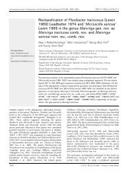
according to 16S rRNA gene similarity. Phylogenetic trees<br />
were constructed with neighbour-joining and maximumparsimony<br />
methods, all trees showed similar topology. The<br />
tree constructed with the neighbour-joining method (Saitou<br />
& Nei, 1987) using Kimura’s two-parameter calculation<br />
model in TREECON W version 1.3b (Van de Peer & De<br />
Wachter, 1994) is shown in Fig. 1. Strain H9 T , together<br />
with P. kongjuensis, P. autotrophica and P. compacta, formed<br />
a phyletic clade with 100 % support.<br />
DNA base composition was determined by thermal denaturation<br />
(Marmur & Doty, 1962) and genomic DNA <strong>from</strong><br />
Escherichia coli DH5a was analysed in parallel for calibration<br />
Table 1. Differential properties of strain H9 T <strong>from</strong> closely<br />
related <strong>Pseudonocardia</strong> <strong>sp</strong>ecies<br />
Strains: 1, P. <strong>ammonioxydans</strong> H9 T ;2,P. kongjuensis DSM 44525 T<br />
(data <strong>from</strong> Prabahar et al., 2004); 3, P. autotrophica IMSNU<br />
20050 T (for columns 3–5, data <strong>from</strong> Lee et al., 2001); 4, P. compacta<br />
IMSNU 20111 T ;5,P. alni IMSNU 20049 T ; 6, P. antarctica<br />
DVS5a1 T (data <strong>from</strong> Prabahar et al., 2004). +, Positive or present;<br />
W, weakly positive; 2, negative or absent; ND, not done.<br />
Characteristic 1 2 3 4 5 6<br />
Acid production <strong>from</strong>:<br />
L-Arabinose + 2 + 2 + 2<br />
D-Cellobiose 2 + + + 2 2<br />
D-Galactose + + + 2 + +<br />
Maltose + + + 2 + +<br />
D-Mannose + + + 2 2 +<br />
D-Melezitose 2 + + 2 + 2<br />
Methyl a-D-glucoside 2 2 + 2 2 ND<br />
Adonitol + + + 2 + 2<br />
myo-Inositol 2 + + 2 2 2<br />
1,2-Propanediol + 2 + 2 + ND<br />
D-Sorbitol 2 2 + 2 + 2<br />
Sucrose 2 + + 2 + 2<br />
D-Trehalose + + + 2 + 2<br />
D-Xylose W + + 2 + +<br />
Decomposition of:<br />
Hypoxanthine 2 + 2 2 + +<br />
Tyrosine + + 2 2 + +<br />
Xanthine 2 2 2 2 + 2<br />
Hydrolysis of:<br />
Gelatin W 2 2 2 + ND<br />
Starch W 2 2 2 + +<br />
Casein + + 2 2 2 2<br />
Urease activity + + + 2 + +<br />
NO { 3 reduction to NO{ 2<br />
+ 2 2 + 2 +<br />
H 2 S production + + + 2 + ND<br />
Oxidation of ammonia + 2 2 + 2 2<br />
Growth at/on:<br />
4 uC + + 2 + 2 2<br />
37 uC + + + 2 + +<br />
7 % NaCl + + + 2 + +<br />
of the T m value. The result showed that the G+C content of<br />
strain H9 T was 69?6 mol% (T m ). DNA–DNA hybridizations<br />
were carried out according to De Ley et al. (1970). Renaturation<br />
rates and relatedness values were calculated as<br />
described by Jahnke (1992). DNA–DNA relatedness values<br />
between strain H9 T and P. kongjuensis DSM 44525 T , P.<br />
autotrophica IMSNU 20050 T and P. compacta IMSNU<br />
20111 T were 42, 13 and 11 %, re<strong>sp</strong>ectively.<br />
Description of <strong>Pseudonocardia</strong> <strong>ammonioxydans</strong><br />
<strong>sp</strong>. <strong>nov</strong>.<br />
<strong>Pseudonocardia</strong> <strong>ammonioxydans</strong> (N.L. n. ammonia ammonia;<br />
N.L. part. adj. oxydans oxidizing; N.L. part. adj. <strong>ammonioxydans</strong><br />
oxidizing ammonia).<br />
556 International Journal of Systematic and Evolutionary Microbiology 56
<strong>Pseudonocardia</strong> <strong>ammonioxydans</strong> <strong>sp</strong>. <strong>nov</strong>.<br />
Fig. 1. Neighbour-joining phylogenetic tree<br />
based on 16S rRNA gene sequences showing<br />
the position of strain H9 T in the genus<br />
<strong>Pseudonocardia</strong>. Numbers at nodes indicate<br />
percentages of bootstrap support based on<br />
a neighbour-joining analysis of 1000 resampled<br />
datasets; only values over 50 % are shown.<br />
Norcardia puris IFM 0583 T was used as the<br />
outgroup. Bar, evolutionary distance (K nuc )of<br />
0?1.<br />
Aerobic, Gram-positive. Forms branched, brown substrate<br />
mycelium and white aerial mycelium on TSB and LB agar.<br />
The mycelium fragments into rod-shaped elements. Smooth<br />
<strong>sp</strong>ores are borne in short chains by acropetal budding <strong>from</strong><br />
the substrate mycelium. No pigment is produced. Growth<br />
occurs at 10–40 uC. Growth occurs on MNM and ammonia<br />
is oxidized to nitrate as the sole energy source. Ammonia is<br />
also oxidized to nitrate during growth on a blend of MNM<br />
and LB media. Growth occurs at NaCl concentrations ranging<br />
<strong>from</strong> 0 to 8 % with an optimum at 3?5 % NaCl. Catalasepositive.<br />
Acid is produced <strong>from</strong> D-fructose, D-glucose, N-<br />
acetyl-D-glucosamine, D-ribose, D-arabitol, D-galacturonic<br />
acid, D-gluconic acid and glycerol, but not <strong>from</strong> N-acetylb-D-mannosamine,<br />
arbutin, L-fucose, gentiobiose, a-Dlactose,<br />
lactulose, maltotriose, methyl a-D-galactoside,<br />
methyl b-D-galactoside, methyl a-D-glucoside, methyl a-<br />
D-mannoside, palatinose, D-psicose, D-raffinose, L-rhamnose,<br />
D-gagatose, turanose or xylitol. Uses acetic acid, b-<br />
hydroxybutyric acid, a-ketoglutaric acid, lactamide, D-lactic<br />
acid methyl ester, L-lactic acid, L-malic acid, pyruvic acid<br />
methyl ester, succinic acid mono-methyl ester, propionic<br />
acid, succinamic acid, succinic acid, N-acetyl-L-glutamic<br />
acid, L-alaninamide, D-alanine, L-alanine, L-alanyl glycine,<br />
L-a<strong>sp</strong>aragine, L-glutamic acid, glycyl L-glutamic acid, L-<br />
pyroglutamic acid, L-serine, putrescine, but not a-hydroxybutyric<br />
acid, c-hydroxybutyric acid, p-hydroxyphenylacetic<br />
acid, a-ketovaleric acid, D-malic acid or pyruvic acid. Other<br />
physiological properties are listed in Table 1. The cellwall<br />
chemotype is type IV. Predominant menaquinone is<br />
MK-8 (H 4 ). It contains pho<strong>sp</strong>hatidylcholine, pho<strong>sp</strong>hatidylglycerol,<br />
pho<strong>sp</strong>hatidylmethylethanolamine, pho<strong>sp</strong>hatidylinositol<br />
mannosides and dipho<strong>sp</strong>hatidylglycerol, but does not<br />
contain glucosamine-containing pho<strong>sp</strong>holipids or mycolic<br />
acids. The major fatty acids are iso-hexadecanoic acid<br />
(16 : 0-iso, 41?1 %), iso-hexadecenoic acid (16 : 1-iso,<br />
15?7 %) and heptadecenoic acid (17 : 1v8c, 12?1 %). The<br />
G+C content of the DNA is 69?6 mol% (T m ).<br />
The type strain, H9 T (=CGMCC 4.1877 T =JCM 12462 T ),<br />
was <strong>isolated</strong> <strong>from</strong> <strong>coastal</strong> sediment collected <strong>from</strong> Jiao-<br />
Dong peninsula near Qingdao, Shandong province, China.<br />
Acknowledgements<br />
This work was supported by projects <strong>from</strong> the Chinese National<br />
Natural Science Foundation (30470024) and <strong>from</strong> the State Key<br />
Laboratory of Marine Ecology and Environmental Science at the<br />
Institute of Oceanology (Tsingdao), Chinese Academy of Sciences.<br />
References<br />
Collins, M. D. (1985). Isoprenoid quinone analysis in classification<br />
and identification. In Chemical Methods in Bacterial Systematics,<br />
pp. 267–287. Edited by M. Goodfellow & D. E. Minnikin. London:<br />
Academic Press.<br />
De Ley, J., Cattoir, H. & Reynaerts, A. (1970). The quantitative<br />
measurement of DNA hybridization <strong>from</strong> renaturation rates. Eur<br />
J Biochem 12, 133–142.<br />
Dong, X.-Z. & Cai, M.-Y. (2001). Determinative Manual for Routine<br />
Bacteriology. Beijing: Scientific Press (English translation).<br />
Gordon, R. E., Barnett, D. A., Handerhan, J. E. & Pang, C. H.-N.<br />
(1974). Norcardia coeliaca, Nocardia autotrophica, and the nocardin<br />
strain. Int J Syst Bacteriol 24, 54–63.<br />
Hasegawa, T., Takizawa, M. & Tanida, S. (1983). A rapid analysis for<br />
chemical grouping of aerobic actinomycetes. J Gen Appl Microbiol<br />
29, 319–322.<br />
Henssen, A. (1957). Beiträge zur morphologie und systematik der<br />
thermophilen actinomyceten. Arch Mikrobiol 26, 373–414 (in German).<br />
Hu, Y.-T., Zhou, P.-J., Zhou, Y.-G., Liu, Z.-H. & Liu, S.-J. (2004).<br />
Saccharothrix xingjiangensis, <strong>sp</strong>. <strong>nov</strong>., a pyrene-degrading actinomycete<br />
<strong>isolated</strong> <strong>from</strong> Tianchi Lake, Xinjiang, China. Int J Syst Evol<br />
Microbiol 54, 2091–2094.<br />
Huang, Y., Wang, L., Lu, Z., Hong, L., Liu, Z., Tan, G. Y. A. &<br />
Goodfellow, M. (2002). Proposal to combine the genera Actinobi<strong>sp</strong>ora<br />
and <strong>Pseudonocardia</strong> in an emended genus <strong>Pseudonocardia</strong>,<br />
and description of <strong>Pseudonocardia</strong> zijingensis <strong>sp</strong>. <strong>nov</strong>. Int J Syst Evol<br />
Microbiol 52, 977–982.<br />
Jahnke, K.-D. (1992). Basic computer program for evaluation of<br />
<strong>sp</strong>ectroscopic DNA renaturation data <strong>from</strong> GILFORD System 2600<br />
<strong>sp</strong>ectrometer on a PC/XT/AT type personal computer. J Microbiol<br />
Methods 15, 61–73.<br />
Kämpfer, P. & Kroppenstedt, R. (2004). <strong>Pseudonocardia</strong> benzenivorans<br />
<strong>sp</strong>. <strong>nov</strong>. Int J Syst Evol Microbiol 54, 749–751.<br />
Lechevalier, M. P. & Lechevalier, H. A. (1970). Chemical composition<br />
as a criterion in the classification of aerobic actinomycetes. Int<br />
J Syst Bacteriol 20, 435–443.<br />
http://ijs.sgmjournals.org 557
Z.-P. Liu and others<br />
Lechevalier, H. A. & Lechevalier, M. P. (1980). The chemotaxonomy<br />
of actinomycetes. In Actinomycete Taxonomy, Special Publication<br />
no. 6, pp. 277–284. Edited by A. Dietz & D. W. Thayer. Arlington,<br />
VA: Society for Industrial Microbiology.<br />
Lechevalier, M. P., De Bièvre, C. & Lechevalier, H. A. (1977).<br />
Chemotaxonomy of aerobic actinomycetes: pho<strong>sp</strong>holipid composition.<br />
Biochem Syst Ecol 5, 249–260.<br />
Lee, S. D., Kim, E. S., Min, K.-L., Lee, W. Y., Kang, S.-O. & Hah, Y. C.<br />
(2001). <strong>Pseudonocardia</strong> kongjuensis <strong>sp</strong>. <strong>nov</strong>., <strong>isolated</strong> <strong>from</strong> a gold<br />
mine cave. Int J Syst Evol Microbiol 51, 1505–1510.<br />
Lee, S.-B., Strand, S. E., Stensel, H. D. & Herwig, R. P. (2004).<br />
<strong>Pseudonocardia</strong> chloroethenivorans <strong>sp</strong>. <strong>nov</strong>., a chloroethene-degrading<br />
actinomycete. Int J Syst Evol Microbiol 54, 131–139.<br />
Marmur, J. & Doty, P. (1962). Determination of the base composition<br />
of deoxyribonucleic acid <strong>from</strong> its thermal denaturation temperature.<br />
J Mol Biol 5, 109–118.<br />
McVeigh, H. P., Munro, J. & Embley, T. M. (1994). The phylogenetic<br />
position of Pseudoamycolata halophobica (Akimov et al. 1989) and a<br />
proposal to reclassify it as <strong>Pseudonocardia</strong> halophobica. Int J Syst<br />
Bacteriol 44, 300–302.<br />
Prabahar, V., Dube, S., Reddy, G. S. N. & Shivaji, S. (2004).<br />
<strong>Pseudonocardia</strong> antarctica <strong>sp</strong>. <strong>nov</strong>., an Actinomycetes <strong>from</strong> McMurdo<br />
Dry Valleys, Antarctica. Syst Appl Microbiol 27, 66–71.<br />
Rand, M. C., Greenberg, A. E. & Taras, M. J. (editors) (1975).<br />
Standard Methods for the Examination of Water and Wastewater,<br />
14th edn, pp. 434–436. Washington, DC: American Public Health<br />
Association.<br />
Reichert, K., Lipski, A., Pradella, S., Stackebrandt, E. & Altendorf, K.<br />
(1998). <strong>Pseudonocardia</strong> asacccharolytica <strong>sp</strong>. <strong>nov</strong>. and <strong>Pseudonocardia</strong><br />
sulfidoxydans <strong>sp</strong>. <strong>nov</strong>., two new dimethyl disulfide-degrading actinomycetes<br />
and emended description of the genus <strong>Pseudonocardia</strong>. Int<br />
J Syst Bacteriol 48, 441–449.<br />
Saitou, N. & Nei, M. (1987). The neighbor-joining method: a new<br />
method for reconstructing phylogenetic trees. Mol Biol Evol 4,<br />
406–425.<br />
Slawyk, G. & Maclsaac, J. J. (1972). Comparison of two automated<br />
ammonium methods in a region of <strong>coastal</strong> upwelling. Deep Sea Res<br />
19, 521–524.<br />
Takamiya, A. & Tubaki, K. (1956). A new form of Streptomyces<br />
capable of growing autotrophically. Arch Mikrobiol 25, 58–64.<br />
Thompson, J. D., Gibson, T. J., Plewniak, F., Jeanmougin, F. &<br />
Higgins, D. G. (1997). The CLUSTAL X windows interface: flexible<br />
strategies for multiple sequence alignment aided by quality analysis<br />
tools. Nucleic Acids Res 25, 4876–4882.<br />
Van de Peer, Y. & De Wachter, R. (1994). TREECON for Windows: a<br />
software package for the construction and drawing of evolutionary<br />
trees for the Microsoft Windows environment. Comput Appl Biosci<br />
10, 569–570.<br />
van’t Riet, J., Stouthamer, A. H. & Planta, R. J. (1968). Regulation of<br />
nitrate assimilation and nitrate re<strong>sp</strong>iration in Aerobacter aerogenes.<br />
J Bacteriol 96, 1455–1464.<br />
Warwick, S., Bowen, T., McVeigh, H. P. & Embley, T. M. (1994). A<br />
phylogenetic analysis of the family <strong>Pseudonocardia</strong>ceae and the<br />
genera Actinokineo<strong>sp</strong>ora and Saccharothrix with 16S rRNA sequences<br />
and a proposal to combine the genera Amycolata and <strong>Pseudonocardia</strong><br />
in an emended genus <strong>Pseudonocardia</strong>. Int J Syst Bacteriol 44,<br />
293–299.<br />
Wu, C., Lu, X., Qin, M., Wang, Y. & Ruan, J. (1989). Analysis of<br />
menaquinone compound in microbial cells by HPLC. Microbiology<br />
(English translation of Mikrobiologiia) 16, 176–178.<br />
Zhang, D., Yang, H., Zhang, W., Huang, Z. & Liu, S.-J. (2003).<br />
Rhodocista pekingensis <strong>sp</strong>. <strong>nov</strong>., a cyst-forming phototrophic bacterium<br />
<strong>from</strong> a municipal wastewater treatment plant. Int J Syst Evol<br />
Microbiol 53, 1111–1114.<br />
558 International Journal of Systematic and Evolutionary Microbiology 56