DEPARTAMENTO DE CIÊNCIAS DA VIDA ... - Estudo Geral
DEPARTAMENTO DE CIÊNCIAS DA VIDA ... - Estudo Geral DEPARTAMENTO DE CIÊNCIAS DA VIDA ... - Estudo Geral
15 I.2.3 - Mechanotransduction and Stem Cells I.2.3.1 - Mechanotransduction and MSCs differentiation Living tissues are known to possess different physiologic characteristics according to their function and cellular type, so considering the elasticity of solid tissues, very different values of elasticity can be found as shown in Figure 4. The solid tissues exhibit a range of stiffness, as measured by the elastic modulus, E (Engler et al., 2006). Figure 4 – Tissue Elasticity. Range of stiffness measured by the elastic modulus, E, of some human solid tissues. Adapted from Engler et al., 2006. Mesenchymal stem cells will differentiate into different phenotypes as a function of substrate stiffness. The lineage they can differentiate into when cultured on a substrate with a certain range of stiffness has phenotypic features similar to cells found in the solid tissues with the same range of stiffness (Figure 4), and MSCs appear to do so in a way that would promote tissue-specific differentiation and healing (Engler et al., 2006). For example, on brain-tissue-like stiffness cells undergo neuronal-like differentiation, whereas muscle-equivalent stiffness promotes myogenesis (Figure 5) (Engler et al., 2006).
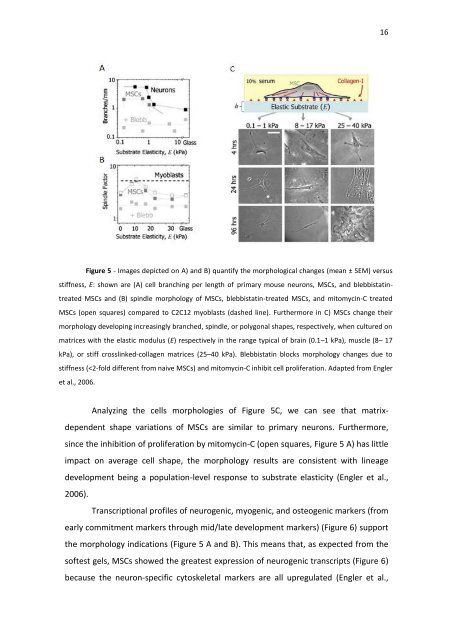
16 Figure 5 - Images depicted on A) and B) quantify the morphological changes (mean ± SEM) versus stiffness, E: shown are (A) cell branching per length of primary mouse neurons, MSCs, and blebbistatintreated MSCs and (B) spindle morphology of MSCs, blebbistatin-treated MSCs, and mitomycin-C treated MSCs (open squares) compared to C2C12 myoblasts (dashed line). Furthermore in C) MSCs change their morphology developing increasingly branched, spindle, or polygonal shapes, respectively, when cultured on matrices with the elastic modulus (E) respectively in the range typical of brain (0.1–1 kPa), muscle (8– 17 kPa), or stiff crosslinked-collagen matrices (25–40 kPa). Blebbistatin blocks morphology changes due to stiffness (
- Page 1 and 2: DEPARTAMENTO DE CIÊNCIAS DA VIDA F
- Page 3 and 4: ii Queria agradecer à minha namora
- Page 5 and 6: iv isolation of hUCM-MSCs was ever
- Page 7 and 8: vi Conseguimos optimizar um novo pr
- Page 9 and 10: viii Table of contents Acknowledgem
- Page 11 and 12: x III. 6 - Influence on MSCs specif
- Page 13 and 14: xii Figure 2| Proteins related to t
- Page 15 and 16: xiv the change in distribution with
- Page 17 and 18: xvi Cells were stained with anti-B-
- Page 19 and 20: xviii List of abbreviations AA - Ac
- Page 22: 1 Chapter I Introduction
- Page 25 and 26: 4 There are many advantages in usin
- Page 27 and 28: 6 only ones MSCs can differentiate
- Page 29 and 30: Figure 1. Mechanotransduction in a
- Page 31 and 32: 10 1C)(Dogterom et al., 2005). Not
- Page 33 and 34: 12 extracellular ligand and the cyt
- Page 35: 14 Figure 3 - The Rigidity Sensing
- Page 39 and 40: 18 Figure 7 - A) Immunofluorescence
- Page 41 and 42: 20 (GFAP) (is a class-III intermedi
- Page 43 and 44: 22 can specify lineage towards cell
- Page 45 and 46: 24 I.3 - Project rationale and expe
- Page 47 and 48: 26 Figure 12 - MSCs cultured on 1 a
- Page 49 and 50: 28 than that in soft substrate. Thi
- Page 52: 31 Chapter II - Materials and Metho
- Page 55 and 56: 34 Covance and DakoCytomation, resp
- Page 57 and 58: 36 clone SAM1; mouse Pe-Cy7 anti-hu
- Page 59 and 60: 38 To functionalize the complete su
- Page 62: 41 Chapter III Results
- Page 65 and 66: 44 2008) and also as previously est
- Page 67 and 68: 12.5% Ac PBS Fold increase of hMSCs
- Page 69 and 70: ≈12 kPa / COL-1 +FN ≈12 kPa / C
- Page 71 and 72: 50 Figure 19 - Immunophenotype of U
- Page 73 and 74: 52 coated with COL-1 (similar to wh
- Page 75 and 76: 54 hydrogels of 1 and 7 kPa (for ce
- Page 77: Figure 23 - hMSCs cultured on TCP (
- Page 81 and 82: 60 Discussion MSCs are widely used
- Page 83 and 84: 62 isolating MSCs on the PA hydroge
- Page 85 and 86: 64 2011) and expanded on TCPs for 5
16<br />
Figure 5 - Images depicted on A) and B) quantify the morphological changes (mean ± SEM) versus<br />
stiffness, E: shown are (A) cell branching per length of primary mouse neurons, MSCs, and blebbistatintreated<br />
MSCs and (B) spindle morphology of MSCs, blebbistatin-treated MSCs, and mitomycin-C treated<br />
MSCs (open squares) compared to C2C12 myoblasts (dashed line). Furthermore in C) MSCs change their<br />
morphology developing increasingly branched, spindle, or polygonal shapes, respectively, when cultured on<br />
matrices with the elastic modulus (E) respectively in the range typical of brain (0.1–1 kPa), muscle (8– 17<br />
kPa), or stiff crosslinked-collagen matrices (25–40 kPa). Blebbistatin blocks morphology changes due to<br />
stiffness (


