PDF (Chapter 27: Frontal Lobes Bedside Testing) - UTas ePrints
PDF (Chapter 27: Frontal Lobes Bedside Testing) - UTas ePrints
PDF (Chapter 27: Frontal Lobes Bedside Testing) - UTas ePrints

Create successful ePaper yourself
Turn your PDF publications into a flip-book with our unique Google optimized e-Paper software.
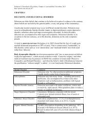
Pridmore S. Download of Psychiatry, <strong>Chapter</strong> <strong>27</strong>. Last modified: December, 2013.<br />
http://eprints.utas.edu.au/287/<br />
1<br />
CHAPTER <strong>27</strong><br />
FRONTAL LOBES – BEDSIDE TESTING<br />
Introduction<br />
The frontal lobes, phylogenetically the youngest part, form over half the brain<br />
volume. Until recent decades the prefrontal areas were referred to as the “silent<br />
areas”, because injury to these regions was not accompanied by sensorimotor signs,<br />
and the function of these areas was not clear. The connections of the frontal lobes<br />
have now been described (Goldman-Rakic, 1987) and their functions have received<br />
much attention (Alvarez & Emory, 2006).<br />
The frontal lobe cortex forms a part of the frontal-subcortical circuits (these have been<br />
described in detail in <strong>Chapter</strong> 2). There are 5 parallel, separate circuits (Alexander et<br />
al, 1986). In brief, each circuit has a “direct” and an “indirect” route. The direct route<br />
has 4 components and is schematically represented:<br />
The indirect routes are a little more complicated, with a projection from the globus<br />
pallidus to the subthalamic nucleus, and another returning from the subthalamic<br />
nucleus to the globus pallidus.<br />
These 5 separate circuits form essentially closed loops, however, they receive input<br />
from other brain regions. Thus, lesions at various sites, both within the closed loops<br />
and outside, may have similar clinical effects. For example, in Alzheimer’s disease<br />
the site of most pathology is the cortex, but Huntington’s disease and Parkinson’s<br />
present related symptoms of dementia due to subcortical pathology.<br />
The 5 frontal-subcortical circuits<br />
1. a motor circuit originating in the motor cortex and pre-motor cortex<br />
2. an oculomotor unit originating in the frontal eye fields<br />
3. the dorsolateral prefrontal circuit, which underpins executive functions<br />
4. the anterior cingulate circuit which underpins motivation<br />
5. the orbitofrontal circuit which underpins impulse control and social behavior.<br />
The motor circuit and the oculomotor unit are of importance, but of greater interest to<br />
neurology than psychiatry. The dorsolateral prefrontal, anterior cingulate and<br />
orbitofrontal circuits are of great interest to psychiatry.
Pridmore S. Download of Psychiatry, <strong>Chapter</strong> <strong>27</strong>. Last modified: December, 2013.<br />
http://eprints.utas.edu.au/287/<br />
2<br />
These parts of the frontal lobes allow the organism to learn from experience, and<br />
organize current information and choose a course of action, to summon drive to<br />
execute the action, and remain attentive and resist distraction.<br />
In the following paragraphs, the “functional regions of the frontal lobes” are<br />
discussed. Beside tests are suggested. The intention is to dispel the notion that the<br />
frontal lobes are the “silent areas” of the brain, and illustrate that they can be<br />
examined, at least to some extent, by an interested doctor.<br />
While there are 5 frontal-subcortical circuits, there are 6 functional regions listed<br />
below. This is because the motor and premotor frontal regions both contribute to the<br />
motor frontal-subcortical circuit.<br />
Functional regions of the frontal lobes<br />
I. Primary motor area<br />
II. Premotor area<br />
III. <strong>Frontal</strong> eye fields<br />
IV. Dorsolateral prefrontal cortex<br />
V. Orbital and basal areas<br />
VI. Supplementary motor area and anterior cingulated gyrus area<br />
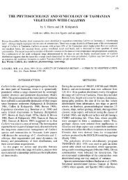
a.<br />
b.<br />
c.<br />
d.<br />
Illustrations. a: Brodmann areas of the left frontal lobe (lateral view); b: functional<br />
regions of the left frontal lobe (lateral view); c: Brodmann areas of the right frontal<br />
lobe (medial view); d: functional regions of the right frontal lobe (medial view).<br />
Primary motor cortex<br />
The primary motor cortex or precentral gyrus (bounded posteriorally by the central<br />
sulcus) is Brodmann area 4. Although designated a “motor” cortex, this area is also<br />
involved with somatosensory perception. Lesions in this area of cortex or the<br />
subcortical elements of the associated circuit result in weakness and incoordination.
Pridmore S. Download of Psychiatry, <strong>Chapter</strong> <strong>27</strong>. Last modified: December, 2013.<br />
http://eprints.utas.edu.au/287/<br />
3<br />
<strong>Bedside</strong> tests:<br />
1. Motor strength of hand grip. The patient is asked to grip the examiners fingers.<br />
Strength should be roughly equal, with greater strength on the dominant side.<br />
It should be difficult for the examiner to free her/his fingers.<br />
2. Motor speed as in finger tapping has also been listed as a useful test (Malloy<br />
& Richardson, 1994) but such tests do not discriminate from the premotor<br />
cortex.<br />
Diagnostically, poor performances suggest local lesions such as vascular or neoplastic<br />
pathology, or a generalized lesion such as a degenerative disease. (Peripheral nerve<br />
lesion must, of course, be excluded.)<br />
Premotor cortex<br />
The premotor cortex, a transverse strip, is Brodmann area 6. It is involved in<br />
sensorimotor integration. Lesions in this area or the subcortical elements of the circuit<br />
cause inability to make use of sensory feedback in the performance of smooth<br />
movements and apraxia. Apraxia may also be a result of lesions of other areas<br />
(parietal lobe).<br />
<strong>Bedside</strong> tests:<br />
1. Sensorimotor abilities are tested by asking the patient touch each finger to the<br />
thumb in succession as rapidly as possible. Watch for speed and dexterity.<br />
2. Apraxia can be tested by asking the patient to "blow a kiss" and to<br />
demonstrate the use of a shovel.<br />
Poor performance carries the diagnostic implications as for the motor cortex above.<br />
<strong>Frontal</strong> eye fields<br />
The frontal eye fields are largely Brodmann area 8, with some area 9 and 6. Eye<br />
movement involves many structures, and a lesion in one may be compensated for by<br />
activity in another.<br />
For present purposes, voluntary eye movements are of two types. Pursuit movement<br />
occurs when the eyes to follow moving objects. Saccadic eye movements are used to<br />
follow imaginary points.<br />
<strong>Bedside</strong> test:<br />
1. Ask the patient to follow the movement of a finger from left to right and up<br />
and down.<br />
2. Ask the patient to look from left to right, up and down (with no finger to<br />
follow).<br />
Note inability to move or jerky movement.
Pridmore S. Download of Psychiatry, <strong>Chapter</strong> <strong>27</strong>. Last modified: December, 2013.<br />
http://eprints.utas.edu.au/287/<br />
4<br />
Dorsolateral prefrontal cortex (DLPC)<br />
The Brodmann areas for the DLPC are a matter of some disagreement. All agree that<br />
area 9 is a large part of the DLPC. Other areas named are 10 and 46. A compromise<br />
position is that the DLPC is composed of Brodmann area 9 and the lateral aspect of 10<br />
and most of area 46.<br />
The DLPC and the subcortical elements of the associated circuit are responsible for<br />
executive functions. The executive functions include the integration of sensory<br />
information, the generation of a range of response alternatives to environmental<br />
challenges, the selection of the most appropriate response, maintenance of task set,<br />
sequential ordering of data, self-evaluation of performance and the selection of a<br />
replacement responses if the first applied response fails.<br />
(The above paragraph is generally accepted. However, as research continues the<br />
complexity of the CNS increases. A recent review indicates that executive functions<br />
may not solely reside in the frontal lobe (Alvarez & Emory, 2006).)<br />
The executive functions largely determine the ability of the individual to cope with<br />
the continuous, but ever changing challenges of the environment. Thus, the patient’s<br />
ability to make an appointment and to arrive on time is valuable information. So too,<br />
is the ability of the patient to give a comprehensive account of her/himself and the<br />
reasons for the consultation.<br />
It is believed by some authors that formal thought disorder arises from a lack of<br />
executive planning and editing (McGrath, 1991). In thought disorder there are<br />
frequent examples of failure to maintain set (distractibility), sequentially order<br />
information, and to ensure that the listener is comprehending. [However, formal<br />
thought disorder is also known to involve the left superior temporal sulcus and the left<br />
temporal pole (Horn et al, 2010).]<br />
<strong>Bedside</strong> tests:<br />
1. Is the patient able to make an appointment and arrive on time?<br />
2. Is the patient able to give a coherent account of current problems and the<br />
reason for the interview? Is there evidence of thought disorder?<br />
3. Digit span, days of the week or months of the year backwards. Here the patient<br />
has to retain the task and simultaneously manipulate information.<br />
4. Controlled oral word association test (COWAT): the patient is asked to<br />
produce as many words as possible, in one minute, starting with F, then A,<br />
then S. Proper nouns and previously used words with a different suffix are<br />
prohibited (Benton, 1968).<br />
Other categorical fluency tests include naming animals, fruits and vegetables<br />
(Monsch et al, 1992).
Pridmore S. Download of Psychiatry, <strong>Chapter</strong> <strong>27</strong>. Last modified: December, 2013.<br />
http://eprints.utas.edu.au/287/<br />
5<br />
For a formal result, it is necessary to test under strict conditions, using norms.<br />
However, valuable information may be obtained without formal testing.<br />
Generally, a normal individual will be able to provide more than ten items for<br />
each of these categories, while a patient with significant deficits will usually<br />
score less than eight. The performance of the task will also provide valuable<br />
information. Common errors include perseveration (repeating words which<br />
have already been given either during the task at hand or an earlier task).<br />
There may also be inappropriate or profane utterances. (These also suggest<br />
disinhibition which is discussed under orbitofrontal cortex, below.)<br />
5. Alternating hand sequences. These can be devised by the examiner. One<br />
example is that one hand is placed palm upwards and the other is place palm<br />
downwards, and the patient is then asked to reverse these positions as rapidly<br />
as possible.<br />
Another example is that the backs of the hands are both placed downwards,<br />
but one hand is clenched and the other is open, then the patients is asked to<br />
close the open hand and open the closed hand, and keep reversing the posture<br />
of the hands as rapidly as possible.<br />
A final example is that the patient taps twice with one fist and once with the<br />
other, then after the rhythm is established, the patient is asked to change over<br />
the number of beats (the fist which first tapped twice now taps only once).<br />
Patients with frontal lobe deficits usually perform poorly on these tests, often<br />
being unable to follow the relatively simple instructions.<br />
6. Formal neuropsychological may be necessary where uncertainty remains.<br />
Commonly employed tests include Controlled Oral Word Association Test<br />
(Benton, 1968) and the Wisconsin Card Sorting Tests (Heaton, 1985).<br />
Head injury and dementing illnesses may result in severe impairment of the executive<br />
functions. Schizophrenia often has thought disorder as a major feature and the<br />
executive functions tests are usually also at least mildly affected. Depressive disorder<br />
may be associated with poor performance on verbal fluency tests during the acute<br />
phase, which normalizes with remission (Trichard, et al., 1995).<br />
Orbital and basal area (Orbitofrontal cortex)<br />
The orbitofrontal cortex is Brodmann areas 10 and 11. (Part of area 10 is mentioned<br />
above as contributing to the DLPC.) It mediates empathic, civil and socially<br />
appropriate behavior (Mega and Cummings, 1994). Much of the personality change<br />
described in cases of frontal lobe injury (Phineas Gage being the most famous) is due<br />
to lesions in this area. Patients may become irritable, labile, disinhibited and fail to<br />
respond to the conventions of acceptable social behavior. Similar changes may occur<br />
with lesions of subcortical element of the frontal-subcortical circuit, as with caudate<br />
damage in Huntington's disease.
Pridmore S. Download of Psychiatry, <strong>Chapter</strong> <strong>27</strong>. Last modified: December, 2013.<br />
http://eprints.utas.edu.au/287/<br />
6<br />
Increased concern about social behavior and contamination has been associated with<br />
increased orbitofrontal and caudate metabolism. This has been reported with lesions<br />
of the globus pallidus and in obsessive compulsive disorder.<br />
<strong>Bedside</strong> tests:<br />
1. Does the patient dress or behave in a way which suggests lack of concern with<br />
the feelings of others or without concern to accepted social customs.<br />
2. Test sense of smell - coffee, cloves etc.<br />
3. Go/no-go Test. The patient is asked to make a response to one signal (the Go<br />
signal) and not to respond to another signal (the no-go signal). The most basic<br />
is to ask the patient to tap their knee when the examiner says, “Go” and to<br />
make no response when the examiner says, “Stop”.<br />
The task may be made more demanding by reversing the customary meaning<br />
of signals. An example is to ask the patient to tap the knee when the examiner<br />
says "Stop" and not to tap when the examiner says "Go" (Malloy and<br />
Richardson, 1994).<br />
4. The Stroop Test (Stroop, 1935). This is a neuropsychological test which<br />
examines the ability of the patient to inhibit responses. Patients are asked to<br />
state the color in which words are printed rather than the words themselves,<br />
e.g., truck may be printed in blue, house may be printed in red.<br />
This task is made difficult by presenting the name of colors printed in different<br />
colored ink. The task is to state the color of the ink, not to read the word. This<br />
is not as easy as it sounds. Try it now. Do it quickly, to increase the pressure.<br />
Green White Purple<br />
Blue Red Green<br />
Pink Orange Blue<br />
Purple Yellow Grey<br />
Red Green Orange<br />
Black Blue Red<br />
White Yellow Pink<br />
Cultural factors are important in making an observation of lack of civility, empathy<br />
and social concern. Eructation following a meal is considered good manners in some<br />
parts of the world, and people of the same race and city will have different sets of<br />
social values depending on socio-economic status.<br />
Failure of inhibition may complicate head injury, other destructive lesions (including<br />
dementing processes) and schizophrenia. Failure of inhibition is found in impulse<br />
control and personality disorder (particularly of the antisocial type). Depressive<br />
disorder may manifest irritability, and has been associated with poor performance on<br />
the Stroop Test (Trichard et al, 1995).
Pridmore S. Download of Psychiatry, <strong>Chapter</strong> <strong>27</strong>. Last modified: December, 2013.<br />
http://eprints.utas.edu.au/287/<br />
7<br />
Obsessive compulsive disorder in which there is excessive concern and caution is<br />
associated with increased metabolism in the orbitofrontal cortex (which may result<br />
from subcortical pathology; Hampson et al, 2012).<br />
Supplementary motor area and anterior cingulate cortex<br />
The supplementary motor area is the medial aspect of Brodmann area 6 (Barker &<br />
Barasi, 1999) and the anterior cingulate gyrus is Brodmann area 24. These areas are<br />
involved in motivated behavior (Mega and Cummings, 1994), initiation and goaldirected<br />
behavior (Devinsky et al, 1995).<br />
At present there are no office or neuropsychological tests to measure the functional<br />
status of these areas.<br />
Akinetic mutism occurs with gross lesions (e.g., meningioma) of the anterior<br />
cingulate. Such patients are profoundly apathetic, generally mute and eat and drink<br />
only when assisted. They do not respond to pain and are indifferent to their<br />
circumstances. Lesions of the supplementary motor area are associated with the alien<br />
hand syndrome (Goldberg & Bloom, 1990).<br />
The apathy of schizophrenia and the immobility of depressive disorder may be<br />
associated with defects in associated circuits.<br />
<strong>Frontal</strong> Release Reflexes<br />
The primitive reflexes are present in normal babies. As the CNS matures, frontal lobe<br />
cells develop and begin to inhibit These may reappear with brain damage or disease -<br />
they may also reappear with normal aging. Their significance is greater when they<br />
appear unilaterally and in young individuals (Ross, 1985).<br />
Grasp<br />
The hand is stroked across the palm toward the thumb by the examiners fingers or the<br />
handle of the patella hammer. When the reflex is present the fingers grasp or the<br />
thumb strongly adducts. The patient may be unable to release the grip. Presence<br />
suggests contralateral frontal lobe disease.<br />
Sucking (pout, snout, rooting)<br />
The sucking reflex is elicited by stroking the lips of the patient with a finger or a<br />
spatula from side to middle and back again. The pouting reflex is elicited by the<br />
examiner placing the index finger of the patient’s closed lips and tapping the finger<br />
with a patella hammer. Sucking or pouting movements of the lips suggest frontal lobe<br />
damage or bilateral lesions above the mid-pons.<br />
Palmar-mental<br />
The palm is scratched firmly with a key or the handle of the patella hammer, from the<br />
fingers, toward the wrist. The positive response is a flicker in the skin on the
Pridmore S. Download of Psychiatry, <strong>Chapter</strong> <strong>27</strong>. Last modified: December, 2013.<br />
http://eprints.utas.edu.au/287/<br />
8<br />
ipsilateral side of the point of the chin. Presence suggests contralateral frontal lobe<br />
damage, however, the true value of the reflex is yet to be clearly determined.<br />
Glabella Tap<br />
The patient is asked to close the eyes and the examiner repeatedly taps (finger tip or<br />
patella hammer) the supraorbital ridge. In the normal individual the orbicularis oris<br />
contracts in response to the first two or three taps and then ceases. In pathological<br />
conditions the orbicularis oris continues to contract with every tap. This reflex is used<br />
in the diagnosis of Parkinson's disease, but it may also occur with frontal damage of<br />
other etiologies.<br />
References<br />
Alexander G, DeLong M, Strick P. Parallel organization of functionally segregated<br />
circuits linking basal ganglia and cortex. Annual Reviews of Neuroscience 1986;<br />
9:357-381.<br />
Alvarez J, Emory E. Executive function and the frontal lobes: a meta-analysis.<br />
Neuropsychology Review 2006; 16:17-42.<br />
Barker R, Barasi S. Neuroscience at a glance. Blackwell Science Ltd: Oxford. 1999.<br />
Benton A. Differential behavioral effects in frontal lobe disease. Neuropsycholigia<br />
1968; 6:53-60.<br />
Devinsky O, Morrell M, Vogt B. Contributions of anterior cingulate cortex to<br />
behavior. Brain 1995; 118:<strong>27</strong>9-306.<br />
Goldberg G, Bloom K. The alien hand sign: localization, lateralization and recovery.<br />
American Journal of Physical Medicine and rehabilitation 1990; 69:228-238.<br />
Goldman-Rakic P. Circuitry of the primate prefrontal cortex and regulation of<br />
behavior by representational memory. In F Plum, V Mountcastle Eds. Handbook of<br />
physiology, the nervous system, and higher functions of the brain (Sec 1, Vol 5, pp<br />
373-417. Bethesda, MD, American Psychological Society, 1987.<br />
Hampson M, Stoica T, Scheinost D, et al. Real-time fMRI biofeedback targeting the<br />
orbitofrontal cortex for contamination anxiety. J Vis Exp 2012; in press.<br />
Heaton R. Wisconsin card sorting test. Odessa, TX: Psychological Assessment<br />
Resources. 1985.<br />
Horn H, Federspiel A, Wirth M. et al. Gray matter volume differences specific to<br />
formalthought disorder in schizophrenia. Psychiatry Research: Neuroimaging 2010;<br />
182:183-186.<br />
McGrath J. Ordering thoughts on thought disorder. British Journal of Psychiatry<br />
1991; 158:683-688.<br />
Malloy P, Richardson E. Assessment of frontal lobe functions. Journal of<br />
Neuropsychiatry and Clinical Neurosciences 1994; 6:399-410.<br />
Mega M, Cummings J. <strong>Frontal</strong>-subcortical circuits and neuropsychiatric disorders.<br />
Journal of Neuropsychiatry and Clinical Neurosciences 1994: 6:358-370.<br />
Monsch A, Bondi M, Butters N. Comparisons of verbal fluency tasks in detection of<br />
dementia of the Alzheimer type. Archives of Neurology 1992; 49:1253-1258.<br />
Trichard C, Martinot J, Alagille M, Masure M, Hardy P, Ginstet D, Feline A. Time<br />
course of prefrontal dysfunction in severely depressed in-patients: a longitudinal<br />
neuropsychological study. Psychological Medicine 1995; 25:79-85.