Chapter 5 HALLUCINATIONS Hallucinations are ... - UTas ePrints
Chapter 5 HALLUCINATIONS Hallucinations are ... - UTas ePrints
Chapter 5 HALLUCINATIONS Hallucinations are ... - UTas ePrints

Create successful ePaper yourself
Turn your PDF publications into a flip-book with our unique Google optimized e-Paper software.
Pridmore S. Download of Psychiatry, <strong>Chapter</strong> 5. Last modified: November, 2013.<br />
http://eprints.utas.edu.au/287/<br />
8<br />
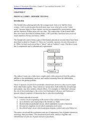
The following 2 illustrations <strong>are</strong> presented as a reminder of the anatomy of these tracts –<br />
they <strong>are</strong> not taken from research on hallucinations.<br />
Illustration: the superior longitudinal fasciculus (of which the arcuate fasciculus is an<br />
anterior component), the cingulum, and the uncinate fasciculus.<br />
Illustration: This diffusion tensor imaging (DTI) image (generously provided for public<br />
use by Aaron G. Filler, MD, PhD) shows the right and left arcuate fasciculus (Raf & Laf),<br />
and the right and left superior longitudinal fasciculus (Rslf & Lslf) as separate entities<br />
(they can be conceptualized as continuous).<br />
In other recent work, Horga et al (2011) studied first-presentation, drug-free patients with<br />
schizophrenia. Using positron emission tomography (PET), they comp<strong>are</strong>d patients with<br />
commenting auditory hallucinations to patients without auditory hallucinations. Patients<br />
with auditory hallucinations demonstrated significantly increased metabolic rates in the<br />
left superior and middle temporal cortices, bilateral medial frontal cortex and the left<br />
caudate nucleus. In addition, there was decreased activity in the hippocampalparahippocampal,<br />
cerebellar and parietal cortices during hallucinations.<br />
This work suggests that failure to deactivate the temporal cortex allows increased<br />
spontaneous activity, and auditory hallucinations. It is possible that decreased activity in