A scanning and transmission electron microscopic study of ... - Digitum
A scanning and transmission electron microscopic study of ... - Digitum
A scanning and transmission electron microscopic study of ... - Digitum

Create successful ePaper yourself
Turn your PDF publications into a flip-book with our unique Google optimized e-Paper software.
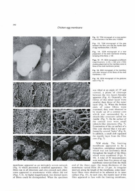
Chicken egg membrane<br />
Fig. 13. TEM micrograph <strong>of</strong> the gap<br />
between the fibre core <strong>and</strong> the mantle layer<br />
<strong>of</strong> egg membrane fibre. x 80,500<br />
Fig. 14. SEM micrograph <strong>of</strong> a tear<br />
preparation <strong>of</strong> the shell mernbrane showing<br />
terraang <strong>of</strong> the fibres. x 135<br />
Figs. 15 - 17. SEM micrographs at different<br />
magnifications. (x 65, x 390 <strong>and</strong> x 550)<br />
showing attachment <strong>of</strong> the rnamillary cores to<br />
fibres <strong>of</strong> the sheli mernbrane (arrow).<br />
Fig. 18. SEM rnicrograph <strong>of</strong> the rnamillary<br />
core after rernoval <strong>of</strong> the fibres <strong>of</strong> the shell<br />
membrane. x 125<br />
Fig. 19. SEM rnicrograph <strong>of</strong> the globules<br />
seen in Fia. 18.<br />
m<br />
membrane appeared as an intricately-woven network<br />
(Fig. 4) which presented a stratified appearance. The<br />
fibres ran in al1 directions <strong>and</strong> criss-crossed each other;<br />
some appeared to anastomose while others did not<br />
(Figs. 5,6). At higher magnification, two distinct layers<br />
<strong>of</strong> fibres could be distinguished. When the specimen<br />
was tilted at an angle <strong>of</strong> 15* <strong>and</strong><br />
viewed, a plane <strong>of</strong> cleavage<br />
between the two layers became<br />
apparent (Fig. 4). Generally, the<br />
fibres <strong>of</strong> the inner layer were<br />
smaller than those <strong>of</strong> the outer<br />
layer (Fig. 5). When the broken<br />
ends <strong>of</strong> some fibres were<br />
examined, they were observed to<br />
be made up <strong>of</strong> a central core<br />
which was surrounded by a<br />
sheath-like structure called the<br />
mantle (Fig. 7). On the surface <strong>of</strong><br />
the central core were small<br />
tubercle-like structures (Fig. 7).<br />
Examination <strong>of</strong> the cut surface <strong>of</strong> a<br />
fibre core showed that it was perforated<br />
by several "holes" (Fig. 8).<br />
In addition, a small cleft separated<br />
the mantle layer from the fibre<br />
- core (Fig. 7).<br />
TEM <strong>study</strong>. The limiting<br />
membrane appeared to be a<br />
featureless, amorphous membrane<br />
which was not uniformly thick<br />
throughout the length <strong>of</strong> the fibres<br />
which were examined (Figs. 9,<br />
10). A total <strong>of</strong> 50 measurements<br />
were made from five pieces <strong>of</strong> egg<br />
membrane removed from the blunt<br />
end <strong>of</strong> the three eggs; the mean thickness <strong>of</strong> the<br />
membrane varied from .O9 to .15 pm. At irregular<br />
intemals along the length <strong>of</strong> the membrane, some innerlayer<br />
fibres were observed to be adherent to its inner<br />
surface (Fig. 10). At such sites, the mantle layer <strong>of</strong> the<br />
fibre appeared to fuse with the limiting membrane,