A scanning and transmission electron microscopic study of ... - Digitum
A scanning and transmission electron microscopic study of ... - Digitum
A scanning and transmission electron microscopic study of ... - Digitum

You also want an ePaper? Increase the reach of your titles
YUMPU automatically turns print PDFs into web optimized ePapers that Google loves.
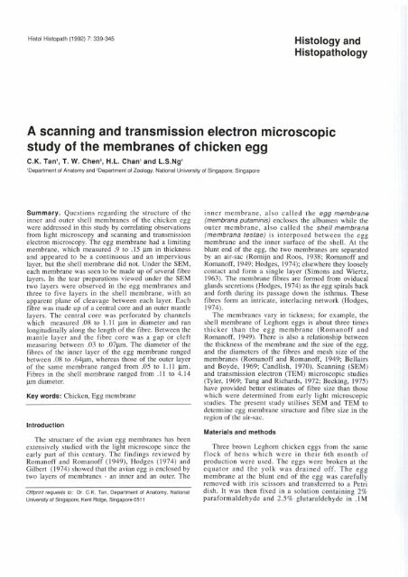
Histol Histopath (1 992) 7: 339-345<br />
Histology <strong>and</strong><br />
Histopathology<br />
A <strong>scanning</strong> <strong>and</strong> <strong>transmission</strong> <strong>electron</strong> <strong>microscopic</strong><br />
<strong>study</strong> <strong>of</strong> the membranes <strong>of</strong> chicken egg<br />
C.K. Tan1, T. W. Chen2, H.L. Chanl <strong>and</strong> L.S.Ngl<br />
'Department <strong>of</strong> Anatomy <strong>and</strong> 2Department <strong>of</strong> Zoology, National University <strong>of</strong> Singapore, Singapore<br />
Summary. Questions regarding the structure <strong>of</strong> the<br />
inner <strong>and</strong> outer shell membranes <strong>of</strong> the chicken egg<br />
were addressed in this <strong>study</strong> by correlating observations<br />
from light microscopy <strong>and</strong> <strong>scanning</strong> <strong>and</strong> <strong>transmission</strong><br />
<strong>electron</strong> microscopy. The egg membrane had a limiting<br />
membrane, which measured .9 to .15 pn in thickness<br />
<strong>and</strong> appeared to be a continuous <strong>and</strong> an impervious<br />
layer, but the shell membrane did not. Under the SEM,<br />
each membrane was seen to be made up <strong>of</strong> severa1 fibre<br />
layers. In the tear preparations viewed under the SEM<br />
two layers were observed in the egg membranes <strong>and</strong><br />
three to five layers in the shell membrane, with an<br />
apparent plane <strong>of</strong> cleavage between each layer. Each<br />
fibre was made up <strong>of</strong> a central core <strong>and</strong> an outer mantle<br />
layers. The central core was perforated by channels<br />
which measured .O8 to 1.1 1 pn in diameter <strong>and</strong> ran<br />
longitudinally along the length <strong>of</strong> the fibre. Between the<br />
mantle layer <strong>and</strong> the fibre core was a gap or cleft<br />
measuring between .O3 to .07pn. The diameter <strong>of</strong> the<br />
fibres <strong>of</strong> the inner layer <strong>of</strong> the egg membrane ranged<br />
between .O8 to .64pn, whereas those <strong>of</strong> the outer layer<br />
<strong>of</strong> the same membrane ranged from .O5 to 1.11 p.m.<br />
Fibres in the shell membrane ranged from .ll to 4.14<br />
pm diameter.<br />
Key words: Chicken, Egg membrane<br />
lntroduction<br />
The structure <strong>of</strong> the avian egg membranes has been<br />
extensively studied with the light microscope since the<br />
early part <strong>of</strong> this century. The findings reviewed by<br />
Roman<strong>of</strong>f <strong>and</strong> Roman<strong>of</strong>f (1949). Hodges (1974) <strong>and</strong><br />
Gilbert (1974) showed that the avian egg is enclosed by<br />
two layers <strong>of</strong> membranes - an inner <strong>and</strong> an outer. The<br />
Onprint requests to: Dr. C.K. Tan, Department <strong>of</strong> Anatomy, National<br />
University <strong>of</strong> Singapore, Kent Ridge, Singapore 051 1<br />
inner membrane, also called the egg membrane<br />
(membrana putaminis) encloses the albumen while the<br />
outer membrane, also called the shell membrana<br />
(membrana testae) is interposed between the egg<br />
membrane <strong>and</strong> the inner surface <strong>of</strong> the shell. At the<br />
blunt end <strong>of</strong> the egg, the two membranes are separated<br />
by an air-sac (Romijn <strong>and</strong> Roos, 1938; Roman<strong>of</strong>f <strong>and</strong><br />
Roman<strong>of</strong>f, 1949; Hodges, 1974); elsewhere they loosely<br />
contact <strong>and</strong> form a single layer (Simons <strong>and</strong> Wiertz,<br />
1963). The membrane fibres are formed from oviducal<br />
gl<strong>and</strong>s secretions (Hodges, 1974) as the egg spirals back<br />
<strong>and</strong> forth during its passage down the isthmus. These<br />
fibres form an intricate, interlacing network (Hodges,<br />
1974).<br />
The membranes vary in tickness; for example, the<br />
shell membrane <strong>of</strong> Leghom eggs is about three times<br />
thicker than the egg membrane (Roman<strong>of</strong>f <strong>and</strong><br />
Roman<strong>of</strong>f, 1949). There is also a relationship between<br />
the thickness <strong>of</strong> the membrane <strong>and</strong> the size <strong>of</strong> the egg,<br />
<strong>and</strong> the diameters <strong>of</strong> the fibres <strong>and</strong> mesh size <strong>of</strong> the<br />
membranes (Roman<strong>of</strong>f <strong>and</strong> Roman<strong>of</strong>f, 1949; Bellairs<br />
<strong>and</strong> Boyde, 1969; C<strong>and</strong>lish, 1970). Scanning (SEM)<br />
<strong>and</strong> <strong>transmission</strong> <strong>electron</strong> (TEM) <strong>microscopic</strong> studies<br />
(Tyler, 1969; Tung <strong>and</strong> Richards, 1972; Becking, 1975)<br />
have provided better estimates <strong>of</strong> fibre size than those<br />
which were determined from early light <strong>microscopic</strong><br />
studies. The present <strong>study</strong> utilises SEM <strong>and</strong> TEM to<br />
determine egg membrane structure <strong>and</strong> fibre size in the<br />
region <strong>of</strong> the air-sac.<br />
Materials <strong>and</strong> methods<br />
Three brown Leghom chicken eggs from the same<br />
flock <strong>of</strong> hens which were in their 6th month <strong>of</strong><br />
production were used. The eggs were broken at the<br />
equator <strong>and</strong> the yolk was drained <strong>of</strong>f. The egg<br />
membrane at the blunt end <strong>of</strong> the egg was carefully<br />
removed with iris scissors <strong>and</strong> transferred to a Petri<br />
dish. It was then fixed in a solution containing 2%<br />
paraformaldehyde <strong>and</strong> 2.5% glutaraldehyde in .lM
Chicken egg membrane<br />
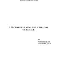
Flg. 3. SEM micrograph showing<br />
str<strong>and</strong>s <strong>of</strong> albumen adherlng to the<br />
lnner su- <strong>of</strong> the limiting membrane.<br />
x400<br />
Fig. 4. SEM micrograph <strong>of</strong> a tear<br />
preparation <strong>of</strong> the egg membrane<br />
showing a terraced appearance <strong>of</strong> the<br />
fibres. (Asterisk<br />
limitlng membrane.<br />
Amm = lnner <strong>and</strong> outer layer deavage<br />
m). x 65<br />
Flg. 5. SEM micrograph <strong>of</strong> the egg<br />
membrane. Asterisk = limiting<br />
mmbtme. (IL = inner layer <strong>of</strong> fibres.<br />
OL = outer layer <strong>of</strong> fibres). x 270<br />
Flg. 6. SEM micrograph showing the<br />
fibra <strong>of</strong> the egg membrane bmching<br />
<strong>and</strong> cfiss-crossing. x 270<br />
Fig. 7. SEM micrograph <strong>of</strong> the fibre<br />
core (C) surrounded by the manthle<br />
layer (M). x 8,500<br />
Fig. 8. SEM micrograph <strong>of</strong> the flbre<br />
phosphate buffer (pH 7.2-7.4), kept at 4OC. After 3<br />
daya, the samples were cut into small pieces, some <strong>of</strong><br />
which were torn into two pieces each using<br />
watchmaker's forceps so as to produce "tear<br />
preparations", Small pieces <strong>of</strong> the shell <strong>and</strong> shell<br />
membranes above the air-sac were also removed <strong>and</strong><br />
fixed for 3 days before they were decalcified in glacial<br />
acetic acid for 1 week. After that, al1 the tissues were<br />
osmicated in 1% osrnium tetroxide in .O1 M phosphate<br />
buffer, pH 7.3, for 2 hours.<br />
For SEM, the tissues were dehydrated with graded<br />
ethanol solution <strong>and</strong> critical point dried with liquid<br />
carbon dioxide in a Polaron E 3100 Series 11 critical<br />
point apparatus (Polaron Equipment Ltd., U.K.). Then<br />
they were mounted on stubs with silver paint <strong>and</strong> goldcoated<br />
in a Polaron E 5100 Series II Cool sputter coater<br />
(Polaron Equipment Ltd., U.K.) <strong>and</strong> viewed in a Philips<br />
SEM 505 <strong>scanning</strong> <strong>electron</strong> microscope.<br />
For TEM <strong>study</strong>, the tissues were embedded in<br />
Araldite after dehydration. Semithin sections (1 pm<br />
thick) were cut on a Reichert OmU3 ultra-microtome<br />
<strong>and</strong> stained with toluidine blue, while ultrathin sections<br />
<strong>of</strong> gold interfererice colour were stained with uranyl<br />
acetate <strong>and</strong> lead citrate <strong>and</strong> viewed under a Philips W<br />
EM. Al1 measurements <strong>of</strong> fibre diameter were made<br />
with a Zeiss Morphomat 10 semi-automatic image<br />
analyser. Since most <strong>of</strong> the fibres were cut obliquely,<br />
only their short diameters were measured. For this
Chicken egg membrane<br />
longitudinal section through lwo fibres <strong>of</strong> the<br />
e(i(i membrane <strong>and</strong> the lonaitudinal channels<br />
rÜñning in the fibre core. x 14,000<br />
used. The means <strong>and</strong> st<strong>and</strong>ard<br />
deviations (SD) <strong>of</strong> the fibre<br />
diameters were calculated <strong>and</strong><br />
significant differences were<br />
determined by the t-test.<br />
Results<br />
Egg membrane<br />
Semithin sections. Light<br />
microscopy revealed that the egg<br />
membrane was made up <strong>of</strong> an inner<br />
<strong>and</strong> outer layer ( ~ i 1). ~ : On its<br />
fibres appeared to adhere to it<br />
whereas the outer layer <strong>of</strong> fibres<br />
confronting the air-sac was not<br />
covered by a membrane.<br />
The thickness <strong>of</strong> each layer <strong>of</strong><br />
fibres was variable although the<br />
outer layer was generally thicker<br />
than the inner (Fig. 1). In addition,<br />
the fibres <strong>of</strong> the outer layer<br />
generally appeared to be large; in<br />
1 diameter than those <strong>of</strong> the inner<br />
purpose, measurements were made on <strong>electron</strong> undulations were due to the presence <strong>of</strong> numerous<br />
micrographs with a print magnification <strong>of</strong> between x humps which were more obvious when the specimen<br />
6,300 <strong>and</strong> x 15,000. For measuring smaller stmctures was viewed at a tilt angle <strong>of</strong> 60"Fig. 2). Str<strong>and</strong>s <strong>of</strong><br />
such as the limiting membrane <strong>and</strong> the cleft between the albumen were frequently observed on the inner surface<br />
mantle layer <strong>and</strong> the fibre core, micrographs <strong>of</strong> print <strong>of</strong> the limiting membrane (Fig. 3).<br />
magnification <strong>of</strong> between x 30000 <strong>and</strong> x 63000 were Under the lower power SEM, fibres <strong>of</strong> egg
Chicken egg membrane<br />
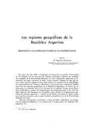
Fig. 13. TEM micrograph <strong>of</strong> the gap<br />
between the fibre core <strong>and</strong> the mantle layer<br />
<strong>of</strong> egg membrane fibre. x 80,500<br />
Fig. 14. SEM micrograph <strong>of</strong> a tear<br />
preparation <strong>of</strong> the shell mernbrane showing<br />
terraang <strong>of</strong> the fibres. x 135<br />
Figs. 15 - 17. SEM micrographs at different<br />
magnifications. (x 65, x 390 <strong>and</strong> x 550)<br />
showing attachment <strong>of</strong> the rnamillary cores to<br />
fibres <strong>of</strong> the sheli mernbrane (arrow).<br />
Fig. 18. SEM rnicrograph <strong>of</strong> the rnamillary<br />
core after rernoval <strong>of</strong> the fibres <strong>of</strong> the shell<br />
membrane. x 125<br />
Fig. 19. SEM rnicrograph <strong>of</strong> the globules<br />
seen in Fia. 18.<br />
m<br />
membrane appeared as an intricately-woven network<br />
(Fig. 4) which presented a stratified appearance. The<br />
fibres ran in al1 directions <strong>and</strong> criss-crossed each other;<br />
some appeared to anastomose while others did not<br />
(Figs. 5,6). At higher magnification, two distinct layers<br />
<strong>of</strong> fibres could be distinguished. When the specimen<br />
was tilted at an angle <strong>of</strong> 15* <strong>and</strong><br />
viewed, a plane <strong>of</strong> cleavage<br />
between the two layers became<br />
apparent (Fig. 4). Generally, the<br />
fibres <strong>of</strong> the inner layer were<br />
smaller than those <strong>of</strong> the outer<br />
layer (Fig. 5). When the broken<br />
ends <strong>of</strong> some fibres were<br />
examined, they were observed to<br />
be made up <strong>of</strong> a central core<br />
which was surrounded by a<br />
sheath-like structure called the<br />
mantle (Fig. 7). On the surface <strong>of</strong><br />
the central core were small<br />
tubercle-like structures (Fig. 7).<br />
Examination <strong>of</strong> the cut surface <strong>of</strong> a<br />
fibre core showed that it was perforated<br />
by several "holes" (Fig. 8).<br />
In addition, a small cleft separated<br />
the mantle layer from the fibre<br />
- core (Fig. 7).<br />
TEM <strong>study</strong>. The limiting<br />
membrane appeared to be a<br />
featureless, amorphous membrane<br />
which was not uniformly thick<br />
throughout the length <strong>of</strong> the fibres<br />
which were examined (Figs. 9,<br />
10). A total <strong>of</strong> 50 measurements<br />
were made from five pieces <strong>of</strong> egg<br />
membrane removed from the blunt<br />
end <strong>of</strong> the three eggs; the mean thickness <strong>of</strong> the<br />
membrane varied from .O9 to .15 pm. At irregular<br />
intemals along the length <strong>of</strong> the membrane, some innerlayer<br />
fibres were observed to be adherent to its inner<br />
surface (Fig. 10). At such sites, the mantle layer <strong>of</strong> the<br />
fibre appeared to fuse with the limiting membrane,
Chicken egg membrane<br />
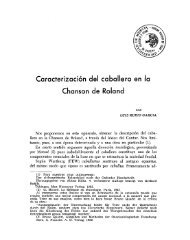
Fig. 20. TEM micrograph <strong>of</strong> a<br />
cross-section <strong>of</strong> the egg<br />
membrane. Four layers can be<br />
distinguished (A - D). Asterisk =<br />
surface facing the air-sac. x<br />
3,600<br />
Fig. 21. TEM micrograph <strong>of</strong> a<br />
decalcified egg sheil showing the<br />
site <strong>of</strong> attachment <strong>of</strong> a mamillary<br />
core to the shell membrane. x<br />
17,500<br />
Fig. 22. TEM micrograph <strong>of</strong> a<br />
fibre near the attachment <strong>of</strong> the<br />
mamillary core. (Arrow = fibrillary<br />
material <strong>and</strong> subsurface<br />
vesicles). x Z,750<br />
were statistically significant<br />
(P
Chicken egg membrane<br />
TEM <strong>study</strong>. At lower power TEM, the fibres were<br />
seen to be organized into groups <strong>of</strong> bundles. In each<br />
bundle, the fibres were al1 oriented similarly; frequently,<br />
a bundle <strong>of</strong> fibres sectioned longitudinally or obliquely<br />
alternated with another in which the fibres were<br />
sections transversely (Fig. 20). The short diameter <strong>of</strong><br />
300 fibres ranged from .ll to 4.14 (mean = 1.37 pm;<br />
SD = .76 pm). The larger fibres tended to be located<br />
more superficially (i.e. nearer the shell). The mean<br />
thickness <strong>of</strong> the mantle layer was .39 pm (SD = .20<br />
pm). The diameters <strong>of</strong> the longitudinal channels in the<br />
fibre core ranged from .O8 to 1.11 pn (mean = .27 pm;<br />
SD = .20 pm). The gaps between the fibre core <strong>and</strong> the<br />
mantle layer varied from .O3 to .O7 p.<br />
At the attachment sites <strong>of</strong> the mamillary cores, many<br />
small pr<strong>of</strong>iles measuring about 3 pm in diameter were<br />
present (Fig. 21). These structures probably<br />
corresponded to the globules observed with the SEM<br />
(see above <strong>and</strong> Fig. 18). Fibrillar material was also<br />
observed on the surface <strong>of</strong> <strong>and</strong> in the intervals between<br />
the fibres (Fig. 22).<br />
Discussion<br />
Terminology. Since the inititation <strong>of</strong> light <strong>microscopic</strong><br />
studies until the more recent ultrastructural studies <strong>of</strong><br />
the structure <strong>of</strong> the avian egg, the term "membrane" has<br />
been traditionally used to denote that part <strong>of</strong> the tissue<br />
which separates the albumen from the shell. It should be<br />
stressed, however, that these "membranes" are not true<br />
biological membranes but are merely investments or<br />
layers <strong>of</strong> material laid down as the egg moves down the<br />
isthmus <strong>of</strong> the oviduct.<br />
The limiting membrane. A thin limiting membrane,<br />
which is continuous <strong>and</strong> impervious, separates the egg<br />
membrane from the albumen <strong>of</strong> the avian egg (Simons<br />
<strong>and</strong> Wiertz, 1963; Bellairs <strong>and</strong> Boyde, 1969). Such a<br />
membrane has also been described in the reptilian egg,<br />
for example in the chelonid (Solomon <strong>and</strong> Baird, 1976),<br />
trionyx (Packard <strong>and</strong> Packard, 1979) <strong>and</strong> kinosterid<br />
turtles (Packard et al., 1984a).<br />
Simons <strong>and</strong> Wiertz (1963) estimated the limiting<br />
membrane to be about 2.7 pm thick, but the present<br />
TEM <strong>study</strong> shows that it is much thinner <strong>and</strong><br />
that it is not <strong>of</strong> uniform thickness but varies from<br />
.O9 to .15 pm. The inner surface (Le. the one which<br />
faces the albumen) is smooth but presents many<br />
undulations. The present TEM observations suggest<br />
that these undulations are, in part, produced by the<br />
fibres which run beneath it. No fenestrations have been<br />
observed. This observation has a functional significance<br />
since it has been suggested that the limiting membrane<br />
not only separates the egg membrane from the<br />
extraembryonic membrane but also may provide defense<br />
against bacterial invasion (Tung <strong>and</strong> Richard, 1972;<br />
Tung et al., 1979).<br />
The egg <strong>and</strong> shell membranes. The existence <strong>of</strong> two<br />
major layers <strong>of</strong> membrane in the avian egg has been<br />
known from early light <strong>microscopic</strong> studies. A double<br />
layer <strong>of</strong> membrane, however, has not been observed in<br />
reptilian eggs, for example, the kinosterid (Packard et<br />
al., 1984a,b) <strong>and</strong> chelonid turtles (Packard, 1980) <strong>and</strong><br />
tuatara, an evolutionarily ancient squamatic reptile<br />
(Packard et al., 1982).<br />
Moran <strong>and</strong> Hale (1936) reported that it was possible<br />
to dissect the egg membrane into two layers <strong>and</strong> the<br />
shell membrane into three layers. This was supported by<br />
Simons <strong>and</strong> Wiertz (1936) who reported that the egg<br />
membrane was made up <strong>of</strong> three layers <strong>and</strong> the shell<br />
membrane <strong>of</strong> six layers. But Simkiss (1958) could not<br />
distinguish separate layers in the two membranes in<br />
histological sections. On the basis <strong>of</strong> SEM observations<br />
<strong>of</strong> tear preparation, the present <strong>study</strong> suggests that the<br />
egg membrane may be composed <strong>of</strong> at least two distinct<br />
layers <strong>of</strong> fibres <strong>and</strong> the shell membrane <strong>of</strong> three or more<br />
layers. However, the possibility that the stratification <strong>of</strong><br />
the fibres seen in the SEM <strong>of</strong> tear preparation could<br />
have been artifactual cannot be ruled out. But if the<br />
stratification is not an artifact, then it would suggest that<br />
the laying down <strong>of</strong> fibres is not one continuous process<br />
but interrupted at intervals as the egg spirals down the<br />
isthmus.<br />
Fibres. A great variability in the size <strong>of</strong> the fibres in<br />
both the egg <strong>and</strong> shell membranes has long been known.<br />
The fibres in the shell membrane show greater<br />
variability than those in the egg membrane. In the<br />
present <strong>study</strong>, the smallest fibres are found to be located<br />
nearest the egg albumen (mean = .3 pm; SD = .12 pm)<br />
<strong>and</strong> the largest ones near the shell (mean = 1.37 pm; SD<br />
= .76). The present results concur with those <strong>of</strong> Simons<br />
<strong>and</strong> Wiertz (1963), C<strong>and</strong>lish (1970), Draper et al.<br />
(1972) <strong>and</strong> Wong et al. (1984) <strong>and</strong> confirm that the<br />
early figures reported by Romankewitsch (1932) <strong>and</strong><br />
Moran <strong>and</strong> Hale (1936) were grossly overestimated. It<br />
also supports the idea that the fibres get larger during<br />
the later stages <strong>of</strong> the passage <strong>of</strong> the egg down the<br />
oviduct (Roman<strong>of</strong>f <strong>and</strong> Roman<strong>of</strong>f, 1949; Draper et al.,<br />
1972; Hodges, 1974). In their immun<strong>of</strong>luorescence<br />
<strong>study</strong>, Wong et al. (1984) concluded that the fibres in<br />
the shell membrane contained q pe 1 collagen whereas<br />
those <strong>of</strong> the egg membrane contained Type 5 collagen,<br />
although the characteristic 67 nm b<strong>and</strong>ing have not been<br />
observed under TEM. Stevenson (1980) has also shown<br />
that these fibres could be digested by a bacterial<br />
protease, Pronase P.<br />
Mash<strong>of</strong>f <strong>and</strong> Stolpmann (1961) described each as<br />
being made up <strong>of</strong> a core surrounded by a mantle with<br />
Bellairs <strong>and</strong> Boyde (1969) called the "medulla" <strong>and</strong><br />
"cortex", respectively. These two components <strong>of</strong> the<br />
fibre have been observed in the present <strong>and</strong> other studies<br />
(Simons <strong>and</strong> Wiertz, 1963; C<strong>and</strong>lish, 1970; Draper et<br />
al., 1972). The two components are separated by a gap<br />
which Draper et al. (1972) called "halo". Under high<br />
power TEM in the present <strong>study</strong>, some fuzzy material<br />
bridging the gap between the mantle layer <strong>and</strong> the fibre
Chicken egg membrane<br />
core was obsewed in the present <strong>study</strong> which Bellairs<br />
<strong>and</strong> Boyde (1968) <strong>and</strong> C<strong>and</strong>lish (1970) described as<br />
"delicate str<strong>and</strong>s". Draper et al. (1972) obsewed that<br />
these width <strong>of</strong> the gap was fairly constant <strong>and</strong> suggested<br />
that the gap might be a shirnkage artifact. Fibrillar<br />
material was also obsewed on the surface <strong>of</strong> the mantle<br />
layer <strong>of</strong> the fibres. Wong et al. (1984) have shown that<br />
these stmctures contain glycosaminoglycans.<br />
The presence <strong>of</strong> "holes" in the fibre core has been<br />
noted by Simons (1971). There has been some<br />
suggestion that these pr<strong>of</strong>iles represented "small holes"<br />
or "hard inclusions" (Simons, 1971; Draper et al.,<br />
1972). It is apparent from the present <strong>study</strong> that these<br />
pr<strong>of</strong>iles, which appear as holes in transverse sections <strong>of</strong><br />
the fibre core, appeared as long channels in longitudinal<br />
sections <strong>of</strong> the fibre core. The functional significance <strong>of</strong><br />
these chameis is not known.<br />
References<br />
Becking J.H. (1975). The ultrastructure <strong>of</strong> the avian egg-shell. Ibis 117,<br />
143-151.<br />
Bellairs R. <strong>and</strong> Boyde A. (1969). Scanning <strong>electron</strong> microscopy <strong>of</strong> the<br />
shell membranes <strong>of</strong> the hen's egg. Z. Zellforsch. Mikros. Anat. 96,<br />
237-249.<br />
C<strong>and</strong>lish J.K. (1970). The outer membrane <strong>of</strong> the avian egg shell as a<br />
reticular structure. Brit. Poult. Sci. 11, 341-344.<br />
Draper M.H., Davidson M.F., Wyburn G.M. <strong>and</strong> Johnston H.S. (1972).<br />
The fine structure <strong>of</strong> the fibrous membrane forming region <strong>of</strong> the<br />
isthmus <strong>of</strong> the oviduct <strong>of</strong> Gallus domesticus. Quart. J. Expt. Physiol.<br />
57, 297-309.<br />
Gilbert A.B. (1970). Female genital organ. In: Form <strong>and</strong> function in Birds,<br />
Vol. 1. King, A.S. <strong>and</strong> McLell<strong>and</strong>. J. (e&.). Academic Press? London.<br />
p~ 237-360.<br />
Hodges R.D. (1974). The histology <strong>of</strong> the fowls. Academic Press. London.<br />
Mash<strong>of</strong>f W. <strong>and</strong> Stolpmann H.J. (1961). Licht-und<br />
Elektronenmikroskopische Untersuchungen an der Schelenhaut und<br />
Kalkschale des Huhnereies. Z. Zellforsch. Mikros. Anat. 55, 81-832.<br />
Moran T. <strong>and</strong> Hale H.P. (1936). Physics <strong>of</strong> the hen's egg. l. Membrane in<br />
the egg. J. Exp. Biol. 13,35-40.<br />
Packard M.J. (1980). Ultrastructural morphology <strong>of</strong> the shell <strong>and</strong> shell<br />
membranes <strong>of</strong> eggs <strong>of</strong> cornmon snapping turtle (Chelydra<br />
serpentina). J. Morphol. 165, 187-204.<br />
Packard M.J. <strong>and</strong> Packard G.C. (1979). Stnicture <strong>of</strong> the shell <strong>and</strong> tertiary<br />
membranes <strong>of</strong> egg <strong>of</strong> soitshell turtle (Trionyx spiniferus). J. Morphol.<br />
159, 131-144.<br />
Packard M.J., Hirsch K.F. <strong>and</strong> Meyer-Rochow V.B. (1982). Structure <strong>of</strong><br />
the sheii from eggs <strong>of</strong> the tuatara, Sphenodon punctatus. J. Morphol.<br />
174, 197-205.<br />
Packard M.J., Hirsch K.F. <strong>and</strong> lverson J.B. (1984a). Structure <strong>of</strong> shells<br />
from eggs <strong>of</strong> Kinostend turtles. J. Morphol. 181,9-20.<br />
Packard M.J., lverson J.B. <strong>and</strong> Packard G.C. (1984b). Morphology <strong>of</strong><br />
shell forrnation in eggs <strong>of</strong> the turtle, Kinosteron flavescens. J.<br />
Morphol. 159, 131-144.<br />
Romankewitsch N.A. (1932). 2. Zellfosch. 21, 110-118. (Cited by<br />
Roman<strong>of</strong>f <strong>and</strong> Roman<strong>of</strong>f, 1949).<br />
Roman<strong>of</strong>f A.L. <strong>and</strong> Roman<strong>of</strong>f A.J. (1949). The avian egg. John Wiley <strong>and</strong><br />
Sons Inc. New York.<br />
Romijn C. <strong>and</strong> Roos J. (1938). The air space <strong>of</strong> the hen's egg <strong>and</strong> its<br />
- -<br />
changes dunng the period <strong>of</strong> incubation. J. Physiol. (Lond.) 94, 365-<br />
379.<br />
Simkiss K. (1958). The structure <strong>of</strong> the eggshell with particular reference<br />
to the hen. PhD Thesis, University <strong>of</strong> Reading. (Cited by Hodges,<br />
1974).<br />
Sirnons P.C.M. <strong>and</strong> Wiertz G. (1963). Notes on the stnicture <strong>of</strong> membrane<br />
<strong>and</strong> shell in the hen's egg. An <strong>electron</strong> <strong>microscopic</strong>al <strong>study</strong>. Z.<br />
Zellforsch. Mikrosk. Anat. 59,555-567.<br />
Simons P.C.M. (1971). Ultrastructure <strong>of</strong> the hen egg-shell <strong>and</strong> its<br />
physiological interpretation. PhD Thesis, L<strong>and</strong>bouwhogeschool,<br />
Wageningen, Netherl<strong>and</strong>s, Centre for Agricultura1 Publishing <strong>and</strong><br />
Documentation.<br />
Solomon S.E. <strong>and</strong> Baird T. (1976). Studies on the egg shell (oviducal <strong>and</strong><br />
oviposited) <strong>of</strong> Chelonia mydas L.J. Exp. Mar. Biol. Ecol. 22, 145-160.<br />
Stevenson J.L. (1980). The removal <strong>of</strong> egg shell membranes by enzyme<br />
treatment to facilitate the <strong>study</strong> <strong>of</strong> shell microstructure. Poultry Sci. 59,<br />
1959-1960.<br />
Tung M.A. Garl<strong>and</strong> M.R. <strong>and</strong> Gill P.K. (1979). A <strong>scanning</strong> <strong>electron</strong><br />
<strong>microscopic</strong> <strong>study</strong> <strong>of</strong> bacteria1 invasion in hen's egg shell. J. Inst. Can.<br />
Sci. Technol. Aliment. 12, 16-22. (Cited by Packard, Hirsch <strong>and</strong><br />
lverson, 1984a).<br />
Tung M. A. <strong>and</strong> Richard J.H. (1972). Ultrastructure <strong>of</strong> the hen's egg shell<br />
membrane by <strong>electron</strong> microscopy. J. Food Sci. 37,277-281.<br />
Tyler C. (1969). Avian egg shells: their structure <strong>and</strong> characteristics. Int.<br />
Rev. Gen. Exp. Zool.4,81-130.<br />
Wong M., Hendtix M.J.C., von der Mark. K., Little C. <strong>and</strong> Stern R. (1984).<br />
Collagen in the egg shell membranes <strong>of</strong> the hen. Dev. Bol. 104,28-36.<br />
Accepted January 22,1992